Atypical Fibroxanthoma-Like Amelanotic Melanoma: A Diagnostic Challenge

 ,
,  ,
,  and
and
Abstract
:1. Introduction
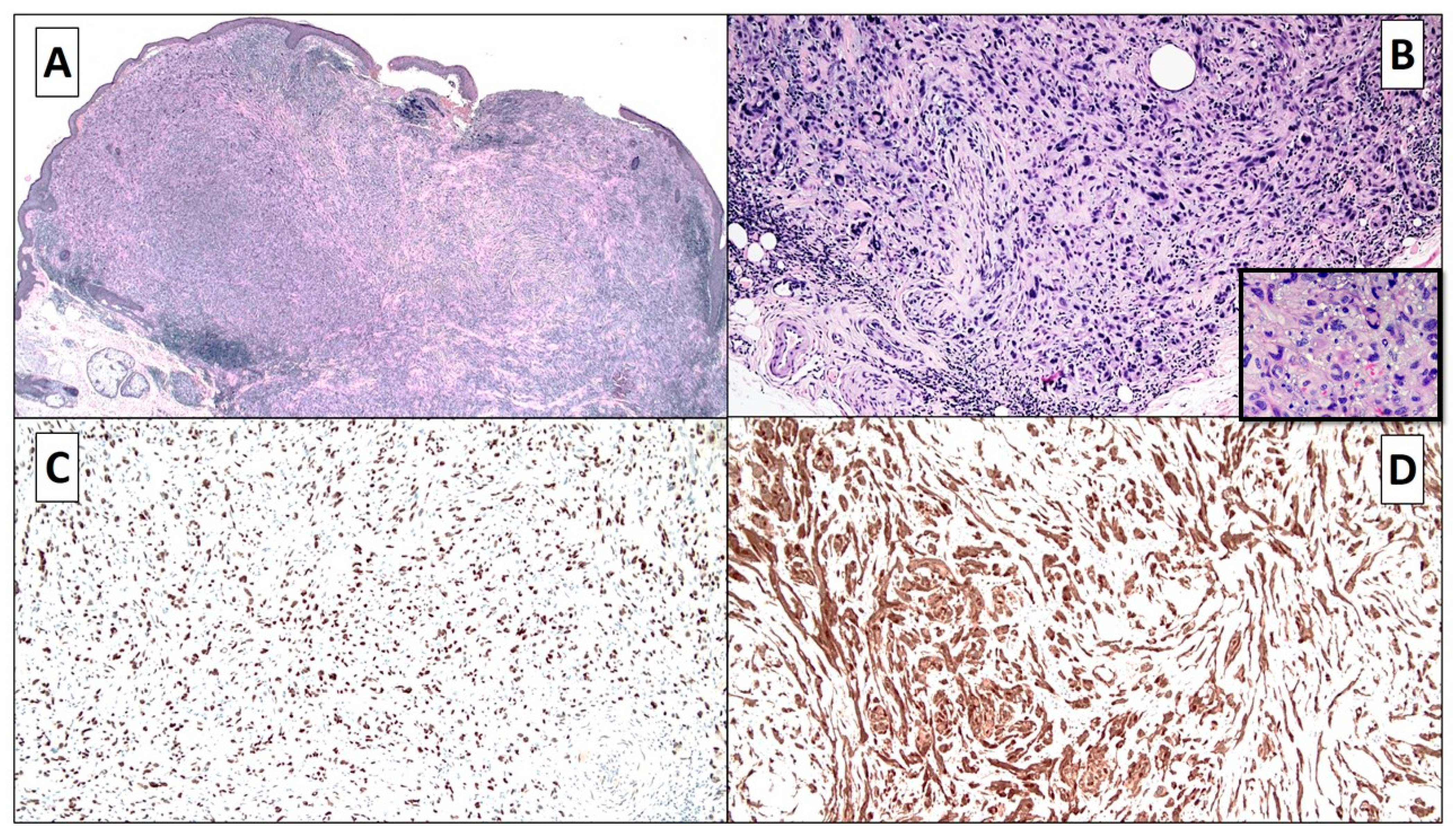
2. Case Report
3. Discussion
4. Conclusions
Author Contributions
Funding
Institutional Review Board Statement
Informed Consent Statement
Conflicts of Interest
References
- Hsu, C.-K.; Chao, S.; Shieh, S.-J.; Lee, J.Y. Atypical fibroxanthoma-like amelanotic malignant melanoma: A case report and literature review. Dermatol. Sin. 2013, 31, 140–144. [Google Scholar] [CrossRef] [Green Version]
- Lee, J.S.-S.; Kossard, S. Atypical Fibroxanthoma-like Melanoma with Touton-like Giant Cells. Am. J. Dermatopathol. 2007, 29, 480–481. [Google Scholar] [CrossRef] [PubMed]
- Sangüeza, M.; Zelger, B. Melanoma Simulating Atypical Fibroxanthoma. Am. J. Dermatopathol. 2007, 29, 551–554. [Google Scholar] [CrossRef] [PubMed]
- Zelger, B.G.; Steiner, H.; Wambacher, B.; Zelger, B. Malignant Melanomas Simulating Various Types of Soft Tissue Tumors. Dermatol. Surg. 1997, 23, 1047–1054. [Google Scholar]
- Melato, M.; Rizzardi, C.; Calacione, R.; Trevisan, G. Atypical fibroxanthoma diagnosed as malignant melanoma. Acta Derm. 2001, 10, 103–106. [Google Scholar]
- Cichewicz, A.W.; Białecka, A.; Męcińska-Jundziłł, K.; Adamska, U.; Neska-Długosz, I.; Grzanka, D.; Czajkowski, R. Atypical fibroxanthoma mimicking amelanotic melanoma in dermoscopy. Adv. Dermatol. Allergol. 2019, 36, 492–494. [Google Scholar] [CrossRef] [PubMed]
- Conic, R.Z.; Ko, J.S.; Allam, S.H.; Mesinkovska, N.A.; Kovalyshyn, I.; Bergfeld, W.; Gastman, B. Mixed Versus Pure Variants of Desmoplastic Melanoma. Ann. Plast. Surg. 2018, 80, 277–281. [Google Scholar] [CrossRef] [PubMed]
- Lefferts, A.J.; Loehrer, A.P.; Yan, S.; Green, D.C.; Deharvengt, S.J.; Leblanc, R.E. CD10 and p63 expression in a sarcomatoid undifferentiated melanoma: A cautionary (and molecularly annotated) tale. J. Cutan. Pathol. 2020, 47, 541–547. [Google Scholar] [CrossRef] [PubMed]


| No. | Age (y)/Gender | Clinical Manifestation | Immunohistochemistry Study | Thickness (Breslow) | Reference |
|---|---|---|---|---|---|
| 1 | 70/M | Two exophytic lesions: left cheek and forehead | Both lesions: S-100 protein: positive, HMB-45: negative | Left cheek: 3.1 mm Forehead: 1.3 mm | Sangueza et al. [3] |
| 2 | 88/F | Amelanotic lesion (1 cm): left forearm | S-100 protein, HMB-45, and melan-A/MART1: positive | Not available | Lee et Al. [2] |
| 3 | 67/F | Ulcerated nodule in the center of a pigmented macule | S-100 protein, HMB-45, and melan-A/MART1: positive CD68: positive in dendritic cells | 2.5 mm | Sangueza et al. [3] |
| 4 | 80/M | Basal cell carcinoma? | S-100 protein, HMB-45, and melan-A/MART1: negative CD68: positive for xanthomatous macrophages and foreign body giant cells | Not available | Sangueza et al. [3] |
| 5 | 72/F | Two separate erythematous nodules within a large hypopigmented patch: left cheek | S-100 protein, HMB-45, and melan-A/MART1: positive CD68: focally positive CD163: negative | 3.4 mm | Chao-Kai Hsu et al. [1] |
| 6 | 93/M | Nodule on the right auricle | S-100 protein, SOX-10: positive melan-A, HMB-45: negative CD68: focally positive | 3.1 mm | Cazzato et al. |
Publisher’s Note: MDPI stays neutral with regard to jurisdictional claims in published maps and institutional affiliations. |
© 2021 by the authors. Licensee MDPI, Basel, Switzerland. This article is an open access article distributed under the terms and conditions of the Creative Commons Attribution (CC BY) license (http://creativecommons.org/licenses/by/4.0/).
Share and Cite
Cazzato, G.; Colagrande, A.; Cimmino, A.; Liguori, G.; Lettini, T.; Serio, G.; Ingravallo, G.; Marzullo, A. Atypical Fibroxanthoma-Like Amelanotic Melanoma: A Diagnostic Challenge. Dermatopathology 2021, 8, 25-28. https://0-doi-org.brum.beds.ac.uk/10.3390/dermatopathology8010004
Cazzato G, Colagrande A, Cimmino A, Liguori G, Lettini T, Serio G, Ingravallo G, Marzullo A. Atypical Fibroxanthoma-Like Amelanotic Melanoma: A Diagnostic Challenge. Dermatopathology. 2021; 8(1):25-28. https://0-doi-org.brum.beds.ac.uk/10.3390/dermatopathology8010004
Chicago/Turabian StyleCazzato, Gerardo, Anna Colagrande, Antonella Cimmino, Giovanni Liguori, Teresa Lettini, Gabriella Serio, Giuseppe Ingravallo, and Andrea Marzullo. 2021. "Atypical Fibroxanthoma-Like Amelanotic Melanoma: A Diagnostic Challenge" Dermatopathology 8, no. 1: 25-28. https://0-doi-org.brum.beds.ac.uk/10.3390/dermatopathology8010004