
Progression of Chronic Kidney Disease and All-Cause Mortality in Patients with Tricuspid Regurgitation

 , ,
, ,
Abstract
:1. Introduction
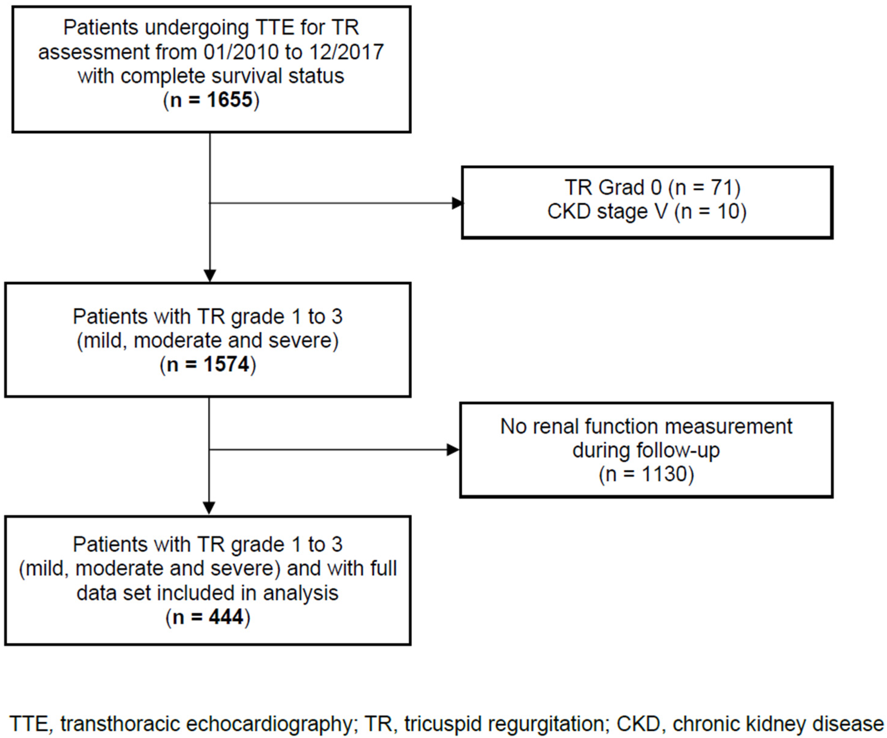
2. Materials and Methods
2.1. Data Collection
2.2. Follow-Up
2.3. Study Endpoint and Definitions
2.4. Statistical Analysis
3. Results
3.1. Patient Characteristics
3.2. Independent Risk Factors for All-Cause Mortality
3.3. Progression of Chronic Kidney Disease
3.4. Independent Risk Factors for CKD Progression
4. Discussion
5. Conclusions
Supplementary Materials
Author Contributions
Funding
Institutional Review Board Statement
Informed Consent Statement
Data Availability Statement
Conflicts of Interest
References
- Reddy, Y.N.V.; Nishimura, R. Intervening for tricuspid regurgitation: Uncertainties in a heterogeneous syndrome. Heart 2019, 105, 1770–1772. [Google Scholar] [CrossRef] [PubMed]
- Mangieri, A.; Montalto, C.; Pagnesi, M.; Jabbour, R.J.; Rodés-Cabau, J.; Moat, N.; Colombo, A.; Latib, A. Mechanism and Implications of the Tricuspid Regurgitation: From the Pathophysiology to the Current and Future Therapeutic Options. Circ. Cardiovasc. Interv. 2017, 10, e005043. [Google Scholar] [CrossRef] [PubMed]
- Wang, N.; Fulcher, J.; Abeysuriya, N.; McGrady, M.; Wilcox, I.; Celermajer, D.; Lal, S. Tricuspid regurgitation is associated with increased mortality independent of pulmonary pressures and right heart failure: A systematic review and meta-analysis. Eur. Heart J. 2019, 40, 476–484. [Google Scholar] [CrossRef] [PubMed]
- Bannehr, M.; Kahn, U.; Okamoto, M.; Kaneko, H.; Hähnel, V.; Neuß, M.; Haase-Fielitz, A.; Butter, C. Post-procedural tricuspid regurgitation predicts long-term survival in patients undergoing percutaneous mitral valve repair. J. Cardiol. 2019, 74, 524–531. [Google Scholar] [CrossRef]
- Singh, J.P.; Evans, J.C.; Levy, D.; Larson, M.; Freed, L.A.; Fuller, D.L.; Lehman, B.; Benjamin, E.J. Prevalence and clinical determinants of mitral, tricuspid, and aortic regurgitation (the Framingham Heart Study). Am. J. Cardiol. 1999, 83, 897–902. [Google Scholar] [CrossRef]
- Topilsky, Y.; Maltais, S.; Medina-Inojosa, J.; Oguz, D.; Michelena, H.; Maalouf, J.; Mahoney, D.W.; Enriquez-Sarano, M. Burden of Tricuspid Regurgitation in Patients Diagnosed in the Community Setting. JACC Cardiovasc. Imaging 2019, 12, 433–442. [Google Scholar] [CrossRef]
- Bannehr, M.; Kahn, U.; Liebchen, J.; Okamoto, M.; Hähnel, V.; Georgi, C.; Dworok, V.; Edlinger, C.; Lichtenauer, M.; Kücken, T.; et al. Right Ventricular Longitudinal Strain Predicts Survival in Patients With Functional Tricuspid Regurgitation. Can. J. Cardiol. 2021, 37, 1086–1093. [Google Scholar] [CrossRef]
- Dietz, M.F.; Prihadi, E.A.; van der Bijl, P.; Goedemans, L.; Mertens, B.J.; Gursoy, E.; van Genderen, O.S.; Marsan, N.A.; Delgado, V.; Bax, J.J. Prognostic Implications of Right Ventricular Remodeling and Function in Patients With Significant Secondary Tricuspid Regurgitation. Circulation 2019, 140, 836–845. [Google Scholar] [CrossRef]
- Murana, G.; Alfonsi, J.; Savini, C.; Mariani, C.; Coppola, G.; Coco, V.L.; Pilato, E.; Pacini, D.; Di Bartolomeo, R. On-X mitral valve replacement: A single-centre experience in 318 patients. Interact. Cardiovasc. Thorac. Surg. 2018, 27, 836–841. [Google Scholar] [CrossRef]
- Gansevoort, R.T.; Correa-Rotter, R.; Hemmelgarn, B.R.; Jafar, T.H.; Heerspink, H.J.L.; Mann, J.F.; Matsushita, K.; Wen, C.P. Chronic kidney disease and cardiovascular risk: Epidemiology, mechanisms, and prevention. Lancet 2013, 382, 339–352. [Google Scholar] [CrossRef]
- Thomas, B.; Matsushita, K.; Abate, K.H.; Al-Aly, Z.; Ärnlöv, J.; Asayama, K.; Atkins, R.; Badawi, A.; Ballew, S.H.; Banerjee, A.; et al. Global Burden of Disease 2013 GFR Collaborators; CKD Prognosis Consortium; Global Burden of Disease Genitourinary Expert Group Global cardiovascular and renal outcomes of reduced GFR. J. Am. Soc. Nephrol. 2017, 28, 2167–2179. [Google Scholar] [CrossRef] [PubMed] [Green Version]
- Go, A.S.; Chertow, G.M.; Fan, D.; McCulloch, C.E.; Hsu, C.-Y. Chronic Kidney Disease and the Risks of Death, Cardiovascular Events, and Hospitalization. N. Engl. J. Med. 2004, 351, 1296–1305. [Google Scholar] [CrossRef] [PubMed]
- Liabeuf, S.; Barreto, D.V.; Barreto, F.C.; Meert, N.; Glorieux, G.; Schepers, E.; Temmar, M.; Choukroun, G.; Vanholder, R.; Massy, Z.A.; et al. Free p-cresylsulphate is a predictor of mortality in patients at different stages of chronic kidney disease. Nephrol. Dial. Transpl. 2010, 25, 1183–1191. [Google Scholar] [CrossRef] [PubMed] [Green Version]
- Mutlak, D.; Aronson, D.; Lessick, J.; Reisner, S.A.; Dabbah, S.; Agmon, Y. Functional tricuspid regurgitation in patients with pulmonary hypertension: Is pulmonary artery pressure the only determinant of regurgitation severity? Chest 2009, 135, 115–121. [Google Scholar] [CrossRef] [PubMed]
- Mullens, W.; Abrahams, Z.; Francis, G.S.; Sokos, G.; Taylor, D.O.; Starling, R.C.; Young, J.B.; Tang, W.W. Importance of Venous Congestion for Worsening of Renal Function in Advanced Decompensated Heart Failure. J. Am. Coll. Cardiol. 2009, 53, 589–596. [Google Scholar] [CrossRef] [Green Version]
- Moradi, H.; Sica, D.A.; Kalantar-Zadeh, K. Cardiovascular Burden Associated with Uremic Toxins in Patients with Chronic Kidney Disease. Am. J. Nephrol. 2013, 38, 136–148. [Google Scholar] [CrossRef] [Green Version]
- Mendoza, F.; Lopez, I.; de Oca, A.M.; Perez, J.; Rodriguez, M.; Aguilera-Tejero, E. Metabolic acidosis inhibits soft tissue calcification in uremic rats. Kidney Int. 2008, 73, 407–414. [Google Scholar] [CrossRef] [Green Version]
- Kyriakidis, N.C.; Cobo, G.; Dai, L.; Lindholm, B.; Stenvinkel, P. Role of Uremic Toxins in Early Vascular Ageing and Calcification. Toxins 2021, 13, 26. [Google Scholar] [CrossRef]
- Galiè, N.; Humbert, M.; Vachiery, J.L.; Gibbs, S.; Lang, I.; Torbicki, A.; Simonneau, G.; Peacock, A.; Vonk Noordegraaf, A.; Beghetti, M.; et al. ESC Scientific Document Group 2015 ESC/ERS Guidelines for the diagnosis and treatment of pulmonary hypertension: The Joint Task Force for the Diagnosis and Treatment of Pulmonary Hypertension of the European Society of Cardiology (ESC) and the European Respiratory Society (ERS): Endorsed by: Association for European Paediatric and Congenital Cardiology (AEPC), International Society for Heart and Lung Transplantation (ISHLT). Eur. Heart J. 2016, 37, 67–119. [Google Scholar]
- Lang, R.M.; Badano, L.P.; Mor-Avi, V.; Afilalo, J.; Armstrong, A.; Ernande, L.; Flachskampf, F.A.; Foster, E.; Goldstein, S.A.; Kuznetsova, T.; et al. Recommendations for Cardiac Chamber Quantification by Echocardiography in Adults: An Update from the American Society of Echocardiography and the European Association of Cardiovascular Imaging. J. Am. Soc. Echocardiogr. 2015, 28, 1–39.e14. [Google Scholar] [CrossRef] [Green Version]
- Nishimura, R.A.; Otto, C.M.; Bonow, R.O.; Carabello, B.A.; Erwin, J.P.; Guyton, R.A.; O’Gara, P.T.; Ruiz, C.E.; Skubas, N.J.; Sorajja, P.; et al. ACC/AHA Task Force Members 2014 AHA/ACC Guideline for the Management of Patients With Valvular Heart Disease: Executive summary: A report of the American College of Cardiology/American Heart Association Task Force on Practice Guidelines. Circulation 2014, 129, 2440–2492. [Google Scholar] [CrossRef] [PubMed]
- Vahanian, A.; Alfieri, O.; Andreotti, F.; Antunes, M.J.; Barón-Esquivias, G.; Baumgartner, H.; Borger, M.; Carrel, T.P.; De Bonis, M.; Evangelista, A.; et al. Guidelines on the management of valvular heart disease (version 2012). Eur. Hear. J. 2012, 33, 2451–2496. [Google Scholar] [CrossRef] [Green Version]
- Levin, A.; Stevens, P.E.; Bilous, R.W.; Coresh, J.; De Francisco, A.L.M.; De Jong, P.E.; Griffith, K.E.; Hemmelgarn, B.R.; Iseki, K.; Lamb, E.J.; et al. Kidney disease: Improving global outcomes (KDIGO) CKD work group. KDIGO 2012 clinical practice guideline for the evaluation and management of chronic kidney disease. Kidney Int. Suppl. 2013, 3, 1–150. [Google Scholar] [CrossRef] [Green Version]
- Levey, A.S.; Stevens, L.A. Estimating GFR Using the CKD Epidemiology Collaboration (CKD-EPI) Creatinine Equation: More Accurate GFR Estimates, Lower CKD Prevalence Estimates, and Better Risk Predictions. Am. J. Kidney Dis. 2010, 55, 622–627. [Google Scholar] [CrossRef] [PubMed] [Green Version]
- Mehr, M.; Taramasso, M.; Besler, C.; Ruf, T.; Connelly, K.A.; Weber, M.; Yzeiraj, E.; Schiavi, D.; Mangieri, A.; Vaskelyte, L.; et al. 1-Year Outcomes After Edge-to-Edge Valve Repair for Symptomatic Tricuspid Regur-gitation: Results From the TriValve Registry. JACC Cardiovasc. Interv. 2019, 12, 1451–1461. [Google Scholar] [CrossRef]
- Ingraham, B.S.; Pislaru, S.; Nkomo, V.T.; Nishimura, R.A.; Stulak, J.M.; A Dearani, J.; Rihal, C.S.; Eleid, M.F. Characteristics and treatment strategies for severe tricuspid regurgitation. Heart 2019, 105, 1244–1250. [Google Scholar] [CrossRef]
- Benfari, G.; Antoine, C.; Miller, W.L.; Thapa, P.; Topilsky, Y.; Rossi, A.; Michelena, H.I.; Pislaru, S.; Enriquez-Sarano, M. Excess Mortality Associated With Functional Tricuspid Regurgitation Complicating Heart Failure With Reduced Ejection Fraction. Circulation 2019, 140, 196–206. [Google Scholar] [CrossRef]
- Agricola, E.; Marini, C.; Stella, S.; Monello, A.; Fisicaro, A.; Tufaro, V.; Slavich, M.; Oppizzi, M.; Castiglioni, A.; Cappelletti, A.; et al. Effects of functional tricuspid regurgitation on renal function and long-term prognosis in patients with heart failure. J. Cardiovasc. Med. 2017, 18, 60–68. [Google Scholar] [CrossRef]
- Bannehr, M.; Edlinger, C.R.; Kahn, U.; Liebchen, J.; Okamoto, M.; Hähnel, V.; Dworok, V.; Schipmann, F.; Kücken, T.; Bramlage, K.; et al. Natural course of tricuspid regurgitation and prognostic implications. Open Heart 2021, 8, e001529. [Google Scholar] [CrossRef]
- Schulman, G.; Berl, T.; Beck, G.J.; Remuzzi, G.; Ritz, E.; Arita, K.; Kato, A.; Shimizu, M. Randomized Placebo-Controlled EPPIC Trials of AST-120 in CKD. J. Am. Soc. Nephrol. 2015, 26, 1732–1746. [Google Scholar] [CrossRef] [Green Version]
- Meijers, B.K.; Claes, K.; Bammens, B.; de Loor, H.; Viaene, L.; Verbeke, K.; Kuypers, D.R.; Vanrenterghem, Y.; Evenepoel, P. p-Cresol and Cardiovascular Risk in Mild-to-Moderate Kidney Disease. Clin. J. Am. Soc. Nephrol. 2010, 5, 1182–1189. [Google Scholar] [CrossRef] [PubMed] [Green Version]
- Wu, C.-L.; Tarng, D.-C. Targeting Uremic Toxins to Prevent Peripheral Vascular Complications in Chronic Kidney Disease. Toxins 2020, 12, 808. [Google Scholar] [CrossRef] [PubMed]
- Besler, C.; Unterhuber, M.; Rommel, K.; Unger, E.; Hartung, P.; Von Roeder, M.; Noack, T.; Zachäus, M.; Halm, U.; Borger, M.; et al. Nutritional status in tricuspid regurgitation: Implications of transcatheter repair. Eur. J. Heart Fail. 2020, 22, 1826–1836. [Google Scholar] [CrossRef] [PubMed]
- Schunk, S.J.; Speer, T.; Petrakis, I.; Fliser, D. Dickkopf 3-a novel biomarker of the ‘kidney injury continuum’. Nephrol Dial Transpl. 2021, 36, 761–767. [Google Scholar] [CrossRef] [PubMed]
- Omote, K.; Nagai, T.; Kamiya, K. Long-term Prognostic Significance of Admission Tricuspid Regurgitation Pressure Gradient in Hospitalized Patients With Heart Failure With Preserved Ejection Fraction: A Report From the Japanese Real-World Multicenter Registry. J. Card. Fail. 2019, 25, 978–985. [Google Scholar] [CrossRef]
- Omrani, H.; Golshani, S.; Vahid Sharifi, V. The Relationship Between Hemodialysis and the Echocardiographic Findings in Patients with Chronic Kidney Disease. Med. Arch. 2016, 70, 328–331. [Google Scholar] [CrossRef] [PubMed] [Green Version]
- Gluba-Brzózka, A.; Michalska-Kasiczak, M.; Franczyk, B. Markers of increased atherosclerotic risk in patients with chronic kidney disease: A preliminary study. J. Lipids Health Dis. 2016, 15, 22. [Google Scholar] [CrossRef] [Green Version]
- Bushyhead, D.; Kirkpatrick, J.N.; Goldberg, D. Pretransplant echocardiographic parameters as markers of posttransplant outcomes in liver transplant recipients. Liver Transpl. 2016, 22, 316–323. [Google Scholar] [CrossRef]
- Taramasso, M.; Maisano, F.; De Bonis, M. Prognostic Impact and Late Evolution of Untreated Moderate (2/4+) Functional Tricuspid Regurgitation in Patients Undergoing Aortic Valve Replacement. J. Card. Surg. 2016, 31, 9–14. [Google Scholar] [CrossRef] [Green Version]
- Navaneethan, S.D.; Roy, J.; Tao, K. Prevalence, Predictors, and Outcomes of Pulmonary Hypertension in CKD. J. Am. Soc. Nephrol. 2016, 27, 877–886. [Google Scholar] [CrossRef] [Green Version]
- Busch, C.; Penov, K.; Amorim, P.A. Risk factors for mortality after pericardiectomy for chronic constrictive pericarditis in a large single-centre cohort. Eur. J. Cardiothorac. Surg. 2015, 48, e110–e116. [Google Scholar] [CrossRef] [PubMed] [Green Version]
- Fernando, M.; Paterson, H.S.; Byth, K. Outcomes of cardiac surgery in chronic kidney disease. J. Thorac. Cardiovasc. Surg. 2014, 148, 2167–2173. [Google Scholar] [CrossRef] [PubMed] [Green Version]
- Ohno, Y.; Attizzani, G.F.; Capodanno, D. Association of tricuspid regurgitation with clinical and echocardiographic outcomes after percutaneous mitral valve repair with the MitraClip System: 30-day and 12-month follow-up from the GRASP Registry. Eur. Heart J. Cardiovasc. Imaging 2014, 15, 1246–1255. [Google Scholar] [CrossRef] [PubMed]
- Ro, S.K.; Kim, J.B.; Jung, S.H. Mild-to-moderate functional tricuspid regurgitation in patients undergoing mitral valve surgery. J. Thorac. Cardiovasc. Surg. 2013, 146, 1092–1097. [Google Scholar] [CrossRef] [PubMed] [Green Version]
- Langanay, T.; Verhoye, J.P.; Ocampo, G. Current hospital mortality of aortic valve replacement in octogenarians. J. Heart Valve Dis. 2006, 15, 630–637. [Google Scholar] [PubMed]


| Died (n = 101) | Survived (n = 343) | p-Value | |
|---|---|---|---|
| Demographics | |||
| Age (years) | 77 (73–82) | 78 (72–82) | 0.698 |
| Female, n (%) | 41 (40.6%) | 161 (46.9%) | 0.260 |
| Body mass index (kg/m2) | 26.5 (24.0–30.5) | 26.7 (24.0–30.4) | 0.878 |
| Type of admission | |||
| Elective case, n (%) | 37 (37.0%) | 172 (52.3%) | 0.012 |
| Transfer from other hospital, n (%) | 19 (19.0%) | 61 (18.5%) | |
| Emergency case, n (%) | 44 (44.0%) | 96 (29.2%) | |
| Comorbidities | |||
| NYHA class, n (%) | |||
| I + II | 53 (52.5%) | 184 (53.6%) | 0.210 |
| III | 47 (46.5%) | 151 (44.0%) | |
| IV | 1 (1.0%) | 8 (2.3%) | |
| Chronic kidney disease, n (%) | |||
| I–II | 37 (36.6%) | 164 (46.9%) | 0.029 |
| III–IV | 64 (63.4%) | 179 (52.2%) | |
| Chronic kidney disease progression, n (%) | 45 (44.6%) | 87 (25.4%) | <0.001 |
| Coronary artery disease, n (%) | 47 (46.5%) | 133 (38.8%) | 0.169 |
| Atrial fibrillation, n (%) | 47 (46.5%) | 108 (31.5%) | 0.006 |
| Arterial hypertension, n (%) | 93 (92.1%) | 325 (94.8%) | 0.259 |
| Pulmonary hypertension, n (%) | 37 (36.6%) | 99 (28.9%) | 0.016 |
| Dilatative cardiomyopathy, n (%) | 11 (10.9%) | 27 (7.9%) | 0.345 |
| Dyslipidaemia, n (%) | 75 (74.3%) | 243 (70.9%) | 0.529 |
| Type 2 diabetes, n (%) | 46 (45.6%) | 107 (31.2%) | 0.008 |
| Chronic obstructive pulmonary disease, n (%) | 23 (22.8%) | 43 (12.5%) | 0.011 |
| Cardiac device, n (%) | 26 (25.7%) | 77 (22.5%) | 0.491 |
| Tricuspid regurgitation, n (%) | |||
| Mild TR | 58 (57.4%) | 268 (78.1%) | <0.001 |
| Moderate TR | 28 (27.7%) | 64 (18.7%) | |
| Severe TR | 15 (14.9%) | 11 (3.2%) | |
| Mitral regurgitation, n (%) | |||
| Mild MR | 38 (37.6%) | 177 (51.6%) | <0.001 |
| Moderate MR | 30 (29.7%) | 93 (27.1%) | |
| Severe MR | 32 (31.7%) | 53 (15.5%) | |
| Aortic regurgitation, n (%) | 35 (34.7%) | 129 (37.6%) | 0.758 |
| Laboratory results at admission | |||
| Estimated glomerular filtration rate (mL/min/1.73) | 58.4 (42.8–74.4) | 68.0 (54.8–83.9) | <0.001 |
| Blood urea nitrogen/Creatinine ratio | 22.4 (17.9–28.0) | 20.7 (17.2–24.3) | 0.032 |
| NT-proBNP (pg/mL) | 3791 (1994–7233) | 2118 (731–4557) | <0.001 |
| Troponin I (µg/L) | 0.05 (0.03–0.08) | 0.03 (0.01–0.05) | 0.052 |
| Haemoglobin (mmol/L) | 7.6 (6.8–8.2) | 7.9 (7.2–8.6) | 0.001 |
| Haematocrit (%) | 37.0 (32.5–39.5) | 38.0 (35.0–42.0) | <0.001 |
| HbA1c (%) | 6.0 (5.7–6.7) | 5.9 (5.5–6.6) | 0.170 |
| Leukocytes (Gpt/L) | 7.9 (6.9–10.1) | 7.6 (6.4–9.4) | 0.433 |
| ALT (µkat/L) | 0.35 (0.28–0.45) | 0.37 (0.27–0.53) | 0.171 |
| Cholesterol (mmol/L) | 4.2 (3.4–5.2) | 4.8 (3.9–5.7) | 0.016 |
| Potassium (mmol/L) | 4.4 (4.1–4.8) | 4.4 (4.1–4.7) | 0.340 |
| Sodium (mmol/L) | 138.5 (136.2–140.4) | 139.0 (136.5–140.6) | 0.614 |
| Diuretics at admission | |||
| Torasemide, n (%) | 69 (68.3%) | 231 (67.4%) | 0.855 |
| Torasemide dose, mg | 20 (10–30) | 10 (10–20) | 0.001 |
| Torasemide dose, mg | |||
| Mild TR | 20 (10–26) | 10 (5–20) | 0.006 |
| Moderate TR | 23 (10–40) | 10 (10–20) | 0.053 |
| Severe TR | 10 (10–50) | 20 (10–20) | 0.555 |
| Aldosterone antagonists, n (%) | 38 (37.6%) | 108 (31.6%) | 0.256 |
| Hydrochlorothiazide, n (%) | 17 (16.8%) | 59 (17.2%) | 0.931 |
| Echocardiographic Parameters | |||
| TAPSE (mm) | 18.0 (15.0–22.0) | 19.0 (16.0–23.0) | 0.097 |
| TR Velocity max (m/s) | 3.2 (2.8–3.7) | 3.0 (2.6–3.4) | 0.009 |
| LVEF (%) | 48.0 (32.0–60.0) | 50.0 (35.0–60.0) | 0.147 |
| LA Dilatation, n (%) | |||
| None | 13 (12.9%) | 88 (25.7%) | <0.001 |
| Mild | 30 (29.7%) | 135 (39.4%) | |
| Moderate | 42 (41.6%) | 90 (26.2%) | |
| Severe | 15 (14.9%) | 30 (8.8%) | |
| RA Dilatation, n (%) | |||
| None | 41 (40.6%) | 196 (57.1%) | <0.001 |
| Mild | 27 (26.7%) | 102 (29.7%) | |
| Moderate | 23 (22.8%) | 31 (9.0%) | |
| Severe | 10 (9.9%) | 13 (3.8%) | |
| RV Dilatation, n (%) | |||
| None | 63 (62.4%) | 287 (83.7%) | <0.001 |
| Mild | 20 (19.8%) | 32 (9.3%) | |
| Moderate | 12 (11.9%) | 19 (5.5%) | |
| Severe | 6 (5.9%) | 5 (1.5%) | |
| All-Cause Mortality | Odds Ratio | 95% CI | p-Value | |
|---|---|---|---|---|
| TR Grade | 2.38 | 1.41 | 4.00 | 0.001 |
| MR Grade | 1.72 | 1.20 | 2.46 | 0.003 |
| CKD progression | 2.38 | 1.30 | 4.35 | 0.005 |
| COPD | 1.70 | 0.81 | 3.57 | 0.162 |
| Haemoglobin at admission | 0.83 | 0.63 | 1.08 | 0.166 |
| NT-proBNP at admission | 1.00 | 1.00 | 1.00 | 0.172 |
| Type 2 diabetes | 1.46 | 0.79 | 2.69 | 0.225 |
| Gender | 1.28 | 0.69 | 2.37 | 0.438 |
| Type of admission | 1.10 | 0.75 | 1.63 | 0.622 |
| Age | 1.00 | 0.97 | 1.04 | 0.710 |
| Atrial fibrillation | 1.09 | 0.57 | 2.07 | 0.792 |
| CKD Progression (n = 132) | No CKD Progression (n = 312) | p-Value | |
|---|---|---|---|
| Demographics | |||
| Age (years) | 77 (70–83) | 78 (73–82) | 0.390 |
| Female, n (%) | 61 (46.2%) | 141 (45.2%) | 0.844 |
| Body mass index (kg/m2) | 26.9 (24.2–30.5) | 26.6 (23.9–30.3) | 0.515 |
| Type of admission | |||
| Elective case, n (%) | 56 (42.4%) | 153 (49.0%) | 0.320 |
| Transfer from other hospital, n (%) | 28 (21.2%) | 52 (16.7%) | |
| Emergency case, n (%) | 45 (34.1%) | 95 (30.5%) | |
| Comorbidities | |||
| NYHA class, n (%) | |||
| I + II | 71 (53.8%) | 166 (53.2%) | 0.261 |
| III | 58 (43.9%) | 140 (44.9%) | |
| IV | 3 (2.3%) | 6 (1.9%) | |
| Chronic kidney disease, n (%) | |||
| I–II | 90 (68.2%) | 111 (34.5%) | <0.001 |
| III–V | 42 (31.8%) | 201 (64.4%) | |
| Coronary artery disease, n (%) | 45 (34.1%) | 135 (43.3%) | 0.068 |
| Atrial fibrillation, n (%) | 46 (34.9%) | 109 (34.9%) | 0.968 |
| Arterial hypertension, n (%) | 125 (94.7%) | 293 (93.9%) | 0.840 |
| Pulmonary hypertension, n (%) | 39 (29.6%) | 97 (31.1%) | 0.682 |
| Dilatative cardiomyopathy, n (%) | 9 (6.8%) | 29 (9.3%) | 0.389 |
| Dyslipidaemia, n (%) | 90 (68.2%) | 228 (73.1%) | 0.273 |
| Type 2 diabetes, n (%) | 58 (43.9%) | 95 (30.5%) | 0.007 |
| Chronic obstructive pulmonary disease, n (%) | 25 (18.9%) | 41 (13.1%) | 0.120 |
| Cardiac device, n (%) | 28 (21.2%) | 75 (24.0%) | 0.519 |
| Tricuspid regurgitation, n (%) | |||
| Mild TR | 92 (69.7%) | 234 (75.0%) | 0.474 |
| Moderate TR | 32 (24.2%) | 60 (19.2%) | |
| Severe TR | 8 (6.1%) | 18 (5.8%) | |
| Mitral regurgitation, n (%) | |||
| Mild MR | 61 (46.2%) | 154 (49.4%) | 0.818 |
| Moderate MR | 37 (28.0%) | 86 (27.6%) | |
| Severe MR | 26 (19.7%) | 59 (18.9%) | |
| Aortic regurgitation, n (%) | 52 (39.4%) | 112 (35.9%) | 0.615 |
| Laboratory results at admission | |||
| Estimated glomerular filtration rate (mL/min/1.73) | 71.5 (54.1–83.8) | 61.6 (50.5–82.4) | 0.039 |
| Blood urea nitrogen/Creatinine ratio | 21.4 (18.2–26.0) | 20.8 (17.1–24.8) | 0.224 |
| NT-proBNP (pg/mL) | 2900 (1140–6175) | 2560 (834–4701) | 0.180 |
| Troponin I (µg/L) | 0.03 (0.01–0.05) | 0.03 (0.01–0.06) | 0.744 |
| Haemoglobin (mmol/L) | 7.7 (6.8–8.5) | 7.9 (7.2–8.6) | 0.087 |
| Haematocrit (%) | 38.0 (33.0–41.0) | 38.0 (35.0–41.8) | 0.121 |
| HbA1c (%) | 6.0 (5.6–6.7) | 5.9 (5.5–6.6) | 0.099 |
| Leukocytes (Gpt/L) | 8.0 (6.7–10.0) | 7.5 (6.3–9.0) | 0.043 |
| ALT (µkat/L) | 0.36 (0.28–0.50) | 0.36 (0.27–0.52) | 0.799 |
| Cholesterol (mmol/L) | 4.7 (3.7–5.4) | 4.7 (3.8–5.8) | 0.594 |
| Potassium (mmol/L) | 4.4 (4.0–4.7) | 4.3 (4.1–4.7) | 0.451 |
| Sodium (mmol/L) | 138.5 (136.3–140.3) | 139.0 (136.5–140.7) | 0.267 |
| Diuretics at admission | |||
| Torasemide, n (%) | 94 (71.2%) | 206 (66.0%) | 0.286 |
| Torasemide dose, mg | 10 (10–20) | 10 (10–20) | 0.907 |
| Torasemide dose, mg | |||
| Mild TR | 10 (6–20) | 10 (5–20) | 0.547 |
| Moderate TR | 10 (10–20) | 20 (10–30) | 0.183 |
| Severe TR | 15 (10–50) | 10 (10–20) | 0.823 |
| Aldosterone antagonists, n (%) | 45 (34.1%) | 101 (32.4%) | 0.725 |
| Hydrochlorothiazide, n (%) | 23 (17.4%) | 53 (17.0%) | 0.911 |
| Echocardiographic Parameters | |||
| TAPSE (mm) | 19.0 (16.0–22.5) | 18.0 (16.0–23.0) | 0.942 |
| TR Velocity max (m/s) | 3.0 (2.7–3.3) | 3.0 (2.6–3.4) | 0.697 |
| LVEF (%) | 50.0 (30.0–60.0) | 50.0 (38.0–60.0) | 0.205 |
| LA Dilatation, n (%) | |||
| None | 25 (18.9%) | 76 (24.4%) | 0.525 |
| Mild | 55 (41.7%) | 110 (35.3%) | |
| Moderate | 39 (29.6%) | 93 (29.8%) | |
| Severe | 13 (9.9%) | 32 (10.3%) | |
| RA Dilatation, n (%) | |||
| None | 69 (52.3%) | 168 (53.9%) | 0.947 |
| Mild | 40 (30.3%) | 89 (28.5%) | |
| Moderate | 17 (12.9%) | 37 (11.9%) | |
| Severe | 6 (4.6%) | 17 (5.5%) | |
| RV Dilatation, n (%) | |||
| None | 100 (75.8%) | 250 (80.1%) | 0.437 |
| Mild | 18 (13.6%) | 34 (10.9%) | |
| Moderate | 12 (9.1%) | 19 (6.1%) | |
| Severe | 2 (1.5%) | 9 (2.9%) | |
| CKD Progression | Odds Ratio | 95% CI | p-Value | |
|---|---|---|---|---|
| Type 2 diabetes | 1.67 | 1.02 | 2.73 | 0.042 |
| Haemoglobin at admission | 0.80 | 0.65 | 0.99 | 0.043 |
| COPD | 1.73 | 0.93 | 3.20 | 0.082 |
| eGFR at admission | 1.01 | 0.99 | 1.02 | 0.113 |
| CAD | 0.72 | 0.45 | 1.17 | 0.190 |
| Age | 0.99 | 0.96 | 1.01 | 0.314 |
| LVEF | 0.99 | 0.98 | 1.01 | 0.337 |
Publisher’s Note: MDPI stays neutral with regard to jurisdictional claims in published maps and institutional affiliations. |
© 2022 by the authors. Licensee MDPI, Basel, Switzerland. This article is an open access article distributed under the terms and conditions of the Creative Commons Attribution (CC BY) license (https://creativecommons.org/licenses/by/4.0/).
Share and Cite
Schipmann, F.; Bannehr, M.; Hähnel, V.; Dworok, V.; Nübel, J.; Edlinger, C.; Lichtenauer, M.; Haase, M.; Zänker, M.; Butter, C.; et al. Progression of Chronic Kidney Disease and All-Cause Mortality in Patients with Tricuspid Regurgitation. Diseases 2022, 10, 16. https://0-doi-org.brum.beds.ac.uk/10.3390/diseases10010016
Schipmann F, Bannehr M, Hähnel V, Dworok V, Nübel J, Edlinger C, Lichtenauer M, Haase M, Zänker M, Butter C, et al. Progression of Chronic Kidney Disease and All-Cause Mortality in Patients with Tricuspid Regurgitation. Diseases. 2022; 10(1):16. https://0-doi-org.brum.beds.ac.uk/10.3390/diseases10010016
Chicago/Turabian StyleSchipmann, Fabian, Marwin Bannehr, Valentin Hähnel, Victoria Dworok, Jonathan Nübel, Christoph Edlinger, Michael Lichtenauer, Michael Haase, Michael Zänker, Christian Butter, and et al. 2022. "Progression of Chronic Kidney Disease and All-Cause Mortality in Patients with Tricuspid Regurgitation" Diseases 10, no. 1: 16. https://0-doi-org.brum.beds.ac.uk/10.3390/diseases10010016