Review

A Role for Acyclic Retinoid in the Chemoprevention of Hepatocellular Carcinoma: Therapeutic Strategy Targeting Phosphorylated Retinoid X Receptor-α

Department of Gastroenterology/Internal Medicine, Gifu University Graduate School of Medicine, Gifu 501-1194, Japan
*
Author to whom correspondence should be addressed.
Submission received: 11 April 2014 / Revised: 10 June 2014 / Accepted: 2 July 2014 / Published: 7 July 2014
(This article belongs to the Special Issue Targeted Therapy of Hepatocellular Carcinoma: Present and Future)

Abstract

:
Hepatocellular carcinoma (HCC) is an aggressive disease with poor prognosis due to its high rate of recurrence after the initial curative treatment. Therefore, development of effective therapeutic strategies that can prevent recurrence and secondary tumor formation is required to improve the clinical outcomes of HCC patients. Malfunctioning of the retinoid X receptor-s (RXRs) of HCC patient by activation of the Ras- mitogen-activated protein kinase (MAPK) signaling pathway is strongly associated with hepatocarcinogenesis. Acyclic retinoid (ACR), a synthetic retinoid, prevents HCC recurrence by inhibiting Ras-MAPK activation and the subsequent RXRα phosphorylation, thereby improving patient prognosis. Here, we have reviewed the detailed effects of ACR on the prevention of HCC development, with particular references to the results of our previous basic and clinical research.

1. Introduction

Liver cancer is the sixth most common neoplasm and the third leading cause of cancer-related deaths in the world. Hepatocellular carcinoma (HCC) accounts for more than 90% of primary liver cancers and is therefore a major health problem worldwide. HCC primarily develops from chronic liver inflammation and subsequent cirrhosis. In Western countries, HCC occurs in a cirrhotic background in up to 90% of the cases [1]. In addition, cirrhosis has been well-established as a major risk factor for HCC, independent of any underlying liver disease [2].
Currently, the surveillance for patients at high risk of HCC has increased the possibility of early diagnosis [1]; however, the overall patient survival rate continues to remain poor (7%, 5-year survival) [3]. Potentially curative therapies, including surgical resection and percutaneous ablation, can provide long-term control in patients with early stage HCC [4,5,6]. However, the 5-year recurrence rates of HCC following these curative treatments exceed 70%, due to intrahepatic metastases (true recurrence) or the development of de novo tumors [2]. In addition, systemic therapies such as those using standard chemotherapeutic agents hvae not had significant effects on HCC in previous randomized trials [7]. Thus, the lack of effective treatments is also one of the main reasons for the poor prognosis of HCC patients, especially with advanced-stage HCC. Therefore, the development of preventative strategies is essential to improve the clinical outcomes for HCC patients.
Several effective strategies for the prevention of development of primary HCC have been presented in clinical trials [8,9,10,11,12,13,14,15,16,17]. It is widely known that chronic infection with hepatitis B virus (HBV) or hepatitis C virus (HCV) causes liver cirrhosis, with both infections together accounting for 75%–80% of the global HCC cases [8]. Primary prevention in the form of HBV vaccination has led to a significant decrease in the number of HBV-related HCC cases [9,10], and antiviral treatment for chronic HBV and HCV infections has reduced the risk of HBV- and HCV-related HCC [11,12,13,14]. Thus, antiviral treatment is a promising approach for the prevention of HCC.
In addition to the studies on antiviral therapies, other important trials using specific agents have been conducted to explore the methods of preventing HCC development. One clinical trial demonstrated that long-term oral supplementation with branched-chain amino acids (BCAA) reduced the frequency of HCC in obese cirrhotic patients [14]. In our randomized controlled study, we found that oral intake of acyclic retinoid (ACR) significantly prevented the development of second primary liver cancer after the initial treatment [15], thereby improving the patient survival [16]. Moreover, a long-term follow-up of the study subjects revealed that 1-year administration of ACR was effective in suppressing secondary liver cancer for up to 3 years [17]. Thus, chemoprevention is one of the key strategies for preventing liver carcinogenesis.
Cancer chemoprevention is defined as an approach in which a natural or synthetic chemical compound regulates premalignant cells via physiological pathways [18]. ACR is a synthetic retinoid developed for the purpose of chemoprevention of HCC [19]. Several experimental studies have reported pleiotropic effects of ACR on either the prevention of HCC development or on the growth suppression of cancer cells [20,21,22]. In this review, we have summarized the important roles of ACR in preventing the development of HCC based on previous basic and clinical studies and the relevant recent literature. We have also discussed the possibility of “combination chemoprevention” using ACR as the key drug, which may be a potential preventive treatment against HCC.

2. Literature Review

2.1. Retinoids and Their Receptors

Retinoids, which are derivatives of vitamin A, are physiological signaling molecules involved in the regulation of cell growth, tissue differentiation, and development of an organism [23,24]. Retinoic acids (RAs) are active metabolites of natural retinoids; they exert their biological functions by regulating the transcription of target genes through two distinct nuclear receptors—RA receptors (RARs) and retinoid X receptors (RXRs). Two isomers of RA, all-trans-RA and 9-cis-RA, have similar binding affinities for RARs, whereas only 9-cis-RA binds to the other RXR nuclear receptors. Both types of nuclear receptors consist of three subtypes (α, β, and γ), characterized by a modular domain structure [23,25]. Different RXRs demonstrate different expression patterns: RXRα is predominantly expressed in the liver, kidney, epidermis, and intestine [26,27,28], RXRβ is ubiquitously distributed and can be detected in almost every tissue [26,28,29,30], while RXRγ is primarily limited to the muscle, certain parts of the brain, and to the pituitary gland [26,28,31,32].
Similar to other members of the nuclear receptor superfamily, the nuclear retinoid receptors regulate target gene transcription in a ligand-dependent manner. After binding with the ligand, RXRs form homodimers or heterodimers with other RARs and then interact with their respective DNA-response elements (RXRE or RARE) located in the promoter region of the target genes to modulate gene expression [23,25,33]. In addition, RXRs are cofactors required for transcriptional activation by several members of the steroid/thyroid hormone nuclear receptor superfamily such as thyroid hormone receptors (TRs), vitamin D receptors (VDRs), and peroxisome proliferator-activated receptors (PPARs) [24,34]. Moreover, interactions with RXRs enhances the DNA-binding efficiency of the partner molecule [33]. Thus, RXRs function as master regulators of nuclear receptors.

2.2. RXRα and Lipid Metabolism in the Liver

Several of the nuclear receptors that form heterodimers with RXRs are implicated as important regulators of genes involved in the liver metabolism [35]. RXRα is the most abundant subtype of RXR in the adult liver [28]. Therefore, it is believed that RXRα plays a prominent role in the regulation of hepatic metabolism. Indeed, RXRα is an obligate heterodimeric partner for nuclear receptors involved in lipid physiology, such as PPARs, liver X receptors (LXRs), and farnesoid X receptors (FXRs) [36,37]. In addition, studies using liver-specific RXRα-deficient mice have revealed that the absence of RXRα reduces the activation of its dimeric partners and results in the impairment of fatty acid and cholesterol metabolism in the liver [35,38,39]. Thus, RXRα and its dimeric receptors are involved in the mediation of normal hepatic lipid metabolism.

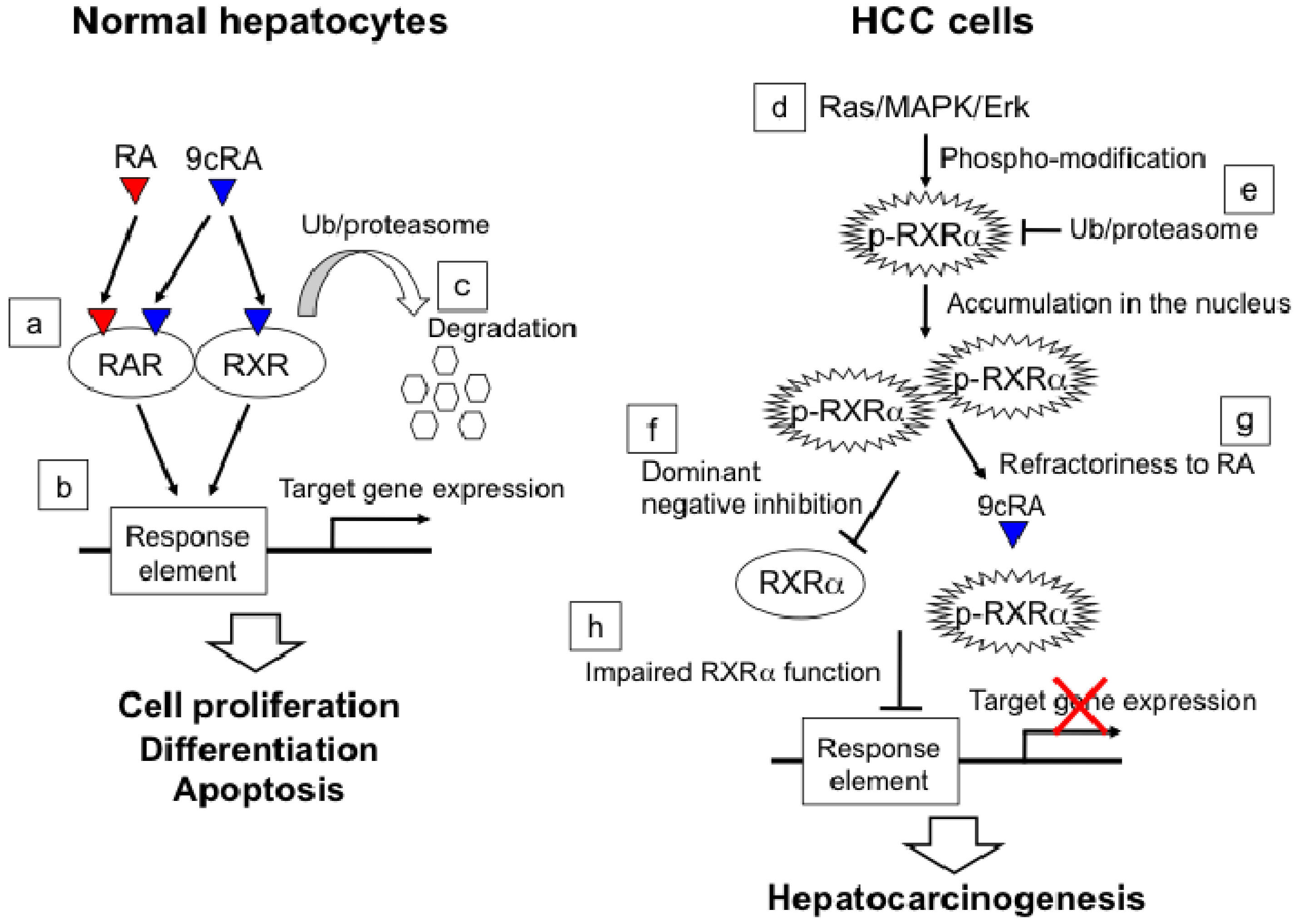
2.3. RXRα Phosphorylation and Hepatocellular Carcinoma

Retinoids and their receptors play important roles in the regulation of normal cell proliferation, differentiation, and apoptosis [40]; therefore, impaired expression or function of these molecules is strongly associated with the development of various human malignancies such as HCC. Indeed, surgically resected HCC tissues contain low levels of vitamin A [41,42]. In a rodent model, 3'-methyl-4-dimethylaminoazobenzene (3'-MeDAB)-induced liver tumors had lower levels of retinol than the surrounding noncancerous liver tissues [19]. Notably, a marked reduction in the levels of both retinol and retinyl ester were observed even at the precancerous, hyperplastic liver nodule stage [42]. In addition, the expression levels of RXRα, which is the most abundant retinoid in normal liver tissues, were also reduced, not only in HCC specimens but also in some precancerous lesions obtained from a 3'-MeDAB-induced rat liver carcinogenesis model [43]. In contrast, increased retinoid signaling of lecithin:retinol acyltransferase-deficient mice suppressed diethylnitrosamine (DEN)-induced hepatocarcinogenesis [44]. Thus, these findings indicate that the expression levels of retinoids or their nuclear receptors, especially RXRα, are closely related to the development of HCC.
Dysfunction of nuclear retinoid receptors is also associated with hepatocarcinogenesis. Our studies showed that the malfunction of RXRα due to post-translational modification by phosphorylation is associated with HCC development [45,46]. It has also been reported that phospho-modification of the nuclear receptors enhances or reduces their transcriptional activity in a context-dependent manner [47]. For instance, phosphorylation of RXRα that occurs in its N-terminal A region induces the expression of several RA-responsive genes and results in RA-induced endodermal differentiation [48]. In contrast, a mitogen-activated protein kinase (MAPK)-mediated phosphorylation of RXRα within its ligand-binding domain impairs the transcriptional activity of RAR-RXR [49] and VDR-RXR heterodimers [50]. Notably, correlation of impaired receptor functions due to phospho-modification and therapy-resistant phenotypes has been reported in several human malignancies [47].
In HCC, Ras-extracellular signal-regulated kinase 1/2 (ERK 1/2) is highly activated, and the phosphorylation of RXRα occurs at both serine 260 and threonine 82, which are recognized as the consensus sites for phosphorylation via the Ras/MAPK/ERK signaling pathways [46]. We found that phosphorylated RXRα is highly accumulated in HCC tissues as well as in HCC cell lines, which prevents its normal degradation through the ubiquitin-proteasome pathway [51]. The accumulated phosphorylated RXRα (non-functional RXRα) abrogates the function of the remaining normal RXRα in a dominant-negative manner, thereby inhibiting the formation of heterodimers with the partner molecules such as RARβ [52]. RARβ has been suggested to be a tumor suppressor gene [53,54,55]. Therefore, impaired RARβ function due to the accumulation of non-functional RXRα may promote the development of HCC. In addition, we have reported in our previous study that phosphorylated RXRα is refractory to its potent ligand, 9cRA, and evades 9cRA-induced apoptosis [56]. These observations suggest that not only the depletion of retinoids but also the dysfunction of retinoid receptors, especially phospho-modification of RXRα, plays a critical role in the development of HCC (Figure 1).

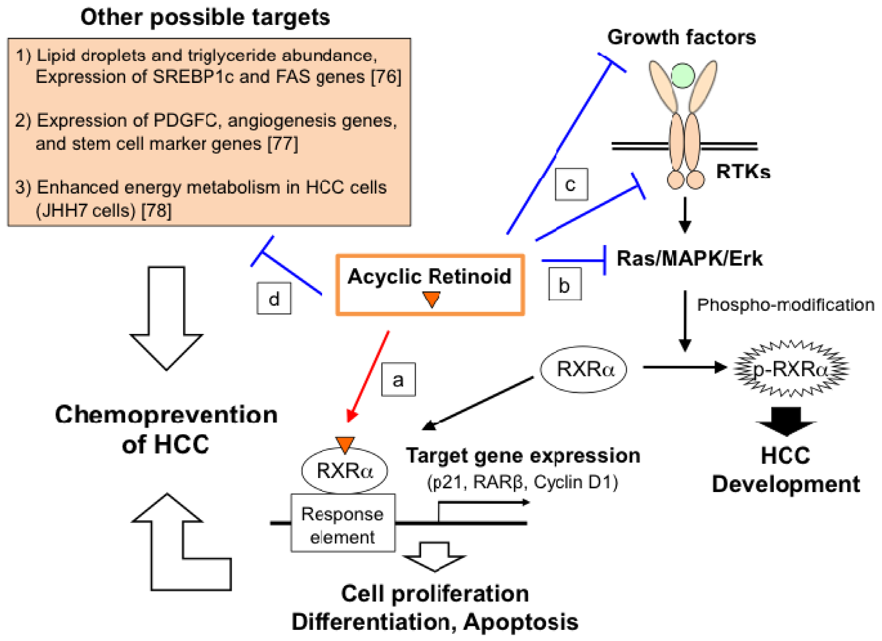
2.4. Molecular Mechanism of ACR in Chemoprevention of Hepatocellular Carcinoma

ACR (equivalent to NIK-333 and Peretinoin; Kowa Pharmaceutical Co., Tokyo, Japan) is a synthetic retinoid developed for chemoprevention of HCC [19]. The chemopreventive effects of ACR have been reported in our previous studies [19,57,58,59,60]. In human HCC cell lines, ACR induces apoptosis and inhibits cell proliferation by inducing cell differentiation or by regulating cell-cycle progression [56,61,62,63,64]. We have found that ACR functions as an agonist for both RAR and RXR and activates RARE and RXRE in hepatoma cells [61,65]. Indeed, increased expression levels of RA-target genes such as RARβ and p21 were observed in ACR-treated HCC cell lines [56,62,63,64,66,67,68,69,70,71]. Thus, ACR inhibits the development of HCC by acting as an effective ligand for nuclear retinoid receptors.
Figure 1. The role of phosphorylated retinoid X receptor (RXR)α in hepatocellular carcinoma (HCC) development. In normal hepatocytes, while all-trans-RA binds only to retinoic acids receptors (RARs), 9-cis-RA binds to both RARs and RXRs as a ligand (a). After activation by their specific ligands, the nuclear receptors bind to their specific response elements and regulate cell proliferation, differentiation, and apoptosis through induction of their target gene expressions (b). Nuclear receptors (RARs and RXRs) cease functioning owing to their degradation through the ubiquitin-proteasome pathway (c). In HCC cells, RXRα is phosphorylated by the Ras/ mitogen-activated protein kinase (MAPK)/Erk pathways, which are constitutively activated in HCC (d), and accumulates in the nucleus, preventing degradation through the ubiquitin-proteasome pathway (e). The phosphorylated RXRα inhibits normal RXRα function in a dominant-negative manner (f) and shows refractoriness to 9-cis-RA (g), thus impairing normal RXRα function (h). The impaired receptor function results in the down-regulation of its target gene expression and leads to hepatocarcinogenesis.
Diseases 02 00226 g001
In addition to its role as a ligand for nuclear retinoid receptors, ACR restores the impaired receptor functions of RXRα by inhibiting RXRα phosphorylation. In HCC, the Ras/MAPK/ERK signaling pathways are highly activated and involved in inducing constitutive phosphorylation within the consensus sites of RXRα [46]. We have reported that ACR inhibits the activated Ras-Erk 1/2 pathways independent of RXRα and consequently prevents phospho-modification of RXRα, thereby restoring the function of RXRα in HCC cells [66]. Moreover, our recent study has revealed that ACR inhibits not only Ras-Erk 1/2 pathways but also several types of growth factors and their corresponding receptor tyrosine kinases (RTKs) in several malignancies, including HCC [58,59,62,72,73,74]. RTKs transmit signals that regulate cell proliferation, differentiation, and survival. The MAPK cascade comprised of Ras-Erk kinases is an essential effector cascade required for most RTK functions [75]. Therefore, the inhibitory effect of ACR on RTKs suppresses the development of HCC via inhibition of the downstream Ras-Erk 1/2 pathways and the subsequent RXRα phosphorylation, in addition to its direct effect on the Ras-Erk 1/2 pathways. Thus, ACR functions not only as a ligand for RXRα but also as a suppressor of the RTK-Ras-MAPK signaling pathways, thereby restoring the function of RXRα and activating the transcriptional activity of its response element.
Recently, a new target of ACR was elucidated in an in vitro study by using Huh-7.5 cells infected with HCV-RNA [76]. This study revealed that ACR inhibits HCV-RNA replication and infectious virus release by modulating several aspects of lipid metabolism, such as triglyceride abundance and the expression of mature sterol regulatory element-binding protein 1c (SREBP1c). Considering that HCV infection is a major cause of HCC development, the inhibitory effect of ACR on HCV infection may be beneficial in addition to its potential for HCC chemoprevention in HCV-positive patients (Figure 2).
Figure 2. The role of acyclic retinoid in the prevention of HCC. Acyclic retinoid (ACR) itself functions as a ligand for RXRα and regulates expression of its downstream genes such as p21, RARβ, and Cyclin D1, thus preventing HCC development through induction of cell proliferation, differentiation, and apoptosis in HCC cells (a). ACR inhibits the activated Ras/MAPK/Erk pathway independent of RXRα (b). ACR also inhibits several types of growth factors and their corresponding receptor tyrosine kinases (RTKs) (c). Thus, ACR restores the impaired RXRα function. Recently, new possible targets of ACR have been reported in either in vivo or in vitro studies [76,77,78] (d). Thus, the pleiotropic responses of ACR target molecules, including phosphorylated RXRα, may play a role in preventing hepatocarcinogenesis.
Diseases 02 00226 g002

2.5. Chemoprevention of Hepatocellular Carcinoma Using ACR

A major concern in HCC patients is the high rate of recurrence and de novo tumors following the initial curative treatments [2], which results in poor prognosis of the malignancy [79]. We have discussed the inhibitory effects of ACR in the development of HCC in our basic studies [19,56,57,58,59,60,61,62,63,64,65,66,67,68,69,70,71,72,73,74]. In order to further investigate the chemopreventive effects of ACR on the recurrent and secondary HCC clinically, a double-blind and placebo-controlled clinical study was performed on patients who had received anticancer treatment for initial HCC [15,16,17]. Oral administration of ACR (n = 44 patients; dose = 600 mg/day) for 12 months significantly reduced the incidence of post-therapeutic recurrence or new HCC development compared to administration of placebo (n = 45 patients) (median follow-up time = 38 months; P = 0.04) [15]. After a median follow-up time of 62 months, ACR administration improved both the recurrence-free survival (P = 0.002) and overall survival (P = 0.04) rates [16]. Moreover, the preventive effects of ACR lasted for up to 3 years following the completion of ACR administration [17]. Thus, a short-term administration of ACR for only 12 months yielded a long-term effect on the prevention of secondary HCC, without causing any severe adverse effects.
A safety-evaluation study was conducted in a Phase I pharmacokinetics clinical trial to determine the dose-limiting toxicities and pharmacokinetics of ACR [80]. In this trial, no adverse effects or dose-limiting toxicities were observed in either of the groups administered with doses of 300 or 600 mg/day, although a dose-limiting toxicity of Grade 3 hypertension was observed in the group receiving a dose of 900 mg/day [80].
Based on the Phase I clinical study of ACR [80], a Phase II/III clinical multicenter, large-scale, randomized, placebo-controlled study (n = 401) was conducted to evaluate the effectiveness of ACR on the prevention of secondary HCC in HCV patients who had received curative treatment for initial HCC [81]. In this study, oral administration of ACR (n = 124 patients, dose = 600 mg/day) showed a reducing trend of incidence of secondary HCC as compared with placebo administration (n = 127 patients), although no significant differences were observed between the two groups at a median follow-up time of 2.5 years. However, at 3 years after treatment, the cumulative recurrence-free survival rate of the ACR-treated group (43.7%) was higher than that of the placebo group (29.3%). Notably, subgroup analysis of this data showed that ACR (n = 100 patients; dose = 600 mg/day) reduced the risk of HCC recurrence or death by approximately 40% as compared to placebo (n = 106 patients), especially in patients with Child-Pugh A and small tumors (size < 20 mm) (p = 0.0347). On the other hand, a 300 mg/day dose of ACR was insufficient for tumor control, showing no substantial difference as compared to placebo. Thus, these studies show that ACR administration to cirrhotic patients effectively inhibits the development of secondary HCC and thereby improves the clinical outcome of the patients. Especially, the inhibitory effects of ACR on secondary HCC were observed in patients with well-preserved liver function (Child-Pugh A) [81]. To this effect, currently, a confirmatory large-scale ACR study focused on Child-Pugh A patients is ongoing.
The inhibitory effects of ACR on the development of secondary HCC have been reported by several clinical studies [15,16,17,81]; however, not much information is available on the mechanisms by which ACR inhibits the secondary HCC in humans in vivo. In response, Honda et al. [77] conducted a gene expression profile analysis under the same study conditions as in previous clinical studies and found that ACR treatment down-regulated the expression of platelet-derived growth factor C (PDGFC), other angiogenesis genes, and cancer stem cell marker genes, thereby successfully preventing HCC recurrence in humans in vivo [77]. This result not only supports the previous clinical data but also suggests the possibility of new targets for ACR for the prevention of HCC recurrence (Figure 2).

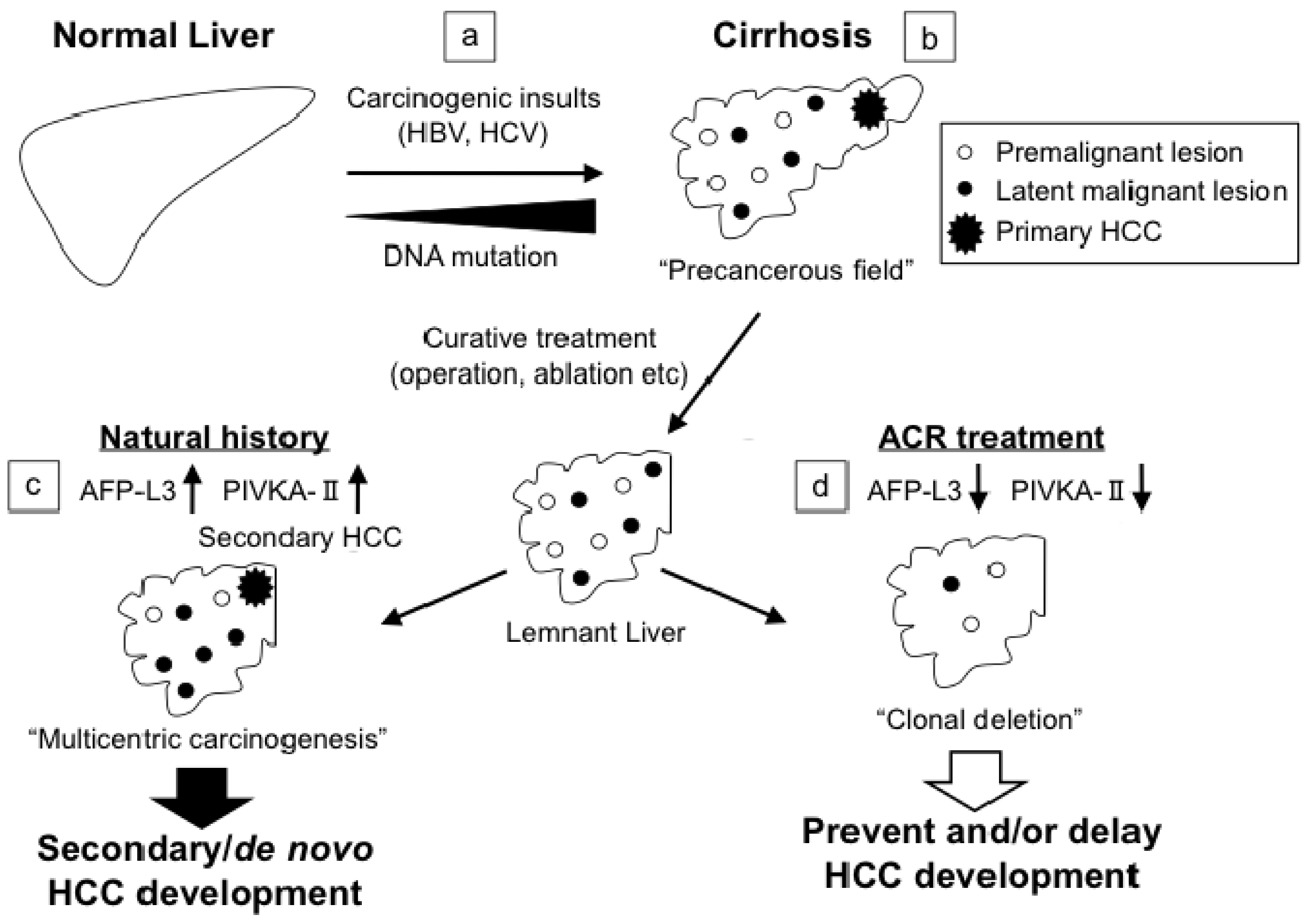
2.6. “Clonal Deletion” in Chemoprevention of Hepatocellular Carcinoma

“Clonal deletion” is a concept in which latent malignant or premalignant cells that are undetected by diagnostic images are removed from the organ in a hyper-carcinogenic state. A typical example of this can be seen in a cirrhosis-HCC sequence. The cirrhotic liver is recognized as a major risk factor for HCC due to a high incidence of tumors in the pathological background [2]. In particular, chronic hepatitis or cirrhosis induced by either HBV or HCV frequently results in the accumulation of DNA mutations across the liver by repeated necrosis and hepatocyte regeneration, and leads to a hyper-carcinogenic state. In cirrhotic patients with HCC, a high incidence of recurrence or de novo tumors is observed after the initial curative treatment [2], which may be explained by the characteristic clinical mode of liver carcinogenesis — “multicentric carcinogenesis” (or “field cancerization”) [82]. Thus, once the liver is exposed to continuous carcinogenic insults such as hepatitis virus infection, the whole liver is regarded as a precancerous field possessing multiple, independent, and premalignant or latent malignant clones.
As reported by some previous studies [15,16,17], ACR effectively inhibits secondary HCC development after initial curative treatment and significantly improves the clinical outcome of HCC patients. These findings suggest that ACR may delete and/or inhibit the “clones of secondary HCC” from the hyper-carcinogenic liver. Indeed, ACR significantly reduced the serum levels of lectin-reactive alpha-fetoprotein factor 3 (AFP-L3) and protein induced by vitamin K absence/antagonist-II (PIVKA-II), both of which indicate the presence of latent HCC cells in the remnant liver [17,83]. This finding suggests that ACR eliminates malignant clones producing AFP-L3 or PIVKA-II before the clones expand to become clinically detectable tumors. Once such latent clones are eliminated from the remnant liver, it takes several years for the secondary HCC to be recognized clinically [17]. In fact, the inhibitory effects of ACR on the development of secondary HCC lasted for about 3 years after termination of its administration [17]. In addition, Zheng et al. [84] recently reported that ACR treatment decreased the emergence of precancerous cells and their progeny in a rat liver carcinogenesis model, consequently leading to suppression of HCC development. This experimental data sufficiently supports the previous clinical reports and the concept of “clonal deletion” [15,16,17]. Thus, we hereby suggest the concept of “clonal deletion” as a new concept in the chemoprevention of HCC using ACR, and that this chemopreventive approach may become a viable cancer therapy for eliminating malignant clones (Figure 3).
Figure 3. The concept of “clonal deletion” and the therapeutic application of ACR on the basis of “clonal deletion.” Continuous exposure to genetic insults such as chronic- hepatitis B virus (HBV) and - hepatitis C virus (HCV) infections induce DNA mutation across the whole liver (a). The multiple premalignant and latent malignant clones occur on the background of this “precancerous field.” Primary HCCs arise from these latent clones in cirrhotic patients (b). After curative treatments, while secondary/de novo HCC development is observed at early time points during the natural history (multicentric carcinogenesis) (c), ACR deletes these latent premalignant clones from the remnant liver by restoring impaired RXRα function and inducing cell differentiation and apoptosis (d). This is the concept of “clonal deletion.” Since lectin-reactive alpha-fetoprotein factor 3 (AFP-L3) and protein induced by vitamin K absence/antagonist-II (PIVKA-II) indicate the presence of latent HCC cells in the remnant liver, reduced levels of these markers observed in ACR treatment sufficiently support this concept.
Diseases 02 00226 g003

2.7. Combination Chemoprevention of Hepatocellular Carcinoma Using ACR

The combined use of two or more agents not only yields the synergistic effects of each agent but also decreases the overall toxicity by enabling treatments with lower clinical dosages [85,86]. Therefore, it is expected that combinatorial treatment of HCC, ACR, and other clinical agents may exert synergistic effects against HCC development. In order to explore this possibility, we conducted studies of “combination chemoprevention” using ACR as the key agent [87,88,89,90]. The combination of ACR and interferon-β (IFN-β) synergistically inhibited cell growth and induced apoptosis in HCC cell lines by inducing the expression of type 1 IFN receptor and STAT1, located downstream of RXRα [68]. In addition, the combinatorial treatment of HCC cells with ACR and OSI-461, a potent derivative of sulindac sulfone, elicited strong synergistic expression levels of RARβ and p21, which are associated with the transcriptional activation of RARE, thereby inducing apoptosis of HCC cells [69]. In addition, the combined use of ACR and vitamin K2 (VK2) synergistically induced apoptosis and inhibited the growth of HCC cells by preventing RXRα phosphorylation through inhibition of the Ras/MAPK/Erk signaling pathway [67]. Thus, the combination of agents with different target sites and action mechanisms enables exertion of the pleiotropic and synergistic inhibitory effects on HCC development and growth of HCC cells. Moreover, if these combination treatments facilitate reduction of the dosage of agents for effective chemoprevention of HCC, the risk of adverse effects and toxicity of the agent would also be reduced. Sorafenib, an oral multi-tyrosine kinase inhibitor, is the first and the only drug that has demonstrated survival benefits in patients with advanced HCC [1]. While improved overall survival has been reported in sorafenib-treated HCC patients, the associated toxicities such as hand-foot skin reaction significantly affect the patients’ quality of life and, occasionally, cause early termination of the treatment [91]. We reported earlier that the combined use of ACR and trastuzumab (the humanized anti-HER2 monoclonal antibody) synergistically inhibited the activation of HER2 and its downstream signaling pathways, including RXRα phosphorylation, and subsequently inhibited the growth of HCC cells [70]. This finding suggests that ACR may be a practical candidate in combination therapy with other RTK antagonists. Considering that sorafenib partially targets the Ras/MAPK/Erk signaling pathway also, the combination of ACR and sorafenib are expected to reduce the probability of adverse events, providing beneficial inhibitory effects against HCC development. Thus, ACR may become a potential candidate for use in a combination therapy with sorafenib against advanced HCC.

3. Discussion

Poor clinical outcomes of HCC patients primarily result from the high incidence of secondary HCC following the initial curative treatment, proving to be a major concern in the treatment of this malignancy. Therefore, the establishment of a new effective strategy to prevent the recurrence of HCC has been recognized as an urgent task worldwide. Accordingly, ACR was originally developed as a chemoprevention agent to accomplish this purpose [19,80] and was administered to patients who had achieved complete cure of HCC in clinical trials [15,16,17,77,80,81]. The preventative effects of ACR on the development of secondary HCC suggest that ACR may play a role beyond that of a “chemopreventive drug”, as a “cancer therapy drug” that actively eliminates latent malignant lesions from the cirrhotic, hyper-carcinogenic liver. This novel therapeutic concept of “clonal deletion” using ACR may be a promising approach in the prevention of HCC.
Our previous reports have shown that ACR exerts its chemopreventive effects by working as a ligand for retinoid receptors as well as by restoring impaired RXRα function through inhibition of Ras/MAPK/Erk activation [61,65,66]. Indeed, restored function of RXRα (unphosphorylated RXRα) regulates the expressions of its downstream genes such as p21, RARβ, and Cyclin D1 and inducts the apoptosis and cell-cycle arrest of HCC cells [64]. In contrast, cells transfected with phospho mimic-RXRα cDNA showed refractoriness to retinoid treatment and association with the acquisition of malignant phenotype [46,52]. These results suggest that phosphorylated RXRα plays an important role in hepatocarcinogenesis. Currently, no clinical agents that can directly target phosphorylated RXRα for the prevention of HCC are commercially available. However, considering its positive role in HCC development, the establishment of a new strategy directly targeting phosphorylated RXRα is highly intriguing.
Although previous evidence suggests that phosphorylated RXRα is a target of ACR, the molecular mechanism by which ACR prevents HCC development is not fully understood [78]. Indeed, recent studies have revealed several targets for ACR that can contribute to the chemoprevention of HCC either in vivo or in vitro [76,77,78] (Figure 2). These reports suggest that the preventative effects of ACR on HCC development may result from the pleiotropic response of ACR target molecules, including phosphorylated RXRα. Further detailed studies are required to fully explore the molecular mechanisms of ACR.
In conclusion, the accumulating evidence discussed in this review suggests that retinoid compounds, especially ACR, may be promising candidates for the chemoprevention of HCC. A Phase III, large-scale, randomized controlled trial on ACR is ongoing to this effect. In addition, ACR-based chemoprevention in combination with other agents may become a promising strategy for HCC chemoprevention.

Acknowledgments

This work was supported by 1) Grants-in-Aid from the Ministry of Education, Science, Sports, and Culture of Japan (No. 22790638 and 25460988), 2) Grant-in-Aid for the 3rd Term Comprehensive 10-Year Strategy for Cancer Control from the Ministry of Health, Labour and Welfare of Japan, and 3) Takeda Science Foundation.

Author Contributions

H. Sakai, M. Shimizu, and H. Moriwaki all participated in designing, writing and editing of the review.

Conflicts of Interest

The authors declare no conflict of interest.

References

  1. Llovet, J.M.; Ducreux, M.; Lencioni, R.; Di Bisceglie, A.M.; Galle, P.R.; Dufour, J.F.; Greten, T.F.; Raymond, E.; Roskams, T.; De Baere, T.; et al. EASL-EORTC clinical practice guidelines: Management of hepatocellular carcinoma. J. Hepatol. 2012, 56, 908–943. [Google Scholar]
  2. El-Serag, H.B. Hepatocellular carcinoma. N. Engl. J. Med. 2011, 365, 1118–1127. [Google Scholar] [CrossRef]
  3. Bosch, F.X.; Ribes, J.; Diaz, M.; Cleries, R. Primary liver cancer: Worldwide incidence and trends. Gastroenterology 2004, 127 (Suppl. 1), S5–S16. [Google Scholar] [CrossRef]
  4. Cho, Y.K.; Kim, J.K.; Kim, M.Y.; Rhim, H.; Han, J.K. Systematic review of randomized trials for hepatocellular carcinoma treated with percutaneous ablation therapies. Hepatology 2009, 49, 453–459. [Google Scholar] [CrossRef]
  5. Llovet, J.M.; Fuster, J.; Bruix, J. Intention-to-treat analysis of surgical treatment for early hepatocellular carcinoma: resection versus transplantation. Hepatology 1999, 30, 1434–1440. [Google Scholar] [CrossRef]
  6. Llovet, J.M.; Schwartz, M.; Mazzaferro, V. Resection and liver transplantation for hepatocellular carcinoma. Semin. Liver Dis. 2005, 25, 181–200. [Google Scholar] [CrossRef]
  7. Llovet, J.M.; Bruix, J. Systematic review of randomized trials for unresectable hepatocellular carcinoma: Chemoembolization improves survival. Hepatology 2003, 37, 429–442. [Google Scholar] [CrossRef]
  8. Yang, J.D.; Roberts, L.R. Hepatocellular carcinoma: A global view. Nat. Rev. Gastroenterol. Hepatol. 2010, 7, 448–458. [Google Scholar] [CrossRef]
  9. Chang, M.H.; You, S.L.; Chen, C.J.; Liu, C.J.; Lee, C.M.; Lin, S.M.; Chu, H.C.; Wu, T.C.; Yang, S.S.; Kuo, H.S.; et al. Decreased incidence of hepatocellular carcinoma in hepatitis B vaccinees: A 20-year follow-up study. J. Natl. Cancer Inst. 2009, 101, 1348–1355. [Google Scholar] [CrossRef]
  10. Zanetti, A.R.; Van Damme, P.; Shouval, D. The global impact of vaccination against hepatitis B: A historical overview. Vaccine 2008, 26, 6266–6273. [Google Scholar] [CrossRef]
  11. Wong, J.S.; Wong, G.L.; Tsoi, K.K.; Wong, V.W.; Cheung, S.Y.; Chong, C.N.; Wong, J.; Lee, K.F.; Lai, P.B.; Chan, H.L. Meta-analysis: The efficacy of anti-viral therapy in prevention of recurrence after curative treatment of chronic hepatitis B-related hepatocellular carcinoma. Aliment. Pharmacol. Ther. 2011, 33, 1104–1112. [Google Scholar] [CrossRef]
  12. Shen, Y.C.; Hsu, C.; Chen, L.T.; Cheng, C.C.; Hu, F.C.; Cheng, A.L. Adjuvant interferon therapy after curative therapy for hepatocellular carcinoma (HCC): A meta-regression approach. J. Hepatol. 2010, 52, 889–894. [Google Scholar] [CrossRef]
  13. Miyake, Y.; Takaki, A.; Iwasaki, Y.; Yamamoto, K. Meta-analysis: Interferon-alpha prevents the recurrence after curative treatment of hepatitis C virus-related hepatocellular carcinoma. J. Viral Hepat. 2010, 17, 287–292. [Google Scholar] [CrossRef]
  14. Singal, A.G.; Volk, M.L.; Jensen, D.; Di Bisceglie, A.M.; Schoenfeld, P.S. A sustained viral response is associated with reduced liver-related morbidity and mortality in patients with hepatitis C virus. Clin. Gastroenterol. Hepatol. 2010, 8, 280–288, 288 e1. [Google Scholar] [CrossRef]
  15. Muto, Y.; Moriwaki, H.; Ninomiya, M.; Adachi, S.; Saito, A.; Takasaki, K.T.; Tanaka, T.; Tsurumi, K.; Okuno, M.; Tomita, E.; et al. Prevention of second primary tumors by an acyclic retinoid, polyprenoic acid, in patients with hepatocellular carcinoma. Hepatoma Prevention Study Group. N. Engl. J. Med. 1996, 334, 1561–1567. [Google Scholar]
  16. Muto, Y.; Moriwaki, H.; Saito, A. Prevention of second primary tumors by an acyclic retinoid in patients with hepatocellular carcinoma. N. Engl. J. Med. 1999, 340, 1046–1047. [Google Scholar] [CrossRef]
  17. Takai, K.; Okuno, M.; Yasuda, I.; Matsushima-Nishiwaki, R.; Uematsu, T.; Tsurumi, H.; Shiratori, Y.; Muto, Y.; Moriwaki, H. Prevention of second primary tumors by an acyclic retinoid in patients with hepatocellular carcinoma. Updated analysis of the long-term follow-up data. Intervirology 2005, 48, 39–45. [Google Scholar] [CrossRef]
  18. Sporn, M.B.; Newton, D.L. Chemoprevention of cancer with retinoids. Fed. Proc. 1979, 38, 2528–2534. [Google Scholar]
  19. Muto, Y.; Moriwaki, H. Antitumor activity of vitamin A and its derivatives. J. Natl. Cancer Inst. 1984, 73, 1389–1393. [Google Scholar]
  20. Moriwaki, H.; Shimizu, M.; Okuno, M.; Nishiwaki-Matsushima, R. Chemoprevention of liver carcinogenesis with retinoids: Basic and clinical aspects. Hepatol. Res. 2007, 37 (Suppl. 2), S299–S302. [Google Scholar] [CrossRef]
  21. Shimizu, M.; Takai, K.; Moriwaki, H. Strategy and mechanism for the prevention of hepatocellular carcinoma: phosphorylated retinoid X receptor alpha is a critical target for hepatocellular carcinoma chemoprevention. Cancer Sci. 2009, 100, 369–374. [Google Scholar] [CrossRef]
  22. Shimizu, M.; Sakai, H.; Moriwaki, H. Chemoprevention of hepatocellular carcinoma by acyclic retinoid. Front. Biosci. (Landmark Ed) 2011, 16, 759–769. [Google Scholar] [CrossRef]
  23. Chambon, P. A decade of molecular biology of retinoic acid receptors. FASEB J. 1996, 10, 940–954. [Google Scholar]
  24. Mangelsdorf, D.J.; Thummel, C.; Beato, M.; Herrlich, P.; Schutz, G.; Umesono, K.; Blumberg, B.; Kastner, P.; Mark, M.; Chambon, P.; et al. The nuclear receptor superfamily: The second decade. Cell 1995, 83, 835–839. [Google Scholar] [CrossRef]
  25. Germain, P.; Chambon, P.; Eichele, G.; Evans, R.M.; Lazar, M.A.; Leid, M.; De Lera, A.R.; Lotan, R.; Mangelsdorf, D.J.; Gronemeyer, H. International Union of Pharmacology. LX. Retinoic acid receptors. Pharmacol. Rev. 2006, 58, 712–725. [Google Scholar] [CrossRef]
  26. Dolle, P.; Fraulob, V.; Kastner, P.; Chambon, P. Developmental expression of murine retinoid X receptor (RXR) genes. Mech. Dev. 1994, 45, 91–104. [Google Scholar] [CrossRef]
  27. Mangelsdorf, D.J.; Ong, E.S.; Dyck, J.A.; Evans, R.M. Nuclear receptor that identifies a novel retinoic acid response pathway. Nature 1990, 345, 224–229. [Google Scholar] [CrossRef]
  28. Mangelsdorf, D.J.; Borgmeyer, U.; Heyman, R.A.; Zhou, J.Y.; Ong, E.S.; Oro, A.E.; Kakizuka, A.; Evans, R.M. Characterization of three RXR genes that mediate the action of 9-cis retinoic acid. Genes Dev. 1992, 6, 329–344. [Google Scholar] [CrossRef]
  29. Hamada, K.; Gleason, S.L.; Levi, B.Z.; Hirschfeld, S.; Appella, E.; Ozato, K. H-2RIIBP, a member of the nuclear hormone receptor superfamily that binds to both the regulatory element of major histocompatibility class I genes and the estrogen response element. Proc. Natl. Acad. Sci. USA 1989, 86, 8289–8293. [Google Scholar] [CrossRef]
  30. Yu, V.C.; Delsert, C.; Andersen, B.; Holloway, J.M.; Devary, O.V.; Naar, A.M.; Kim, S.Y.; Boutin, J.M.; Glass, C.K.; Rosenfeld, M.G. RXR beta: A coregulator that enhances binding of retinoic acid, thyroid hormone, and vitamin D receptors to their cognate response elements. Cell 1991, 67, 1251–1266. [Google Scholar] [CrossRef]
  31. Chiang, M.Y.; Misner, D.; Kempermann, G.; Schikorski, T.; Giguere, V.; Sucov, H.M.; Gage, F.H.; Stevens, C.F.; Evans, R.M. An essential role for retinoid receptors RARbeta and RXRgamma in long-term potentiation and depression. Neuron 1998, 21, 1353–1361. [Google Scholar] [CrossRef]
  32. Haugen, B.R.; Brown, N.S.; Wood, W.M.; Gordon, D.F.; Ridgway, E.C. The thyrotrope-restricted isoform of the retinoid-X receptor-gamma1 mediates 9-cis-retinoic acid suppression of thyrotropin-beta promoter activity. Mol. Endocrinol. 1997, 11, 481–489. [Google Scholar]
  33. Germain, P.; Chambon, P.; Eichele, G.; Evans, R.M.; Lazar, M.A.; Leid, M.; De Lera, A.R.; Lotan, R.; Mangelsdorf, D.J.; Gronemeyer, H. International Union of Pharmacology. LXIII. Retinoid X receptors. Pharmacol. Rev. 2006, 58, 760–772. [Google Scholar] [CrossRef]
  34. Pfahl, M. Signal transduction by retinoid receptors. Skin Pharmacol. 1993, 6 (Suppl. 1), 8–16. [Google Scholar] [CrossRef]
  35. Wan, Y.J.; An, D.; Cai, Y.; Repa, J.J.; Hung-Po Chen, T.; Flores, M.; Postic, C.; Magnuson, M.A.; Chen, J.; Chien, K.R.; et al. Hepatocyte-specific mutation establishes retinoid X receptor alpha as a heterodimeric integrator of multiple physiological processes in the liver. Mol. Cell. Biol. 2000, 20, 4436–4444. [Google Scholar] [CrossRef]
  36. Tontonoz, P.; Graves, R.A.; Budavari, A.I.; Erdjument-Bromage, H.; Lui, M.; Hu, E.; Tempst, P.; Spiegelman, B.M. Adipocyte-specific transcription factor ARF6 is a heterodimeric complex of two nuclear hormone receptors, PPAR gamma and RXR alpha. Nucl. Acids Res. 1994, 22, 5628–5634. [Google Scholar] [CrossRef]
  37. Repa, J.J.; Turley, S.D.; Lobaccaro, J.A.; Medina, J.; Li, L.; Lustig, K.; Shan, B.; Heyman, R.A.; Dietschy, J.M.; Mangelsdorf, D.J. Regulation of absorption and ABC1-mediated efflux of cholesterol by RXR heterodimers. Science 2000, 289, 1524–1529. [Google Scholar] [CrossRef]
  38. Wan, Y.J.; Cai, Y.; Lungo, W.; Fu, P.; Locker, J.; French, S.; Sucov, H.M. Peroxisome proliferator-activated receptor alpha-mediated pathways are altered in hepatocyte-specific retinoid X receptor alpha-deficient mice. J. Biol. Chem. 2000, 275, 28285–28290. [Google Scholar]
  39. Gyamfi, M.A.; Tanaka, Y.; He, L.; Klaassen, C.D.; Wan, Y.J. Hepatic effects of a methionine-choline-deficient diet in hepatocyte RXRalpha-null mice. Toxicol. Appl. Pharmacol. 2009, 234, 166–178. [Google Scholar] [CrossRef]
  40. Hansen, L.A.; Sigman, C.C.; Andreola, F.; Ross, S.A.; Kelloff, G.J.; De Luca, L.M. Retinoids in chemoprevention and differentiation therapy. Carcinogenesis 2000, 21, 1271–1279. [Google Scholar] [CrossRef]
  41. Muto, Y.; Omori, M.; Sugawara, K. Demonstration of a novel cellular retinol-binding protein, F-type, in hepatocellular carcinoma. Gann 1979, 70, 215–222. [Google Scholar]
  42. Muto, Y.; Omori, M. A novel cellular retinoid-binding protein, F-type, in hepatocellular carcinom. Ann. NY Acad. Sci. 1981, 359, 91–103. [Google Scholar] [CrossRef]
  43. Ando, N.; Shimizu, M.; Okuno, M.; Matsushima-Nishiwaki, R.; Tsurumi, H.; Tanaka, T.; Moriwaki, H. Expression of retinoid X receptor alpha is decreased in 3'-methyl-4-dimethylaminoazobenzene-induced hepatocellular carcinoma in rats. Oncol. Rep. 2007, 18, 879–884. [Google Scholar]
  44. Shirakami, Y.; Gottesman, M.E.; Blaner, W.S. Diethylnitrosamine-induced hepatocarcinogenesis is suppressed in lecithin:retinol acyltransferase-deficient mice primarily through retinoid actions immediately after carcinogen administration. Carcinogenesis 2012, 33, 268–274. [Google Scholar] [CrossRef]
  45. Matsushima-Nishiwaki, R.; Shidoji, Y.; Nishiwaki, S.; Yamada, T.; Moriwaki, H.; Muto, Y. Aberrant metabolism of retinoid X receptor proteins in human hepatocellular carcinoma. Mol. Cell. Endocrinol. 1996, 121, 179–190. [Google Scholar]
  46. Matsushima-Nishiwaki, R.; Okuno, M.; Adachi, S.; Sano, T.; Akita, K.; Moriwaki, H.; Friedman, S.L.; Kojima, S. Phosphorylation of retinoid X receptor alpha at serine 260 impairs its metabolism and function in human hepatocellular carcinoma. Cancer Res. 2001, 61, 7675–7682. [Google Scholar]
  47. Rochette-Egly, C. Nuclear receptors: Integration of multiple signalling pathways through phosphorylation. Cell Signal. 2003, 15, 355–366. [Google Scholar] [CrossRef]
  48. Bastien, J.; Adam-Stitah, S.; Plassat, J.L.; Chambon, P.; Rochette-Egly, C. The phosphorylation site located in the A region of retinoic X receptor alpha is required for the antiproliferative effect of retinoic acid (RA) and the activation of RA target genes in F9 cells. J. Biol. Chem. 2002, 277, 28683–28689. [Google Scholar]
  49. Lee, H.Y.; Suh, Y.A.; Robinson, M.J.; Clifford, J.L.; Hong, W.K.; Woodgett, J.R.; Cobb, M.H.; Mangelsdorf, D.J.; Kurie, J.M. Stress pathway activation induces phosphorylation of retinoid X receptor. J. Biol. Chem. 2000, 275, 32193–32199. [Google Scholar] [CrossRef]
  50. Solomon, C.; White, J.H.; Kremer, R. Mitogen-activated protein kinase inhibits 1,25-dihydroxyvitamin D3-dependent signal transduction by phosphorylating human retinoid X receptor alpha. J. Clin. Invest. 1999, 103, 1729–1735. [Google Scholar] [CrossRef]
  51. Adachi, S.; Okuno, M.; Matsushima-Nishiwaki, R.; Takano, Y.; Kojima, S.; Friedman, S.L.; Moriwaki, H.; Okano, Y. Phosphorylation of retinoid X receptor suppresses its ubiquitination in human hepatocellular carcinoma. Hepatology 2002, 35, 332–340. [Google Scholar] [CrossRef]
  52. Yoshimura, K.; Muto, Y.; Shimizu, M.; Matsushima-Nishiwaki, R.; Okuno, M.; Takano, Y.; Tsurumi, H.; Kojima, S.; Okano, Y.; Moriwaki, H. Phosphorylated retinoid X receptor alpha loses its heterodimeric activity with retinoic acid receptor beta. Cancer Sci. 2007, 98, 1868–1874. [Google Scholar] [CrossRef]
  53. Lippman, S.M.; Lotan, R. Advances in the development of retinoids as chemopreventive agents. J. Nutr. 2000, 130 (Suppl. 2S), 479S–482S. [Google Scholar]
  54. Sun, S.Y.; Lotan, R. Retinoids and their receptors in cancer development and chemoprevention. Crit. Rev. Oncol. Hematol. 2002, 41, 41–55. [Google Scholar] [CrossRef]
  55. Altucci, L.; Gronemeyer, H. The promise of retinoids to fight against cancer. Nat. Rev. Cancer 2001, 1, 181–193. [Google Scholar] [CrossRef]
  56. Yasuda, I.; Shiratori, Y.; Adachi, S.; Obora, A.; Takemura, M.; Okuno, M.; Shidoji, Y.; Seishima, M.; Muto, Y.; Moriwaki, H. Acyclic retinoid induces partial differentiation, down-regulates telomerase reverse transcriptase mRNA expression and telomerase activity, and induces apoptosis in human hepatoma-derived cell lines. J. Hepatol. 2002, 36, 660–671. [Google Scholar] [CrossRef]
  57. Moriwaki, H.; Muto, Y.; Ninomiya, M.; Kawai, K.; Suzuki, Y.; Seto, T. Inhibitory effects of synthetic acidic retinoid and polyprenoic acid on the development of hepatoma in rats induced by 3'-methyl-N,N-dimethyl-4-aminoazobenzene. Gastroenterol. Jpn. 1988, 23, 546–552. [Google Scholar]
  58. Kagawa, M.; Sano, T.; Ishibashi, N.; Hashimoto, M.; Okuno, M.; Moriwaki, H.; Suzuki, R.; Kohno, H.; Tanaka, T. An acyclic retinoid, NIK-333, inhibits N-diethylnitrosamine-induced rat hepatocarcinogenesis through suppression of TGF-alpha expression and cell proliferation. Carcinogenesis 2004, 25, 979–985. [Google Scholar] [CrossRef]
  59. Sano, T.; Kagawa, M.; Okuno, M.; Ishibashi, N.; Hashimoto, M.; Yamamoto, M.; Suzuki, R.; Kohno, H.; Matsushima-Nishiwaki, R.; Takano, Y.; et al. Prevention of rat hepatocarcinogenesis by acyclic retinoid is accompanied by reduction in emergence of both TGF-alpha-expressing oval-like cells and activated hepatic stellate cells. Nutr. Cancer 2005, 51, 197–206. [Google Scholar] [CrossRef]
  60. Shimizu, M.; Sakai, H.; Shirakami, Y.; Iwasa, J.; Yasuda, Y.; Kubota, M.; Takai, K.; Tsurumi, H.; Tanaka, T.; Moriwaki, H. Acyclic retinoid inhibits diethylnitrosamine-induced liver tumorigenesis in obese and diabetic C57BLKS/J- +(db)/+Lepr(db) mice. Cancer Prev. Res. (Phila) 2011, 4, 128–136. [Google Scholar] [CrossRef]
  61. Yamada, Y.; Shidoji, Y.; Fukutomi, Y.; Ishikawa, T.; Kaneko, T.; Nakagama, H.; Imawari, M.; Moriwaki, H.; Muto, Y. Positive and negative regulations of albumin gene expression by retinoids in human hepatoma cell lines. Mol. Carcinog. 1994, 10, 151–158. [Google Scholar] [CrossRef]
  62. Nakamura, N.; Shidoji, Y.; Yamada, Y.; Hatakeyama, H.; Moriwaki, H.; Muto, Y. Induction of apoptosis by acyclic retinoid in the human hepatoma-derived cell line, HuH-7. Biochem. Biophys. Res. Commun. 1995, 207, 382–388. [Google Scholar] [CrossRef]
  63. Fukutomi, Y.; Omori, M.; Muto, Y.; Ninomiya, M.; Okuno, M.; Moriwaki, H. Inhibitory effects of acyclic retinoid (polyprenoic acid) and its hydroxy derivative on cell growth and on secretion of alpha-fetoprotein in human hepatoma-derived cell line (PLC/PRF/5). Jpn. J. Cancer Res. 1990, 81, 1281–1285. [Google Scholar] [CrossRef]
  64. Suzui, M.; Masuda, M.; Lim, J.T.; Albanese, C.; Pestell, R.G.; Weinstein, I.B. Growth inhibition of human hepatoma cells by acyclic retinoid is associated with induction of p21(CIP1) and inhibition of expression of cyclin D1. Cancer Res. 2002, 62, 3997–4006. [Google Scholar]
  65. Araki, H.; Shidoji, Y.; Yamada, Y.; Moriwaki, H.; Muto, Y. Retinoid agonist activities of synthetic geranyl geranoic acid derivatives. Biochem. Biophys. Res. Commun. 1995, 209, 66–72. [Google Scholar]
  66. Matsushima-Nishiwaki, R.; Okuno, M.; Takano, Y.; Kojima, S.; Friedman, S.L.; Moriwaki, H. Molecular mechanism for growth suppression of human hepatocellular carcinoma cells by acyclic retinoid. Carcinogenesis 2003, 24, 1353–1359. [Google Scholar] [CrossRef]
  67. Kanamori, T.; Shimizu, M.; Okuno, M.; Matsushima-Nishiwaki, R.; Tsurumi, H.; Kojima, S.; Moriwaki, H. Synergistic growth inhibition by acyclic retinoid and vitamin K2 in human hepatocellular carcinoma cells. Cancer Sci. 2007, 98, 431–437. [Google Scholar] [CrossRef]
  68. Obora, A.; Shiratori, Y.; Okuno, M.; Adachi, S.; Takano, Y.; Matsushima-Nishiwaki, R.; Yasuda, I.; Yamada, Y.; Akita, K.; Sano, T.; et al. Synergistic induction of apoptosis by acyclic retinoid and interferon-beta in human hepatocellular carcinoma cells. Hepatology 2002, 36, 1115–1124. [Google Scholar] [CrossRef]
  69. Shimizu, M.; Suzui, M.; Deguchi, A.; Lim, J.T.; Xiao, D.; Hayes, J.H.; Papadopoulos, K.P.; Weinstein, I.B. Synergistic effects of acyclic retinoid and OSI-461 on growth inhibition and gene expression in human hepatoma cells. Clin. Cancer Res. 2004, 10, 6710–6721. [Google Scholar] [CrossRef]
  70. Tatebe, H.; Shimizu, M.; Shirakami, Y.; Tsurumi, H.; Moriwaki, H. Synergistic growth inhibition by 9-cis-retinoic acid plus trastuzumab in human hepatocellular carcinoma cells. Clin. Cancer Res. 2008, 14, 2806–2812. [Google Scholar] [CrossRef]
  71. Suzui, M.; Shimizu, M.; Masuda, M.; Lim, J.T.; Yoshimi, N.; Weinstein, I.B. Acyclic retinoid activates retinoic acid receptor beta and induces transcriptional activation of p21(CIP1) in HepG2 human hepatoma cells. Mol. Cancer Ther. 2004, 3, 309–316. [Google Scholar]
  72. Komi, Y.; Sogabe, Y.; Ishibashi, N.; Sato, Y.; Moriwaki, H.; Shimokado, K.; Kojima, S. Acyclic retinoid inhibits angiogenesis by suppressing the MAPK pathway. Lab. Invest. 2010, 90, 52–60. [Google Scholar]
  73. Shimizu, M.; Suzui, M.; Deguchi, A.; Lim, J.T.; Weinstein, I.B. Effects of acyclic retinoid on growth, cell cycle control, epidermal growth factor receptor signaling, and gene expression in human squamous cell carcinoma cells. Clin. Cancer Res. 2004, 10, 1130–1140. [Google Scholar] [CrossRef]
  74. Shao, R.X.; Otsuka, M.; Kato, N.; Taniguchi, H.; Hoshida, Y.; Moriyama, M.; Kawabe, T.; Omata, M. Acyclic retinoid inhibits human hepatoma cell growth by suppressing fibroblast growth factor-mediated signaling pathways. Gastroenterology 2005, 128, 86–95. [Google Scholar] [CrossRef]
  75. McKay, M.M.; Morrison, D.K. Integrating signals from RTKs to ERK/MAPK. Oncogene 2007, 26, 3113–3121. [Google Scholar] [CrossRef]
  76. Shimakami, T.; Honda, M.; Shirasaki, T.; Takabatake, R.; Liu, F.; Murai, K.; Shiomoto, T.; Funaki, M.; Yamane, D.; Murakami, S.; et al. The acyclic retinoid Peretinoin inhibits hepatitis C virus replication and infectious virus release in vitro. Sci. Rep. 2014, 4, 4688. [Google Scholar]
  77. Honda, M.; Yamashita, T.; Arai, K.; Sakai, Y.; Sakai, A.; Nakamura, M.; Mizukoshi, E.; Kaneko, S. Peretinoin, an acyclic retinoid, improves the hepatic gene signature of chronic hepatitis C following curative therapy of hepatocellular carcinoma. BMC Cancer 2013, 13, 191. [Google Scholar] [CrossRef]
  78. Qin, X.Y.; Wei, F.; Tanokura, M.; Ishibashi, N.; Shimizu, M.; Moriwaki, H.; Kojima, S. The effect of acyclic retinoid on the metabolomic profiles of hepatocytes and hepatocellular carcinoma cells. PLoS One 2013, 8, e82860. [Google Scholar]
  79. Mazzaferro, V.; Romito, R.; Schiavo, M.; Mariani, L.; Camerini, T.; Bhoori, S.; Capussotti, L.; Calise, F.; Pellicci, R.; Belli, G.; et al. Prevention of hepatocellular carcinoma recurrence with alpha-interferon after liver resection in HCV cirrhosis. Hepatology 2006, 44, 1543–1554. [Google Scholar] [CrossRef]
  80. Okusaka, T.; Ueno, H.; Ikeda, M.; Morizane, C. Phase I and pharmacokinetic clinical trial of oral administration of the acyclic retinoid NIK-333. Hepatol. Res. 2011, 41, 542–552. [Google Scholar] [CrossRef]
  81. Okita, K.; Izumi, N.; Matsui, O.; Tanaka, K.; Kaneko, S.; Moriwaki, H.; Ikeda, K.; Osaki, Y.; Numata, K.; Nakachi, K.; et al. Peretinoin after curative therapy of hepatitis C-related hepatocellular carcinoma: A randomized double-blind placebo-controlled study. J. Gastroenterol. 2014. [Google Scholar] [CrossRef]
  82. Slaughter, D.P.; Southwick, H.W.; Smejkal, W. Field cancerization in oral stratified squamous epithelium; clinical implications of multicentric origin. Cancer 1953, 6, 963–968. [Google Scholar] [CrossRef]
  83. Moriwaki, H.; Yasuda, I.; Shiratori, Y.; Uematsu, T.; Okuno, M.; Muto, Y. Deletion of serum lectin-reactive alpha-fetoprotein by acyclic retinoid: a potent biomarker in the chemoprevention of second primary hepatoma. Clin. Cancer Res. 1997, 3, 727–731. [Google Scholar]
  84. Zheng, Y.W.; Tsuchida, T.; Shimao, T.; Li, B.; Takebe, T.; Zhang, R.R.; Sakurai, Y.; Ueno, Y.; Sekine, K.; Ishibashi, N.; et al. The CD133+CD44+ Precancerous Subpopulation of Oval Cells is a Therapeutic Target for Hepatocellular Carcinoma. Stem Cells Dev. 2014. [Google Scholar] [CrossRef]
  85. Yamazaki, K.; Shimizu, M.; Okuno, M.; Matsushima-Nishiwaki, R.; Kanemura, N.; Araki, H.; Tsurumi, H.; Kojima, S.; Weinstein, I.B.; Moriwaki, H. Synergistic effects of RXR alpha and PPAR gamma ligands to inhibit growth in human colon cancer cells—Phosphorylated RXR alpha is a critical target for colon cancer management. Gut 2007, 56, 1557–1563. [Google Scholar] [CrossRef]
  86. Shimizu, M.; Moriwaki, H. Synergistic Effects of PPARgamma Ligands and Retinoids in Cancer Treatment. PPAR Res. 2008, 2008, 181047. [Google Scholar]
  87. Tatebe, H.; Shimizu, M.; Shirakami, Y.; Sakai, H.; Yasuda, Y.; Tsurumi, H.; Moriwaki, H. Acyclic retinoid synergises with valproic acid to inhibit growth in human hepatocellular carcinoma cells. Cancer Lett. 2009, 285, 210–217. [Google Scholar] [CrossRef]
  88. Ohno, T.; Shirakami, Y.; Shimizu, M.; Kubota, M.; Sakai, H.; Yasuda, Y.; Kochi, T.; Tsurumi, H.; Moriwaki, H. Synergistic growth inhibition of human hepatocellular carcinoma cells by acyclic retinoid and GW4064, a farnesoid X receptor ligand. Cancer Lett. 2012, 323, 215–222. [Google Scholar] [CrossRef]
  89. Shimizu, M.; Shirakami, Y.; Sakai, H.; Iwasa, J.; Shiraki, M.; Takai, K.; Naiki, T.; Moriwaki, H. Combination of acyclic retinoid with branched-chain amino acids inhibits xenograft growth of human hepatoma cells in nude mice. Hepatol. Res. 2012, 42, 1241–1247. [Google Scholar] [CrossRef]
  90. Baba, A.; Shimizu, M.; Ohno, T.; Shirakami, Y.; Kubota, M.; Kochi, T.; Terakura, D.; Tsurumi, H.; Moriwaki, H. Synergistic growth inhibition by acyclic retinoid and phosphatidylinositol 3-kinase inhibitor in human hepatoma cells. BMC Cancer 2013, 13, 465. [Google Scholar] [CrossRef]
  91. Lee, J.H.; Chung, Y.H.; Kim, J.A.; Shim, J.H.; Lee, D.; Lee, H.C.; Shin, E.S.; Yoon, J.H.; Kim, B.I.; Bae, S.H.; et al. Genetic predisposition of hand-foot skin reaction after sorafenib therapy in patients with hepatocellular carcinoma. Cancer 2013, 119, 136–142. [Google Scholar] [CrossRef]

