
Whole-Exome Sequencing of Rare Site Endometriosis-Associated Cancer

Abstract
:1. Introduction
2. Materials and Methods
2.1. Tumor Samples and DNA Extraction
2.2. Exome Sequencing
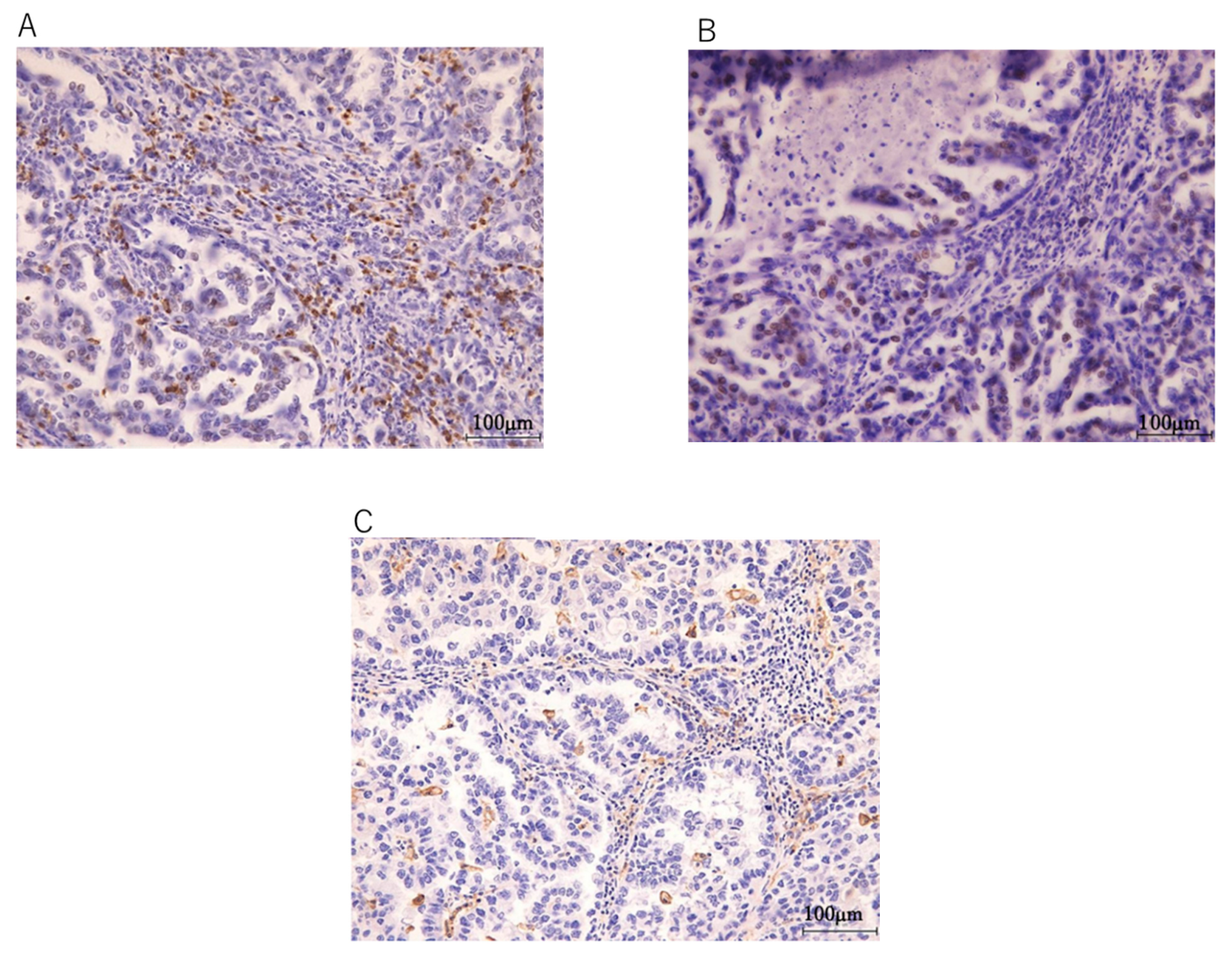
2.3. Immunohistochemical Analysis
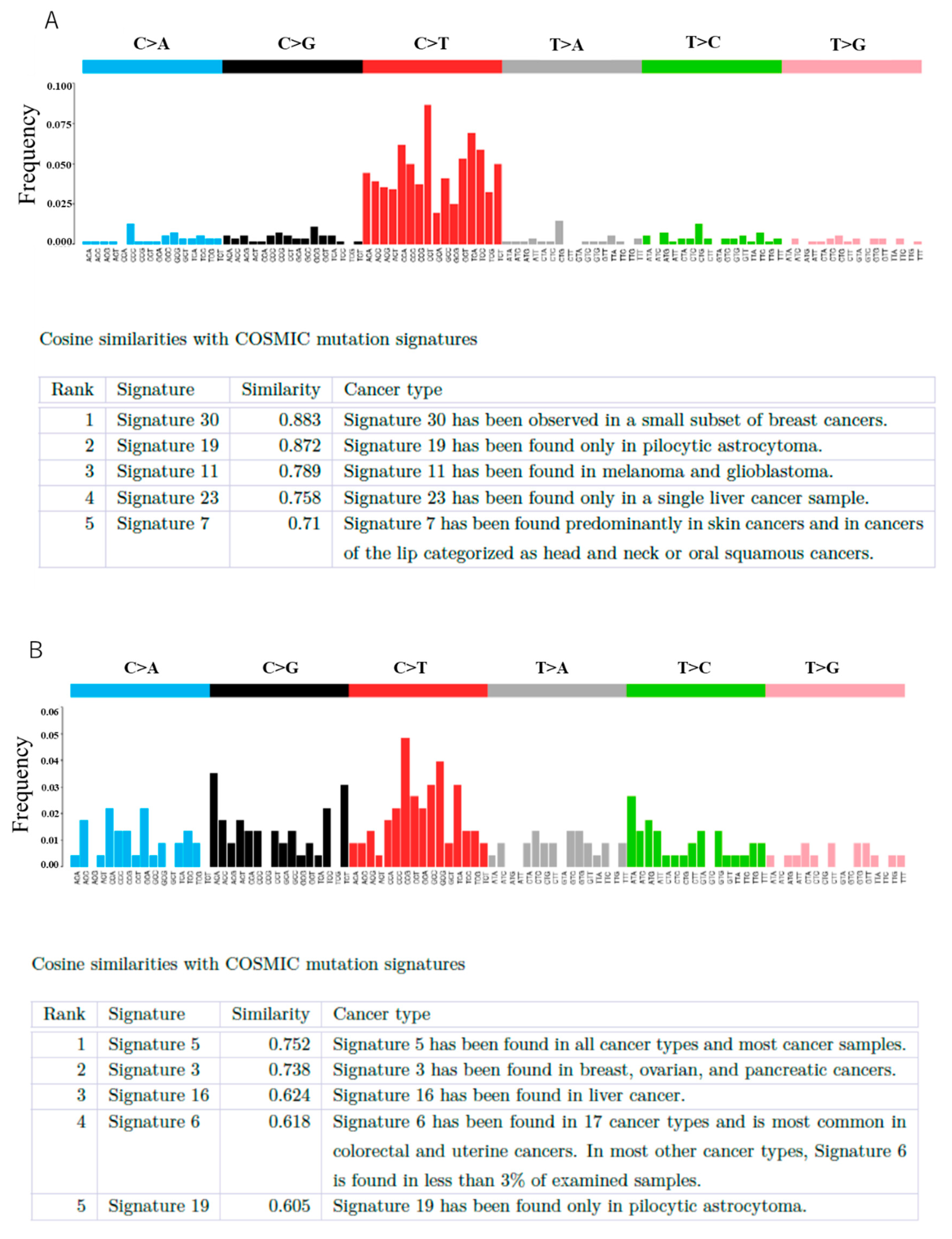
3. Results
4. Discussion
5. Conclusions
Supplementary Materials
Author Contributions
Funding
Institutional Review Board Statement
Informed Consent Statement
Data Availability Statement
Conflicts of Interest
Abbreviations
| TAH | Total abdominal hysterectomy |
| BSO | Bilateral salping oohprectomy |
| TLH | Total laparoscopic hysterectomy |
| LLAR | Laparoscopic low anterior resection |
| TC | Taxan + carboplatin |
| PLD | Doxorubicin hydrochloride liposome |
| DOD | Died of disease |
| AWD | Alive without disease |
| VAF | Variant allele frequency |
| CN | Copy number |
References
- Giudice, L.C. Clinical practice. Endometriosis. N. Engl. J. Med. 2010, 362, 2389–2398. [Google Scholar] [CrossRef]
- Shigeru, K.; Morio, S.; Tatsuya, G.; Naoki, A.; Masayuki, S.; Takahisa, S.; Kenji, T.; Shigetaka, Y.; Shiro, U. Endometrioid adenocarcinoma arising from endometriosis of the rectosigmoid. Dig. Endosc. 2010, 22, 59–63. [Google Scholar]
- Wei, J.J.; William, J.; Bulun, S. Endometriosis and ovarian cancer: A review of clinical, pathologic, and molecular aspects. Int. J. Gynecol. Pathol. 2011, 30, 553–568. [Google Scholar] [CrossRef] [Green Version]
- Kobayashi, H.; Sumimoto, K.; Moniwa, N.; Imai, M.; Takakura, K.; Kuromaki, T.; Morioka, E.; Arisawa, K.; Terao, T. Risk of developing ovarian cancer among women with ovarian endometrioma: A cohort study in Shizuoka, Japan. Int. J. Gynecol. Cancer 2007, 17, 37–43. [Google Scholar] [CrossRef]
- Heaps, J.M.; Nieberg, R.K.; Berek, J.S. Malignant neoplasms arising in endometriosis. Obstet. Gynecol. 1990, 75, 1023–1028. [Google Scholar] [CrossRef]
- Ishikawa, M.; Nakayama, K.; Nakamura, K.; Ono, R.; Sanuki, K.; Yamashita, H.; Ishibashi, T.; Minamoto, T.; Iida, K.; Razia, S.; et al. Affinity-purified DNA-based mutation profiles of endometriosis-related ovarian neoplasms in Japanese patients. Oncotarget 2018, 9, 14754–14763. [Google Scholar] [CrossRef] [PubMed]
- Itamochi, H.; Oishi, T.; Oumi, N.; Takeuchi, S.; Yoshihara, K.; Mikami, M.; Yaegashi, N.; Terao, Y.; Takehara, K.; Ushijima, K.; et al. Whole-genome sequencing revealed novel prognostic biomarkers and promising targets for therapy of ovarian clear cell carcinoma. Br. J. Cancer 2017, 117, 717–724. [Google Scholar] [CrossRef]
- Murakami, R.; Matsumura, N.; Brown, J.B.; Higasa, K.; Tsutsumi, T.; Kamada, M.; Abou-Taleb, H.; Hosoe, Y.; Kitamura, S.; Yamaguchi, K.; et al. Exome sequencing landscape analysis in ovarian clear cell carcinoma shed light on key chromosomal regions and mutation gene networks. Am. J. Pathol. 2017, 187, 2246–2258. [Google Scholar] [CrossRef] [Green Version]
- Moga, M.A.; Bălan, A.; Dimienescu, O.G.; Burtea, V.; Dragomir, R.M.; Anastasiu, C.V. Circulating miRNAs as biomarkers for endometriosis and endometriosis-related ovarian cancer—An overview. J. Clin. Med. 2019, 8, 735. [Google Scholar] [CrossRef] [PubMed] [Green Version]
- Judd, H.L.; Judd, G.E.; Lucas, W.E.; Yen, S.S. Endocrine function of the postmenopausal ovary: Concentration of androgens and estrogens in ovarian and peripheral vein blood. J. Clin. Endocrinol. Metab. Actions 1974, 39, 1020–1024. [Google Scholar] [CrossRef]
- Mandai, M.; Osuga, Y.; Hirata, T.; Enomoto, T.; Nakai, H.; Honda, R.; Taniguchi, F.; Katabuchi, H. Cancers associated with extraovarian endometriosis at less common/rare sites: A nationwide survey in Japan. J. Obstet. Gynaecol. Res. 2020, 46, 917–923. [Google Scholar] [CrossRef] [PubMed]
- Nakamura, K.; Aimono, E.; Tanishima, S.; Imai, M.; Nagatsuma, A.K.; Hayashi, H.; Yoshimura, Y.; Nakayama, K.; Kyo, S.; Nishihara, H. Intratumoral genomic heterogeneity may hinder precision medicine strategies in patients with serous ovarian carcinoma. Diagnostics 2020, 10, 200. [Google Scholar] [CrossRef] [Green Version]
- Ishikawa, M.; Nakayama, K.; Nakamura, K.; Yamashita, H.; Ishibashi, T.; Minamoto, T.; Iida, K.; Razia, S.; Ishikawa, N.; Nakayama, S.; et al. High PD-1 expression level is associated with an unfavorable prognosis in patients with cervical adenocarcinoma. Arch. Gynecol. Obstet. 2020, 302, 209–218. [Google Scholar] [CrossRef]
- Slavin, R.E.; Krum, R.; Van Dinh, T. Endometriosis-associated intestinal tumors: A clinical and pathological study of 6 cases with a review of the literature. Hum. Pathol. 2000, 31, 456–463. [Google Scholar] [CrossRef]
- Li, N.; Zhou, W.; Zhao, L.; Zhou, J. Endometriosis-associated recto-sigmoid cancer: A case report. BMC Cancer 2018, 18, 905. [Google Scholar] [CrossRef]
- Okimura, H.; Tatsumi, H.; Ito, F.; Yamashita, S.; Kokabu, T.; Kitawaki, J.; Okimura, H. Endometrioid carcinoma arising from diaphragmatic endometriosis treated with laparoscopy: A case report. J. Obstet. Gynaecol. Res. 2018, 44, 972–977. [Google Scholar] [CrossRef] [PubMed]
- Yazawa, H.; Imaizumi, K.; Kato, A.; Takiguchi, K.; Yazawa, H. Extragonadal giant endometrial cyst with endometrioid borderline tumor. Gynecol. Minim. Invasive Ther. 2019, 8, 179–184. [Google Scholar] [CrossRef] [PubMed]
- Tsuruga, T.; Hirata, T.; Akiyama, I.; Matsumoto, Y.; Oda, K.; Fujii, T.; Osuga, Y. Mixed endometrioid and clear cell carcinoma arising from laparoscopic trocar site endometriosis. J. Obstet. Gynaecol. Res. 2019, 45, 1613–1618. [Google Scholar] [CrossRef]
- Poon, C.; Rome, R. Malignant extra-ovarian endometriosis: A case series of ten patients and review of the literature. Aust. N. Z. J. Obstet. Gynaecol. 2020, 60, 585–591. [Google Scholar] [CrossRef]
- Angiolo, G.; Zannoni, G.F. Endometriosis-associated extraovarian malignancies: A challenging question for the clinician and the pathologist. Anticancer Res. 2020, 40, 2429–2438. [Google Scholar]
- Efthymiou, C.A. Endometriosis-associated intestinal tumours: A consequence of long-term unopposed oestrogen? Ann. R. Coll. Surg. Engl. 2009, 91, 259–260. [Google Scholar] [CrossRef]
- Heidemann, L.N.; Hartwell, D.; Heidemann, C.H.; Jochumsen, K.M. The relation between endometriosis and ovarian cancer: A review. Acta Obstet. Gynecol. Scand. 2014, 93, 20–31. [Google Scholar] [CrossRef]
- Brinton, L.A.; Gridley, G.; Persson, I.; Baron, J.; Bergqvist, A. Cancer risk after a hospital discharge diagnosis of endometriosis. Am. J. Obstet. Gynecol. 1997, 176, 572–579. [Google Scholar] [CrossRef]
- Bala, P.; Singh, A.K.; Kavadipula, P.; Kotapalli, V.; Sabarinathan, R.; Bashyam, M.D. Exome sequencing identifies ARID2 as a novel tumor suppressor in early-onset sporadic rectal cancer. Oncogene 2020, 40, 863–874. [Google Scholar] [CrossRef] [PubMed]
- Tomasetti, C.; Marchionni, L.; Nowak, M.A.; Parmigiani, G.; Vogelstein, B. Only three driver gene mutations are required for the development of lung and colorectal cancers. Proc. Natl. Acad. Sci. USA 2015, 112, 118–123. [Google Scholar] [CrossRef] [Green Version]
- Vogelstein, B.; Kinzler, K.W. The path to cancer –Three strikes and you’re out. N. Engl. J. Med. 2015, 373, 1895–1898. [Google Scholar] [CrossRef]
- Suda, K.; Nakaoka, H.; Yoshihara, K.; Ishiguro, T.; Tamura, R.; Mori, Y.; Yamawaki, K.; Adachi, S.; Takahashi, T.; Kase, H.; et al. Clonal expansion and diversification of cancer-associated mutations in endometriosis and normal endometrium. Cell Rep. 2018, 24, 1777–1789. [Google Scholar] [CrossRef] [Green Version]
- Suda, K.; Nakaoka, H.; Yoshihara, K.; Ishiguro, T.; Adachi, S.; Kase, H.; Motoyama, T.; Inoue, I.; Enomoto, T. Different mutation profiles between epithelium and stroma in endometriosis and normal endometrium. Hum. Reprod. 2019, 34, 1899–1905. [Google Scholar] [CrossRef]
- Suda, K.; Cruz Diaz, L.A.; Yoshihara, K.; Nakaoka, H.; Yachida, N.; Motoyama, T.; Inoue, I.; Enomoto, T. Clonal lineage from normal endometrium to ovarian clear cell carcinoma through ovarian endometriosis. Cancer Sci. 2020, 111, 3000–3009. [Google Scholar] [CrossRef]
- Anglesio, M.S.; Papadopoulos, N.; Ayhan, A.; Nazeran, T.M.; Noë, M.; Horlings, H.M.; Lum, A.; Jones, S.; Senz, J.; Seckin, T.; et al. Cancer-associated mutations in endometriosis without cancer. N. Engl. J. Med. 2017, 376, 1835–1848. [Google Scholar] [CrossRef] [Green Version]
- Lac, V.; Verhoef, L.; Aguirre-Hernandez, R.; Nazeran, T.M.; Tessier-Cloutier, B.; Praetorius, T.; Orr, N.L.; Noga, H.; Lum, A.; Khattra, J.; et al. Iatrogenic endometriosis harbors somatic cancer-driver mutations. Hum. Reprod. 2019, 34, 69–78. [Google Scholar] [CrossRef]
- Lac, V.; Nazeran, T.M.; Tessier-Cloutier, B.; Aguirre-Hernandez, R.; Albert, A.; Lum, A.; Khattra, J.; Praetorius, T.; Mason, M.; Chiu, D.; et al. Oncogenic mutations in histologically normal endometrium: The new normal? J. Pathol. 2019, 249, 173–181. [Google Scholar] [CrossRef]
- Moore, L.; Leongamornlert, D.; Coorens, T.H.; Sanders, M.A.; Ellis, P.; Dentro, S.C.; Dawson, K.J.; Butler, T.; Rahbari, R.; Mitchell, T.J.; et al. The mutational landscape of normal human endometrial epithelium. Nature 2020, 580, 640–646. [Google Scholar] [CrossRef]
- Inoue, S.; Hirota, Y.; Ueno, T.; Fukui, Y.; Yoshida, E.; Hayashi, T.; Kojima, S.; Takeyama, R.; Hashimoto, T.; Kiyono, T.; et al. Uterine adenomyosis is an oligoclonal disorder associated with KRAS mutations. Nat. Commun. 2019, 10, 5785. [Google Scholar] [CrossRef] [PubMed]
- Inoue, S.; Yoshida, E.; Fukui, Y.; Ueno, T.; Kawazu, M.; Takeyama, R.; Ikemura, M.; Osuga, Y.; Terao, Y.; Hirota, Y.; et al. KRAS mutations in uterine endometrium are associated with gravidity and parity. Cell Death Dis. 2020, 11, 347. [Google Scholar] [CrossRef]
- Kondo, E.; Maki, S.; Nii, M.; Yoshida, K.; Tabata, T.; Ikeda, T. Long-term survival of a patient with malignant transformation of extragonadal endometriosis treated solely with chemotherapy: A case report. J. Obstet. Gynaecol. Res. 2018, 44, 2186–2189. [Google Scholar] [CrossRef]
- Osaku, D.; Taniguchi, F.; Moriyama, M.; Sato, S.; Oishi, T.; Harada, T. Retroperitoneal endometrioid carcinoma arising from ureteral endometriosis. Case Rep. Obstet. Gynecol. 2019, 2019, 9273858. [Google Scholar] [CrossRef]
- Nakamura, K.; Aimono, E.; Tanishima, S.; Nomura, H.; Imai, M.; Hayashi, H.; Nishihara, H. Genetic profiling of patients with adenoid cystic carcinoma of the Bartholin’s glands reveals potential new routes for targeted therapies: A case report. Diagn. Pathol. 2020, 15, 64. [Google Scholar] [CrossRef]
- Hongo, H.; Kosaka, T.; Aimono, E.; Nishihara, H.; Oya, M.; Hongo, H. Aggressive prostate cancer with somatic loss of the homologous recombination repair gene FANCA: A case report. Diagn. Pathol. 2020, 15, 5. [Google Scholar] [CrossRef] [Green Version]
- Musacchio, L.; Boccia, S.M.; Caruso, G.; Santangelo, G.; Fischetti, M.; Tomao, F.; Perniola, G.; Palaia, I.; Muzii, L.; Pignata, S.; et al. Immune checkpoint inhibitors: A promising choice for endometrial cancer patients? J. Clin. Med. 2020, 9, 1721. [Google Scholar] [CrossRef]
- Yamashita, H.; Nakayama, K.; Ishikawa, M.; Nakamura, K.; Ishibashi, T.; Sanuki, K.; Ono, R.; Sasamori, H.; Minamoto, T.; Iida, K.; et al. Microsatellite instability is a biomarker for immune checkpoint inhibitors in endometrial cancer. Oncotarget 2017, 9, 5652–5664. [Google Scholar] [CrossRef] [Green Version]
- Choi, E.; Chang, M.S.; Byeon, S.J.; Jin, H.; Jung, K.C.; Kim, H.; Lee, K.L.; Kim, W.; Park, J.H.; Kim, K.H.; et al. Prognostic perspectives of PD-L1 combined with tumor-infiltrating lymphocytes, Epstein-Barr virus, and microsatellite instability in gastric carcinomas. Diagn. Pathol. 2020, 15, 69. [Google Scholar] [CrossRef] [PubMed]


| Case No. | Age | Parity | Hormone Therapy | Location | Surgical Operation | Chemotherapy | Histology | Survival Time | Prognosis |
|---|---|---|---|---|---|---|---|---|---|
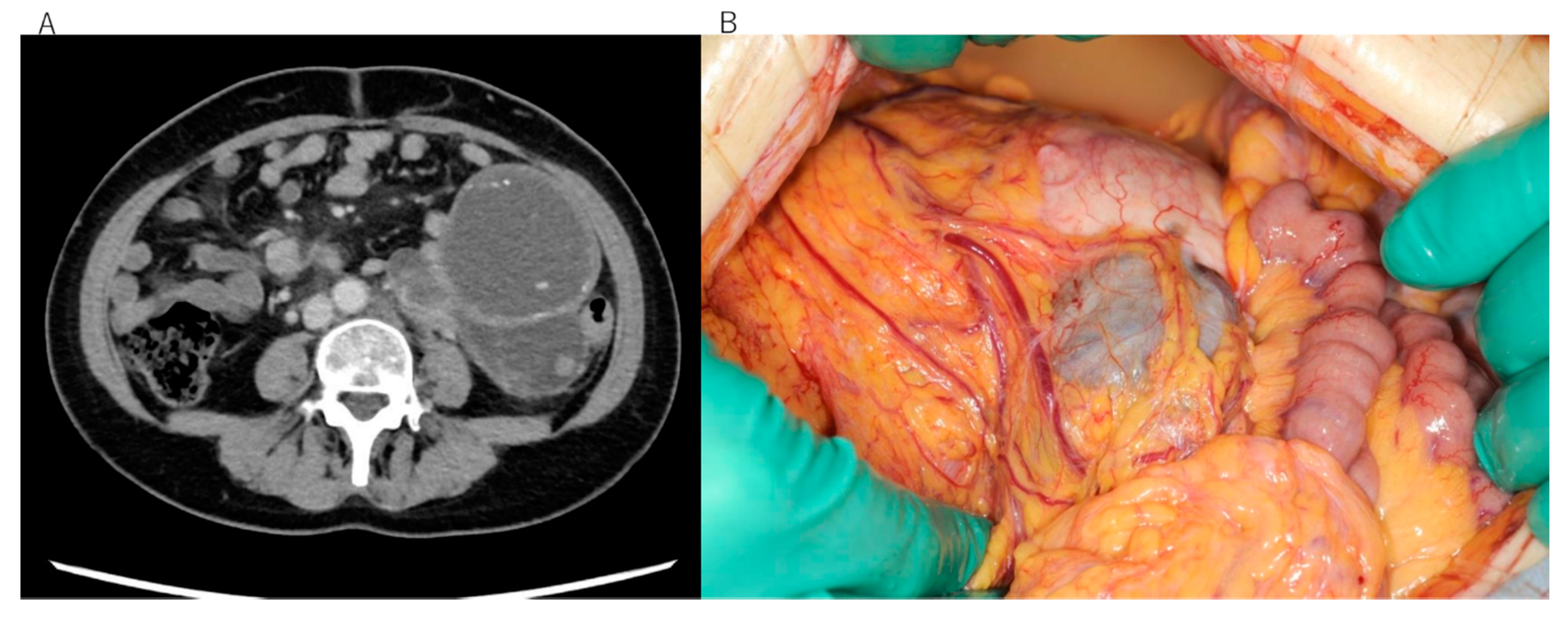
| 1 | 52 | 0 | None | Descending colon | TAH+BSO+ Omentectomy +left hemicolectomy | TC 9course, PLD +Bev 3 course | Clear cell carcinoma | 15 months | DOD |
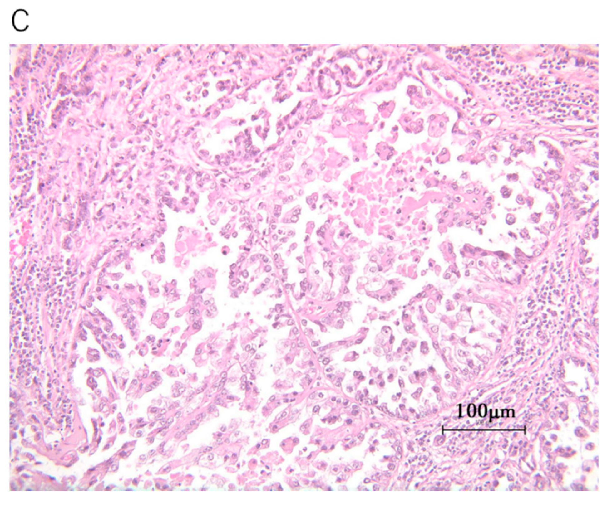
| 2 | 68 | 2 | None | Rectum | TLH+BSO+LLAR | TC 3course | Undifferentiated carcinoma | 6 months | AWD |
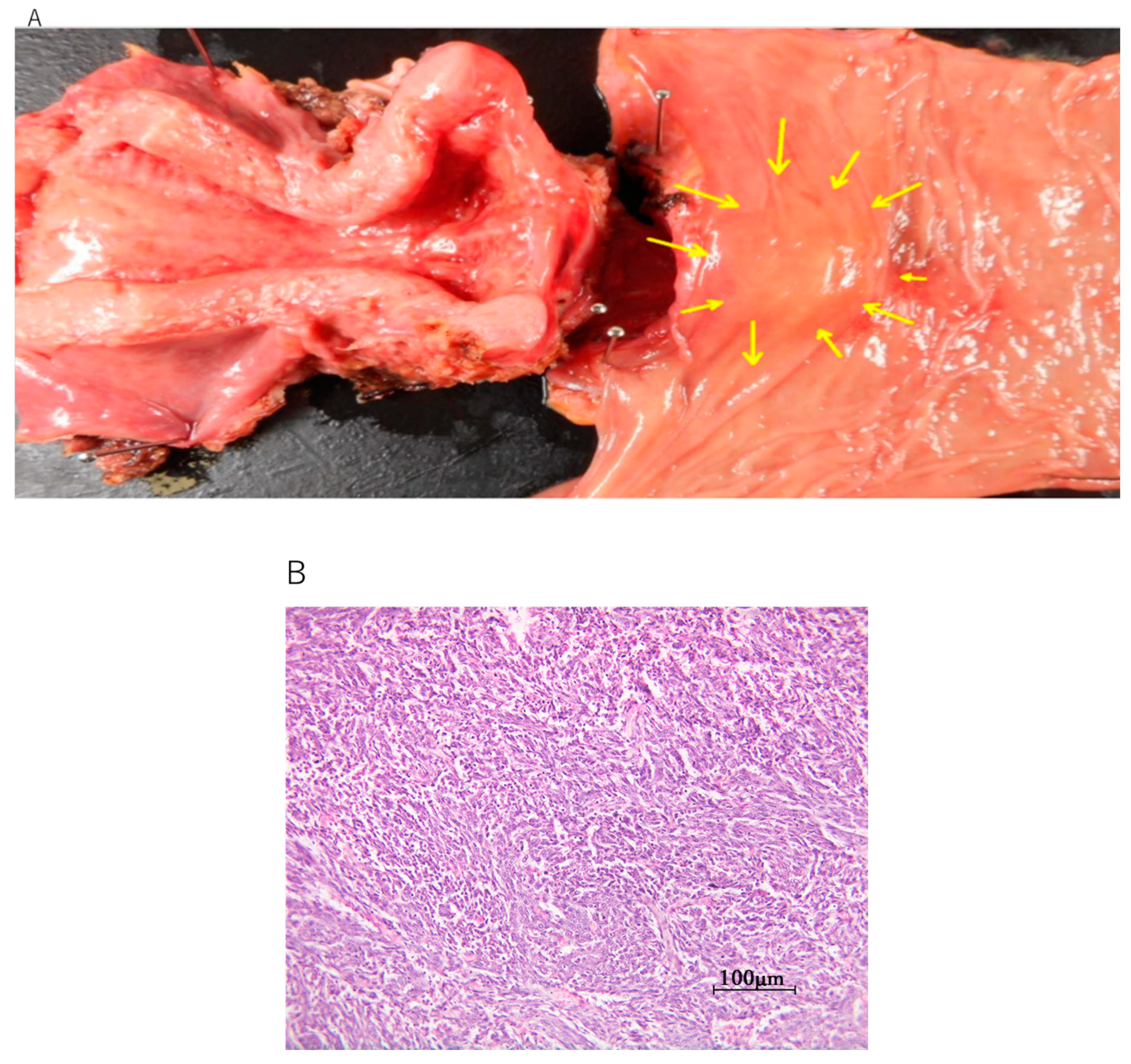
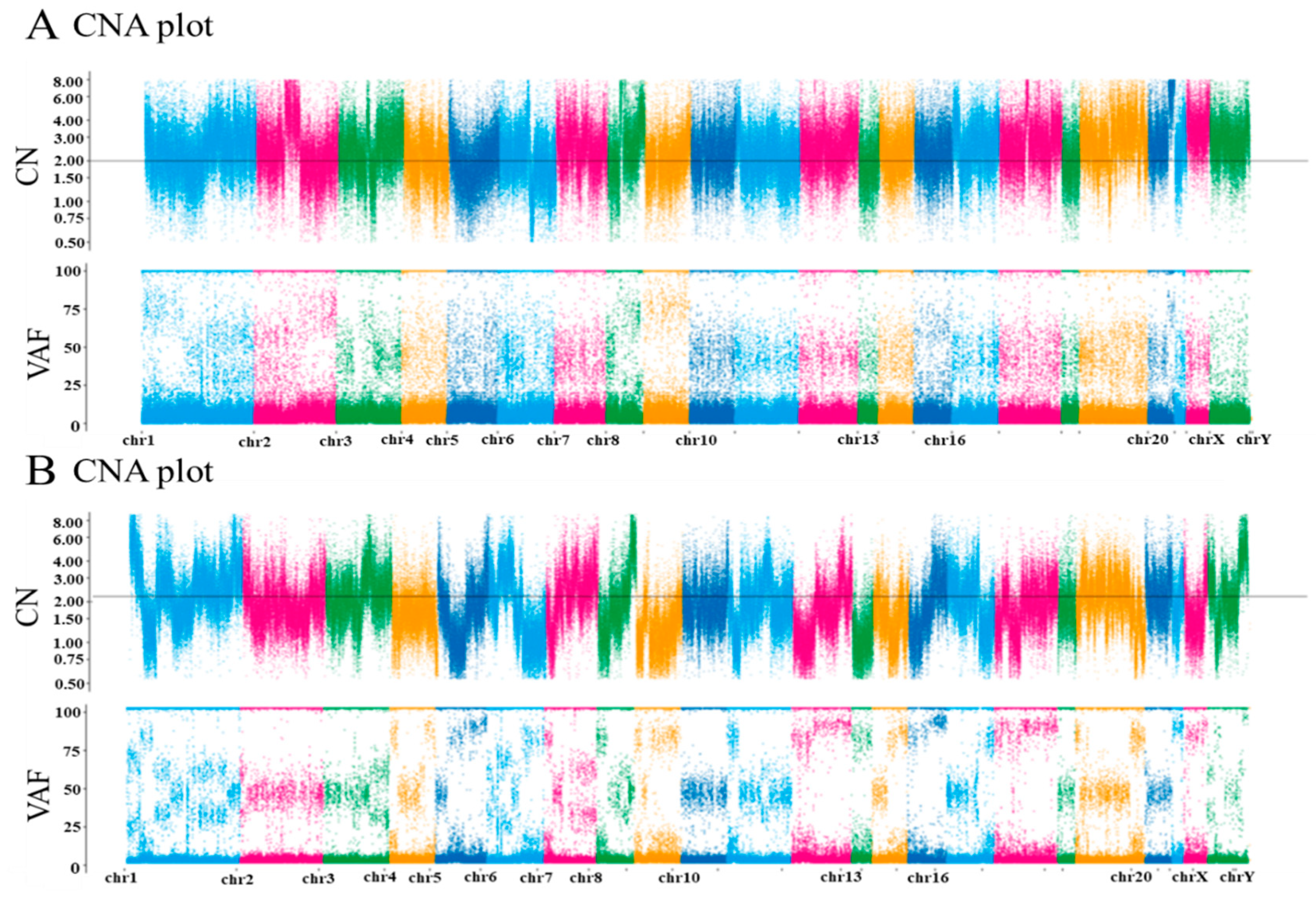
| Case No. | Actionable Gene Variants | Variant Allele Frequency (%) | Copy Number |
|---|---|---|---|
| 1 | ARID1A M835Wfs*24 | 51.3 | 2 |
| PTEN D19Rfs*26 | 32.5 | 2.3 | |
| 2 | p53 R259fs*6 | 76.6 | 1.9 |
Publisher’s Note: MDPI stays neutral with regard to jurisdictional claims in published maps and institutional affiliations. |
© 2021 by the authors. Licensee MDPI, Basel, Switzerland. This article is an open access article distributed under the terms and conditions of the Creative Commons Attribution (CC BY) license (http://creativecommons.org/licenses/by/4.0/).
Share and Cite
Kurose, S.; Nakayama, K.; Razia, S.; Ishikawa, M.; Ishibashi, T.; Yamashita, H.; Sato, S.; Sakiyama, A.; Yoshioka, S.; Kobayashi, M.; et al. Whole-Exome Sequencing of Rare Site Endometriosis-Associated Cancer. Diseases 2021, 9, 14. https://0-doi-org.brum.beds.ac.uk/10.3390/diseases9010014
Kurose S, Nakayama K, Razia S, Ishikawa M, Ishibashi T, Yamashita H, Sato S, Sakiyama A, Yoshioka S, Kobayashi M, et al. Whole-Exome Sequencing of Rare Site Endometriosis-Associated Cancer. Diseases. 2021; 9(1):14. https://0-doi-org.brum.beds.ac.uk/10.3390/diseases9010014
Chicago/Turabian StyleKurose, Sonomi, Kentaro Nakayama, Sultana Razia, Masako Ishikawa, Tomoka Ishibashi, Hitomi Yamashita, Seiya Sato, Asuka Sakiyama, Shinya Yoshioka, Misa Kobayashi, and et al. 2021. "Whole-Exome Sequencing of Rare Site Endometriosis-Associated Cancer" Diseases 9, no. 1: 14. https://0-doi-org.brum.beds.ac.uk/10.3390/diseases9010014