Article

Clinical and Radiological Outcome of Titanium Implants in Clinical Practice: A 5 Year, Prospective, Multicenter Case Series

Department for Cranio-Maxillofacial Surgery, University Hospital Basel, Spitalstrasse 21, Basel CH-4031, Switzerland
*
Author to whom correspondence should be addressed.
These authors contributed equally to this work.
Submission received: 12 September 2014 / Revised: 3 November 2014 / Accepted: 12 November 2014 / Published: 20 November 2014

Abstract

:
The objective of this prospective clinical case series was to confirm the 5-year clinical and radiographic performance of titanium implants occlusally loaded after 12 weeks. Periapical X-rays for quantitative bone level assessment were taken immediately post-surgery, after loading, 6 and 12 months, and 5 years after loading. Pocket depth (PD) and clinical attachment level (CAL) were measured. One-hundred and fourteen implants were inserted in 71 consecutive patients; 111 (97%) implants/69 (97%) patients were followed for 5 years. One implant was lost before loading (acute infection of unknown origin). Another patient developed peri-implantitis in the healing phase, and underwent successful surgical revision. Mesial median bone level at 6 and 12 months, and 5 years was 1.2 (SD ± 0.7), 1.3 (SD ± 0.7) and 1.1 (SD ± 0.6) mm; distal values were: 1.1 (SD ± 0.7), 1.4 (SD ± 0.7) and 1.2 (SD ± 0.6) mm; below implant shoulder. Stable CAL (1.2–1.6 mm) and PD (2.0–3.0 mm) were found. After 5 years no implant was lost. Three crowns and one abutment screw were defective and replaced. Implant survival (1 failure/114) as well as minimal peri-implant bone loss confirmed the favorable outcome of the used implant line. Five years post-loading, crestal bone stabilized slightly below the machined/rough border.

1. Introduction

Dental implants manufactured from pure titanium with rough endosseous surfaces are currently the gold standard for tooth replacement. In the past 30 years, replacement of missing teeth with implant-supported restorations has become a widely-accepted treatment modality for fully- or partially-edentulous patients. Clinical results with up to 10 years follow-up have demonstrated excellent survival and success rates using titanium implants [1]. Initially, implants with smooth “machined” endosseous surfaces were inserted and left to integrate into surrounding bone for some 3–6 months before occlusal loading. Today, implants with rough surfaces are standard: Increased roughness leads to a larger surface area and an ensuing higher potential for bone contact [2]. Additionally, improved osteoconductive properties stimulate bone formation near the implant surface, and result in accelerated osseointegration, thereby allowing for shorter healing times: from the classic 3–6 months, down to functional loading within 6 weeks after implant placement [3]. Several titanium implant brands are currently available, and vary considerably in their geometry, macrodesign and roughness. The tested implant line covers the entire range of indications, and is suitable for sub- as well as trans-gingival healing. Identical platforms and lengths allow easy selection and preparation of prosthetic parts. Informal reports from users in general practice have suggested their excellent survival rate and minimal peri-implant bone loss. These positive results are based on the design of the implant and the osteoconductive properties of the implant surface manufactured including sandblasting and thermic acid etching that promotes excellent implant-to-bone contact [4]. It was the objective of this prospective, observational study to investigate whether this clinical experience holds true under routine practice conditions. All patients amenable to implant supported rehabilitation were included i.e., no study-specific inclusion or exclusion criteria have been defined. The results of 114 evaluable implant reports inserted in 71 consecutive patients (from five general practices) are presented. The prospective assessment of peri-implant tissues from a clinical and radiological point of view is shown.

2. Materials and Methods

2.1. Ethical Conduct

The Investigations have been carried out following the rules of the Declaration of Helsinki (current revision). Ethics Committee approval was not sought for as the tested products are commercially available (CE certified). All treatments have been within the approved indication range i.e., the conducted cases series did not include any research aspect.

2.2. Materials and Methods

Between February 2006 and January 2009, 71 partially edentulous patients were recruited from five general practices in Switzerland. The first author acted as the coordinating investigator. Implant placement had to be the treatment of choice over conventional fixed or removable prosthetic treatments. All patients were asked to provide an informed consent to participate in the study so that their data could be used in an anonymized form for publication purposes. They were free to withdraw at any time, without any disadvantage to their further treatment. Patients were recruited consecutively from the general population when tooth replacement appeared to be the treatment of choice. No specific selection criteria were defined to ensure that the patient population was representative of that found in general practice. The participating dentists had been asked to report the cases immediately after surgery to avoid post-hoc case selection, i.e. positive or negative under-reporting. Reports were to be collected only for patients suitable for treatment with the ELEMENT® implant line (endosseous diameters: 3.5, 4.0, 4.5, and 5 mm; lengths: 8.0, 9.5, 11.0, 12.5, and 14 mm; Thommen Medical AG, Grenchen, Switzerland), according to the approved indication range (package insert).
Pre-surgical evaluation included assessment of general oral health, consisting of clinical and radiological documentation. Patients having risk factors (smoking and previously treated periodontitis) were not excluded from the study, thereby rendering the result of the study better applicable to the general patient population. It was intended to exclude implants which would not have achieved primary stability (none reported). Before implant insertion, the patient’s risk factors were recorded, together with periodontitis pre-treatment, if applicable, and planned implant position. Prior to surgery, patients used a chlorhexidine (0.2%) mouthwash for at least one-half minute. Previous and simultaneous implant site augmentation and bone quality (I–IV clinical assessment), along with the implant healing procedure (two stage, submerged versus one stage, non-submerged), was recorded after surgery. In selected cases, soft tissue augmentation using connective tissue graft harvested from the palatal mucosa, was performed at least two to three months prior to implant surgery. Insertion was performed into fully healed alveolar ridges, according to the manufacturer’s recommendations. Together with implant placement, bony augmentation procedure using osteoconductive material covered by collagen membrane was added if necessary. At the implantation, insertion torque force was recorded as an indicator of primary implant stability [5]. This was either a reading from the implant machine or estimated by the MONO torque ratchet (Thommen Medical, Grenchen, Switzerland). Implants were to be loaded 12-weeks post-surgery. The restoration type was recorded, i.e., single tooth replacement (cemented or screwed) or primary or secondary blocking of implants (fixed or removable prosthetic restoration, respectively). Data collection also included information if implant loading was only axial or extra-axial as well. Peri-implant parameters, i.e. clinical attachment level (CAL) and pocket depth (PD) were assessed at four sites around the implant [6], and evaluated 6 and 12 months, and 5 years after loading, along with the manual assessment of implant mobility. At the 1 and 5year assessments, the prosthetic reconstruction was checked and possible damage recorded. Periapical x-rays were taken post-surgery [7] and repeated immediately after functional loading, and at 6 and 12 months as well as 5 years post-implant placement using the parallel-perpendicular technique (standardized x-ray). The resulting non-digital radiographs were scanned, and the mesial and distal bone level of each individual implant was assessed using the following method: radiographs depicting dental implants were imported into ImageJ (Version1.42, National Institutes of Health, Washington, DC, USA). All images were scaled separately based on the 1.0 mm thread height of the ELEMENT® implant line. A dedicated plug-in (PeriImplant) that correct inhomogeneities among the different radiographs (light, direction, acquisition set-up, etc.) was written (AP) and used to provide objective measurements. Subsequently, the slope of each implant was calculated by selecting two points along the implant longitudinal axis on the same side of the implant (either mesial or distal; Figure 1).
Figure 1. Quantitative X-ray Evaluation: 1: The height of implant thread (vertical white bar = 1.00 mm); 2: Upper implant shoulder (horizontal white arrow); 3: The most coronal bone-to-implant contact point (horizontal red arrow).
Dentistry 02 00106 g001
Perpendicular to this slope, a line at the minimal point of contact between implant and bone was calculated (PeriImplant). To evaluate the mesial and distal bone levels, the vertical distance between the top level of implant shoulder i.e. the edge of implant-abutment connection (common landmark on all radiographs) and the previously-determined line was calculated (PeriImplant). Results from all evaluated images were imported into Excel and represented graphically using the boxplot function (Matlab® Statistics Toolbox®, MathWorks, Bern, Switzerland). All radiographs have been evaluated by a single evaluator.

3. Results

For this study, 71 patients (both genders) with a mean age of 58 years (range 20–83 years) were treated with 114 implants, according to the requirements of the case collection. To enhance the validity of this study, patients with risk factors (smokers, previous periodontitis treatment) were also included (Table 1).
Table 1. Risk factors.
Risk factorNo. of patients
Smokers (patient self-assessment)5 of 71 (7%)
Previous periodontitis treatment12 of 71 (17%)
More than 50% of the implants were inserted in combination with an augmentation procedure for treating bone defects [8] and to support soft tissue for achieving an aesthetic reconstruction [9]. Bone quality was estimated to be sufficient and the terminal insertion torque was ≤50 Ncm to avoid tissue compression. According to clinical requirements, implants were placed either submerged or non-submerged (Table 2).
Table 2. Surgical characteristics.
InsertionSubmergedNon-submergedNot known
No. of sites (%)*86 (75)12 (11)16 (14)
Simultaneous bone augmentationYesNoNot known
No. of sites (%)*67 (59)46 (40)1 (1)
Bone qualityIIIIIIIV
No. of sites (%)*2 (2)69 (61)41 (36)0 (0)
Terminal insertion torque (Ncm)<2020–3031–40>40
No. of sites (%)*16 (14)53 (46)11 (10)19 (17)
Notes: * 100% = 114 implants; not known 2 (2%); not known 15 (13%).
Implants were followed-up clinically and radiographically, up to 5 years post-loading. One implant (#31, Patient 69.5 years) was lost in the early healing phase due to infection of unknown etiology. Re-implantation was performed successfully 6 months later. Another patient developed a peri-implant infection (#21, 74.5 years old patient). The site was surgically revised, healed successfully and the implant was deemed to be a survivor at the 12-month assessment. Single tooth replacement and integration of at least two implants within a fixed reconstruction were the most common prosthetic rehabilitations (Table 3).
Table 3. Prosthetic parameters.
RestorationNo. of implants (100% = 114)
Single tooth replacement43 (38%)
Fixed bridge47 (42%)
Removable restoration15 (13%)
Not known9 (7%)
Within the observation period, three fractures of ceramic crowns and one fracture of a prosthetic screw occurred. These minor prosthetic problems were solved by minimal interventions; prosthetic parts were replaced immediately. PD and CAL were recorded 6 and 12 months and 5 years after loading (Figure 2a,b). Differences measured within the observation period did not reveal statistical significance.
Radiological changes (peri-implant bone level) post-loading, 6 and 12 months and 5 years after loading are shown in Figure 3a,b.
This prospective clinical study involved 71 patients from five different private dental practices, into whom 114 ELEMENT® (Thommen Medical AG, Grenchen, Switzerland) implants were inserted. The results presented here reflect the conditions in private practices, including patients with risk factors. All the treating dentists had many years of experience in implantology and use more than one implant system. The integration time was based on the manufacturer’s recommendations and included 12 weeks of unloaded osseointegration. During the observation period, only one implant was lost and another implant had to be revised due to a peri-implant infection, after which the further course was uneventful. Additionally, three crowns and one abutment screw were defective and were replaced without further complication.
Figure 2. Peri-implant Parameters: CAL (a) and PD (b) as determined at four different sites around implants, 6 months, 12 months and 5 years post-loading.
Dentistry 02 00106 g002
Figure 3. Radiological Parameters: Mesial (a) and distal (b) bone levels as determined by x-ray at baseline (immediately after loading), 6 months, 12 months and 5 years later.
Dentistry 02 00106 g003

4. Discussion

4.1. Risk Factors

The selection of patients reflects the general population found in private practices in Switzerland. Due to Swiss insurance regulations, most patients pay for their own implant reconstructions. Patients with risk factors were therefore not excluded from implant surgery [10]. In the presented study, 7% (5 of 71) of patients were smokers and 17% (12 of 71) had a history of previous periodontitis treatment. Smoking affects wound healing and alters microcirculation, which influences the quality and quantity of bone-to-implant contact [11]. Cessation of smoking, however, decreases the risk of implant failure to that observed in non-smokers [10]. Despite successful treatment, long-term implant success rates in patients with previous periodontitis are still lower than those of periodontaly-healthy individuals [12,13]. This indicates that implant survival rate in these patients is not only affected by local factors, but is also influenced by the innate host parameters. Only one implant was lost after 5 years of functional loading, which corresponds to an early implant failure ratio of <1% (1 of 114) [14]. However, the implant success rate has to be monitored over the long-term in order to definitely assess the failure rate in the context of the risk factors discussed above.

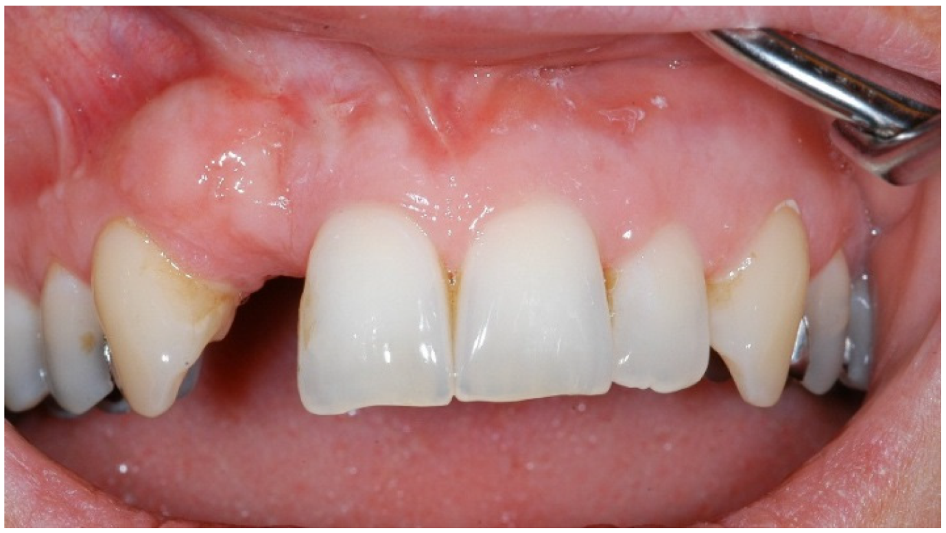
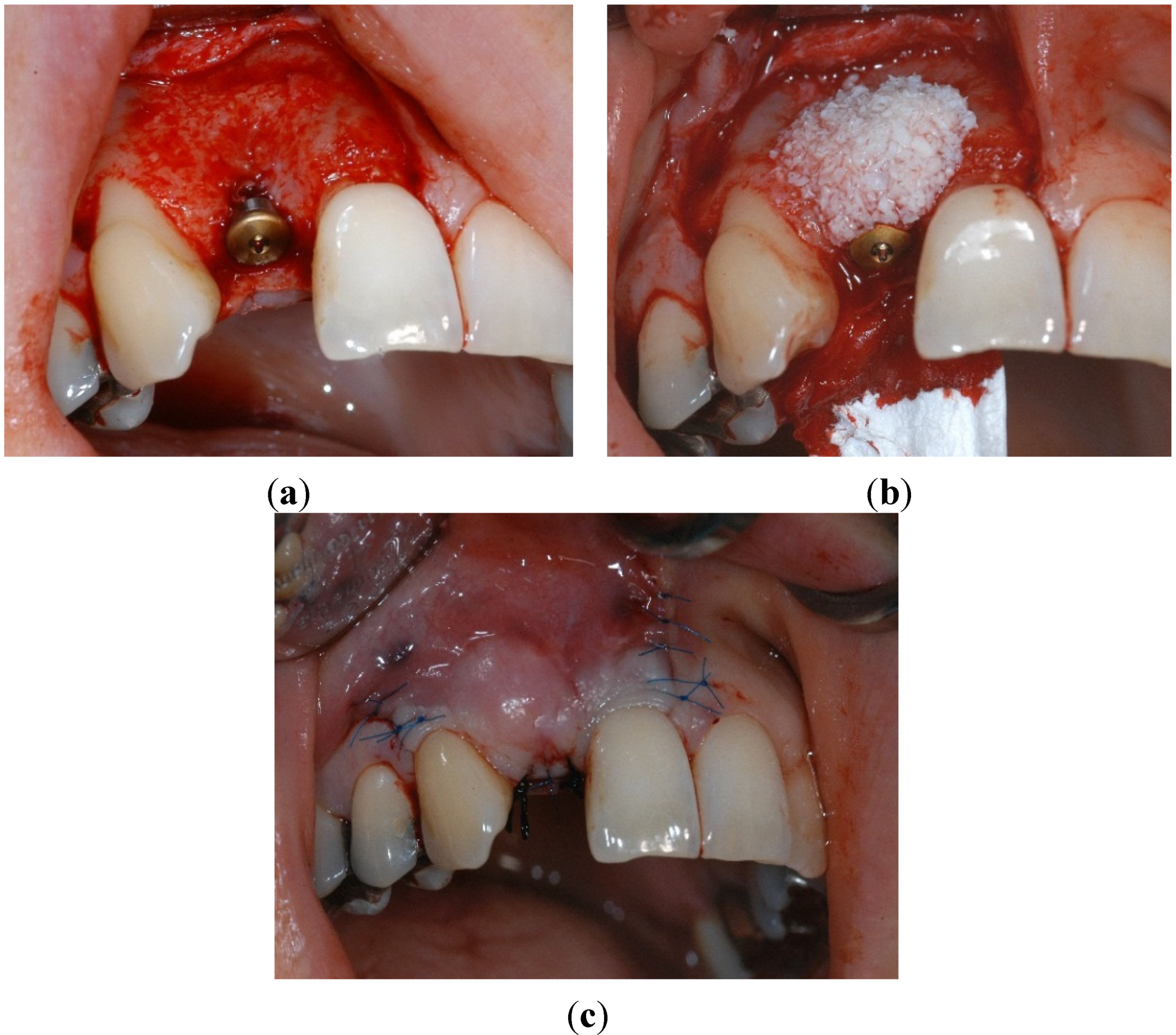
4.2. Surgical Characteristics

Implants have to be placed in such a way that a natural and harmonic emergence profile can be expected [15]. This requires submerged positioning of the fixture, often necessitating a soft tissue graft [16]. It is known that the implant success rate is not influenced by the integration modality (submerged, non-submerged) [17]. The relatively high percentage of submerged implants in our study reflected the preference of this surgical protocol by the authors. The favorable effect of soft tissue augmentation is shown in Figure 4.
Figure 4. Integrated autologous soft tissue graft before exposure of the implant.
Dentistry 02 00106 g004
Simultaneous bone augmentation is mandatory to regenerate bony defects during placement of implants. It is also indicated for supporting soft tissues with aesthetic restorations [18]. The considerable number of simultaneous hard tissue augmentations can be attributed to the preference of implant sites mentioned above (Figure 5a–c).
One of the favorable characteristics of the used implants is its self-tapping thread that provides excellent primary stability, even in bone of lower quality. A strong relationship between bone density and primary stability has been demonstrated [19], whereas terminal insertion torque is directly correlated with implant failure rate [20]. In our study, about 35% of implants were placed in bone of low density (Class III); nevertheless, sufficient primary stability of >20 Ncm, was achieved.

4.3. Prosthetic Parameters

In our study, 79% (90) of the implants were reconstructed by single crowns (support of single tooth replacement) or were incorporated within a fixed prosthesis. Only 13% (15) were used in the context of removable prosthetic reconstruction. The long-term success rate of implants in fixed prosthetic reconstructions has been demonstrated to be better than that of implants that support removable prostheses [21]. In addition, implants that support single tooth replacement improve the long-term success rate of those remaining teeth that are used as pillars for removable prosthetic reconstructions [22]. Patients in our study were selected randomly according to clinical requirements for implant reconstruction. The results shown here clearly confirm that single tooth replacement or fixed prosthetic reconstructions are to be preferred (Figure 6).
Figure 5. Vestibular bone defect after implant placement in position 12 (a); Augmentation procedure using osteoconductive ceramic material and resorbable semipermeable membrane, fixed at the palatal mucosa (b); Wound closure (c).
Dentistry 02 00106 g005
Figure 6. Final single crown restoration (cemented) three months after functional loading.
Dentistry 02 00106 g006

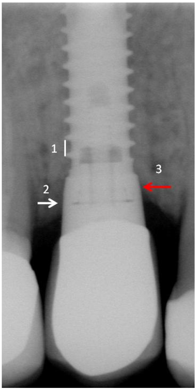
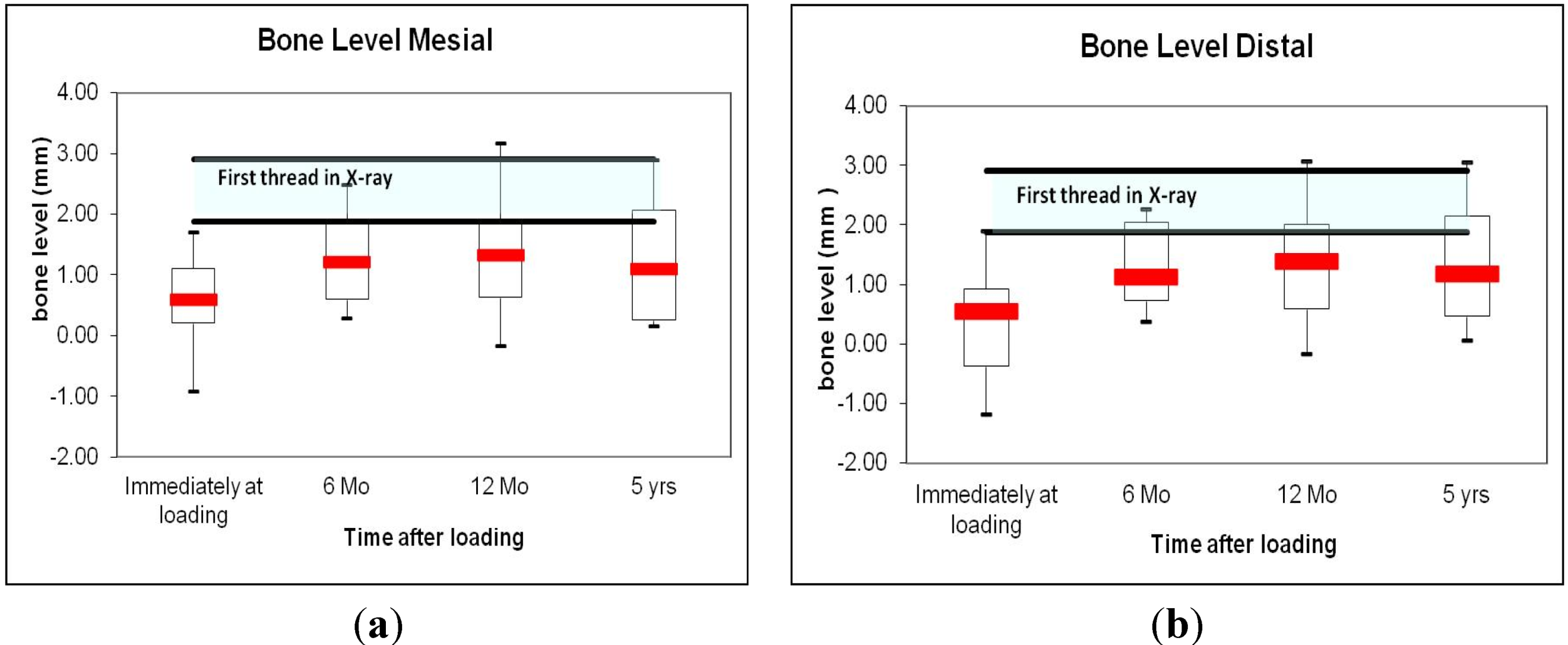
4.4. Periodontal and Radiologic Parameters

PD and CAL were measured and recorded 6 and 12 months, and 5 years after functional loading. Mesial and distal measurements were, in general, slightly higher than buccal and lingual results. No significant changes could be demonstrated over time, thereby indicating stable soft tissue conditions during the observation period.
The dimensions of the peri-implant soft tissue, i.e. the biologic width, depend on: (i) positioning of the implant in relation to the alveolar crest; and (ii) the presence of a microgap between the implant and the abutment [23]. According to the manufacturer’s protocol, the implant can be placed at bone level or slightly below bone level. This implies an increase in the biologic width when compared to natural teeth, but it nevertheless should not jeopardize the position of the marginal soft tissue for the final restoration. Radiographic measurements were performed by using the standardized parallel-perpendicular technique. The accuracy of each measurement was improved by digitalization and calibration of each single x-ray, according to the method described above [24]. In our study, radiographic measurements immediately after implant loading (Figure 3) revealed an average level of bone to implant contact of 0.59 ± 0.68 mesially, and 0.55 ± 0.81 distally, below the implant shoulder (implant-abutment connection). Given that the dimension of the smooth implant collar is exactly 1.0 mm, the smooth/rough border of the fixture was placed slightly below bone level, as recommended before study start. During functional loading, the average level of the bone to implant contact increased to 1.3 ± 0.7 and to 1.4 ± 0.7, respectively. The mean bone level after one and five years of functional loading was therefore located 0.3–0.4 mm and 0.1–0.2 mm respectively below the rough/smooth border, slightly less than that reported by Hänggi and co-workers who used a similar type of implant [25], but more than was reported by Shin and co-workers who evaluated an implant with microthreads at the collar zone [26]. In this study, the finding of minimal, statistically not significant changes in the bone to implant contact distance over time was confirmed by stable peri-implant soft tissue, represented by PD and CAL at 6 months to 5 years of function. There is an average, statistically not significant increase of PD between 12 months and 5 years. However, the peri-implant bone remained stable at the level above the first thread during this period. Some individual sites with higher measurements of PD will be re-evaluated on a regular basis. The results of this quantitative assessment further support the clinical finding that the bone to implant contact stabilizes above or at the level of the first implant thread in the implants used in this study (Figure 2 and Figure 3). Similar observation was also concluded from a prospective clinical trial conducted with edentulous and partially dentate patients after followed 5 years follow up [27].
In the treated population, most of the restorations were cemented rather than being fixed with screws (Table 3). By using a screwed abutment connection, it seems to be possible to minimize the gap between the implant and the corresponding superstructure [28]. However, according to clinical requirements, crowns and other prosthetic elements often have to be cemented, which does not allow for gaps of less than 40 μm [29]. Finally, immunohistochemical evaluation of peri-implant soft tissue of cemented or screwed abutments did not reveal significant differences in the dimensions of the microgaps, but did reveal inflammatory tissue in the presence of screw loosening [29].

5. Conclusions

Within the limitations of this study, the excellent implant survival as well as the minimal peri-implant bone loss observed confirms the favorable outcome achieved with the tested implant line. Up to five years after loading, crestal bone stabilized slightly below the smooth/rough implant border. Similar outcome was recently shown for the tested implant line in a different study performed in a university setting [30].

Acknowledgments

Implants were provided free of charge by Thommen Medical AG, Grenchen, Switzerland. No other funds have been received.

Author Contributions

All authors made a significant contribution to the article. Claude Jaquiéry acted as the Coordinating Investigator; Patients were recruited by Bernd Ilgenstein, Markus Jungo, Konrad Rüeger, Stephan Chenaux, and Kurt Jäger; Adam Papadimitropoulos is the author of PeriImplant software.

Conflicts of Interest

The authors declare no conflict of interest. Claude Jaquiéry is the Medical Reviewer of Thommen Medical AG.

References

  1. Buser, D.; Mericske-Stern, R.; Bernard, J.P.; Behneke, A.; Behneke, N.; Hirt, H.P.; Belser, U.C.; Lang, N.P. Long-term evaluation of non-submerged ITI implants. Part 1: 8-year life table analysis of a prospective multi-center study with 2359 implants. Clin. Oral Implants Res. 1997, 8, 161–172. [Google Scholar] [CrossRef] [PubMed]
  2. Buser, D.; Nydegger, T.; Oxland, T.; Cochran, D.L.; Schenk, R.K.; Hirt, H.P.; Snétivy, D.; Nolte, L.-P. Interface shear strength of titanium implants with a sandblasted and acid-etched surface: A biomechanical study in the maxilla of miniature pigs. J. Biomed. Mater. Res. 1999, 45, 75–83. [Google Scholar]
  3. Ferguson, S.J.; Broggini, N.; Wieland, M.; de Wild, M.; Rupp, F.; Geis-Gerstorfer, J.; Cochran, D.L.; Buser, D. Biomechanical evaluation of the interfacial strength of a chemically modified sandblasted and acid-etched titanium surface. J. Biomed. Mater. Res. A. 2006, 78, 291–297. [Google Scholar] [CrossRef]
  4. Langhoff, J.D.; Voelter, K.; Scharnweber, D.; Schnabelrauch, M.; Schlottig, F.; Hefti, T.; Kalchofner, K.; Nuss, K.; von Rechenberg, B. Comparison of chemically and pharmaceutically modified titanium and zirconia implant surfaces in dentistry: A study in sheep. Int. J. Oral Maxillofac. Surg. 2008, 37, 1125–1132. [Google Scholar] [CrossRef]
  5. Degidi, M.; Daprile, G.; Piattelli, A. Primary stability determination by means of insertion torque and RFA in a sample of 4,135 implants. Clin. Implant Dent. Relat. Res. 2012, 17, 501–507. [Google Scholar] [CrossRef]
  6. Engelberger, T.; Hefti, A.; Kallenberger, A.; Rateitschak, K.H. Correlations among Papilla Bleeding Index, other clinical indices and histologically determined inflammation of gingival papilla. J. Clin. Periodontol. 1983, 10, 579–589. [Google Scholar] [CrossRef] [PubMed]
  7. Whaites, E.; Brown, J. An update on dental imaging. Br. Dent. J. 1998, 185, 166–172. [Google Scholar] [CrossRef] [PubMed]
  8. Klinge, B.; Flemmig, T.F. Tissue augmentation and esthetics (Working Group 3). Clin. Oral Implants Res. 2009, 20 (Suppl 4), 166–170. [Google Scholar] [CrossRef]
  9. Paolantoni, G.; Marenzi, G.; Fusco, A.; Sammartino, G. Implant rehabilitation of central incisor: A staged approach. Implant Dent. 2007, 16, 349–355. [Google Scholar] [CrossRef] [PubMed]
  10. Johnson, G.K.; Hill, M. Cigarette smoking and the periodontal patient. J. Periodontol. 2004, 75, 196–209. [Google Scholar] [CrossRef] [PubMed]
  11. Levin, L.; Schwartz-Arad, D. The effect of cigarette smoking on dental implants and related surgery. Implant Dent. 2005, 14, 357–361. [Google Scholar] [PubMed]
  12. Mengel, R.; Behle, M.; Flores-de-Jacoby, L. Osseointegrated implants in subjects treated for generalized aggressive periodontitis: 10-year results of a prospective, long-term cohort study. J. Periodontol. 2007, 78, 2229–2237. [Google Scholar] [CrossRef] [PubMed]
  13. Karoussis, I.K.; Salvi, G.E.; Heitz-Mayfield, L.J.; Brägger, U.; Hämmerle, C.H.; Lang, N.P. Long-term implant prognosis in patients with and without a history of chronic periodontitis: A 10-year prospective cohort study of the ITI Dental Implant System. Clin. Oral Implants Res. 2003, 14, 329–339. [Google Scholar] [CrossRef]
  14. Cochran, D.L.; Buser, D.; ten Bruggenkate, C.M.; Weingart, D.; Taylor, T.M.; Bernard, J.-P.; Peters, F.; Simpson, J.P. The use of reduced healing times on ITI implants with a sandblasted and acid-etched (SLA) surface: Early results from clinical trials on ITI SLA implants. Clin. Oral Implants Res. 2002, 13, 144–153. [Google Scholar] [CrossRef]
  15. Kessler-Liechti, G.; Mericske-Stern, R. Anterior teeth esthetics with the SPI implant system. Schweiz Monatsschr. Zahnmed. 2006, 116, 275–286. [Google Scholar] [PubMed]
  16. Buser, D.; von Arx, T. Surgical procedures in partially edentulous patients with ITI implants. Clin. Oral Implants Res. 2000, 11 (Suppl 1), 83–100. [Google Scholar] [PubMed]
  17. Buser, D.; Weber, H.P.; Lang, N.P. Tissue integration of non-submerged implants. 1-year results of a prospective study with 100 ITI hollow-cylinder and hollow-screw implants. Clin. Oral Implants Res. 1990, 1, 33–40. [Google Scholar] [CrossRef] [PubMed]
  18. Buser, D.; Halbritter, S.; Hart, C.; Bornstein, M.M.; Grütter, L.; Chappuis, V.; Belser, U.C. Early implant placement with simultaneous guided bone regeneration following single-tooth extraction in the esthetic zone: 12-month results of a prospective study with 20 consecutive patients. J. Periodontol. 2009, 80, 152–162. [Google Scholar] [CrossRef] [PubMed]
  19. Turkyilmaz, I.; Aksoy, U.; McGlumphy, E.A. Two alternative surgical techniques for enhancing primary implant stability in the posterior maxilla: A clinical study including bone density, insertion torque, and resonance frequency analysis data. Clin. Implant Dent. Relat. Res. 2008, 10, 231–237. [Google Scholar] [PubMed]
  20. Ottoni, J.M.; Oliveira, Z.F.; Mansini, R.; Cabral, A.M. Correlation between placement torque and survival of single-tooth implants. Int. J. Oral Maxillofac. Implants 2005, 20, 769–776. [Google Scholar] [PubMed]
  21. Moheng, P.; Feryn, J.M. Clinical and biologic factors related to oral implant failure: A 2-year follow-up study. Implant Dent. 2005, 14, 281–288. [Google Scholar] [CrossRef] [PubMed]
  22. Priest, G. Single-tooth implants and their role in preserving remaining teeth: A 10-year survival study. Int. J. Oral Maxillofac. Implants 1999, 14, 181–188. [Google Scholar] [PubMed]
  23. Hermann, J.S.; Buser, D.; Schenk, R.K.; Higginbottom, F.L.; Cochran, D.L. Biologic width around titanium implants. A physiologically formed and stable dimension over time. Clin. Oral Implants Res. 2000, 11, 1–11. [Google Scholar] [CrossRef] [PubMed]
  24. Salvi, G.E.; Lang, N.P. Diagnostic parameters for monitoring peri-implant conditions. Int. J. Oral Maxillofac. Implants 2004, 19, 116–127. [Google Scholar] [PubMed]
  25. Hänggi, M.P.; Hänggi, D.C.; Schoolfield, J.D.; Meyer, J.; Cochran, D.L.; Hermann, J.S. Crestal bone changes around titanium implants. Part I: A retrospective radiographic evaluation in humans comparing two non-submerged implant designs with different machined collar lengths. J. Periodontol. 2005, 76, 791–802. [Google Scholar] [CrossRef] [PubMed]
  26. Shin, Y.K.; Han, C.H.; Heo, S.J.; Kim, S.; Chun, H.J. Radiographic evaluation of marginal bone level around implants with different neck designs after 1 year. Int. J. Oral Maxillofac. Implants 2006, 21, 789–794. [Google Scholar] [PubMed]
  27. Gholami, H.; Mericske-Stern, R.; Kessler-Liechti, R.; Katsoulis, J. Radiographic bone level changes of implant-supported restorations in edentulous and partially dentate patients: 5-year results. Int. J. Oral Maxillofac. Implants 2014, 29, 898–904. [Google Scholar] [CrossRef] [PubMed]
  28. Besimo, D.; Bodenschatz, V.; Guggenheim, R.; Hassell, T. Marginal fit of prefabricated crowns of the Ha-Ti implant system: An in vitro scanning electron microscopic study. Int. J. Prosthodont. 1996, 9, 87–94. [Google Scholar] [PubMed]
  29. Scarano, A.; Assenza, B.; Piattelli, M.; Iezzi, G.; Leghissa, G.C.; Quaranta, A.; Tortora, P.; Piattelli, A. A 16-year study of the microgap between 272 human titanium implants and their abutments. J. Oral Implantol. 2005, 31, 269–275. [Google Scholar] [CrossRef] [PubMed]
  30. Assenza, B.; Artese, L.; Scarano, A.; Rubini, C.; Perrotti, V.; Piattelli, M.; Thams, U.; San Roman, F.; Piccirilli, M.; Piattelli, A. Screw vs cement-implant-retained restorations: An experimental study in the beagle. Part 2. Immunohistochemical evaluation of the peri-implant tissues. J. Oral Implantol. 2006, 32, 1–7. [Google Scholar] [CrossRef] [PubMed]

