
Root Fracture and Extrusive Luxation in Primary Teeth and Their Management: A Case Report

 ,
,  , , and
Abstract
:1. Introduction
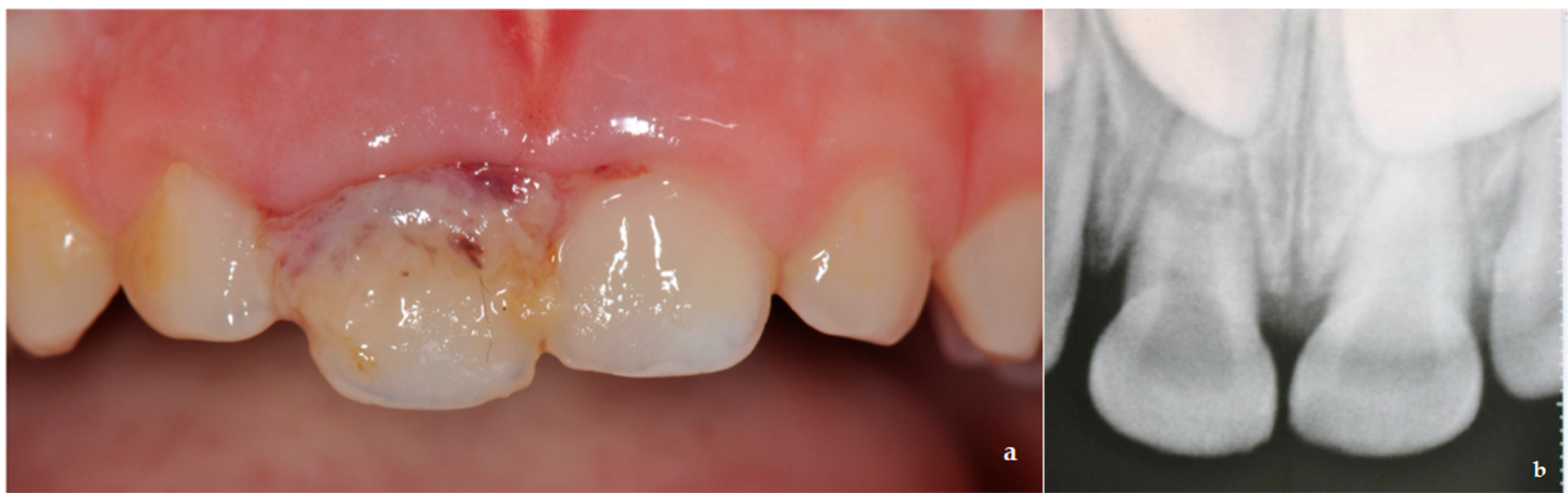
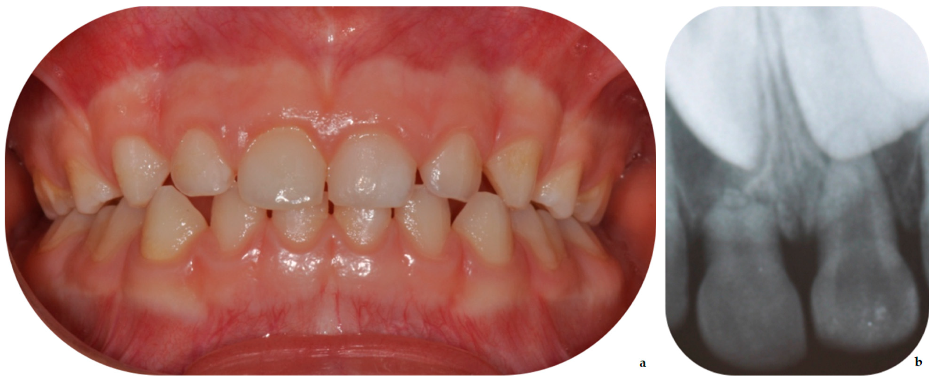
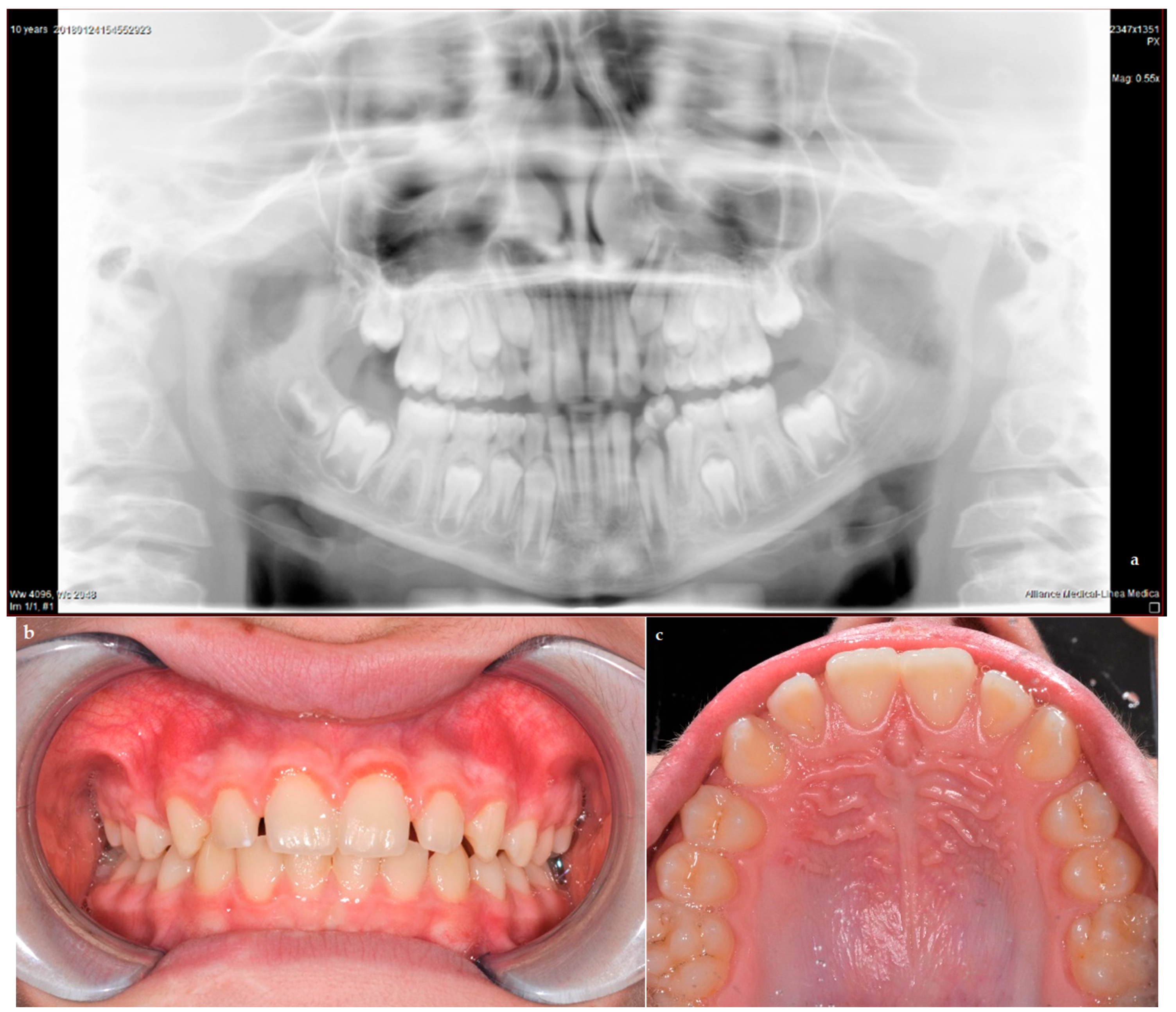
2. Case Report
3. Discussion
4. Conclusions
Author Contributions
Funding
Institutional Review Board Statement
Informed Consent Statement
Data Availability Statement
Conflicts of Interest
References
- Andreasen, J.O.; Lauridsen, E.; Daugaard-Jensen, J. Dental traumatology: An orphan in pediatric dentistry? Pediatr. Dent. 2009, 31, 153–156. [Google Scholar] [PubMed]
- Sleet, D.A. The global challenge of child injury prevention. Int. J. Environ. Res. Public Health 2018, 15, 1921. [Google Scholar] [CrossRef] [Green Version]
- Huang, B.; Marcenes, W.; Croucher, R.; Hector, M. Activities related to the occurrence of traumatic dental injuries in 15- to 18-year-olds. Dent. Traumatol. 2009, 25, 64–68. [Google Scholar] [CrossRef] [PubMed]
- Tham, R.C.; Cassell, E.; Calache, H. Traumatic orodental injuries and the development of an orodental injury surveillance system: A pilot study in Victoria, Austwralia. Dent. Traumatol. 2009, 25, 103–109. [Google Scholar] [CrossRef]
- Day, P.F.; Flores, M.T.; O’Connell, A.C.; Abbott, P.V.; Tsilingaridis, G.; Fouad, A.F.; Cohenca, N.; Lauridsen, E.; Bourguignon, C.; Hicks, L.; et al. International Association of Dental Traumatology guidelines for the management of traumatic dental injuries: 3. Injuries in the primary dentition. Dent. Traumatol. 2020, 36, 343–359. [Google Scholar] [CrossRef]
- Petti, S.; Glendor, U.; Andersson, L. World traumatic dental injury prevalence and incidence, a meta-analysis-One billion living people have had traumatic dental injuries. Dent. Traumatol. 2018, 34, 71–86. [Google Scholar] [CrossRef] [Green Version]
- Glendor, U. Epidemiology of traumatic dental injuries-A 12 year review of the literature. Dent. Traumatol. 2008, 24, 603–611. [Google Scholar] [CrossRef]
- Andersson, L.; Petti, S.; Day, P.; Kenny, K.; Glendor, U.; Andreasen, J.O. Classification, Epidemiology and Etiology. In Textbook and Color Atlas of Traumatic Injuries to the Teeth, 5th ed.; Andreasen, J.O., Andreasen, F.M., Andersson, L., Eds.; Wiley Blackwell: Copenhagen, Danmark, 2019; pp. 252–294. [Google Scholar]
- Arraj, G.P.; Rossi-Fedele, G.; Doğramacı, E.J. The association of overjet size and traumatic dental injuries—A systematic review and meta-analysis. Dent. Traumatol. 2019, 35, 217–232. [Google Scholar] [CrossRef] [Green Version]
- Levin, L.; Day, P.F.; Hicks, L.; O´Connell, A.; Fouad, A.F.; Bourguignon, C.; Abbott, P. International Association of Dental Traumatology guidelines for the management of traumatic dental injuries: General Introduction. Dent. Traumatol. 2020, 36, 309–313. [Google Scholar] [CrossRef]
- Santos, L.V.; da Hora, K.C.; Alve, A.C. Successful minimally invasive intervention in a primary central incisor after root fracture: A case report. Dent. Traumatol. 2021, 1–6. [Google Scholar] [CrossRef]
- Cully, J.L.; Zeeb, K.; Sahay, R.D.; Gosnell, E.; Morris, H.; Thikkurissy, S. Prevalence of primary teeth injuries presenting to a pediatric emergency department. Pediatr. Dent. 2019, 41, 136–139. [Google Scholar] [PubMed]
- Andreasen, J.O. Luxation of permanent teeth due to trauma A clinical and radiographic follow-up study of 189 injured teeth. Eur. J. Oral Sci. 1970, 78, 273–286. [Google Scholar] [CrossRef]
- Spinas, E.; Pipi, L.; Dettori, C. Extrusive Luxation Injuries in Young Patients: A Retrospective Study with 5-Year Follow-Up. Dent. J. 2020, 8, 136. [Google Scholar] [CrossRef]
- Flores, M.T. Traumatic injuries in the primary dentition. Dent. Traumatol. 2002, 18, 287–298. [Google Scholar] [CrossRef] [PubMed]
- Faria, L.V.; Chaves, H.G.D.S.; Silva, E.A.B.; Antunes, L.D.S.; Antunes, L.A.A. Minimally invasive treatment of an extruded deciduous tooth-Case report. Dent. Traumatol. 2019, 36, 303–306. [Google Scholar] [CrossRef]
- Antunes, L.A.A.; Lemos, H.M.; Milani, A.J.; Guimarães, L.S.; Küchler, E.C.; Antunes, L.S. Does traumatic dental injury impact oral health-related to quality of life of children and adolescents? Systematic review and meta-analysis. Int. J. Dent. Hyg. 2020, 18, 142–162. [Google Scholar] [CrossRef] [PubMed]
- Abreu, G.V.L.; Milani, A.J.; Oliveira, F.T.; Gomes, C.C.; Antunes, L.S.; Antunes, L.A.A. Dental trauma in primary dentition, its effect on permanent successors and on Oral Health-Related Quality of Life: A 4-year follow-up case report. Int. J. Burn. Trauma 2020, 10, 201–209. [Google Scholar]
- Frujeri, M.L.; Frujeri, J.A.; Bezerra, A.C.; Cortes, M.I.; Costa, E.D., Jr. Socio-economic indicators and predisposing factors associated with traumatic dental injuries in schoolchildren at Brasília, Brazil: A cross-sectional, population-based study. BMC Oral Health 2014, 14, 1–7. [Google Scholar] [CrossRef] [PubMed] [Green Version]
- Berger, T.D.; Kenny, D.J.; Casas, M.J.; Barrett, E.J.; Lawrence, H.P. Effects of severe dentoalveolar trauma on the quality-of-life of children and parents. Dent. Traumatol. 2009, 25, 462–469. [Google Scholar] [CrossRef]
- Mello-Moura, A.C.; Bonini, G.A.; Suga, S.S.; Navarro, R.S.; Wanderley, M.T. Multidisciplinary approach on rehabilitation of primary teeth traumatism repercussion on the permanent successor: 6-year follow-up case report. J. Indian Soc. Pedod. Prev. Dent. 2009, 27, 125–130. [Google Scholar] [PubMed]
- Karataş, M.S.; Sönmez, I. Developmental disturbances of a maxillary central incisor due to trauma to its predecessor: A case report. Med. Princ. Pract. 2013, 22, 590–592. [Google Scholar] [CrossRef] [Green Version]
- Arenas, M.; Barbería, E.; Lucavechi, T.; Maroto, M. Severe trauma in the primary dentition-diagnosis and treatment of sequelae in permanent dentition. Dent. Traumatol. 2006, 22, 226–230. [Google Scholar] [CrossRef]
- Sennhenn-Kirchner, S.; Jacobs, H.G. Traumatic injuries to the primary dentition and effects on the permanent successors-A clinical follow-up study. Dent. Traumatol. 2006, 22, 237–241. [Google Scholar] [CrossRef] [PubMed]
- Altun, C.; Cehreli, Z.C.; Güven, G.; Acikel, C. Traumatic intrusion of primary teeth and its effects on the permanent successors: A clinical follow-up study. Oral Surg. Oral Med. Oral Pathol. Oral Radiol. Endod. 2009, 107, 493–498. [Google Scholar] [CrossRef]
- Korolenkova, M.V.; Rakhmanova, M.S. Outcomes of traumatic dental injuries in children. Stomatologiia 2019, 98, 116–122. [Google Scholar] [CrossRef]
- Kratunova, E.; Silva, D. Pulp therapy for primary and immature permanent teeth: An overview. Gen. Dent. 2018, 66, 30–38. [Google Scholar]
- Borges, T.S.; Vargas-Ferreira, F.; Kramer, P.F.; Feldens, C.A. Impact of traumatic dental injuries on oral health-related quality of life of pre school children: A systematic review and meta-analysis. PLoS ONE 2017, 12, e0172235. [Google Scholar] [CrossRef] [Green Version]
- Magno, M.B.; Jural, L.A.; Nogueira, A.D.V.; Lenzi, M.M.; Pithon, M.M.; Maia, L.C. Impact of crown frac ture treatment on oral health-related quality of life of children, adolescents, and their families: A prospective clinical study. Int. J. Paediatr. Dent. 2019, 29, 86–93. [Google Scholar] [CrossRef] [PubMed] [Green Version]
- Cunha, R.F.; Pugliesi, D.M.; Vieira, A.E. Oral trauma in Brazilian patients aged 0–3 years. Dent. Traumatol. 2001, 17, 210–212. [Google Scholar] [CrossRef]
- Da Fidalgo, T.K.; Maia, L.C. Minimally invasive intervention of acute trauma in the primary dentition: Successful five-year follow-up. Gen. Dent. 2012, 60, 158–161. [Google Scholar]
- Ruviére, D.B.; Costa, M.M.; Cunha, R.F. Conservative management of severe intrusion in a primary tooth: A 4-year follow-up. J. Dent. Child. 2009, 76, 87–91. [Google Scholar]
- Charone, S.; Kuchler, E.C.; Costa, M.C.; Maia, L.C. A successful outcome using a minimal invasive approach to manage a severe trauma to the primary maxillary incisor in a toddler. Dent. Traumatol. 2010, 26, 294–297. [Google Scholar] [CrossRef]
- Spinas, E.; Melis, A.; Savasta, A. Therapeutic approach to intrusive luxation injuries in primary dentition. A clinical follow-up study. Eur. J. Paediatr. Dent. 2006, 7, 179–186. [Google Scholar] [PubMed]
- La Monaca, G.; Pranno, N.; Vozza, I.; Annibali, S.; Polimeni, A.; Bossù, M.; Cristalli, M.P. Sequelae in permanent teeth after traumatic injuries to primary dentition. Minerva Stomatol. 2019, 68, 332–340. [Google Scholar] [CrossRef]
- Cho, W.C.; Nam, O.H.; Kim, M.S.; Lee, H.S.; Choi, S.C. A retrospective study of traumatic dental injuries in primary dentition: Treatment outcomes of splinting. Acta Odontol. Scand. 2018, 76, 253–256. [Google Scholar] [CrossRef] [PubMed]
- Lopes, T.S.; Santin, G.C.; Marengoni, L.A.; Crispim, J.B.; Ceron, L.C.; Fracasso, M.L.C. Clinical and radiographic sequelae in primary teeth due to dental trauma. Pesqui. Bras. Odontopediatria Clín. Integr. 2019, 19, 1–10. [Google Scholar] [CrossRef]
- Santos, B.Z.; Cardoso, M.; Almeida, I.C. Pulp canal obliteration following trauma to primary incisors: A 9-year clinical study. Pediatr. Dent. 2011, 33, 399–402. [Google Scholar] [PubMed]
- Nam, O.H.; Kim, M.S.; Kim, G.T.; Choi, S.C. Atypical root resorption following root fractures in primary teeth. Quintessence Int. 2017, 48, 793–797. [Google Scholar] [CrossRef] [PubMed]


Publisher’s Note: MDPI stays neutral with regard to jurisdictional claims in published maps and institutional affiliations. |
© 2021 by the authors. Licensee MDPI, Basel, Switzerland. This article is an open access article distributed under the terms and conditions of the Creative Commons Attribution (CC BY) license (https://creativecommons.org/licenses/by/4.0/).
Share and Cite
Di Giorgio, G.; Zumbo, G.; Saccucci, M.; Luzzi, V.; Ierardo, G.; Biagi, R.; Bossù, M. Root Fracture and Extrusive Luxation in Primary Teeth and Their Management: A Case Report. Dent. J. 2021, 9, 107. https://0-doi-org.brum.beds.ac.uk/10.3390/dj9090107
Di Giorgio G, Zumbo G, Saccucci M, Luzzi V, Ierardo G, Biagi R, Bossù M. Root Fracture and Extrusive Luxation in Primary Teeth and Their Management: A Case Report. Dentistry Journal. 2021; 9(9):107. https://0-doi-org.brum.beds.ac.uk/10.3390/dj9090107
Chicago/Turabian StyleDi Giorgio, Gianni, Giulia Zumbo, Matteo Saccucci, Valeria Luzzi, Gaetano Ierardo, Roberto Biagi, and Maurizio Bossù. 2021. "Root Fracture and Extrusive Luxation in Primary Teeth and Their Management: A Case Report" Dentistry Journal 9, no. 9: 107. https://0-doi-org.brum.beds.ac.uk/10.3390/dj9090107