Review

Natural Killer Cell Mobilization in Breast and Prostate Cancer Survivors: The Implications of Altered Stress Hormones Following Acute Exercise

1
Department of Exercise and Sport Science, University of North Carolina, Chapel Hill, NC 27599, USA
2
Human Movement Science Curriculum, University of North Carolina, Chapel Hill, NC 27517, USA
3
Department of Physical Therapy Education, Elon University, Elon, NC 27244, USA
*
Author to whom correspondence should be addressed.
Submission received: 20 March 2021 / Revised: 23 April 2021 / Accepted: 7 May 2021 / Published: 19 May 2021
(This article belongs to the Special Issue Exercise Endocrinology)

Abstract

:
Natural killer (NK) cells from the innate immune system are integral to overall immunity and also in managing the tumor burden during cancer. Breast (BCa) and prostate cancer (PCa) are the most common tumors in U.S. adults. Both BCa and PCa are frequently treated with hormone suppression therapies that are associated with numerous adverse effects including direct effects on the immune system. Regular exercise is recommended for cancer survivors to reduce side effects and improve quality of life. Acute exercise is a potent stimulus for NK cells in healthy individuals with current evidence indicating that NK mobilization in individuals with BCa and PCa is comparable. NK cell mobilization results from elevations in shear stress and catecholamine levels. Despite a normal NK cell response to exercise, increases in epinephrine are attenuated in BCa and PCa. The significance of this potential discrepancy still needs to be determined. However, alterations in adrenal hormone signaling are hypothesized to be due to chronic stress during cancer treatment. Additional compensatory factors induced by exercise are reviewed along with recommendations on standardized approaches to be used in exercise immunology studies involving oncology populations.

1. Overview of Natural Killer Cells

The primary role of the immune system is to manage infections by reducing the initial pathogen growth and the development of long-term immunity to reduce the risk or length of future exposures. The immune response is classified into innate and adaptive immunity [1] with innate cells providing an immediate response whereas the adaptive immune function is slower and more prolonged. As in many physiological systems, the immune function is age-dependent as the susceptibility to disease and infection increases in older individuals [2]. This higher degree of immunosenescence likely contributes to several of the common causes of death in older populations including cancers, sepsis, influenza/pneumonia and nephritis [3].
Natural killer (NK) cells are part of the innate immune system and consist of 5–15% of circulating lymphocytes. NK cells secrete cytotoxic proteins such as perforin and granzyme B that lyse target cells, which include virally infected and tumor cells. Identified as CD3 and in combination with CD56 and CD16, NK cells have inflammatory, regulatory and cytotoxic functions. CD56bright cells are less frequent and produce high levels of cytokines [4]. CD56dim cells have a greater cytotoxic function and are preferentially mobilized [5]. With aging, NK cells are redistributed to favor the highly differentiated CD56dimCD57+ population with a lower proliferation capacity [6]. However, the absolute NK cell number and frequency are maintained in healthy older adults whereas decreased cytotoxicity coincides with greater risks of infection and inflammation [6].
Regular exercise stimulates increased immune cell numbers and functions. The acute and chronic responses of immune cells (including NK cells) in healthy individuals of all ages have been extensively reviewed elsewhere [2,7,8,9]. In brief, acute exercise increases NK cell counts immediately post-exercise with cellular egress causing circulating levels to drop (even below baseline) as cells migrate into the tissue before returning to resting levels [10,11,12]. This biphasic response (see Figure 1) is affected by the exercise dose with higher intensity [13,14] and aerobic exercise [15] producing a greater NK mobilization. The NK cell type also impacts the response with CD56bright being less responsive to exercise than the more cytotoxic CD56dim population [5].

2. NK Cells and Hormone-Dependent Cancers

With tumor recognition amongst their primary functions, NK cells play critical roles in preventing and controlling cancer although both conventional and unconventional T cells also are involved in this process [16]. Collectively, these cells are important in managing the risk of cancer recurrence or secondary cancers associated with treatment [17]. As exercise enhances the immune function, these effects have been investigated across numerous oncology populations in recent years. As breast (BCa) and prostate cancer (PCa) are the two most common tumors in women and men [18], respectively, data from these groups are more prevalent amongst the exercise oncology and immunology literature. Moreover, there is evidence of immune dysregulation within these patients (discussed in Section 3) that may be improved using regular exercise [19,20]. However, exercise immunology in oncology populations has several areas that require additional research to improve exercise prescription. Beneficial adaptations to the clinically relevant outcomes (e.g., musculoskeletal, cardiovascular, metabolic function) have direct impacts on physical function and quality of life; however, consideration for more subtle changes such as inflammation and endocrine and immune function are generally understudied [20]. Additionally, comparisons vs. that of healthy controls using an acute stimulus (i.e., exercise) provide an important context, highlighting potential deficiencies in the response in cancer patients that may be less evident at rest. In the following sections, a background of the side effects of BCa and PCa with an emphasis on endocrine therapies and their effects on NK cells is provided. The NK cell response to acute exercise is then described along with the factors known to influence immune cell mobilization and egress. Lastly, we highlight the discordance between the immune and endocrine responses to acute aerobic exercise and provide alternative mechanisms that may compensate for the attenuated stress hormone response in BCa and PCa.

3. Side Effects of Breast and Prostate Cancer Treatments

3.1. General Side Effects

Cancer treatments are known to be associated with a variety of physiological and psychological side effects. Treatment side effects vary according to treatment type (e.g., surgery, chemotherapy, radiation therapy, hormonal therapy and other targeted agents) and the mode of delivery (e.g., local vs. systemic therapy) and can be influenced by other disease-related and patient-specific factors [21]. Common potential side effects include cardiopulmonary system damage, decreased blood counts, gastrointestinal distress, development or worsening of a metabolic syndrome, peripheral neuropathy, cognitive changes, fatigue, weakness, lymphedema, pain, skin irritation and a reduced joint range of motion [21]. As such, cancer treatments can affect multiple aspects of physical and psychological functions thus impacting quality of life.

3.2. Endocrine Therapy Side Effects

Individuals with a history of hormone receptor positive BCa tend to experience improved cancer-related outcomes with the use of adjuvant endocrine therapies such as aromatase inhibitors and tamoxifen [22]. These agents are generally administered for 5–10 years and reduce the risks of cancer recurrence, development of metastases and cancer mortality [22,23]. Their mechanisms of action involve either the suppression of estrogen production or the inhibition of estrogen receptor activity, thereby inhibiting tumor cell growth [24]. While aromatase inhibitors and tamoxifen do improve cancer survival outcomes, they are also associated with a variety of short- and long-term adverse effects. Commonly-discussed sequelae include musculoskeletal, vasomotor and cardiovascular health concerns, sexual dysfunction, weight gain, mood swings and fatigue [22,23,24,25,26].
Similarly, individuals with a history of PCa tend to experience improved cancer-related outcomes with the use of androgen deprivation therapy (ADT) [27]. Adjuvant ADT may include gonadotropin-releasing hormone (GnRH) agonists or antagonists, anti-androgens and adrenal gland CYP17 inhibitors [28]. Similar to aromatase inhibitors and tamoxifen, ADT mechanisms of action involve either the suppression of circulating androgen levels or the inhibition of androgen entry into prostate cancer cells, thereby inhibiting tumor cell growth [28,29]. Commonly-discussed adverse effects of ADT also include musculoskeletal, vasomotor and cardiometabolic health concerns and sexual dysfunction [27,28,29,30,31]. These sequelae, particularly those related to the musculoskeletal function (e.g., sarcopenia, decreased bone mineral density), body composition and fatigue can negatively impact functional independence, quality of life and adherence to these adjuvant endocrine therapies [26,27].

3.3. Endocrine Therapy Helps Mediate Chemotherapy and Radiation Side Effects on Immunity

Chemotherapy and radiation therapy are associated with decreased blood counts and the activity of several leukocyte parameters in individuals with a history of BCa largely due to their suppressive effects on leukopoiesis, the inhibition of proliferating lymphocytes, the suppression of antibody responses and a reduction in the activity of selective leukocyte subpopulations [32,33,34,35,36]. Interestingly, several investigations have indicated that adjuvant endocrine therapy, more specifically tamoxifen, may mitigate these decreases in leukocyte counts and activity [32,36,37,38]. Bone marrow samples obtained from individuals with BCa 24 months post-surgery and systemic therapy found that individuals who had received tamoxifen did not display reductions in cells associated with anti-tumor activity (e.g., activated NK cells and CD4 T lymphocytes) whereas bone marrow samples taken from individuals who had received chemotherapy had reductions in these immune cell numbers [32]. Compared with individuals who had received chemotherapy alone, the addition of tamoxifen attenuated decreases in circulating total lymphocyte, T lymphocyte and NK cell counts [37,38]. Furthermore, individuals who received adjuvant endocrine therapy (e.g., tamoxifen or aromatase inhibitors) alone experienced increases in circulating counts of various leukocyte subpopulations including increases in circulating total, T and B lymphocyte and NK cell counts [36,37,38].
The observation that endocrine therapy may be associated with attenuated decreases or even increases in immune cell numbers in BCa is interesting particularly when considering that these effects in immune cells are known for their surveillance against tumor cells. However, underlying mechanisms explaining the stimulant-like effect of tamoxifen on immune cell numbers or its ability to mitigate the effects of chemotherapy are not well-established. Although clinical outcomes from a delayed recovery of the immune system after chemotherapy or radiation therapy for BCa are generally not life-threatening, they may still diminish quality of life [34]. Thus, additional investigations into the impact of endocrine therapy on the immune system function along with other biological, behavioral and psychosocial factors that may contribute to immune recovery in individuals with BCa should be considered [34].

3.4. Androgen Deprivation Therapy Side Effects on Immunity

While estrogen suppression provides beneficial effects on immune cell counts, ADT to treat PCa has similar effects to that of tamoxifen that indicate the importance of both androgens and estrogens on immune health. Prostate tissue samples from men on ADT displayed an increased presence of T lymphocytes and NK cells [39]. One potential mechanism is that androgen deprivation may reverse the suppressive effect of testosterone on thymus function, thus allowing for an increased T lymphocyte proliferation [40,41]. Indeed, T lymphocyte expansion and activation has been shown to occur in benign and malignant prostate cells within ~1 month of ADT [41,42]. ADT may also act locally to elicit T lymphocyte infiltration and NK cell presence within prostate tissue as well as to modulate local cytokine production to facilitate antigen-specific T lymphocyte activation [39,41,42]. In doing so, ADT may augment the desired effects of prostate cancer-directed immunotherapies and may prime systemic immune responses against metastases and cancer recurrence [40,41,42].

4. Exercise during Cancer Treatment

Exercise training is commonly prescribed to mitigate the side effects of breast and prostate cancer treatment [43,44]. Current guidelines for exercise during hormone-dependent cancer treatment recommend a medical history where health concerns and priorities are determined followed by identifying patient capacity such that the exercise prescription aligns with goals [45]. Combined training (aerobic and resistance) is recommended with at least two resistance training sessions and multiple bouts of 20+ min of aerobic activity dispersed across the week to permit adequate recovery while aiming to accumulate 150 minutes of moderate intensity exercise. Strong evidence supports multiple expected benefits from combined training that include reduced anxiety, depression and fatigue with an improved physical function that leads to an enhanced quality of life [21]. However, several outcomes contain only limited evidence supporting the potential benefits of exercise such as chemotherapy-induced neuropathy, cardiotoxicity, falls and cognitive function while others (e.g., inflammation, immune function) that have direct effects on the tumor control and management are not included at this stage due to insufficient evidence. However, recent evidence suggests that combined exercise training induces small to moderate reductions in the pro-inflammatory cytokines C-reactive protein and tumor necrosis factor in BCa and PCa [20]. While potentially promising for improving the tumor microenvironment following exercise, the clinical implications of these changes remain unclear. As such, additional high quality investigations that include standardized immune outcomes and functional assays in oncology populations are required prior to immune and inflammation outcomes being integrated into exercise oncology guidelines.

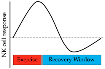
5. Mobilization of Natural Killer Cells during Hormone-Dependent Cancer

The mobilization of immune cells is multi-factorial. During moderate to vigorous efforts, catecholamine levels and hemodynamic shear stress increase with the latter the result of a greater cardiac output necessary to meet the physiological demands of exercise. Collectively, these changes induce alterations in immune cell adhesion molecules that causes immune cells to enter the circulation [46,47] leading to elevated cell numbers during and immediately following exercise (see Figure 1). During recovery, NK cell numbers drop rapidly [12] as they move out of the circulation with levels normalizing within a few hours except after particularly vigorous or prolonged exercise [8].
In laboratory-controlled settings in BCa and PCa, NK cell mobilization is relatively consistent with studies demonstrating robust increases in the absolute number (cells/µL) and cell frequency (relative to total lymphocytes) immediately following exercise. With intermittent cycling at 60% of VO2peak, CD3CD16+CD56+ NK cells increased by 103 cells/µL compared with a 178 cells/µL change in non-cancer controls that led to a difference in counts being reported at 0 h [10]. In contrast, the NK cell frequency increased from 9.3% to 18.1% and was similar between groups, indicating that higher NK cell counts at 0 h were due to higher total lymphocytes in the controls. With 67% of the BCa cohort receiving chemotherapy, this treatment may have contributed to the group differences. Chemotherapy reduces the total lymphocyte numbers during BCa with particularly detrimental effects on conventional T cells [48] although NK cells appear to be less affected [49,50]. Using a similar cycling protocol as BCa but a higher exercise intensity, PCa receiving ADT demonstrated increases in CD3CD56+ NK cell absolute counts of 306 cells/µL and 230 cells/µL in non-cancer controls that were similar between groups [11]. NK cell frequency increased from 8.6% to 15.8% and was comparable between groups and also with the changes seen in BCa [10]. The greater mobilization of absolute cell numbers in PCa is likely attributed to a greater exercise intensity (60% of peak power output (~80% of VO2peak) vs. BCa) with exercise intensity having a large impact on the immune cell mobilization [14].
Using a real-world exercise stimulus, participation in a half marathon had substantial impacts on immune cell frequency [51]. Both non-cancer controls and BCa had NK frequencies of ~18% that decreased to ~10% when acquired 15 minutes after the run. As NK cell egress begins immediately following exercise cessation with counts falling to ~40% of post-exercise levels within 10 minutes [12], the timing of the sample is the likely and logical explanation. However, NK frequencies also remained suppressed at 24 h [51], which is contrary to the laboratory-based work [10,11]. There are several possible explanations regarding this finding. A half marathon completion time exceeds that of the intermittent cycling protocols and may have also been at a higher relative effort, with the authors describing this bout as “long-lasting and exhaustive endurance exercise”. Vigorous exercise has been shown to suppress NK cell counts for up to a week [52] and may account for the lower frequency at 24 h although recovery strategies that include sleep and post-exercise nutrition are additional factors [8]. As neither the half marathon duration nor the intensity were reported [51], the classification of exhaustive exercise remains somewhat speculative. Importantly, BCa and the controls demonstrated consistent responses in terms of NK cells but also other lymphocyte subpopulations. This suggests that a longer duration or more intense acute exercise does not increase the risk to cancer survivors with normal immune cell frequency distribution. Subsequent exercise prescription should take into account the potential need for an extended recovery from more substantial acute exercise bouts as well as the need to replicate these findings using larger sample sizes.

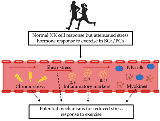
5.1. Attenuated Catecholamine Response

One mechanism that contributes to NK cell mobilization is catecholamine secretion. Catecholamine release into the circulation is both intensity- and duration-dependent [53] with higher levels leading to greater NK cell mobilization [14] that is diminished in the presence of a β2 adrenergic receptor blockade [54]. BCa treatment has previously been shown to alter carbohydrate and lipid metabolism [55,56] with the mechanisms leading to lower lactate concentrations and higher fat oxidation being unknown. As such, the metabolic effects of the adrenal hormones during acute cycling were examined. Cortisol and norepinephrine demonstrated similar post-exercise responses between BCa and non-cancer controls whereas increases in epinephrine were attenuated in BCa [57]. With norepinephrine being primarily derived from sympathetic neurons, this finding suggests a greater chronic adrenal stress reactivity in BCa. In support of this finding, men with PCa also demonstrated suppressed exercise-induced rises in epinephrine only [58] that also occurred independently of hormone therapy. These findings are intriguing for multiple reasons. Despite different exercise intensities, cancer types and sex of the participants, the slower rise in epinephrine was consistently ~50% lower in both BCa and PCa. While cancer treatment is associated with increased stress [59], adrenal hormone differences were not detectable at rest and only in response to a physical stress. Lastly, epinephrine contributes to the mobilization of NK cells in both humans [14] and mice [60] yet the attenuated response did not affect the NK cell count or frequency. Moreover, no correlations were observed between NK cell counts and changes in epinephrine [11]. This then begs the questions, if attenuated catecholamine levels are not affecting NK mobilization, is this suppressed level meaningful? Is there another mechanism that is compensating?

5.2. Other Potential Mechanisms

We hypothesized that normal NK cell mobilization observed in BCa and PCa, despite an attenuated catecholamine response, may be attributed to (i) increased shear stress during exercise, (ii) myokines released during muscle contractions that include IL-6 and/or (iii) chronic stress exposure (see Figure 2). A brief investigation into each of these potential mechanistic factors is provided. However, it is important to consider that these pathways are not functioning independently. It may be that all three (or others) are contributing to the normal NK cell mobilization to counteract the lower catecholamine response to exercise.

5.2.1. Shear Stress

Blood flow across the endothelial surface of the blood vessel creates shear stress [61] that may influence immune cell adhesion to the vascular wall. Endothelial cells may be altered by shear stress, thereby facilitating immune cell mobilization via decreased adhesion molecules on endothelial cells and less oxidative stress [62]. For example, nitric oxide (NO) produced following shear stress decreased neutrophil adhesion that was reversed using NO inhibitors [63]. The balance between oxidative and anti-oxidative molecules that is altered with acute exercise influences cellular adhesion to the vascular wall [47]. Additionally, exercise-induced NK cell mobilization has been reported, potentially due to an improved endothelial function via an inhibition of the integrin-mediated adhesion to blood vessels [64,65,66,67]. In addition to the immune cell mobilization, shear stress may directly affect the cancer cell apoptosis and proliferation, thereby reducing the invasion and metastasis of circulating tumor cells [68]. An in vitro shear stress equivalent to strenuous exercise demonstrated a poor viability across several tumor cell lines [69] while continuous shear stress levels induced 1.6-fold increases in colon cancer cells that became arrested in the G1 phase of the cell cycle [70]. While it is unclear if shear stress is able to compensate for a reduced catecholamine secretion, exercise improves vascular function [71,72] and therefore has the potential to permit a greater release of NK cells into the circulation following stress-induced shear stress [73]. In addition, the direct effects on tumor cell viability from an elevated laminar flow provide a compelling argument for the use of regular bouts of acute exercise to help manage cancer recurrence and metastases.

5.2.2. Myokines

Contracting skeletal muscles release myokines into the circulation [74]; amongst them is IL-6, which influences NK cell mobilization [60,75]. In mouse models, both exercise and epinephrine infusion contributed to increased NK mobilization and tumor infiltration while reducing the overall tumor volume with an IL-6 blockade reducing the benefits of exercise training [60]. In humans, a chronic inhibition of IL-6 receptor signaling abrogated increases in CD56dim but not CD56bright NK cells during exercise while also increasing the epinephrine release [75]. This latter finding suggests an inverse relationship between IL-6 and epinephrine. IL-6 levels are elevated in BCa and PCa in untreated individuals [76], with radiation [77] or tamoxifen [76] but not ADT [78], indicating this pathway is disrupted by cancer or cancer treatment and may be partially responsible for the attenuated rise in epinephrine during exercise. IL-6 release during exercise also plays a vital role in promoting NK cell proliferation, differentiation and maturation [79]. Furthermore, the redistribution of NK cells following exercise may contribute to the control of tumor growth [60] although it is unclear if IL-6 contributes to tumor suppression/cancer prevention in humans [75]. However, serum from BCa following acute exercise reduced in vitro breast tumor cell viability [80] with increased IL-6, IL-8, TNF and lactate concentrations being reported. Other myokines may also contribute to the regulation of NK cell mobilization and activation following exercise including IL-7 and IL-15 [73,81]. Myokine release during exercise contributes to the immune cell mobilization and immunosurveillance of cancer although how this response may be impacted by aging in cancer survivors remains unknown [81].

5.2.3. Chronic Stress Exposure

Cancer diagnosis, treatment and survivorship may result in chronic psychological stress in addition to the physiological stress of fighting the disease [82,83]. Psychological stresses are associated with the hypersecretion of adrenal hormones such as epinephrine [84]. We hypothesize that BCa and PCa have chronically low-grade increases in circulating epinephrine [85] potentially due to a stress-induced overactivated hypothalamic-pituitary-adrenal axis [86]. Therefore, exercise may not elicit the expected epinephrine secretion with several possible explanations. First, the epinephrine responses in BCa and PCa are normal but are bound to β2 adrenergic receptors that gives the appearance of reduced circulating levels. Alternatively, a more vigorous physical stressor is required for a normal response. We speculate that chronic stress exposure may desensitize epinephrine secretion but individuals with chronic stress have elevated stress levels that over time decrease immune cell adrenergic sensitivity [87], which would require greater (not lower) epinephrine responses. It is worth noting that elevated stress and plasma catecholamines have repeatedly been shown to be associated with tumor progression [88] so the lower catecholamine response to acute exercise in BCa [57] and PCa [58] could actually be beneficial. Lastly, cancer treatments (e.g., immune checkpoint and/or tyrosine kinase inhibitors) are associated with adrenal insufficiency [89] but this is rarely reported in BCa and PCa [90], making this unlikely. On the surface, chronic stress exposure appears plausible yet evidence to support this hypothesis is quite limited. As such, explanations for a normal NK response despite an attenuated stress hormone remain speculative.

5.3. Interaction between Acute and Chronic Exercise

The focus of this review has been on the acute response to exercise with little regard to exercise training. The effects of exercise training on immune outcomes in cancer survivors have been reviewed elsewhere [19,20,91]. Briefly, using resistance, aerobic or combined training, NK cell counts and frequency in BCa were unchanged [91,92,93,94] with a few [94,95] but not all [93] studies reporting greater NK cell cytotoxicity in the trained state. While acute exercise is a potent stimulus for mobilizing NK (and other immune) cells, it is unclear if this response changes with training as this approach is rarely used. To our knowledge, only two studies combined acute and regular training in the same study [96,97] but neither examined NK cells. One benefit of this approach is an increased ability to identify deficiencies in the immune function that are less apparent in the resting state. Indeed, our group recently demonstrated that conventional and unconventional T cells [98] and also neutrophils [99] from BCa were mobilized to a lesser extent than healthy controls that were partially rescued by 16 weeks of community-based exercise training. While these data also do not specifically examine NK cells, they do highlight deficiencies within aspects of innate immunity that appear to improve following training. While promising, this preliminary work requires replication and further investigations to establish if these findings extend to other immune cells (including NK cells).

6. Implications and Future Directions

The number of studies that examine acute exercise, NK cells and stress hormone responses in cancer are limited, which affected our ability to interpret these data. Instead, the key conflict that is identified in this review is the normal mobilization of NK cells following exercise in BCa and PCa despite an attenuated stress hormone response. One possibility is the altered activity within the HPA axis from the chronic stress of BCa and PCa [82,83] with chronic sympathetic nervous system activity linked to cancer progression [100]. As such, the additional stress from physical activity could compound existing psychological stress and is most likely to occur in patients undergoing active treatment. However, a single bout of aerobic or resistance exercise during chemotherapy has demonstrated beneficial effects on stress, energy and nausea [101], which argues against this possibility. Moreover, how the acute response may vary over longer timeframes (e.g., one microcycle) is unknown as well as how mobilization of other immune cells beyond NK cells may be affected by attenuated increases in epinephrine. Other compensatory factors have been proposed as mechanisms to maintain a normal immune response but need to be tested before more definite conclusions are possible. Finally, the standardized reporting of immune outcomes (e.g., counts, frequencies, lytic activity) would allow for a better consolidation of the existing literature [20] and to examine if different immune populations have a normal response to acute exercise.

Author Contributions

Conceptualization, E.D.H., L.C.B., K.M. and E.S.E.; literature searches, L.C.B. and K.M.; data curation, E.D.H.; figure development, L.C.B. and K.M.; writing—original draft preparation E.D.H., L.C.B. and E.S.E.; writing—review and editing, K.M. All authors have read and agreed to the published version of the manuscript.

Funding

This research received no external funding.

Data Availability Statement

Not applicable.

Conflicts of Interest

The authors declare no conflict of interest.

References

  1. Dranoff, G. Cytokines in cancer pathogenesis and cancer therapy. Nat. Rev. Cancer 2004, 4, 11–22. [Google Scholar] [CrossRef] [PubMed]
  2. Simpson, R.J.; Lowder, T.W.; Spielmann, G.; Bigley, A.B.; LaVoy, E.C.; Kunz, H. Exercise and the aging immune system. Ageing Res. Rev. 2012, 11, 404–420. [Google Scholar] [CrossRef] [PubMed]
  3. Leading Causes of Death (U.S.). Centers for Disease Control and Prevention. Available online: https://www.cdc.gov/nchs/fastats/leading-causes-of-death.htm (accessed on 15 March 2021).
  4. De Maria, A.; Bozzano, F.; Cantoni, C.; Moretto, L. Revisiting human natural killer cell subset function revealed cytolytic CD56(dim)CD16+ NK cells as rapid producers of abundant IFN-gamma on activation. Proc. Natl. Acad. Sci. USA 2011, 108, 728–732. [Google Scholar] [CrossRef] [PubMed] [Green Version]
  5. Bigley, A.B.; Rezvani, K.; Chew, C.; Sekine, T.; Pistillo, M.; Crucian, B.; Bollard, C.M.; Simpson, R.J. Acute exercise preferentially redeploys NK-cells with a highly-differentiated phenotype and augments cytotoxicity against lymphoma and multiple myeloma target cells. Brain Behav. Immun. 2014, 39, 160–171. [Google Scholar] [CrossRef]
  6. Gayoso, I.; Sanchez-Correa, B.; Campos, C.; Alonso, C.; Pera, A.; Casado, J.G.; Morgado, S.; Tarazona, R.; Solana, R. Immunosenescence of human natural killer cells. J. Innate. Immun. 2011, 3, 337–343. [Google Scholar] [CrossRef]
  7. Campbell, J.P.; Turner, J.E. Debunking the Myth of Exercise-Induced Immune Suppression: Redefining the Impact of Exercise on Immunological Health Across the Lifespan. Front. Immunol. 2018, 9, 648. [Google Scholar] [CrossRef]
  8. Peake, J.M.; Neubauer, O.; Walsh, N.P.; Simpson, R.J. Recovery of the immune system after exercise. J. Appl. Physiol. (1985) 2017, 122, 1077–1087. [Google Scholar] [CrossRef]
  9. Walsh, N.P.; Gleeson, M.; Shephard, R.J.; Gleeson, M.; Woods, J.A.; Bishop, N.C.; Fleshner, M.; Green, C.; Pedersen, B.K.; Hoffman-Goetz, L.; et al. Position statement. Part one: Immune function and exercise. Exerc. Immunol. Rev. 2011, 17, 6–63. [Google Scholar]
  10. Evans, E.S.; Hackney, A.C.; McMurray, R.G.; Randell, S.H.; Muss, H.B.; Deal, A.M.; Battaglini, C.L. Impact of Acute Intermittent Exercise on Natural Killer Cells in Breast Cancer Survivors. Integr. Cancer Ther. 2015, 14, 436–445. [Google Scholar] [CrossRef] [Green Version]
  11. Hanson, E.D.; Sakkal, S.; Que, S.; Cho, E.; Spielmann, G.; Kadife, E.; Violet, J.A.; Battaglini, C.L.; Stoner, L.; Bartlett, D.B.; et al. Natural killer cell mobilization and egress following acute exercise in men with prostate cancer. Exp. Physiol. 2020, 105, 1524–1539. [Google Scholar] [CrossRef]
  12. Rooney, B.V.; Bigley, A.B.; LaVoy, E.C.; Laughlin, M.; Pedlar, C.; Simpson, R.J. Lymphocytes and monocytes egress peripheral blood within minutes after cessation of steady state exercise: A detailed temporal analysis of leukocyte extravasation. Physiol. Behav. 2018, 194, 260–267. [Google Scholar] [CrossRef] [Green Version]
  13. Nieman, D.C.; Miller, A.R.; Henson, D.A.; Warren, B.J.; Gusewitch, G.; Johnson, R.L.; Davis, J.M.; Butterworth, D.E.; Herring, J.L.; Nehlsen-Cannarella, S.L. Effect of high- versus moderate-intensity exercise on lymphocyte subpopulations and proliferative response. Int. J. Sports Med. 1994, 15, 199–206. [Google Scholar] [CrossRef]
  14. Anane, L.H.; Edwards, K.M.; Burns, V.E.; Drayson, M.T.; Riddell, N.E.; van Zanten, J.J.; Wallace, G.R.; Mills, P.J.; Bosch, J.A. Mobilization of gammadelta T lymphocytes in response to psychological stress, exercise, and beta-agonist infusion. Brain Behav. Immun. 2009, 23, 823–829. [Google Scholar] [CrossRef]
  15. Natale, V.M.; Brenner, I.K.; Moldoveanu, A.I.; Vasiliou, P.; Shek, P.; Shephard, R.J. Effects of three different types of exercise on blood leukocyte count during and following exercise. Sao Paulo Med. J. 2003, 121, 9–14. [Google Scholar] [CrossRef] [Green Version]
  16. Hanson, E.D.; Bates, L.C.; Bartlett, D.B.; Campbell, J.P. Does exercise attenuate age- and disease-associated dysfunction in unconventional T cells? Shining a light on overlooked cells in exercise immunology. Eur. J. Appl. Physiol. 2021. [Google Scholar] [CrossRef]
  17. Standish, L.J.; Sweet, E.S.; Novack, J.; Wenner, C.A.; Bridge, C.; Nelson, A.; Martzen, M.; Torkelson, C. Breast cancer and the immune system. J. Soc. Integr. Oncol. 2008, 6, 158–168. [Google Scholar]
  18. Siegel, R.L.; Miller, K.D.; Jemal, A. Cancer statistics, 2020. CA Cancer J. Clin. 2020, 70, 7–30. [Google Scholar] [CrossRef]
  19. Kruijsen-Jaarsma, M.; Revesz, D.; Bierings, M.B.; Buffart, L.M.; Takken, T. Effects of exercise on immune function in patients with cancer: A systematic review. Exerc. Immunol. Rev. 2013, 19, 120–143. [Google Scholar]
  20. Khosravi, N.; Stoner, L.; Farajivafa, V.; Hanson, E.D. Exercise training, circulating cytokine levels and immune function in cancer survivors: A meta-analysis. Brain Behav. Immun. 2019, 81, 92–104. [Google Scholar] [CrossRef]
  21. Campbell, K.L.; Winters-Stone, K.M.; Wiskemann, J.; May, A.M.; Schwartz, A.L.; Courneya, K.S.; Zucker, D.S.; Matthews, C.E.; Ligibel, J.A.; Gerber, L.H.; et al. Exercise Guidelines for Cancer Survivors: Consensus Statement from International Multidisciplinary Roundtable. Med. Sci. Sports Exerc. 2019, 51, 2375–2390. [Google Scholar] [CrossRef] [Green Version]
  22. von Blanckenburg, P.; Schuricht, F.; Albert, U.S.; Rief, W.; Nestoriuc, Y. Optimizing expectations to prevent side effects and enhance quality of life in breast cancer patients undergoing endocrine therapy: Study protocol of a randomized controlled trial. BMC Cancer 2013, 13, 426. [Google Scholar] [CrossRef] [PubMed] [Green Version]
  23. Ramchand, S.K.; Lim, E.; Grossmann, M. Adjuvant endocrine therapy in women with oestrogen-receptor-positive breast cancer: How should the skeletal and vascular side effects be assessed and managed? Clin. Endocrinol. 2016, 85, 689–693. [Google Scholar] [CrossRef] [PubMed]
  24. Choo, S.B.; Saifulbahri, A.; Zullkifli, S.N.; Fadzil, M.L.; Redzuan, A.M.; Abdullah, N.; Bustamam, R.S.A.; Ahmad, H.Z.; Shah, N.M. Adjuvant endocrine therapy side-effects among postmenopausal breast cancer patients in Malaysia. Climacteric 2019, 22, 175–181. [Google Scholar] [CrossRef] [PubMed]
  25. Berkowitz, M.J.; Thompson, C.K.; Zibecchi, L.T.; Lee, M.K.; Streja, E.; Berkowitz, J.S.; Wenziger, C.M.; Baker, J.L.; DiNome, M.L.; Attai, D.J. How patients experience endocrine therapy for breast cancer: An online survey of side effects, adherence, and medical team support. J. Cancer Surviv. 2021, 15, 29–39. [Google Scholar] [CrossRef]
  26. Condorelli, R.; Vaz-Luis, I. Managing side effects in adjuvant endocrine therapy for breast cancer. Expert Rev. Anticancer. Ther. 2018, 18, 1101–1112. [Google Scholar] [CrossRef]
  27. Bargiota, A.; Oeconomou, A.; Zachos, I.; Samarinas, M.; Pisters, L.L.; Tzortzis, V. Adverse effects of androgen deprivation therapy in patients with prostate cancer: Focus on muscle and bone health. J. BUON 2020, 25, 1286–1294. [Google Scholar]
  28. Agarwal, M.; Canan, T.; Glover, G.; Thareja, N.; Akhondi, A.; Rosenberg, J. Cardiovascular Effects of Androgen Deprivation Therapy in Prostate Cancer. Curr. Oncol. Rep. 2019, 21, 91. [Google Scholar] [CrossRef]
  29. Fang, D.; Zhou, L. Androgen deprivation therapy in nonmetastatic prostate cancer patients: Indications, treatment effects, and new predictive biomarkers. Asia Pac. J. Clin. Oncol. 2019, 15, 108–120. [Google Scholar] [CrossRef] [Green Version]
  30. Edmunds, K.; Tuffaha, H.; Scuffham, P.; Galvao, D.A.; Newton, R.U. The role of exercise in the management of adverse effects of androgen deprivation therapy for prostate cancer: A rapid review. Support. Care Cancer 2020, 28, 5661–5671. [Google Scholar] [CrossRef]
  31. Mohamad, N.V.; Soelaiman, I.N.; Chin, K.Y. A Review on the Effects of Androgen Deprivation Therapy (ADT) on Bone Health Status in Men with Prostate Cancer. Endocr. Metab. Immune. Disord. Drug Targets 2017, 17, 276–284. [Google Scholar] [CrossRef]
  32. Solomayer, E.F.; Feuerer, M.; Bai, L.; Umansky, V.; Beckhove, P.; Meyberg, G.C.; Bastert, G.; Schirrmacher, V.; Diel, I.J. Influence of adjuvant hormone therapy and chemotherapy on the immune system analysed in the bone marrow of patients with breast cancer. Clin. Cancer Res. 2003, 9, 174–180. [Google Scholar]
  33. Mozaffari, F.; Lindemalm, C.; Choudhury, A.; Granstam-Bjorneklett, H.; Helander, I.; Lekander, M.; Mikaelsson, E.; Nilsson, B.; Ojutkangas, M.L.; Osterborg, A.; et al. NK-cell and T-cell functions in patients with breast cancer: Effects of surgery and adjuvant chemo- and radiotherapy. Br. J. Cancer 2007, 97, 105–111. [Google Scholar] [CrossRef] [Green Version]
  34. Kang, D.H.; Weaver, M.T.; Park, N.J.; Smith, B.; McArdle, T.; Carpenter, J. Significant impairment in immune recovery after cancer treatment. Nurs Res. 2009, 58, 105–114. [Google Scholar] [CrossRef] [Green Version]
  35. Zavadova, E.; Vocka, M.; Spacek, J.; Konopasek, B.; Fucikova, T.; Petruzelka, L. Cellular and humoral immunodeficiency in breast cancer patients resistant to hormone therapy. Neoplasma 2014, 61, 90–98. [Google Scholar] [CrossRef] [Green Version]
  36. Larsson, A.M.; Roxa, A.; Leandersson, K.; Bergenfelz, C. Impact of systemic therapy on circulating leukocyte populations in patients with metastatic breast cancer. Sci. Rep. 2019, 9, 1–10. [Google Scholar] [CrossRef] [Green Version]
  37. Sabbioni, M.E.; Castiglione, M.; Hurny, C.; Siegrist, H.P.; Bacchi, M.; Bernhard, J.; Thurlimann, B.; Bonnefoi, H.; Perey, L.; Goldhirsch, A.; et al. Interaction of tamoxifen with concurrent cytotoxic adjuvant treatment affects lymphocytes and lymphocyte subsets counts in breast cancer patients. Support. Care Cancer 1999, 7, 149–153. [Google Scholar] [CrossRef] [PubMed]
  38. Sheard, C.R.; Reilly, F.; Tee, D.E.; Vergani, D.; Lowe, D.; Baum, M.; Cameron, A.E. The effect of adjuvant cyclophosphamide or tamoxifen on the numbers of lymphocytes bearing T cell or NK cell markers. Br. J. Cancer 1986, 54, 705–709. [Google Scholar] [CrossRef] [Green Version]
  39. Gannon, P.O.; Poisson, A.O.; Delvoye, N.; Lapointe, R.; Mes-Masson, A.M.; Saad, F. Characterization of the intra-prostatic immune cell infiltration in androgen-deprived prostate cancer patients. J. Immunol. Methods 2009, 348, 9–17. [Google Scholar] [CrossRef]
  40. Aragon-Ching, J.B.; Williams, K.M.; Gulley, J.L. Impact of androgen-deprivation therapy on the immune system: Implications for combination therapy of prostate cancer. Front. Biosci 2007, 12, 4957–4971. [Google Scholar] [CrossRef] [Green Version]
  41. Morse, M.D.; McNeel, D.G. Prostate cancer patients on androgen deprivation therapy develop persistent changes in adaptive immune responses. Hum. Immunol. 2010, 71, 496–504. [Google Scholar] [CrossRef] [Green Version]
  42. Mercader, M.; Bodner, B.K.; Moser, M.T.; Kwon, P.S.; Park, E.S.; Manecke, R.G.; Ellis, T.M.; Wojcik, E.M.; Yang, D.; Flanigan, R.C.; et al. T cell infiltration of the prostate induced by androgen withdrawal in patients with prostate cancer. Proc. Natl. Acad. Sci. USA 2001, 98, 14565–14570. [Google Scholar] [CrossRef] [PubMed] [Green Version]
  43. Battaglini, C.L.; Mills, R.C.; Phillips, B.L.; Lee, J.T.; Story, C.E.; Nascimento, M.G.; Hackney, A.C. Twenty-five years of research on the effects of exercise training in breast cancer survivors: A systematic review of the literature. World J. Clin. Oncol 2014, 5, 177–190. [Google Scholar] [CrossRef] [PubMed]
  44. Hanson, E.D.; Hurley, B.F. Intervening on the side effects of hormone-dependent cancer treatment: The role of strength training. J. Aging Res. 2011, 2011, 1–8. [Google Scholar] [CrossRef] [PubMed] [Green Version]
  45. Hayes, S.C.; Newton, R.U.; Spence, R.R.; Galvao, D.A. The Exercise and Sports Science Australia position statement: Exercise medicine in cancer management. J. Sci. Med. Sport 2019, 22, 1175–1199. [Google Scholar] [CrossRef] [Green Version]
  46. Shephard, R.J. Adhesion molecules, catecholamines and leucocyte redistribution during and following exercise. Sports Med. 2003, 33, 261–284. [Google Scholar] [CrossRef]
  47. Sen, C.K.; Roy, S. Antioxidant regulation of cell adhesion. Med. Sci. Sports Exerc. 2001, 33, 377–381. [Google Scholar] [CrossRef]
  48. Dusseaux, M.; Martin, E.; Serriari, N.; Peguillet, I.; Premel, V.; Louis, D.; Milder, M.; Le Bourhis, L.; Soudais, C.; Treiner, E.; et al. Human MAIT cells are xenobiotic-resistant, tissue-targeted, CD161hi IL-17-secreting T cells. Blood 2011, 117, 1250–1259. [Google Scholar] [CrossRef]
  49. Kay, N.E.; Oken, M.M.; Kyle, R.; Van Ness, B.; Kalish, L.; Leong, T.; Greipp, P. Sequential phenotyping of myeloma patients on chemotherapy: Persistence of activated T-cells and natural killer cells. Leuk. Lymphoma 1995, 16, 351–354. [Google Scholar] [CrossRef]
  50. Waidhauser, J.; Schuh, A.; Trepel, M.; Schmalter, A.K.; Rank, A. Chemotherapy markedly reduces B cells but not T cells and NK cells in patients with cancer. Cancer Immunol. Immunother. 2020, 69, 147–157. [Google Scholar] [CrossRef]
  51. Zimmer, P.; Baumann, F.T.; Bloch, W.; Zopf, E.M.; Schulz, S.; Latsch, J.; Schollmayer, F.; Shimabukuro-Vornhagen, A.; von Bergwelt-Baildon, M.; Schenk, A. Impact of a half marathon on cellular immune system, pro-inflammatory cytokine levels, and recovery behavior of breast cancer patients in the aftercare compared to healthy controls. Eur. J. Haematol. 2016, 96, 152–159. [Google Scholar] [CrossRef]
  52. Shek, P.N.; Sabiston, B.H.; Buguet, A.; Radomski, M.W. Strenuous exercise and immunological changes: A multiple-time-point analysis of leukocyte subsets, CD4/CD8 ratio, immunoglobulin production and NK cell response. Int. J. Sports Med. 1995, 16, 466–474. [Google Scholar] [CrossRef]
  53. Zouhal, H.; Jacob, C.; Delamarche, P.; Gratas-Delamarche, A. Catecholamines and the effects of exercise, training and gender. Sports Med. 2008, 38, 401–423. [Google Scholar] [CrossRef]
  54. Graff, R.M.; Kunz, H.E.; Agha, N.H.; Baker, F.L.; Laughlin, M.; Bigley, A.B.; Markofski, M.M.; LaVoy, E.C.; Katsanis, E.; Bond, R.A.; et al. beta2-Adrenergic receptor signaling mediates the preferential mobilization of differentiated subsets of CD8+ T-cells, NK-cells and non-classical monocytes in response to acute exercise in humans. Brain Behav. Immun. 2018, 74, 143–153. [Google Scholar] [CrossRef]
  55. Evans, E.S.; Battaglini, C.L.; Groff, D.G.; Hackney, A.C. Aerobic exercise intensity in breast cancer patients: A preliminary investigation. Integr. Cancer Ther. 2009, 8, 139–147. [Google Scholar] [CrossRef]
  56. Tosti, K.P.; Hackney, A.C.; Battaglini, C.L.; Evans, E.S.; Groff, D. Exercise in patients with breast cancer and healthy controls: Energy substrate oxidation and blood lactate responses. Integr. Cancer Ther. 2011, 10, 6–15. [Google Scholar] [CrossRef] [Green Version]
  57. Evans, E.S.; Hackney, A.C.; Pebole, M.M.; McMurray, R.G.; Muss, H.B.; Deal, A.M.; Battaglini, C.L. Adrenal Hormone and Metabolic Biomarker Responses to 30 min of Intermittent Cycling Exercise in Breast Cancer Survivors. Int. J. Sports Med. 2016, 37, 921–929. [Google Scholar] [CrossRef] [Green Version]
  58. Hanson, E.D.; Sakkal, S.; Evans, W.S.; Violet, J.A.; Battaglini, C.L.; McConell, G.K.; Hayes, A. Altered stress hormone response following acute exercise during prostate cancer treatment. Scand. J. Med. Sci. Sports 2018, 28, 1925–1933. [Google Scholar] [CrossRef]
  59. Carlson, L.E.; Angen, M.; Cullum, J.; Goodey, E.; Koopmans, J.; Lamont, L.; MacRae, J.H.; Martin, M.; Pelletier, G.; Robinson, J.; et al. High levels of untreated distress and fatigue in cancer patients. Br. J. Cancer 2004, 90, 2297–2304. [Google Scholar] [CrossRef]
  60. Pedersen, L.; Idorn, M.; Olofsson, G.H.; Lauenborg, B.; Nookaew, I.; Hansen, R.H.; Johannesen, H.H.; Becker, J.C.; Pedersen, K.S.; Dethlefsen, C.; et al. Voluntary running suppresses tumor growth through epinephrine-and il-6-dependent nk cell mobilization and redistribution. Cell Metab. 2016, 23, 554–562. [Google Scholar] [CrossRef] [Green Version]
  61. Paszkowiak, J.J.; Dardik, A. Arterial wall shear stress: Observations from the bench to the bedside. Vasc. Endovasc. Surg. 2003, 37, 47–57. [Google Scholar] [CrossRef]
  62. Evans, W. NK cell recruitment and exercise: Potential immunotherapeutic role of shear stress and endothelial health. Med. Hypotheses 2017, 109, 170–173. [Google Scholar] [CrossRef]
  63. Provost, P.; Lam, J.Y.; Lacoste, L.; Merhi, Y.; Waters, D. Endothelium-derived nitric oxide attenuates neutrophil adhesion to endothelium under arterial flow conditions. Arter. Thromb. 1994, 14, 331–335. [Google Scholar] [CrossRef] [Green Version]
  64. Benschop, R.J.; Nijkamp, F.P.; Ballieux, R.E.; Heijnen, C.J. The effects of beta-adrenoceptor stimulation on adhesion of human natural killer cells to cultured endothelium. Br. J. Pharm. 1994, 113, 1311–1316. [Google Scholar] [CrossRef]
  65. Benschop, R.J.; Oostveen, F.G.; Heijnen, C.J.; Ballieux, R.E. Beta 2-adrenergic stimulation causes detachment of natural killer cells from cultured endothelium. Eur. J. Immunol. 1993, 23, 3242–3247. [Google Scholar] [CrossRef]
  66. Benschop, R.J.; Schedlowski, M.; Wienecke, H.; Jacobs, R.; Schmidt, R.E. Adrenergic control of natural killer cell circulation and adhesion. Brain Behav. Immun. 1997, 11, 321–332. [Google Scholar] [CrossRef] [Green Version]
  67. Capellino, S.; Claus, M.; Watzl, C. Regulation of natural killer cell activity by glucocorticoids, serotonin, dopamine, and epinephrine. Cell Mol. Immunol. 2020, 17, 705–711. [Google Scholar] [CrossRef]
  68. Huang, Q.; Hu, X.; He, W.; Zhao, Y.; Hao, S.; Wu, Q.; Li, S.; Zhang, S.; Shi, M. Fluid shear stress and tumor metastasis. Am. J. Cancer Res. 2018, 8, 763–777. [Google Scholar]
  69. Regmi, S.; Fu, A.; Luo, K.Q. High Shear Stresses under Exercise Condition Destroy Circulating Tumor Cells in a Microfluidic System. Sci. Rep. 2017, 7, 1–12. [Google Scholar] [CrossRef] [Green Version]
  70. Avvisato, C.L.; Yang, X.; Shah, S.; Hoxter, B.; Li, W.; Gaynor, R.; Pestell, R.; Tozeren, A.; Byers, S.W. Mechanical force modulates global gene expression and beta-catenin signaling in colon cancer cells. J. Cell Sci. 2007, 120, 2672–2682. [Google Scholar] [CrossRef] [Green Version]
  71. Schadler, K.L.; Thomas, N.J.; Galie, P.A.; Bhang, D.H.; Roby, K.C.; Addai, P.; Till, J.E.; Sturgeon, K.; Zaslavsky, A.; Chen, C.S.; et al. Tumor vessel normalization after aerobic exercise enhances chemotherapeutic efficacy. Oncotarget 2016, 7, 65429–65440. [Google Scholar] [CrossRef] [Green Version]
  72. Ashcraft, K.A.; Warner, A.B.; Jones, L.W.; Dewhirst, M.W. Exercise as Adjunct Therapy in Cancer. Semin. Radiat. Oncol. 2019, 29, 16–24. [Google Scholar] [CrossRef] [PubMed]
  73. Idorn, M.; Hojman, P. Exercise-Dependent Regulation of NK Cells in Cancer Protection. Trends Mol. Med. 2016, 22, 565–577. [Google Scholar] [CrossRef] [PubMed] [Green Version]
  74. Pedersen, B.K.; Febbraio, M.A. Muscle as an endocrine organ: Focus on muscle-derived interleukin-6. Physiol. Rev. 2008, 88, 1379–1406. [Google Scholar] [CrossRef] [PubMed] [Green Version]
  75. Bay, M.L.; Heywood, S.; Wedell-Neergaard, A.S.; Schauer, T.; Lehrskov, L.L.; Christensen, R.H.; Legard, G.E.; Jensen, P.O.; Krogh-Madsen, R.; Ellingsgaard, H. Human immune cell mobilization during exercise: Effect of IL-6 receptor blockade. Exp. Physiol. 2020, 105, 2086–2098. [Google Scholar] [CrossRef] [PubMed]
  76. Premkumar, V.G.; Yuvaraj, S.; Vijayasarathy, K.; Gangadaran, S.G.; Sachdanandam, P. Serum cytokine levels of interleukin-1beta, -6, -8, tumour necrosis factor-alpha and vascular endothelial growth factor in breast cancer patients treated with tamoxifen and supplemented with co-enzyme Q(10), riboflavin and niacin. Basic Clin. Pharm. Toxicol. 2007, 100, 387–391. [Google Scholar] [CrossRef]
  77. Sprod, L.K.; Palesh, O.G.; Janelsins, M.C.; Peppone, L.J.; Heckler, C.E.; Adams, M.J.; Morrow, G.R.; Mustian, K.M. Exercise, sleep quality, and mediators of sleep in breast and prostate cancer patients receiving radiation therapy. Community Oncol. 2010, 7, 463–471. [Google Scholar] [CrossRef] [Green Version]
  78. Hawley, J.E.; Pan, S.; Figg, W.D.; Lopez-Bujanda, Z.A.; Strope, J.D.; Aggen, D.H.; Dallos, M.C.; Lim, E.A.; Stein, M.N.; Hu, J.; et al. Association between immunosuppressive cytokines and PSA progression in biochemically recurrent prostate cancer treated with intermittent hormonal therapy. Prostate 2020, 80, 336–344. [Google Scholar] [CrossRef]
  79. Hojman, P. Exercise protects from cancer through regulation of immune function and inflammation. Biochem Soc. Trans. 2017, 45, 905–911. [Google Scholar] [CrossRef]
  80. Dethlefsen, C.; Lillelund, C.; Midtgaard, J.; Andersen, C.; Pedersen, B.K.; Christensen, J.F.; Hojman, P. Exercise regulates breast cancer cell viability: Systemic training adaptations versus acute exercise responses. Breast Cancer Res. Treat. 2016, 159, 469–479. [Google Scholar] [CrossRef]
  81. Hwang, J.H.; McGovern, J.; Minett, G.M.; Della Gatta, P.A.; Roberts, L.; Harris, J.M.; Thompson, E.W.; Parker, T.J.; Peake, J.M.; Neubauer, O. Mobilizing serum factors and immune cells through exercise to counteract age-related changes in cancer risk. Exerc. Immunol. Rev. 2020, 26, 80–99. [Google Scholar]
  82. Antoni, M.H.; Dhabhar, F.S. The impact of psychosocial stress and stress management on immune responses in patients with cancer. Cancer 2019, 125, 1417–1431. [Google Scholar] [CrossRef]
  83. Pasquini, M.; Biondi, M. Depression in cancer patients: A critical review. Clin. Pr. Epidemiol Ment. Health 2007, 3, 2. [Google Scholar] [CrossRef] [Green Version]
  84. Wurtman, R.J. Stress and the adrenocortical control of epinephrine synthesis. Metabolism 2002, 51, 11–14. [Google Scholar] [CrossRef] [Green Version]
  85. Hassan, S.; Karpova, Y.; Flores, A.; D’Agostino, R., Jr.; Danhauer, S.C.; Hemal, A.; Kulik, G. A pilot study of blood epinephrine levels and CREB phosphorylation in men undergoing prostate biopsies. Int. Urol. Nephrol. 2014, 46, 505–510. [Google Scholar] [CrossRef] [Green Version]
  86. Thornton, L.M.; Andersen, B.L.; Blakely, W.P. The pain, depression, and fatigue symptom cluster in advanced breast cancer: Covariation with the hypothalamic-pituitary-adrenal axis and the sympathetic nervous system. Health Psychol. 2010, 29, 333–337. [Google Scholar] [CrossRef] [Green Version]
  87. Mausbach, B.T.; Aschbacher, K.; Mills, P.J.; Roepke, S.K.; von Kanel, R.; Patterson, T.L.; Dimsdale, J.E.; Ziegler, M.G.; Ancoli-Israel, S.; Grant, I. A 5-year longitudinal study of the relationships between stress, coping, and immune cell beta(2)-adrenergic receptor sensitivity. Psychiatry Res. 2008, 160, 247–255. [Google Scholar] [CrossRef] [Green Version]
  88. Krizanova, O.; Babula, P.; Pacak, K. Stress, catecholaminergic system and cancer. Stress 2016, 19, 419–428. [Google Scholar] [CrossRef]
  89. Wright, J.J.; Powers, A.C.; Johnson, D.B. Endocrine toxicities of immune checkpoint inhibitors. Nat. Rev. Endocrinol 2021. [Google Scholar] [CrossRef]
  90. Grouthier, V.; Lebrun-Vignes, B.; Moey, M.; Johnson, D.B.; Moslehi, J.J.; Salem, J.E.; Bachelot, A. Immune Checkpoint Inhibitor-Associated Primary Adrenal Insufficiency: WHO VigiBase Report Analysis. Oncologist 2020, 25, 696–701. [Google Scholar] [CrossRef]
  91. Hagstrom, A.D.; Marshall, P.W.; Lonsdale, C.; Papalia, S.; Cheema, B.S.; Toben, C.; Baune, B.T.; Fiatarone Singh, M.A.; Green, S. The effect of resistance training on markers of immune function and inflammation in previously sedentary women recovering from breast cancer: A randomized controlled trial. Breast Cancer Res. Treat. 2016, 155, 471–482. [Google Scholar] [CrossRef]
  92. Hutnick, N.A.; Williams, N.I.; Kraemer, W.J.; Orsega-Smith, E.; Dixon, R.H.; Bleznak, A.D.; Mastro, A.M. Exercise and lymphocyte activation following chemotherapy for breast cancer. Med. Sci. Sports Exerc. 2005, 37, 1827–1835. [Google Scholar] [CrossRef]
  93. Nieman, D.C.; Cook, V.D.; Henson, D.A.; Suttles, J.; Rejeski, W.J.; Ribisl, P.M.; Fagoaga, O.R.; Nehlsen-Cannarella, S.L. Moderate exercise training and natural killer cell cytotoxic activity in breast cancer patients. Int. J. Sports Med. 1995, 16, 334–337. [Google Scholar] [CrossRef] [PubMed]
  94. Peters, C.; Lotzerich, H.; Niemeier, B.; Schule, K.; Uhlenbruck, G. Influence of a moderate exercise training on natural killer cytotoxicity and personality traits in cancer patients. Anticancer. Res. 1994, 14, 1033–1036. [Google Scholar]
  95. Fairey, A.S.; Courneya, K.S.; Field, C.J.; Bell, G.J.; Jones, L.W.; Mackey, J.R. Randomized controlled trial of exercise and blood immune function in postmenopausal breast cancer survivors. J. Appl. Physiol. 2005, 98, 1534–1540. [Google Scholar] [CrossRef] [Green Version]
  96. Galvao, D.A.; Nosaka, K.; Taaffe, D.R.; Peake, J.; Spry, N.; Suzuki, K.; Yamaya, K.; McGuigan, M.R.; Kristjanson, L.J.; Newton, R.U. Endocrine and immune responses to resistance training in prostate cancer patients. Prostate Cancer Prostatic Dis. 2008, 11, 160–165. [Google Scholar] [CrossRef]
  97. Khosravi, N.; Hanson, E.D.; Farajivafa, V.; Evans, W.S.; Lee, J.T.; Danson, E.; Wagoner, C.W.; Harrell, E.P.; Sullivan, S.A.; Nyrop, K.A.; et al. Exercise-induced modulation of monocytes in breast cancer survivors. Brain Behav. Immun. Health 2021. in Press. [Google Scholar]
  98. Hanson, E.D.; Bates, L.C.; Harrell, E.P.; Bartlett, D.B.; Lee, J.T.; Wagoner, C.W.; Alzer, M.S.; Amatuli, D.J.; Jensen, B.C.; Deal, A.M.; et al. Exercise Training Partially Rescues Impaired Mucosal Associated Invariant T-Cell Mobilization in Breast Cancer Survivors Compared to Healthy Older Women; University of North Carolina: Chapel Hill, NC, USA, Unpublished work; 2021. [Google Scholar]
  99. Bartlett, D.B.; Hanson, E.D.; Lee, J.T.; Wagoner, C.W.; Harrell, E.P.; Sullivan, S.A.; Bates, L.C.; Deal, A.M.; Jensen, B.C.; MacDonald, G.; et al. The Effects of 16-Weeks of Exercise Training on Neutrophil Functions in Breast Cancer Survivors; Duke University: Durham, NC, USA, Unpublished work; 2021. [Google Scholar]
  100. Conceicao, F.; Sousa, D.M.; Paredes, J.; Lamghari, M. Sympathetic activity in breast cancer and metastasis: Partners in crime. Bone Res. 2021, 9, 1–11. [Google Scholar] [CrossRef]
  101. Johnsson, A.; Demmelmaier, I.; Sjovall, K.; Wagner, P.; Olsson, H.; Tornberg, A.B. A single exercise session improves side-effects of chemotherapy in women with breast cancer: An observational study. BMC Cancer 2019, 19, 1–9. [Google Scholar] [CrossRef] [PubMed] [Green Version]
Figure 1. Biphasic response to exercise that results in an increase in NK counts immediately post-exercise followed by cellular egress below resting levels.
Endocrines 02 00012 g001
Figure 2. Hypothesized mechanisms that compensate for the attenuated stress hormone response following acute exercise that permits normal NK mobilization in breast (BCa) and prostate cancer (PCa) patients.
Endocrines 02 00012 g002
Publisher’s Note: MDPI stays neutral with regard to jurisdictional claims in published maps and institutional affiliations.

