Effects of Mixtures of Engineered Nanoparticles and Metallic Pollutants on Aquatic Organisms


1
Department F.-A. Forel for Environmental and Aquatic Sciences, Earth and Environment Sciences, Faculty of Sciences, University of Geneva, 1211 Geneva, Switzerland
2
Institute for Environmental Sciences, Uni Carl Vogt, 1211 Geneva, Switzerland
*
Author to whom correspondence should be addressed.
Environments 2020, 7(4), 27; https://0-doi-org.brum.beds.ac.uk/10.3390/environments7040027
Submission received: 23 February 2020
/
Revised: 27 March 2020
/
Accepted: 30 March 2020
/
Published: 1 April 2020
(This article belongs to the Special Issue New Insights into Impacts of Toxic Metals in Aquatic Environments)
Abstract
:In aquatic environment, engineered nanoparticles (ENPs) are present as complex mixtures with other pollutants, such as trace metals, which could result in synergism, additivity or antagonism of their combined effects. Despite the fact that the toxicity and environmental risk of the ENPs have received extensive attention in the recent years, the interactions of ENPs with other pollutants and the consequent effects on aquatic organisms represent an important challenge in (nano)ecotoxicology. The present review provides an overview of the state-of-the-art and critically discusses the existing knowledge on combined effects of mixtures of ENPs and metallic pollutants on aquatic organisms. The specific emphasis is on the adsorption of metallic pollutants on metal-containing ENPs, transformation and bioavailability of ENPs and metallic pollutants in mixtures. Antagonistic, additive and synergistic effects observed in aquatic organisms co-exposed to ENPs and metallic pollutants are discussed in the case of “particle-proof” and “particle-ingestive” organisms. This knowledge is important in developing efficient strategies for sound environmental impact assessment of mixture exposure in complex environments.
1. Introduction
Engineered nanomaterials with significantly enhanced properties showed a lot of promises for direct and indirect benefits in almost all sphere of the modern society [1]. However, with increasing production and ever-expanding applications, engineered nanoparticles (ENPs) are unavoidably released to water, soil and atmosphere throughout their lifecycle [2,3,4]. Hence, the concerns about environmental safety of ENPs have significantly raised. Indeed, the abundant information on the environmental implications of ENPs can be found in recent review papers [5,6,7,8,9,10,11,12,13], evidencing the potential of ENPs to harm aquatic organisms if present in enough high concentrations. Nevertheless, in most cases the existing information is obtained considering pollution by ENPs individually, neglecting that in the environment the organisms are exposed simultaneously to mixtures of diverse pollutants. Therefore, evaluation of the potential risk of ENPs can be significantly under- or over-estimated if the cocktail effects are neglected.
Assessing the effects of mixtures of environmental pollutants, and more generally multiple stressors, is a central topic in the modern ecotoxicology [14,15]. Approaches to assess mixture effects were thoroughly reviewed for toxic metals, pharmaceutical products or pesticides [16,17]. Fundamental concepts in describing the mixture effects can be divided into two major categories depending on the existence or not of the interactions between the components in the mixture and their mode-of-toxic-action [16,18]. In a case when no interaction between the mixture components exists, two major concepts are used to predict mixture effects: the concentration addition (CA) and the independent action (IA). CA applies when the sum of the effects of individual components is equal to the toxicity of a mixture as a whole. It assumes identical mechanisms of toxicity and same target sites for the individual components. By contrast, IA applies when the components of the mixture are acting independently and do not influence the toxicity of each other. It assumes that individual components have different mode-of-toxic-action and different biological target sites. In a case where interactions between the mixture components exist, the observed effects will differ from the CA and IA predictions. In a case that the observed toxicity is stronger than predicted one, the mixture toxicity effects are described as synergistic. By contrast, when the toxicity of a mixture is less than expected by CA or IA, the mixture effects are described as antagonistic.
ENPs possess enhanced physical and chemical properties, and very high reactivity because of their smaller size and larger specific surface area, which favor the interactions with various biotic and abiotic components, including pollutants in the aquatic systems [19]. Therefore, similarly to the naturally present nanoparticles and colloids [20], ENPs could affect significantly the behavior and effects of pollutants in the aquatic environment. For example, based on the free metal ion and biotic ligand models [21], ENPs could be expected to decrease the concentration of the free metal ions in the exposure medium, thus to reduce metal bioavailability and toxicity. Naasz et al. recently reviewed the knowledge on the co-exposure to ENPs and different micropollutants in in-vitro and in-vivo biological test systems with a special focus on the Trojan-horse phenomenon and its ecotoxicological relevance [19]. The authors assigned the effects in mixtures containing ENPs to 6 groups, including Trojan horse (+) and (−), surface enrichment, retention, inertism and coalism [19], and underlined the necessity of considering different processes, such as adsorption and desorption, bioavailability and internalization in which individual components in mixtures are involved, to better understand and predict the mixture toxicity.
In such a context, the present review paper provides an overview and discusses relevant processes driving the interactions in mixtures of metal-containing ENPs and metallic pollutants with aquatic organisms. The specific focus is on: (i) adsorption of metallic pollutants on metal-containing ENPs; (ii) transformation processes in exposure medium containing mixture of ENPs and metallic pollutant; and (iii) adsorption and internalization of ENPs and metallic pollutants by aquatic organisms. The evidences of the toxicity to aquatic organisms co-exposed to mixtures of metal-containing ENPs and metallic pollutants as well as conceptualization of the toxic outcome are discussed. The important knowledge gaps and the further research needs are identified, too. Such information is critical in developing efficient strategies for assessment of environmental impact of ENPs and metal co-exposure in complex environments.
2. Key Processes at The Exposure Medium—Organism Interface
In the aquatic environment the ENPs and metallic pollutants interact with different biotic and abiotic constituents. As a result, they are present as complex and dynamic mixture of aggregates, different dissolved metal forms (free metal ions, bound to inorganic and organic ligands, forming labile or inert complexes), metal adsorbed on ENPs, natural nanoparticles, colloids and their aggregates. All these entities shape physicochemical speciation in the exposure medium and exhibit different reactivity towards biota. The key chemo- and bio-dynamic processes driving the interactions in the mixtures of metal-containing ENPs and metallic pollutants with aquatic (micro)organisms are shown in Figure 1. As these processes were recently reviewed for metal-containing ENPs [13], here we will focus specifically on the adsorption of the metallic pollutants on ENPs, the transformations of mixtures in exposure medium, the adsorption and internalization of ENPs and metallic pollutants by aquatic organisms.
2.1. Adsorption of Metallic Pollutants on Metal-Containing ENPs
The adsorption of metallic pollutants on ENPs is widely studied from different perspectives, for example from nanocatalysis [24,25], wastewater treatment [26,27] and remediation [28,29]. The most studied metal-containing ENPs include metallic ENPs, metal oxide ENPs, quantum dots (QDs) and mixtures of different ENPs. Strong sorption on metallic ENPs is due to the presence of metal coordination sites (terrace, edge, kink or corner sites) on the particle surface which exhibit quite different coordination chemistry towards reactants, intermediates and products [30]. Atoms on the surface of metallic ENPs, especially the ones at the edges and corners are active center of the catalyst and the adsorption [31,32]. For example, a distinct decrease of AgNPs major peaks observed after adsorption of HgCl2 suggested that one of the adsorption mechanisms could be the formation of an amalgam between the two metals AgmHgn [33]. Adsorption of Au(III), Pd (II) and Pt(IV) on CuSNPs was related to high affinity between exposed sulfur atoms on CuSNPs and noble metal ions [34]. Several articles focused on the interaction between QDs and dissolved metals. For instance, CdSe/ZnS QDs could be used to detect Hg(II) and Cu(II) [35]. PEG-ZnSQD @ZIF-67 hybrid nanocomposite could be used as fluorescence probe for Cu(II) [36]. CdSe/ZnS QDs exhibited ultrasensitive and highly selective detection of Zn(II) ions [37]. Carboxyl-terminal groups coupled polymer coated core/shell CdSe/ZnSQDs adsorbed significant amounts of dissolved Cu and Pb and affected their uptake in green algae Chlorella kesslerii and Chlamydomonas reinhardtii [38] and in metal-resistant bacterium Cupriavidus metallidurance [39].
In addition to the high specific surface area, many metal oxide ENPs, such as ZnONPs, Al2O3NPs and TiO2NPs possess properties of both Lewis bases and acids [40,41,42]. Residual hydroxyl groups and anions/cations holes increase the surface activity of metal oxide ENPs [43,44]. Among the metal oxide ENPs, the most studied is TiO2NPs [45,46,47,48]. Selected examples demonstrated its strong adsorption capacity: TiO2NPs (Degussa P25) with primary size of 21 nm sorbed rapidly (equilibrium within 30 min) Cd(II) and As(V) [3,49]; P25, TiO2NPs (anatase synthesized by sol-gel method) and titanate nanotubes adsorbed >99% of inorganic mercury (Hg) present in the medium [50]. Similarly, TiO2NPs (Rutile/Anatase: 87/13) with size of 10 nm retained 96% of IHg in 45 min [51], whereas TiO2NPs (Rutile/Anatase: 85/15) of 20 nm removed 98.6% of IHg [52]. Comparative study of four metal oxide ENPs revealed that the adsorption capacity of TiO2NPs (5–10 nm) and Al2O3NPs (50-100 nm) to As(V) were higher than that of ZnONPs (10–20 nm) and CuONPs (<10 nm) [53]. In some cases, the adoption of metallic pollutants is also accompanied with an oxidation process. For example, As(III) adsorbed on TiO2NPs was oxidized to As(V) on the particle surface in the presence of UV light (Yan and Jing 2019).
Recent studies demonstrated that binary metal oxides show enhanced adsorption of metals as compared with single metal oxides [54,55]. For example, Al-Mn binary oxides exhibited higher adsorption capacity for Cd(II) [56]. Fe3O4-TiO2NPs completely removed As(V) and 93% of As(III) from the medium in 1h [57]. Fe-Mn binary oxides combining the oxidation property of manganese dioxide and the high adsorption features of iron oxides were highly efficient in the removal of both As(V) and As(III) [58]. Fe-Mn binary oxides have also high adsorption capacity for Cr(VI) with the carboxyl- and the hydroxyl-groups participating in the adsorption process [59]. Superparamagnetic iron oxide nanoparticles functionalized with 3-mercaptopropionic acid were very efficient in sorbing metals: Ag(I) and Hg(II) interact strongly with the thiol groups, while the Pb(II) seems to adsorb mostly via electrostatic interaction [60]. When present in mixture with dissolving ENPs, the stable ENPs were also shown to adsorb the released ions. For example, the combined effects of ZnONPs dissolution and Zn(II) adsorption onto TiO2NPs controlled the concentration of dissolved Zn(II) in the ZnONPs and TiO2NPs mixture [61].
Overall, these studies revealed that the characteristics of ENPs, metallic pollutants and exposure medium influence the amount of adsorbed metallic pollutants. The adsorption of metallic pollutants on ENPs could results in their surface modification, which could change their properties and the way they interact with aquatic biota [62]. Nevertheless, the sorption of metallic pollutants on the ENPs has received only limited attention in (nano)ecotoxicology context and systematic studies on how the adsorption of the metallic pollutants on the ENPs will affect their reactivity towards the organisms are still missing.
2.2. Transformations of Mixture of ENPs and Metallic Pollutants in The Aquatic Environment
In the environment ENPs are subject to different transformations including dissolution, agglomeration, aggregation and sedimentation, [63] which determine their stability and affect the interactions with aquatic organisms (Figure 1). The transformations of ENPs (and adsorbed metallic pollutants) strongly depend on: (i) Characteristics of ENPs (composition, size, shape, surface properties, crystal structure, etc.); for example, TiO2NPs, SiO2NPs and Al2O3NPs of smaller size aggregate easier than larger ones [64]; Rutile TiO2NPs aggregate stronger than anatase of comparable size [65]; TiO2NPs with sodium citrate were more stable than polyvinylpyrrolidone, sodium dodecyl sulfate and polyethylene glycol [66]. (ii) Exposure medium variables (ionic strength, pH, composition and concentration of dissolved organic matter (DOM) etc.); for example, TiO2NPs or Al2O3NPs and cells significantly aggregated at neutral pH or low ionic strength, while SiO2NPs at low pH or high ionic strength [64,67]. Extracellular polymeric substance (EPS) extracted from bacterium Bacillus subtilis stabilized TiO2NPs [68].
Furthermore, once adsorbed on the ENPs metallic pollutants could alter the surface properties of ENPs and thus influence their aggregation and dissolution. For example, TiO2NPs sorbed Zn(II) released from ZnONPs and also co-aggregated with ZnONPs [69]. Hetero-aggregates formed by electrostatic interaction between AgNPs and hematite prevented AgNPs to have a direct contact with Escherichia coli cells [70]. CeO2NPs and TiO2NPs aggregated when present as a mixture in bacterial culture medium due to their opposite surface charges, while CeO2NPs had no effect on ZnONPs stability [71]. Magnetic Fe3O4NPs with different surface functionalities adsorbed various metal ions (Cr(III), Co(II), Ni(II), Cu(II), Cd(II), Pb(II) and As(III)) by different mechanisms (chelation, ion exchange or/and electrostatic interaction etc.) [72,73].
2.3. Adsorption and Internalization of ENPs and Metallic Pollutants in Mixtures by Aquatic Organisms
Adsorption of ENPs (and associated metallic pollutants) onto the surface of aquatic organisms is a key step determining their bioavailability [13,23]. Different interaction forces could drive the adsorption process, including Van der Waals forces, hydrophobic forces, electrostatic attraction, hydrogen bonding and receptor-ligand interactions [23,74,75]. For the specific case of “particle-proof” microalgae, examples include adsorption of SiO2NPs and CeO2NPs on Raphidocelis subcapitata [76], TiO2NPs on Phaeodactylum tricornutum [77], CuONPs [78] and TiO2NPs [13] on Chlamydomonas reinhardtii, AgNPs on Euglena gracilis [79]. AuNPs adsorbed on the carapace and appendages of water flea Daphnia magna, inducing mechanical disruptions of the feeding appendages [80]. Given the enhanced adsorption of metallic pollutant by ENPs (Section 2.1), ENPs can play a role of carriers facilitating the entry of metallic pollutants into the “particle-ingestive” organisms (“Trojan-horse effect”). For example, Pb(II) sorption on TiO2NPs facilitated its accumulation by freshwater bivalve Corbicula fluminea [81]. Binding of Cd(II) on TiO2NPs resulted in an alleviation of Cd(II) toxicity to Chlamydomonas reinhardtii [82], however has no consequences for the uptake and toxicity of Cd(II) by Daphnia magna and Lumbriculus variegatus [83]. The above examples demonstrated the complexity of the interactions between biota and mixtures of ENPs and metallic pollutants, as well as the ENPs, metals and species dependence.
ENPs (and associated metallic pollutants) adsorbed onto the organisms can undergo translocation into the intracellular environment via different mechanisms, such as diffusion and endocytosis, as evidenced by several studies [13,75]. Endocytosis occurs by multiple mechanisms that fall into two broad categories, “phagocytosis” and “pinocytosis” [84]. Different examples demonstrated the relevance of endocytosis for various microorganisms: internalization of AgNPs [85] and thioglycolic acid stabilized CdTe QDs [86] by the golden-brown alga Ochromonas danica, TiO2NPs by the blue-green alga Anabaena variabilis [87]. The 3.4 nm AuNPs were taken up into macrophage cells via pinocytosis [88]. Carboxylic CdSe/ZnS QDs were shown to enter into the ciliate Tetrahymena thermophila by phagocytosis and clathrin-mediated endocytosis [89], as did AgNPs, AuNPs, CuONPs and TiO2NPs [90]. Nonetheless, it is still unclear to what extent the majority of microorganisms have specific mechanisms of ENPs internalization.
The internalization of ENPs by zooplankton is still poorly understood [12]. The water flea and shrimp can feed on particles of 0.4–40 μm, and ENPs and their aggregates are taken up via water filtration. The uptake of AuNPs by Gammarus pulex varied depending on the surface coatings, with Au-mercaptoundecanoic acid and Au-citrate NPs being taken up to a greater extent than Au-NH2 and Au-PEG NPs in a range of standard ecotoxicity media and natural waters. No relationships were observed between the aggregation state of different AuNPs and the uptake, suggesting that the widely accepted assumption that AuNP uptake is related to particle size is, in fact, not verified [91]. No similar studies exist on the uptake of ENPs-metallic pollutant complexes, but in principle the mechanisms known for ENPs should be relevant.
It is still unclear how the processes of adsorption of metallic pollutants on ENPs and their aggregation could influence the bioavailability of ENPs-metallic pollutants’ complexes. The recent work of Tan et al. evidenced that the biouptake of Cd(II) in Daphnia magna may either increase or reduce depending on the aggregation state of the TiO2NPs [92]. This study highlighted the linkage between the aggregation of ENPs, adsorption of metallic pollutants and bioaccumulation. The ENPs compositions and concentrations were shown to impact the metallic pollutants uptake. For example, TiO2NPs enhanced Cr(VI) bioavailability to freshwater alga Scenedesmus obliquus, while Al2O3NPs had no significant effect [93]. The bioavailability of ENPs and the feeding behavior of the aquatic organisms could also modulate metal uptake. For example, filter feeders, such as daphnids and shrimps can actively take up ENPs into their guts, thus facilitating accumulation of the adsorbed metal [92].
Adsorption and consequent internalization of dissolved metal is another key step in the overall biouptake process in co-exposure to ENPs and metallic pollutants. Dissolved fraction contains various species originating from the metallic pollutants and ENPs exhibiting different reactivities towards biota [94,95]. They bind to different functional groups (e.g., carboxyl-, hydroxyl-, phosphate, amino- and sulfhydryl-) in the cell wall and membrane constituents of aquatic microorganisms, such as algae [96] and bacteria [97]. Since the majority of metal species are hydrophilic, their transport through the biological membrane is mediated by specific proteins through the channels, channel carriers and pumps [98]. Common transporter families in eukaryotes and bacteria are well studied [99,100,101,102]. There is a consensus that for the majority of metals, the uptake is driven by the free metal ions [94,95] and the amount of metal bound to the biotic ligands [21]. Therefore, any environmental variables (including the ENPs) that could affect the speciation in the exposure medium as well as the competition between the metals for the biotic ligands will influence the amount of the metal taken up by the organisms.
Based on the available literature for ENPs and metallic pollutants, as well as limited literature about the mixtures, the processes at the medium–organism interface in mixtures could depend on: (i) the physicochemical properties of the ENPs (e.g. size, shape, surface modification, etc.); (ii) the nature and characteristics of the metallic pollutants; (iii) the water quality variables (e.g., pH, ionic strength and the type/concentration of DOM); (iv) the characteristics of the aquatic organisms (e.g., membrane permeability, differentiation stage and cellular uptake pathways).
3. Toxicity of Mixtures of ENPs and Metallic Pollutants to Aquatic Organisms
Current knowledge on the co-exposure of aquatic organisms to mixtures of ENPs and metallic pollutants was nicely summarized in the literature [19]. Here we provide some more recent examples in Table 1. In general, the studies were designed in a way to consider the effect of mixture exposure by making comparison with the effect of individual components. Thus, toxicity outcomes of mixture exposure can be grouped in three categories: (i) ENPs have no significant effect on the toxicity of metallic pollutants; (ii) ENPs increase the toxicity of metallic pollutants; (iii) ENPs reduce toxicity of metallic pollutants (Table 1). However, this categorization is operational and dependent on different experimental factors, including species, exposure duration, etc. For instance, co-exposure of two Bacillus species to TiO2NPs and Cu(II) under the same test conditions resulted in different effects, showing the species dependence of the observed effects [103]. Acute and chronic exposure of Tigriopus japonicus to mixtures of ZnONPs and Cd(II) or Pb(II) resulted in the contrasting effects [104]. However, chronic exposure of Daphnia magna to mixture of TiO2NPs and As(Ⅲ)/As(V) led to comparable toxicity [105].
3.1. No Significant Effect of ENPs on The Toxicity of Metallic Pollutants
ENPs have no significant effect on the toxicity of other metallic pollutants when there was no interaction between ENPs and metallic pollutants, and ENPs itself had no effect on the organisms [19]. Vale et al. states that Cd(II) uptake and antioxidant enzyme activities by bivalve Corbicula fluminea were comparable in the absence and presence TiO2NPs [106]. Similarly, TiO2NPs did not affect specific metal-induced responses (metallothionein induction) and Cd(II) accumulation in tissues of marine bivalve Mytilus galloprovincialis [107]. AgNPs had no influence on the Cd(II) toxicity to green alga Chlamydomonas reinhardtii [108]. The lack of modulation of the metal toxicity in the presence of ENPs could be also related with the lowering of the concentrations of the suspended ENPs which can interact with planktonic organisms due to the formation of the big aggregates that sedimented. For example, the real concentration of dispersed TiO2NPs was by far lower than the nominal one, mainly due to particle concentration dependent aggregation and sedimentation [109].
Moreover, Canesi et al. suggested that ENPs interact with organic matter produced by the organisms (mucus, fecal pellets, gametes), promoting the adsorption on mussel shell and byssal threads, as well as the sedimentation, thus affecting ENPs capability to adsorb contaminants [116]. All these processes may account for the low ENPs concentrations found in water samples collected during exposure, clearly indicating that only a minor fraction of the ENPs (and associated pollutants) in the dispersed form is available to the test organisms.
There are some evidences showing that ENPs had no significant effect on the toxicity of other metallic pollutants even though there was interaction between ENPs and metallic pollutants, and ENPs had an effect on the organism. For instance, the acute toxicities of As(V) and Cu(II) were unaffected by citrate-coated AgNPs despite the fact that their bioaccumulation was reduced [117].
3.2. Increase of Toxicity/Bioavailability of Metallic Pollutants by ENPs
Since ENPs are efficient adsorbents for metallic pollutants, they can play a role of carriers facilitating the entry of metallic pollutants into the organisms (“Trojan-horse effect”), which may lead to enhancement of metallic pollutant toxicity to aquatic organisms. Indeed, TiO2NPs (21 nm) greatly promoted the accumulation of Cd(II) in Cyprinus carpio and the cumulative Cd(II) concentrations in the presence of TiO2NPs were 2.5 times higher than in the absence [3]. Similarly, TiO2NPs increased As(V) accumulation in Cyprinus carpio by 44% [118], suggesting that TiO2NPs played a role of carrier promoting the uptake. TiO2NPs increased the bioconcentration of Pb(II), which led to the disruption of thyroid endocrine and neuronal system in zebrafish larvae [119]. Toxicity and bioavailability of Cd(II) and Zn(II) to Daphnia magna increased significantly after TiO2NPs pre-exposure [120], as was observed for Cu(II) accumulation [121]. The presence of hydrophobic TiO2NPs increased simultaneously the bioaccumulation of Cu(II) and Ti(IV) and induced high oxidative stress injury [114]. TiO2NPs enhanced Cd uptake in the “particle-ingestive” protozoa Tetrahymena thermophila, with 46.3% of internalized Cd corresponded to Cd-TiO2NPs complex [122].
The presence of ENPs can also increase the uptake and toxicity of different pollutants to unicellular organisms with no endocytosis. For example, Hartmann et al. showed that despite of the decrease of Cd(II) concentration due to the adsorption to TiO2NPs, algal growth inhibition increased [123]. This observation was explained by: (i) adsorption of Cd(II) on TiO2NPs resulting in an increase of Cd(II) concentration in algal cells surface; the main component of the algal cell wall is cellulose, which also usually contains glycoproteins and polysaccharides [124]. These components can act as binding sites to promote the adsorption of ENPs by algae [125,126]. Large aggregates of TiO2NPs entrapped almost all Pseudokirchneriella subcapitata cells [127]. It was also observed that algae Chlorella vulgaris and Raphidocelis subcapitata absorb large amount of AgNPs [128]. TiO2NPs at low concentrations (<1.0 mgL−1) significantly enhanced the toxicity of Zn(II) to Anabaena sp. [129]. (ii) TiO2NPs changed the permeability of the cells and increased the bioavailability of Cd(II). Indeed, various ENPs such as TiO2NPs, CuONPs and QD were shown to induce membrane damage to different unicellular organisms, such as algae or bacteria [78,130,131,132]. (iii) TiO2NPs can transfer Cd into the algae cells as a carrier. The algae cell walls are semipermeable, and their structure is usually porous [124]. The diameter of these pores is in the range of 5–20 nm [133,134], which makes the ENPs in the size smaller than the maximal pore size can pass through the cell wall easily. Once the ENPs pass through the cell wall, ENPs will encounter the second barrier—the plasma membrane. Endocytosis and passive diffusion are considered to be the main pathway for ENPs to cross the bilayer lipid membrane [135,136]. In addition, the permeability of the cell wall may change during cell cycling, with the newly synthesized cell wall being more permeable to ENPs, thereby increasing the uptake of ENPs by algal cells [128,137]. Al2O3NPs loaded with Pb(II) penetrated Isochrysis galbana cells and a synergistic effect was also found for the toxic effect of Pb(II) [110].
ENPs adhered to the bacterial surface and enhanced the membrane damage and metal accumulation in bacterial cell [138]. Co-exposure to TiO2NPs and Cu(II) resulted in an increased uptake of Cu(II) by Bacillus thuringiensis, leading to increase of growth inhibition and oxidative stress [103]. ENPs mainly affected cell membrane by the oxidative damage and membrane integrity [139]. AgNPs induced an excessive generation of ROS in the cell membrane and the production of membrane protein crystals of Dunaliella salina [140]. ENPs can also accumulate in the cell membrane and lead to cell wall depression, which causes changes in cell membrane permeability, until cell apoptosis occurs [141].
3.3. Reduction of The Toxicity/Bioavailability of Metallic Pollutants by ENPs
Most of the existing studies on reduced combined toxic effects of ENPs and metallic pollutants were based on the adsorption consideration and reduction of metallic pollutants bioavailability in a way similar to those of natural nanoparticles and colloids [20]. For instance, TiO2NPs reduced the growth inhibitory effect of Cd(II) on Chlamydomonas reinhardtii due to the adsorption of Cd(II) by TiO2NPs and reduction of the concentrations of free Cd(II) ions [82,142]. High TiO2NPs concentration effectively reduced the soluble Zn(II) (and its toxicity) concentrations by adsorbing Zn(II) on ENPs and settling to the bottom of the reactor [129]. Growth inhibition, oxidative stress and Cu(II) uptake by Scenedesmus obliquus were lower in mixture exposure with Al2O3NPs [143]. TiO2NPs reduced the toxicity of Cd(II) to bacterium Escherichia coli and the combined toxicity decreased with the increasing TiO2NPs concentrations, which can be explained by the reduction of the free Cd(II) concentration in the exposure medium [111]. Furthermore, ENPs may interfere with the metabolism of co-existed metals. Indeed, TiO2NPs altered the metabolization of arsenic by polychaete Laeonereis acuta and increased the proportions of dimethyl arsenic forms [144].
The well dispersed TiO2NPs and their μm-sized aggregates lessened Cd(II) toxicity to Daphnia magna, however the alleviation mechanisms were different. The μm-sized aggregates were actively ingested by Daphnia magna which resulted in an accumulation of Cd(II) in the daphnid gut. Whereas, the well dispersed TiO2NPs were taken up mainly by endocytosis which resulted in their accumulation in abdominal areas and the gut of Daphnia magna. Accumulation of Cd(II) was considerably impaired because of the metal’s rapid dissociation from the ENPs surfaces during endocytosis of the metal-ENPs complexes [92].
Li et al. showed that ENPs can modulate the toxicity of metallic pollutants by influencing their subcellular distribution in the test organisms, too. TiO2NPs (25 nm) decreased the toxicity of As(III) to Daphnia magna, as TiO2NPs accumulated in intestinal tract may act as a barrier, blocking the intestinal absorption of As(III) [145]. Excretion is another important process that may impact the eventually toxicological effects of the mixtures. It was found that organisms may excrete compounds as a feedback response, to alter the ENPs’ toxicity [146,147,148]. The excretion behavior of the mixtures may differ among the organisms as a function of their physiology, exposure routes, ENPs and metallic pollutants. For instance, the main elimination routes for AgNPs in Daphnia magna were excretion (63%) and fecal production [37]. In addition, the excretion rate constant of AgNPs in daphnids was much lower than that of Ag, suggesting the difficulty of eliminating AgNPs [149]. The majority of CdSe/ZnSQDs adhered to the external carapace or free floating within the gastrointestinal tract were excreted, whereas there were still a significant number of QDs retained by Ceriodaphnia dubia within the digestive tract [150]. No studies are currently available about ENPs excretion for microalgae. Only studies about the biosynthesis of ENPs in microalgae have been reported [151,152,153].
Overall, the existing research on combined effects of mixtures containing ENPs and metallic pollutants focused on the effects of fixed concentrations of ENPs on the toxicity of metallic pollutants, without considering the varied proportions of ENPs and metallic pollutants in the real environment.
4. Possible Bioaccumulation and Toxicity Outcome Scenarios during Co-Exposure to ENPs and Metallic Pollutants
4.1. Exposure of “Particle-Proof” Organisms to Mixtures of ENPs and Metallic Pollutants
Based on the available literature, we propose 10 possible toxicity outcome scenarios of ENPs-metallic pollutants mixtures on the “particle-proof” organisms (Figure 2).
- No interaction between ENPs and metallic pollutants; ENPs do not adsorb or penetrate algal cells. ENPs have no significant effect on algae. Metallic pollutants’ bioaccumulation and toxicity to the algae are unchanged.
- No interaction between ENPs and metallic pollutants; ENPs adsorb and penetrate into algal cells. Metallic pollutants’ bioaccumulation in the algae is unchanged. Effects of ENPs and metallic pollutants on the organism are independent. Combined toxicity may remain the same or increase depending on the species and concentrations of ENPs.
- No interaction between ENPs and metallic pollutants; ENPs and metallic pollutants compete for the same binding sites on algal surface. Under this condition, the metallic pollutants’ bioaccumulation decreases. Whereas the combined toxicity may increase, decrease or remain the same depending on the toxicity of ENPs.
- No interaction between ENPs and metallic pollutants; ENPs adsorb, but do not enter algal cells. ENPs affect cell membrane permeability, resulting in increase of bioaccumulation of metallic pollutants.
- No interaction between ENPs and metallic pollutants; ENPs adsorb and alter cell membrane permeability. ENPs and metallic pollutants enter the algal cells independently. Bioaccumulation of ENPs and metallic pollutants both increase.
- Metallic pollutants adsorb onto ENPs; ENPs do not interact with algal cells. Bioaccumulation and effect of metallic pollutants decrease.
- Metallic pollutants adsorb onto ENPs; ENPs with adsorbed metallic pollutants accumulate on the surface of algal cells. Bioaccumulation of metallic pollutants increases whereas the toxicity decreases.
- Metallic pollutants adsorb onto ENPs; ENPs affect membrane permeability and enter algal cells. There is no desorption of metallic pollutants from ENPs. Bioaccumulation of metallic pollutants increases whereas the toxicity decreases.
- Metallic pollutants adsorb onto ENPs; ENPs alter cell membrane permeability and enter algal cells. Metallic pollutants desorb from ENPs. In this case, the bioaccumulation of metallic pollutants increases. Desorption of chemical from ENPs is an important process for microorganisms with the food vacuoles. The pH in the food vacuoles becomes acidic (pH<4) within 1h after vacuole formation [154]. ENPs tend to release adsorbed environmental pollutants under the acidic condition [155].
- Free metal ions released from ENPs compete with metallic pollutants for algal cell binding and internalization sites. Accumulation of metallic pollutants in algal cells decreases. The biological outcome is uncertain depending the species and concentration of ENPs.
4.2. Exposure of “Particle-Ingestive” Organisms to Mixtures of ENPs and Metallic Pollutants
Based on the available literature here we summarized 6 possible toxicity outcome scenarios for mixtures of ENPs and metallic pollutants (Figure 3). These scenarios are proposed considering several dominating processes that determine the toxicity outcome of ENPs metallic pollutants mixtures, namely (i) aggregation, (ii) type of interaction between ENPs and metallic pollutants, (iii) internalization of ENPs by test organisms, (iv) desorption of metallic pollutants from ENPs upon contact to organisms and (v) excretion of ENPs, metallic pollutants and their complexes. We also assumed that invertebrates like Daphnia magna and shrimps take up more metallic pollutants adsorbed on ENPs via water filtration than exposed to metallic pollutants alone, and monodispersed ENPs can easier get deep inside the organisms and harder to be excreted compared with aggregated ENPs.
- No aggregation of ENPs. No interaction between ENPs and metallic pollutants. Bioaccumulation and effect of metallic pollutants are unchanged.
- No aggregation of ENPs. Adsorption of metallic pollutants onto ENPs. No desorption of metallic pollutants from ENPs in the organism. Metallic pollutants uptake is facilitated by ENPs but the bioavailability of metallic pollutants decreases because of the reduction of the concentration of free metallic pollutants.
- Aggregation of ENPs. Adsorption of metallic pollutants onto ENPs in medium. Desorption of metallic pollutant from ENPs in the organism. Increased body burden and bioavailability as a result of desorption of metallic pollutants from ENPs.
- Aggregation of ENPs. No interaction between ENPs and metallic pollutants. Bioaccumulation and toxicity of metallic pollutants are unchanged as the metallic pollutants and ENPs act independently.
- Aggregation of ENPs. Adsorption of metallic pollutants onto ENPs in medium. No desorption of metallic pollutants from ENPs in the organism. Reduced bioavailability of the metallic pollutants due to the decline in the concentration of free metallic pollutants.
- Aggregation of ENPs. Adsorption of metallic pollutants onto ENPs in medium. No desorption of metallic pollutants from ENPs in the organism. Increased body burden and toxicity as a result of desorption of metallic pollutants from ENPs.
5. Conclusions and Outlook
Aquatic organisms are typically exposed to multi-component mixtures of environmental pollutants, including ENPs. However, the current environmental risk assessment considers the effects of single pollutant which might be over-simplistic and this results to underestimation the possible risks. Since it is not realistic to test every possible combination of ENPs and environmental pollutants, development of the conceptual frames allowing to predict the cocktail effects is very important. The existing concepts of CA and IA were developed for dissolved pollutant mixtures; therefore, it is necessary to verify their applicability and to make an adjustment (if necessary) in the specific case of mixtures containing ENPs.
Significant progress was achieved in the understanding of the bioavailability and cocktail effects of mixtures containing ENPs and metallic pollutants. There is a consensus that ENPs can either increase, decrease or have no effect on the trace metal bioavailability and toxic effect depending on the feeding pattern of the organisms (e.g., “particle-proof” or “particle-ingestive” organism). Among the metallic ENPs, the most studied is TiO2NPs given its wide use [156,157], and among the metallic pollutants Cd(II) due to its high toxicity [158]. All the studies that analyzed the joint action of defined mixtures were combinations of only 2 components—ENPs and one metallic pollutant. The results on the combined exposure of model species, such as Daphnia magna and different algae, to ENPs and metallic pollutants revealed no interactions, positive or negative interactions between them. However, the limited data availability and low comparability make difficult the comparison between metals, ENPs and species.
Despite the growing literature dealing with combined effects of ENPs and metallic pollutants on phyto- and zoo-plankton, numerous knowledge gaps still need to be filled. Coming studies should focus on the better understanding of the interactions between ENPs and metallic pollutants and go beyond of the sorption equilibrium considerations. Cocktail effects of mixtures containing low doses of ENPs and metallic pollutants, when individual components have no effects are to be explored. Most of the results are obtained in well controlled lab conditions with quite high concentrations of ENPs and metallic pollutants with unrealistic ratios, which make difficult the extrapolation of the obtained results to natural environments. Better understanding of the mechanisms driving bioavailability and effects of mixtures containing ENPs and metals in the complex environment settings (e.g., presence of dissolved organic matter), more realistic ENPs/ metallic pollutant ratios will enable relevant lab-to-field extrapolation of toxicity data. Incorporation of the time-dependent size distribution of dispersed and internalized ENPs in the bioavailability modeling [159] was proposed. Overall, a more systematic approach considering the combined pressure of multiple stressors [160] and reconceptualization of exposure and effects of chemical cocktails containing ENPs is necessary and will be in the center of forthcoming nanoecotoxicology agenda.
Author Contributions
Conceptualization, V.I.S.; writing—original draft preparation, M.L., W.L., V.I.S.; writing—review and editing, M.L., W.L., V.I.S.; funding acquisition, M.L., V.I.S. All authors have read and agreed to the published version of the manuscript.
Funding
This research was funded by China Scholarship Council grant [2016]3100.
Acknowledgments
M.L. and V.I.S. are grateful for the financial support of the China Scholarship Council.
Conflicts of Interest
The authors declare no conflict of interest.
References
- Nanotechnology Market: By Type (Nanocomposites, Nanofibers, Nanoceramics, Nanomagnetics); By Application (Medical Diagnosis, Energy, ICT, Nano-EHS); By End-Users (Electronics, Pharmaceuticals, Biotechnology, Textile, Military)—Forecast (2016–2021); Industry ARC: Telangana, India, 2016.
- Quigg, A.; Chin, W.-C.; Chen, C.-S.; Zhang, S.; Jiang, Y.; Miao, A.-J.; Schwehr, K.A.; Xu, C.; Santschi, P.H. Direct and indirect toxic effects of engineered nanoparticles on algae: Role of natural organic matter. ACS Sustain. Chem. Eng. 2013, 1, 686–702. [Google Scholar] [CrossRef]
- Zhang, X.; Sun, H.; Zhang, Z.; Niu, Q.; Chen, Y.; Crittenden, J.C. Enhanced bioaccumulation of cadmium in carp in the presence of titanium dioxide nanoparticles. Chemosphere 2007, 67, 160–166. [Google Scholar] [CrossRef] [PubMed]
- Hu, J.; Wang, D.; Wang, J.; Wang, J. Toxicity of lead on Ceriodaphnia dubia in the presence of nano-CeO2 and nano-TiO2. Chemosphere 2012, 89, 536–541. [Google Scholar] [CrossRef] [PubMed]
- Akter, M.; Sikder, M.T.; Rahman, M.M.; Ullah, A.; Hossain, K.F.B.; Banik, S.; Hosokawa, T.; Saito, T.; Kurasaki, M. A systematic review on silver nanoparticles-induced cytotoxicity: Physicochemical properties and perspectives. J. Adv. Res. 2018, 9, 1–16. [Google Scholar] [CrossRef]
- Holden, P.A.; Gardea-Torresdey, J.L.; Klaessig, F.; Turco, R.F.; Mortimer, M.; Hund-Rinke, K.; Hubal, E.A.C.; Avery, D.; Barcelo, D.; Behra, R.; et al. Considerations of environmentally relevant test conditions for improved evaluation of ecological hazards of engineered nanomaterials. Environ. Sci. Technol. 2016, 50, 6124–6145. [Google Scholar] [CrossRef]
- Ivask, A.; Juganson, K.; Bondarenko, O.; Mortimer, M.; Aruoja, V.; Kasemets, K.; Blinova, I.; Heinlaan, M.; Slaveykova, V.; Kahru, A. Mechanisms of toxic action of Ag, ZnO and CuO nanoparticles to selected ecotoxicological test organisms and mammalian cells in vitro: A comparative review. Nanotoxicology 2014, 8, 57–71. [Google Scholar] [CrossRef]
- McGillicuddy, E.; Murray, I.; Kavanagh, S.; Morrison, L.; Fogarty, A.; Cormican, M.; Dockery, P.; Prendergast, M.; Rowan, N.; Morris, D. Silver nanoparticles in the environment: Sources, detection and ecotoxicology. Sci. Total Environ. 2017, 575, 231–246. [Google Scholar] [CrossRef]
- Zhang, W.C.; Xiao, B.D.; Fang, T. Chemical transformation of silver nanoparticles in aquatic environments: Mechanism, morphology and toxicity. Chemosphere 2018, 191, 324–334. [Google Scholar] [CrossRef]
- Joonas, E.; Aruoja, V.; Olli, K.; Kahru, A. Environmental safety data on CuO and TiO2 nanoparticles for multiple algal species in natural water: Filling the data gaps for risk assessment. Sci. Total Environ. 2019, 647, 973–980. [Google Scholar] [CrossRef]
- Kumar, R.; Umar, A.; Kumar, G.; Nalwa, H.S. Antimicrobial properties of ZnO nanomaterials: A review. Ceram. Int. 2017, 43, 3940–3961. [Google Scholar] [CrossRef]
- Lead, J.R.; Batley, G.E.; Alvarez, P.J.J.; Croteau, M.N.; Handy, R.D.; McLaughlin, M.J.; Judy, J.D.; Schirmer, K. Nanomaterials in the environment: Behavior, fate, bioavailability, and effects. An updated review. Environ. Toxicol. Chem. 2018, 37, 2029–2063. [Google Scholar] [CrossRef] [PubMed]
- Slaveykova, V.I.; Li, M.; Worms, I.A.; Liu, W. When environmental chemistry meets ecotoxicology: Bioavailability of inorganic nanoparticles to phytoplankton. Chimia 2020. [Google Scholar] [CrossRef] [PubMed]
- Fischer, B.B.; Pomati, F.; Eggen, R.I.L. The toxicity of chemical pollutants in dynamic natural systems: The challenge of integrating environmental factors and biological complexity. Sci. Total Environ. 2013, 449, 253–259. [Google Scholar] [CrossRef] [PubMed]
- Segner, H.; Schmitt-Jansen, M.; Sabater, S. Assessing the impact of multiple stressors on aquatic biota: The receptor’s side matters. Environ. Sci. Technol. 2014, 48, 7690–7696. [Google Scholar] [CrossRef]
- Heys, K.A.; Shore, R.F.; Pereira, M.G.; Jones, K.C.; Martin, F.L. Risk assessment of environmental mixture effects. RSC Adv. 2016, 6, 47844–47857. [Google Scholar] [CrossRef] [Green Version]
- Godoy, A.A.; Kummrow, F. What do we know about the ecotoxicology of pharmaceutical and personal care product mixtures? A critical review. Crit. Rev. Env. Sci. Technol. 2017, 47, 1453–1496. [Google Scholar] [CrossRef]
- Escher, B.I.; Hermens, J.L.M. Modes of Action in Ecotoxicology: Their role in body burdens, species sensitivity, QSARs, and mixture effects. Environ. Sci. Technol. 2002, 36, 4201–4217. [Google Scholar] [CrossRef]
- Naasz, S.; Altenburger, R.; Kühnel, D. Environmental mixtures of nanomaterials and chemicals: The Trojan-horse phenomenon and its relevance for ecotoxicity. Sci. Total Environ. 2018, 635, 1170–1181. [Google Scholar] [CrossRef]
- Hartland, A.; Lead, J.R.; Slaveykova, V.; O’Carroll, D.; Valsami-Jones, E. The environmental significance of natural nanoparticles. Nat. Educ. Knowl. 2013, 4, 7. [Google Scholar]
- Slaveykova, V.I.; Wilkinson, K.J. Predicting the bioavailability of metals and metal complexes: Critical review of the biotic ligand model. Environ. Chem. 2005, 2, 9–24. [Google Scholar] [CrossRef]
- Ma, S.; Lin, D. The biophysicochemical interactions at the interfaces between nanoparticles and aquatic organisms: Adsorption and internalization. Env. Sci. Process. Impacts 2013, 14, 145–160. [Google Scholar] [CrossRef] [PubMed]
- Von Moos, N.; Bowen, P.; Slaveykova, V.I. Bioavailability of inorganic nanoparticles to planktonic bacteria and aquatic microalgae in freshwater. Environ. Sci. Nano 2014, 1, 214–232. [Google Scholar] [CrossRef]
- Nelson, J.; Bargar, J.R.; Wasylenki, L.; Brown Jr, G.E.; Maher, K. Effects of nano-confinement on Zn (II) adsorption to nanoporous silica. Geochimica Cosmochimica Acta 2018, 240, 80–97. [Google Scholar] [CrossRef]
- Kumari, P.; Alam, M.; Siddiqi, W.A. Usage of nanoparticles as adsorbents for waste water treatment: An emerging trend. Sustain. Mater. Technol. 2019, 22, e00128. [Google Scholar] [CrossRef]
- Hokkanen, S.; Sillanpää, M. Nano-and microcellulose-based adsorption materials in water treatment. In Advanced Water Treatment; Elsevier: Amsterdam, The Netherlands, 2020; pp. 1–83. [Google Scholar]
- Abdugaffarova, K.K.; Dorogov, M.V.; Vikarchuk, A.A.; Zabolotskikh, V.V.; Firsov, V.S. New sorption materials on the basis of aluminosilicates for wasterwater treatment. In Nano Hybrids and Composites; Trans Tech Publications Ltd.: Zurich, Switzerland, 2017; pp. 190–196. [Google Scholar]
- Xue, W.; Peng, Z.; Huang, D.; Zeng, G.; Wan, J.; Xu, R.; Cheng, M.; Zhang, C.; Jiang, D.; Hu, Z. Nanoremediation of cadmium contaminated river sediments: Microbial response and organic carbon changes. J. Hazard. Mater. 2018, 359, 290–299. [Google Scholar] [CrossRef]
- Fajardo, C.; Sánchez-Fortún, S.; Costa, G.; Nande, M.; Botías, P.; García-Cantalejo, J.; Mengs, G.; Martín, M. Evaluation of nanoremediation strategy in a Pb, Zn and Cd contaminated soil. Sci. Total Environ. 2020, 706, 136041. [Google Scholar] [CrossRef]
- Zecchina, A.; Bordiga, S.; Groppo, E. Selective Nanocatalysts and Nanoscience: Concepts for Heterogeneous and Homogeneous Catalysis; John Wiley & Sons: Hoboken, NJ, USA, 2011. [Google Scholar]
- O’Brien, N.J.; Cummins, E.J. A risk assessment framework for assessing metallic nanomaterials of environmental concern: Aquatic exposure and behavior. Risk Anal. 2011, 31, 706–726. [Google Scholar] [CrossRef]
- Sethi, M.; Pacardo, D.B.; Knecht, M.R. Biological surface effects of metallic nanomaterials for applications in assembly and catalysis. Langmuir 2010, 26, 15121–15134. [Google Scholar] [CrossRef]
- Tauanov, Z.; Shah, D.; Inglezakis, V. Silver nanoparticles impregnated zeolites derived from coal fly ash: Effect of the silver loading on adsorption of mercury (II). In Proceedings of the Multidisciplinary Digital Publishing Institute, Lefkada Island, Greece, 27–30 June 2018; p. 647. [Google Scholar]
- Yao, C.; Chen, S.; Wang, L.; Deng, H.; Tong, S. Low cost and rapid fabrication of copper sulfides nanoparticles for selective and efficient capture of noble metal ions. Chem. Eng. J. 2019, 373, 1168–1178. [Google Scholar] [CrossRef]
- Zhu, S.; Zhao, F.; Deng, M.; Zhang, T.; Lü, C. Construction of β-cyclodextrin derived CDs-coupled block copolymer micelles loaded with CdSe/ZnS QDs via host-guest interaction for ratiometric fluorescence sensing of metal ions. Dye. Pigment. 2019, 168, 369–380. [Google Scholar] [CrossRef]
- Asadi, F.; Azizi, S.N.; Chaichi, M.J. Green synthesis of fluorescent PEG-ZnS QDs encapsulated into Co-MOFs as an effective sensor for ultrasensitive detection of copper ions in tap water. Mater. Sci. Eng. C 2019, 105, 110058. [Google Scholar] [CrossRef]
- Yang, L.; Zhang, X.; Wang, J.; Sun, H.; Jiang, L. Double-decrease of the fluorescence of CdSe/ZnS quantum dots for the detection of zinc (II) dimethyldithiocarbamate (ziram) based on its interaction with gold nanoparticles. Microchim. Acta 2018, 185, 472. [Google Scholar] [CrossRef]
- Worms, I.A.M.; Boltzman, J.; Garcia, M.; Slaveykova, V.I. Cell-wall-dependent effect of carboxyl-CdSe/ZnS quantum dots on lead and copper availability to green microalgae. Environ. Pollut. 2012, 167, 27–33. [Google Scholar] [CrossRef]
- Slaveykova, V.I.; Pinheiro, J.P.; Floriani, M.; Garcia, M. Interactions of core–shell quantum dots with metal resistant bacterium Cupriavidus metallidurans: Consequences for Cu and Pb removal. J. Hazard. Mater. 2013, 261, 123–129. [Google Scholar] [CrossRef]
- Cumbal, L.; SenGupta, A.K. Arsenic removal using polymer-supported hydrated iron (III) oxide nanoparticles: Role of Donnan membrane effect. Environ. Sci. Technol. 2005, 39, 6508–6515. [Google Scholar] [CrossRef]
- Qiao, R.; Yang, C.; Gao, M. Superparamagnetic iron oxide nanoparticles: From preparations to in vivo MRI applications. J. Mater. Chem. 2009, 19, 6274–6293. [Google Scholar] [CrossRef]
- Lucas, E.; Decker, S.; Khaleel, A.; Seitz, A.; Fultz, S.; Ponce, A.; Li, W.; Carnes, C.; Klabunde, K.J. Nanocrystalline metal oxides as unique chemical reagents/sorbents. Chem. A Eur. J. 2001, 7, 2505–2510. [Google Scholar] [CrossRef]
- Deiana, C.; Fois, E.; Coluccia, S.; Martra, G. Surface structure of TiO2 P25 nanoparticles: Infrared study of hydroxy groups on coordinative defect sites. J. Phys. Chem. C 2010, 114, 21531–21538. [Google Scholar] [CrossRef]
- Wang, C.-Y.; Groenzin, H.; Shultz, M.J. Molecular species on nanoparticulate anatase TiO2 film detected by sum frequency generation: Trace hydrocarbons and hydroxyl groups. Langmuir 2003, 19, 7330–7334. [Google Scholar] [CrossRef]
- Pavlović, V.P.; Vujančević, J.D.; Mašković, P.; Ćirković, J.; Papan, J.M.; Kosanović, D.; Dramićanin, M.D.; Petrović, P.B.; Vlahović, B.; Pavlović, V.B. Structure and enhanced antimicrobial activity of mechanically activated nano TiO2. J. Am. Ceram. Soc. 2019, 102, 7735–7745. [Google Scholar] [CrossRef]
- Jhang, J.-H.; Chang, S.-J.; Pedaballi, S.; Li, C.-C. A new porous structure with dispersed nano-TiO2 in a three-dimensional carbon skeleton for achieving high photocatalytic activity. Microporous Mesoporous Mater. 2019, 276, 62–67. [Google Scholar] [CrossRef]
- Galiano, F.; Song, X.; Marino, T.; Boerrigter, M.; Saoncella, O.; Simone, S.; Faccini, M.; Chaumette, C.; Drioli, E.; Figoli, A. Novel photocatalytic PVDF/nano-TiO2 hollow fibers for environmental remediation. Polymers 2018, 10, 1134. [Google Scholar] [CrossRef] [Green Version]
- Li, H.; Ding, S.; Zhang, L.; Ouyang, J.; Han, B. Effects of particle size, crystal phase and surface treatment of nano-TiO2 on the rheological parameters of cement paste. Constr. Build. Mater. 2020, 239, 117897. [Google Scholar] [CrossRef]
- Sun, H.; Zhang, X.; Niu, Q.; Chen, Y.; Crittenden, J.C. Enhanced accumulation of arsenate in carp in the presence of titanium dioxide nanoparticles. Water Air Soil Pollut. 2007, 178, 245–254. [Google Scholar] [CrossRef]
- Lopez-Munoz, M.J.; Arencibia, A.; Cerro, L.; Pascual, R.; Melgar, A. Adsorption of Hg (II) from aqueous solutions using TiO2 and titanate nanotube adsorbents. Appl. Surf. Sci. 2016, 367, 91–100. [Google Scholar] [CrossRef]
- Ghasemi, Z.; Seif, A.; Ahmadi, T.S.; Zargar, B.; Rashidi, F.; Rouzbahani, G.M. Thermodynamic and kinetic studies for the adsorption of Hg (II) by nano-TiO2 from aqueous solution. Adv. Powder Technol. 2012, 23, 148–156. [Google Scholar] [CrossRef]
- Afshar, E.; Mohammadi-Manesh, H.; Khavidaki, H.D. Removal of Hg (I) and Hg (II) ions from aqueous solutions, using TiO2 nanoparticles. Pollution 2017, 3, 505–516. [Google Scholar] [CrossRef]
- Zhan, H.; Jiang, Y.; Ma, Q. Determination of adsorption characteristics of metal oxide nanomaterials: Application as adsorbents. Anal. Lett. 2014, 47, 871–884. [Google Scholar] [CrossRef]
- Islam, A.; Awual, R.; Angove, M.J. A review on nickel (II) adsorption in single and binary component systems and future path. J. Environ. Chem. Eng. 2019, 103305. [Google Scholar] [CrossRef]
- Ma, Y.; Mu, B.; Zhang, X.; Yuan, D.; Ma, C.; Xu, H.; Qu, Z.; Fang, S. Graphene enhanced Mn-Ce binary metal oxides for catalytic oxidation and adsorption of elemental mercury from coal-fired flue gas. Chem. Eng. J. 2019, 358, 1499–1506. [Google Scholar] [CrossRef]
- Zheng, L.; Peng, D.; Meng, P. Corncob-supported aluminium-manganese binary oxide composite enhanced removal of cadmium ions. Colloids Surf. A Physicochem. Eng. Asp. 2019, 561, 109–119. [Google Scholar] [CrossRef]
- Beduk, F. Superparamagnetic nanomaterial Fe3O4–TiO2 for the removal of As (V) and As (III) from aqueous solutions. Environ. Technol. 2016, 37, 1790–1801. [Google Scholar] [CrossRef]
- Zhang, G.; Qu, J.; Liu, H.; Liu, R.; Wu, R. Preparation and evaluation of a novel Fe–Mn binary oxide adsorbent for effective arsenite removal. Water Res. 2007, 41, 1921–1928. [Google Scholar] [CrossRef]
- Liang, M.; Xu, S.; Zhu, Y.; Chen, X.; Deng, Z.; Yan, L.; He, H. Preparation and characterization of Fe-Mn binary oxide/mulberry stem biochar composite adsorbent and adsorption of Cr (VI) from aqueous solution. Int. J. Environ. Res. Public Health 2020, 17, 676. [Google Scholar] [CrossRef] [Green Version]
- Lanas, S.G.; Valiente, M.; Tolazzi, M.; Melchior, A. Thermodynamics of Hg2+ and Ag+ adsorption by 3-mercaptopropionic acid-functionalized superparamagnetic iron oxide nanoparticles. J. Therm. Anal. Calorim. 2019, 136, 1153–1162. [Google Scholar] [CrossRef] [Green Version]
- Tong, T.; Wilke, C.M.; Wu, J.; Binh, C.T.T.; Kelly, J.J.; Gaillard, J.-F.O.; Gray, K.A. Combined toxicity of nano-ZnO and nano-TiO2: From single-to multinanomaterial systems. Environ. Sci. Technol. 2015, 49, 8113–8123. [Google Scholar] [CrossRef]
- Hartmann, N.B.; Baun, A. The nano cocktail: Ecotoxicological effects of engineered nanoparticles in chemical mixtures. Integr. Environ. Assess. Manag. 2010, 6, 311. [Google Scholar] [CrossRef]
- Peng, C.; Zhang, W.; Gao, H.; Li, Y.; Tong, X.; Li, K.; Zhu, X.; Wang, Y.; Chen, Y. Behavior and potential impacts of metal-based engineered nanoparticles in aquatic environments. Nanomaterials 2017, 7. [Google Scholar] [CrossRef]
- Ma, S.; Zhou, K.; Yang, K.; Lin, D. Heteroagglomeration of oxide nanoparticles with algal cells: Effects of particle type, ionic strength and pH. Environ. Sci. Technol. 2014, 49, 932–939. [Google Scholar] [CrossRef]
- Nur, Y.; Lead, J.; Baalousha, M. Evaluation of charge and agglomeration behavior of TiO2 nanoparticles in ecotoxicological media. Sci. Total Environ. 2015, 535, 45–53. [Google Scholar] [CrossRef]
- Raza, G.; Amjad, M.; Kaur, I.; Wen, D. Stability and aggregation kinetics of Titania nanomaterials under environmentally realistic conditions. Environ. Sci. Technol. 2016, 50, 8462–8472. [Google Scholar] [CrossRef] [PubMed]
- Hartmann, N.I.B.; Skjolding, L.M.; Hansen, S.F.; Baun, A.; Kjølholt, J.; Gottschalk, F. Environmental Fate and Behaviour of Nanomaterials: New Knowledge on Important Transfomation Processes; Environmental Project; Miljøministeriet: Copenhagen, Denmark, 2014. [Google Scholar]
- Lin, D.; Story, S.D.; Walker, S.L.; Huang, Q.; Cai, P. Influence of extracellular polymeric substances on the aggregation kinetics of TiO2 nanoparticles. Water Res. 2016, 104, 381–388. [Google Scholar] [CrossRef] [PubMed]
- Yu, R.; Wu, J.; Liu, M.; Chen, L.; Zhu, G.; Lu, H. Physiological and transcriptional responses of Nitrosomonas europaea to TiO2 and ZnO nanoparticles and their mixtures. Environ. Sci. Pollut. Res. 2016, 23, 13023–13034. [Google Scholar] [CrossRef] [PubMed]
- Huynh, K.A.; McCaffery, J.M.; Chen, K.L. Heteroaggregation reduces antimicrobial activity of silver nanoparticles: Evidence for nanoparticle–cell proximity effects. Environ. Sci. Technol. Lett. 2014, 1, 361–366. [Google Scholar] [CrossRef]
- Yu, R.; Wu, J.; Liu, M.; Zhu, G.; Chen, L.; Chang, Y.; Lu, H. Toxicity of binary mixtures of metal oxide nanoparticles to Nitrosomonas europaea. Chemosphere 2016, 153, 187–197. [Google Scholar] [CrossRef]
- Singh, S.; Barick, K.C.; Bahadur, D. Surface engineered magnetic nanoparticles for removal of toxic metal ions and bacterial pathogens. J. Hazard Mater. 2011, 192, 1539–1547. [Google Scholar] [CrossRef]
- Ge, F.; Li, M.-M.; Ye, H.; Zhao, B.-X. Effective removal of heavy metal ions Cd2+, Zn2+, Pb2+, Cu2+ from aqueous solution by polymer-modified magnetic nanoparticles. J. Hazard. Mater. 2012, 211, 366–372. [Google Scholar] [CrossRef]
- Verma, A.; Uzun, O.; Hu, Y.; Hu, Y.; Han, H.-S.; Watson, N.; Chen, S.; Irvine, D.J.; Stellacci, F. Surface-structure-regulated cell-membrane penetration by monolayer-protected nanoparticles. Nat. Mater. 2008, 7, 588. [Google Scholar] [CrossRef]
- Liu, W.; Worms, I.A.; Slaveykova, V.I. Interactions of metal-containing nanomaterials with microorganisms. In Interfaces Between Nanomaterials and Microbes; Khare, S.K., Gupta, M.N., Sinha, R., Eds.; SCIENCE PUBLISHERS (An Imprint of CRC Press/Taylor & Francis Group): London, UK, 2020. [Google Scholar]
- Rodea-Palomares, I.; Boltes, K.; Fernandez-Pinas, F.; Leganes, F.; Garcia-Calvo, E.; Santiago, J.; Rosal, R. Physicochemical characterization and ecotoxicological assessment of CeO2 nanoparticles using two aquatic microorganisms. Toxicol. Sci. 2011, 119, 135–145. [Google Scholar] [CrossRef]
- Wang, Y.; Zhu, X.; Lao, Y.; Lv, X.; Tao, Y.; Huang, B.; Wang, J.; Zhou, J.; Cai, Z. TiO2 nanoparticles in the marine environment: Physical effects responsible for the toxicity on algae Phaeodactylum tricornutum. Sci. Total Environ. 2016, 565, 818–826. [Google Scholar] [CrossRef]
- Cheloni, G.; Marti, E.; Slaveykova, V.I. Interactive effects of copper oxide nanoparticles and light to green alga Chlamydomonas reinhardtii. Aquat. Toxicol. 2016, 170, 120–128. [Google Scholar] [CrossRef] [PubMed]
- Yue, Y.; Li, X.M.; Sigg, L.; Suter, M.J.F.; Pillai, S.; Behra, R.; Schirmer, K. Interaction of silver nanoparticles with algae and fish cells: A side by side comparison. J. Nanobiotechnology 2017, 15, s12951–s13017. [Google Scholar] [CrossRef] [PubMed] [Green Version]
- Botha, T.L.; Boodhia, K.; Wepener, V. Adsorption, uptake and distribution of gold nanoparticles in Daphnia magna following long term exposure. Aquat. Toxicol. 2016, 170, 104–111. [Google Scholar] [CrossRef] [PubMed]
- Fan, X.; Wang, P.; Wang, C.; Hu, B.; Wang, X. Lead accumulation (adsorption and absorption) by the freshwater bivalve Corbicula fluminea in sediments contaminated by TiO2 nanoparticles. Environ. Pollut. 2017, 231, 712–721. [Google Scholar] [CrossRef] [PubMed]
- Yang, W.-W.; Miao, A.-J.; Yang, L.-Y. Cd2 + toxicity to a green alga Chlamydomonas reinhardtii as influenced by its adsorption on TiO2 engineered nanoparticles. PLoS ONE 2012, 7, e32300. [Google Scholar] [CrossRef] [PubMed]
- Hartmann, N.B.; Legros, S.; von der Kammer, F.; Hofmann, T.; Baun, A. The potential of TiO2 nanoparticles as carriers for cadmium uptake in Lumbriculus variegatus and Daphnia magna. Aquat. Toxicol. 2012, 118, 1–8. [Google Scholar] [CrossRef]
- Conner, S.D.; Schmid, S.L. Regulated portals of entry into the cell. Nature 2003, 422, 37. [Google Scholar] [CrossRef]
- Miao, A.-J.; Luo, Z.; Chen, C.-S.; Chin, W.-C.; Santschi, P.H.; Quigg, A. Intracellular uptake: A possible mechanism for silver engineered nanoparticle toxicity to a freshwater alga Ochromonas danica. PLoS ONE 2010, 5, e15196. [Google Scholar] [CrossRef] [Green Version]
- Wang, Y.; Miao, A.J.; Luo, J.; Wei, Z.B.; Zhu, J.J.; Yang, L.Y. Bioaccumulation of Cd te quantum dots in a freshwater alga Ochromonas danica: A kinetics study. Environ. Sci. Technol. 2013, 47, 10601–10610. [Google Scholar] [CrossRef]
- Cherchi, C.; Chernenko, T.; Diem, M.; Gu, A.Z. Impact of nano titanium dioxide exposure on cellular structure of anabaena variabilis and evidence of internalization. Environ. Toxicol Chem. 2011, 30, 861–869. [Google Scholar] [CrossRef]
- Shukla, R.; Bansal, V.; Chaudhary, M.; Basu, A.; Bhonde, R.R.; Sastry, M. Biocompatibility of gold nanoparticles and their endocytotic fate inside the cellular compartment: A microscopic overview. Langmuir 2005, 21, 10644–10654. [Google Scholar] [CrossRef] [PubMed]
- Mortimer, M.; Kahru, A.; Slaveykova, V.I. Uptake, localization and clearance of quantum dots in ciliated protozoa Tetrahymena thermophila. Environ. Pollut. 2014, 190, 58–64. [Google Scholar] [CrossRef] [PubMed]
- Mortimer, M.; Gogos, A.; Bartolomé, N.; Kahru, A.; Bucheli, T.D.; Slaveykova, V.I. Potential of hyperspectral imaging microscopy for semi-quantitative analysis of nanoparticle uptake by Protozoa. Environ. Sci. Technol. 2014, 48, 8760–8767. [Google Scholar] [CrossRef] [PubMed]
- Park, S.; Woodhall, J.; Ma, G.; Veinot, J.G.; Boxall, A.B. Do particle size and surface functionality affect uptake and depuration of gold nanoparticles by aquatic invertebrates? Environ. Toxicol. Chem. 2015, 34, 850–859. [Google Scholar] [CrossRef]
- Tan, L.-Y.; Huang, B.; Xu, S.; Wei, Z.-B.; Yang, L.-Y.; Miao, A.-J. Aggregation reverses the carrier effects of TiO2 nanoparticles on cadmium accumulation in the waterflea Daphnia magna. Environ. Sci. Technol. 2017, 51, 932–939. [Google Scholar] [CrossRef]
- Dalai, S.; Pakrashi, S.; Bhuvaneshwari, M.; Iswarya, V.; Chandrasekaran, N.; Mukherjee, A. Toxic effect of Cr (VI) in presence of n-TiO2 and n-Al2O3 particles towards freshwater microalgae. Aquat. Toxicol. 2014, 146, 28–37. [Google Scholar] [CrossRef]
- Worms, I.; Simon, D.F.; Hassler, C.S.; Wilkinson, K.J. Bioavailability of trace metals to aquatic microorganisms: Importance of chemical, biological and physical processes on biouptake. Biochimie 2006, 88, 1721–1731. [Google Scholar] [CrossRef]
- Tercier Waeber, M.-L.; Stoll, S.; Slaveykova, V.I. Trace metal behavior in surface waters: Emphasis on dynamic speciation, sorption processes and bioavailability. Arch. Des. Sci. 2012, 65, 119–142. [Google Scholar]
- Domozych, D. Algal Cell Walls; Wiley Online Library: Hoboken, NJ, USA, 2006. [Google Scholar] [CrossRef]
- Kuhn, A.E. Bacterial Cell Walls and Membranes; Springer Nature: London, UK, 2019. [Google Scholar]
- Campbell, P.G.C. Interactions between trace metals and aquatic organisms: A critique of the free-ion activity model. In Metal Speciation and Bioavailability in Aquatic Systems; Tessier, A., Turner, D., Eds.; John Wiley & Son: Chichester, UK, 1995. [Google Scholar]
- Blaby-Haas, C.E.; Merchant, S.S. The ins and outs of algal metal transport. Biochim. Biophys. Acta BBA Mol. Cell Res. 2012, 1823, 1531–1552. [Google Scholar] [CrossRef] [Green Version]
- Voica, D.M.; Bartha, L.; Banciu, H.L.; Oren, A. Heavy metal resistance in Halophilic bacteria and Archaea. FEMS Microbiol. Lett. 2016, 363. [Google Scholar] [CrossRef] [Green Version]
- Rosenzweig, A.C.; Arguello, J.M. Toward a molecular understanding of metal transport by P-1B-type ATPases. In Metal Transporters; Lutsenko, S., Arguello, J.M., Eds.; Academic Press: Cambridge, MA, USA, 2012; Volume 69, pp. 113–136. [Google Scholar]
- Nelson, N. Metal ion transporters and homeostasis. EMBO J. 1999, 18, 4361–4371. [Google Scholar] [CrossRef] [PubMed] [Green Version]
- Li, X.; Ma, Q.; Liu, T.; Dong, Z.; Fan, W. Effect of TiO2-nanoparticles on copper toxicity to bacteria: Role of bacterial surface. RSC Adv. 2020, 10, 5058–5065. [Google Scholar] [CrossRef] [Green Version]
- Yi, X.; Chi, T.; Liu, B.; Liu, C.; Feng, G.; Dai, X.; Zhang, K.; Zhou, H. Effect of nano zinc oxide on the acute and reproductive toxicity of cadmium and lead to the marine copepod Tigriopus japonicus. Comp. Biochem. Physiol. Part C Toxicol. Pharmacol. 2019, 222, 118–124. [Google Scholar] [CrossRef] [PubMed]
- Fan, W.; Liang, D.; Wang, X.; Ren, J.; Xiao, S.; Zhou, T. Two-generational effects and recovery of arsenic and arsenate on Daphnia magna in the presence of nano-TiO2. Ecotoxicol. Environ. Saf. 2019, 172, 136–143. [Google Scholar] [CrossRef]
- Vale, G.; Franco, C.; Diniz, M.S.; Santos, M.M.C.D.; Domingos, R.F. Bioavailability of cadmium and biochemical responses on the freshwater bivalve Corbicula fluminea—The role of TiO2 nanoparticles. Ecotoxicol. Environ. Saf. 2014, 109, 161–168. [Google Scholar] [CrossRef]
- Balbi, T.; Smerilli, A.; Fabbri, R.; Ciacci, C.; Montagna, M.; Grasselli, E.; Brunelli, A.; Pojana, G.; Marcomini, A.; Gallo, G. Co-exposure to n-TiO2 and Cd2+ results in interactive effects on biomarker responses but not in increased toxicity in the marine bivalve M. galloprovincialis. Sci. Total Environ. 2014, 493, 355–364. [Google Scholar] [CrossRef]
- Yu, Z.; Hao, R.; Zhang, L.; Zhu, Y. Effects of TiO2, SiO2, Ag and CdTe/CdS quantum dots nanoparticles on toxicity of cadmium towards Chlamydomonas reinhardtii. Ecotoxicol. Environ. Saf. 2018, 156, 75–86. [Google Scholar] [CrossRef]
- Brunelli, A.; Pojana, G.; Callegaro, S.; Marcomini, A. Agglomeration and sedimentation of titanium dioxide nanoparticles (n-TiO2) in synthetic and real waters. J. Nanoparticle Res. 2013, 15, 1684. [Google Scholar] [CrossRef]
- Hu, J.; Zhang, Z.; Zhang, C.; Liu, S.; Zhang, H.; Li, D.; Zhao, J.; Han, Z.; Liu, X.; Pan, J. Al2O3 nanoparticle impact on the toxic effect of Pb on the marine microalga Isochrysis galbana. Ecotoxicol. Environ. Saf. 2018, 161, 92–98. [Google Scholar] [CrossRef]
- Li, M.; Pei, J.; Tang, X.; Guo, X. Effects of surfactants on the combined toxicity of TiO2 nanoparticles and cadmium to Escherichia coli. J. Environ. Sci. 2018, 74, 126–133. [Google Scholar] [CrossRef]
- Hu, S.; Han, J.; Yang, L.; Li, S.; Guo, Y.; Zhou, B.; Wu, H. Bioconcentration, depuration and toxicity of Pb in the presence of titanium dioxide nanoparticles in zebrafish larvae. Aquat. Toxicol. 2019, 214, 105257. [Google Scholar] [CrossRef] [PubMed]
- Wang, J.; Dai, H.; Nie, Y.; Wang, M.; Yang, Z.; Cheng, L.; Liu, Y.; Chen, S.; Zhao, G.; Wu, L. TiO2 nanoparticles enhance bioaccumulation and toxicity of heavy metals in Caenorhabditis elegans via modification of local concentrations during the sedimentation process. Ecotoxicol. Environ. Saf. 2018, 162, 160–169. [Google Scholar] [CrossRef] [PubMed]
- Liu, S.; Cui, M.; Li, X.; Thuyet, D.Q.; Fan, W. Effects of hydrophobicity of titanium dioxide nanoparticles and exposure scenarios on copper uptake and toxicity in Daphnia magna. Water Res. 2019, 154, 162–170. [Google Scholar] [CrossRef] [PubMed]
- Fajardo, C.; Costa, G.; Nande, M.; Martín, C.; Martín, M.; Sánchez-Fortún, S. Heavy metals immobilization capability of two iron-based nanoparticles (nZVI and Fe3O4): Soil and freshwater bioassays to assess ecotoxicological impact. Sci. Total Environ. 2019, 656, 421–432. [Google Scholar] [CrossRef]
- Canesi, L.; Fabbri, R.; Gallo, G.; Vallotto, D.; Marcomini, A.; Pojana, G. Biomarkers in Mytilus galloprovincialis exposed to suspensions of selected nanoparticles (Nano carbon black, C60 fullerene, Nano-TiO2, Nano-SiO2). Aquat. Toxicol. 2010, 100, 168–177. [Google Scholar] [CrossRef]
- Kim, I.; Lee, B.-T.; Kim, H.-A.; Kim, K.-W.; Kim, S.D.; Hwang, Y.-S. Citrate coated silver nanoparticles change heavy metal toxicities and bioaccumulation of Daphnia magna. Chemosphere 2016, 143, 99–105. [Google Scholar] [CrossRef]
- Nel, A.E.; Mädler, L.; Velegol, D.; Xia, T.; Hoek, E.M.; Somasundaran, P.; Klaessig, F.; Castranova, V.; Thompson, M. Understanding biophysicochemical interactions at the nano–bio interface. Nat. Mater. 2009, 8, 543–557. [Google Scholar] [CrossRef]
- Miao, W.; Zhu, B.; Xiao, X.; Li, Y.; Dirbaba, N.B.; Zhou, B.; Wu, H. Effects of titanium dioxide nanoparticles on lead bioconcentration and toxicity on thyroid endocrine system and neuronal development in zebrafish larvae. Aquat. Toxicol. 2015, 161, 117–126. [Google Scholar] [CrossRef]
- Tan, C.; Wang, W.-X. Modification of metal bioaccumulation and toxicity in Daphnia magna by titanium dioxide nanoparticles. Environ. Pollut. 2014, 186, 36–42. [Google Scholar] [CrossRef]
- Fan, W.; Peng, R.; Li, X.; Ren, J.; Liu, T.; Wang, X. Effect of titanium dioxide nanoparticles on copper toxicity to Daphnia magna in water: Role of organic matter. Water Res. 2016, 105, 129–137. [Google Scholar] [CrossRef]
- Yang, W.-W.; Wang, Y.; Huang, B.; Wang, N.-X.; Wei, Z.-B.; Luo, J.; Miao, A.-J.; Yang, L.-Y. TiO2 nanoparticles act as a carrier of Cd bioaccumulation in the ciliate Tetrahymena thermophila. Environ. Sci. Technol. 2014, 48, 7568–7575. [Google Scholar] [CrossRef] [PubMed]
- Hartmann, N.B.; von der Kammer, F.; Hofmann, T.; Baalousha, M.; Ottofuelling, S.; Baun, A. Algal testing of titanium dioxide nanoparticles—Testing considerations, inhibitory effects and modification of cadmium bioavailability. Toxicology 2010, 269, 190–197. [Google Scholar] [CrossRef] [PubMed]
- Navarro, E.; Baun, A.; Behra, R.; Hartmann, N.B.; Filser, J.; Miao, A.-J.; Quigg, A.; Santschi, P.H.; Sigg, L. Environmental behavior and ecotoxicity of engineered nanoparticles to algae, plants, and fungi. Ecotoxicology 2008, 17, 372–386. [Google Scholar] [CrossRef] [PubMed] [Green Version]
- Chen, P.; Powell, B.A.; Mortimer, M.; Ke, P.C. Adaptive interactions between zinc oxide nanoparticles and Chlorella sp. Environ. Sci. Technol. 2012, 46, 12178–12185. [Google Scholar] [CrossRef] [PubMed]
- Vinopal, S.; Ruml, T.; Kotrba, P. Biosorption of Cd2+ and Zn2+ by cell surface-engineered Saccharomyces cerevisiae. Int. Biodeterior. Biodegrad. 2007, 60, 96–102. [Google Scholar] [CrossRef]
- Aruoja, V.; Dubourguier, H.-C.; Kasemets, K.; Kahru, A. Toxicity of nanoparticles of CuO, ZnO and TiO2 to microalgae Pseudokirchneriella subcapitata. Sci. Total Environ. 2009, 407, 1461–1468. [Google Scholar] [CrossRef]
- Kalman, J.; Paul, K.B.; Khan, F.R.; Stone, V.; Fernandes, T.F. Characterisation of bioaccumulation dynamics of three differently coated silver nanoparticles and aqueous silver in a simple freshwater food chain. Environ. Chem. 2015, 12, 662–672. [Google Scholar] [CrossRef] [Green Version]
- Tang, Y.; Li, S.; Qiao, J.; Wang, H.; Li, L. Synergistic effects of nano-sized titanium dioxide and zinc on the photosynthetic capacity and survival of Anabaena sp. Int. J. Mol. Sci. 2013, 14, 14395–14407. [Google Scholar] [CrossRef]
- Von Moos, N.; Maillard, L.; Slaveykova, V.I. Dynamics of sub-lethal effects of nano-CuO on the microalga Chlamydomonas reinhardtii during short-term exposure. Aquat. Toxicol. 2015, 161, 267–275. [Google Scholar] [CrossRef]
- Regier, N.; Cosio, C.; von Moos, N.; Slaveykova, V.I. Effects of copper-oxide nanoparticles, dissolved copper and ultraviolet radiation on copper bioaccumulation, photosynthesis and oxidative stress in the aquatic macrophyte Elodea nuttallii. Chemosphere 2015, 128, 56–61. [Google Scholar] [CrossRef]
- Leroueil, P.R.; Hong, S.; Mecke, A.; Baker, J.R., Jr.; Orr, B.G.; Banaszak Holl, M.M. Nanoparticle interaction with biological membranes: Does nanotechnology present a Janus face? Acc. Chem. Res. 2007, 40, 335–342. [Google Scholar] [CrossRef] [PubMed] [Green Version]
- Chen, Q.; Hu, X.; Yin, D.; Wang, R. Effect of subcellular distribution on nC60 uptake and transfer efficiency from Scenedesmus obliquus to Daphnia magna. Ecotoxicol. Environ. Saf. 2016, 128, 213–221. [Google Scholar] [CrossRef] [PubMed]
- Zouzelka, R.; Cihakova, P.; Ambrozova, J.R.; Rathousky, J. Combined biocidal action of silver nanoparticles and ions against Chlorococcales (Scenedesmus quadricauda, Chlorella vulgaris) and filamentous algae (Klebsormidium sp.). Environ. Sci. Pollut. Res. 2016, 23, 8317–8326. [Google Scholar] [CrossRef]
- England, C.G.; Gobin, A.M.; Frieboes, H.B. Evaluation of uptake and distribution of gold nanoparticles in solid tumors. Eur. Phys. J. Plus 2015, 130, 231. [Google Scholar] [CrossRef] [PubMed] [Green Version]
- Nambara, K.; Niikura, K.; Mitomo, H.; Ninomiya, T.; Takeuchi, C.; Wei, J.; Matsuo, Y.; Ijiro, K. Reverse size dependences of the cellular uptake of triangular and spherical gold nanoparticles. Langmuir 2016, 32, 12559–12567. [Google Scholar] [CrossRef] [PubMed]
- Xia, B.; Chen, B.; Sun, X.; Qu, K.; Ma, F.; Du, M. Interaction of TiO2 nanoparticles with the marine microalga Nitzschia closterium: Growth inhibition, oxidative stress and internalization. Sci. Total Environ. 2015, 508, 525–533. [Google Scholar] [CrossRef]
- Simon-Deckers, A.; Loo, S.; Mayne-L’hermite, M.; Herlin-Boime, N.; Menguy, N.; Reynaud, C.; Gouget, B.; Carriere, M. Size-, composition-and shape-dependent toxicological impact of metal oxide nanoparticles and carbon nanotubes toward bacteria. Environ. Sci. Technol. 2009, 43, 8423–8429. [Google Scholar] [CrossRef]
- Abdolahpur Monikh, F.; Arenas-Lago, D.; Porcal, P.; Grillo, R.; Zhang, P.; Guo, Z.; Vijver, M.G.; JGM Peijnenburg, W. Do the joint effects of size, shape and ecocorona influence the attachment and physical eco (cyto) toxicity of nanoparticles to algae? Nanotoxicology 2019, 14, 1–16. [Google Scholar] [CrossRef]
- Bahador, E.; Einali, A.; Azizian-Shermeh, O.; Sangtarash, M.H. Metabolic responses of the green microalga Dunaliella salina to silver nanoparticles-induced oxidative stress in the presence of salicylic acid treatment. Aquat. Toxicol. 2019, 217, 105356. [Google Scholar] [CrossRef]
- Fazelian, N.; Movafeghi, A.; Yousefzadi, M.; Rahimzadeh, M. Cytotoxic impacts of CuO nanoparticles on the marine microalga Nannochloropsis oculata. Environ. Sci. Pollut. Res. 2019, 26, 17499–17511. [Google Scholar] [CrossRef]
- Yang, W.-W.; Li, Y.; Miao, A.-J.; Yang, L.-Y. Cd2+ toxicity as affected by bare TiO2 nanoparticles and their bulk counterpart. Ecotoxicol. Environ. Saf. 2012, 85, 44–51. [Google Scholar] [CrossRef] [PubMed]
- Li, X.; Zhou, S.; Fan, W. Effect of nano-Al2O3 on the toxicity and oxidative stress of copper towards Scenedesmus obliquus. Int. J. Environ. Res. Public Health 2016, 13, 575. [Google Scholar] [CrossRef] [PubMed] [Green Version]
- Nunes, S.M.; Josende, M.E.; Ruas, C.P.; Gelesky, M.A.; da Silva Júnior, F.M.R.; Fattorini, D.; Regoli, F.; Monserrat, J.M.; Ventura-Lima, J. Biochemical responses induced by co-exposition to arsenic and titanium dioxide nanoparticles in the estuarine polychaete Laeonereis acuta. Toxicology 2017, 376, 51–58. [Google Scholar] [CrossRef] [PubMed]
- Li, M.; Luo, Z.; Yan, Y.; Wang, Z.; Chi, Q.; Yan, C.; Xing, B. Arsenate accumulation, distribution, and toxicity associated with titanium dioxide nanoparticles in Daphnia magna. Environ. Sci. Technol. 2016, 50, 9636–9643. [Google Scholar] [CrossRef]
- Ma, H.; Lenz, K.A.; Gao, X.; Li, S.; Wallis, L.K. Comparative toxicity of a food additive TiO2, a bulk TiO2, and a nano-sized P25 to a model organism the nematode C. elegans. Environ. Sci. Pollut. Res. 2019, 26, 3556–3568. [Google Scholar] [CrossRef]
- He, E.; Qiu, H.; Huang, X.; Van Gestel, C.A.; Qiu, R. Different dynamic accumulation and toxicity of ZnO nanoparticles and ionic Zn in the soil sentinel organism Enchytraeus crypticus. Environ. Pollut. 2019, 245, 510–518. [Google Scholar] [CrossRef]
- Shang, Y.; Lan, Y.; Liu, Z.; Kong, H.; Huang, X.; Wu, F.; Liu, L.; Hu, M.; Huang, W.; Wang, Y. Synergistic effects of nano-ZnO and low pH of sea water on the physiological energetics of the thick shell mussel Mytilus coruscus. Front. Physiol. 2018, 9, 757. [Google Scholar] [CrossRef]
- Zhao, C.-M.; Wang, W.-X. Biokinetic uptake and efflux of silver nanoparticles in Daphnia magna. Environ. Sci. Technol. 2010, 44, 7699–7704. [Google Scholar] [CrossRef]
- Feswick, A.; Griffitt, R.J.; Siebein, K.; Barber, D. Uptake, retention and internalization of quantum dots in Daphnia is influenced by particle surface functionalization. Aquat. Toxicol. 2013, 130, 210–218. [Google Scholar] [CrossRef]
- Gu, H.; Chen, X.; Chen, F.; Zhou, X.; Parsaee, Z. Ultrasound-assisted biosynthesis of CuO-NPs using brown alga Cystoseira trinodis: Characterization, photocatalytic AOP, DPPH scavenging and antibacterial investigations. Ultrason. Sonochemistry 2018, 41, 109–119. [Google Scholar] [CrossRef]
- Dahoumane, S.A.; Mechouet, M.; Wijesekera, K.; Filipe, C.D.; Sicard, C.; Bazylinski, D.A.; Jeffryes, C. Algae-mediated biosynthesis of inorganic nanomaterials as a promising route in nanobiotechnology—A review. Green Chem. 2017, 19, 552–587. [Google Scholar] [CrossRef]
- Rahman, A.; Kumar, S.; Nawaz, T. Biosynthesis of nanomaterials using algae. In Microalgae Cultivation for Biofuels Production; Elsevier: Amsterdam, The Netherlands, 2020; pp. 265–279. [Google Scholar]
- Mortimer, M.; Kasemets, K.; Kahru, A. Toxicity of ZnO and CuO nanoparticles to ciliated protozoa Tetrahymena thermophila. Toxicology 2010, 269, 182–189. [Google Scholar] [CrossRef] [PubMed]
- Lin, M.S.; Leu, H.J. A Fe3O4-based chemical sensor for cathodic determination of hydrogen peroxide. Electroanal. Int. J. Devoted Fundam. Pract. Asp. Electroanal. 2005, 17, 2068–2073. [Google Scholar] [CrossRef]
- Haider, A.J.; Jameel, Z.N.; Al-Hussaini, I.H. Review on: Titanium dioxide applications. Energy Procedia 2019, 157, 17–29. [Google Scholar] [CrossRef]
- Baranowska-Wójcik, E.; Szwajgier, D.; Oleszczuk, P.; Winiarska-Mieczan, A. Effects of titanium dioxide nanoparticles exposure on human health—A review. Biol. Trace Elem. Res. 2020, 193, 118–129. [Google Scholar] [CrossRef] [PubMed] [Green Version]
- Zhang, H.; Reynolds, M. Cadmium exposure in living organisms: A short review. Sci. Total Environ. 2019, 678, 761–767. [Google Scholar] [CrossRef]
- Vijver, M.G.; Zhai, Y.J.; Wang, Z.; Peijnenburg, W. Emerging investigator series: The dynamics of particle size distributions need to be accounted for in bioavailability modelling of nanoparticles. Environ. Sci. Nano 2018, 5, 2473–2481. [Google Scholar] [CrossRef] [Green Version]
- Kaushal, S.S.; Gold, A.J.; Bernal, S.; Johnson, T.A.N.; Addy, K.; Burgin, A.; Burns, D.A.; Coble, A.A.; Hood, E.; Lu, Y.H.; et al. Watershed “chemical cocktails”: Forming novel elemental combinations in Anthropocene fresh waters. Biogeochemistry 2018, 141, 281–305. [Google Scholar] [CrossRef]
Figure 1.
Schematic representation of key processes at the exposure medium—organisms’ interface, which govern the interactions in the mixtures of metal-containing engineered nanoparticles (ENPs) and metallic pollutants. Different forms of ENPs and metallic pollutants are transported towards the organism surface via diffusion (1). This process is size-dependent and differs for metal ions, metal complexes, ENPs and aggregates. Once at the vicinity of the organism, different species are adsorbed (5) on the organisms’ surface. ENPs and their aggregates (and sorbed metallic pollutants) adhere on the cell walls and cell membrane. Then ENPs could penetrate (6) through the membrane (but not necessarily) by different pathways, including endocytosis for the case of “particle-ingestive” organisms, alteration in cell membrane permeability for the “particle-proof” organism or other possible mechanisms [7,13,22,23]. Dissolved metallic pollutants first adsorb to different sites of the organism surface. Next, they could cross the membrane by facilitated (e.g., via channel mediated diffusion) or active (e.g., through the essential trace metal transporters) transport. Once inside the cell, ENPs and metallic pollutants interact with intracellular structures and biomolecules, such as proteins, lipids and DNA, and could affect vital cellular processes; ENPs or metallic pollutants can be transformed (8) via different processes such as intracellular complexation, dissolution of ENPs, sulfidation or excreted (9). The complex interplay between all the processes will determine the overall effect of the mixture of ENPs and metallic pollutants.

Figure 2.
Conceptual presentation of different scenarios concerning combined effects of mixtures of ENPs and metallic pollutants to “particle-proof” microorganisms like Chlamydomonas reinhardtii.
Figure 2.
Conceptual presentation of different scenarios concerning combined effects of mixtures of ENPs and metallic pollutants to “particle-proof” microorganisms like Chlamydomonas reinhardtii.

Figure 3.
Conceptual presentation of different scenarios concerning combined effects of mixtures of ENPs and metallic pollutants to ecologically important freshwater zooplankton species Daphnia magna.
Figure 3.
Conceptual presentation of different scenarios concerning combined effects of mixtures of ENPs and metallic pollutants to ecologically important freshwater zooplankton species Daphnia magna.


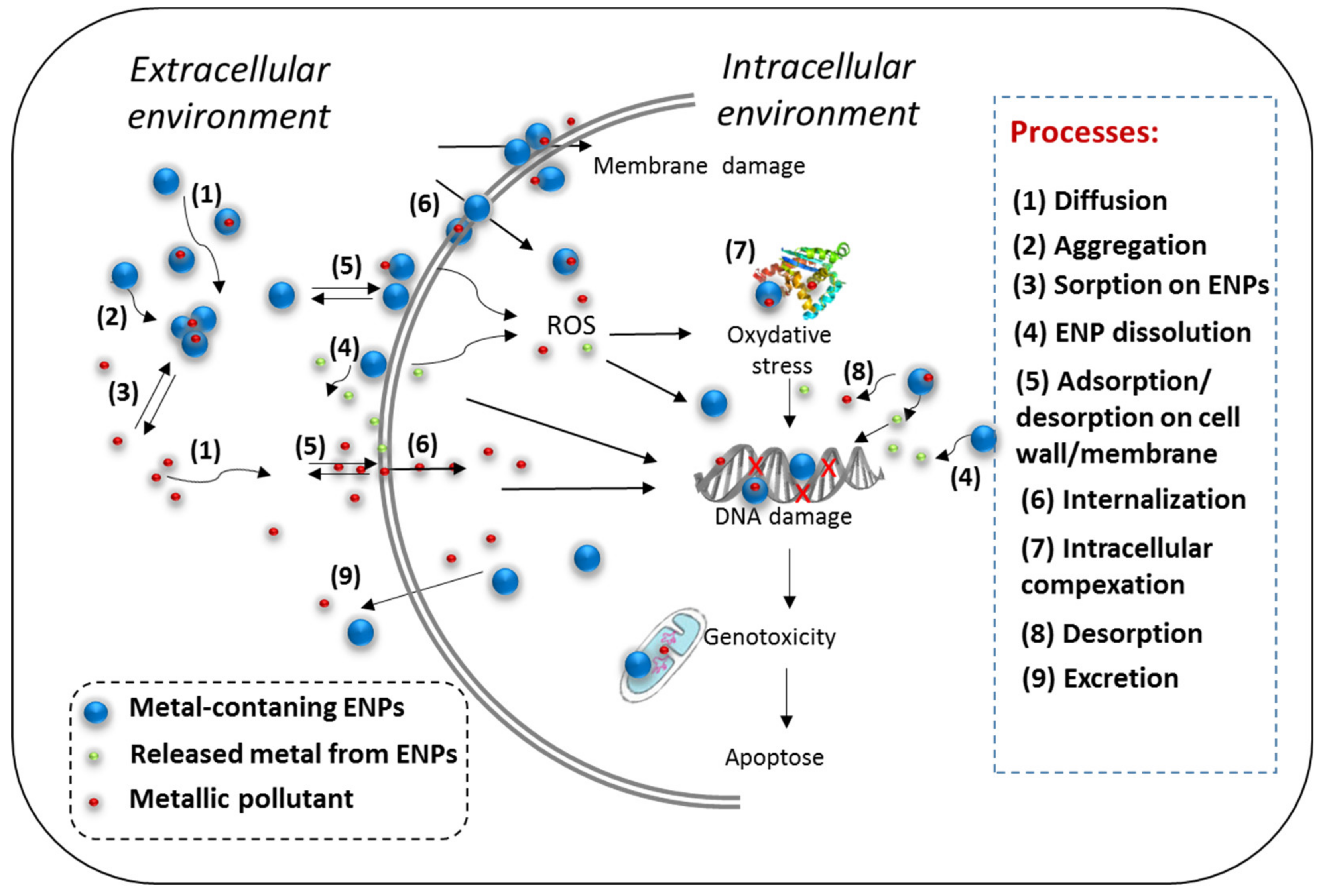
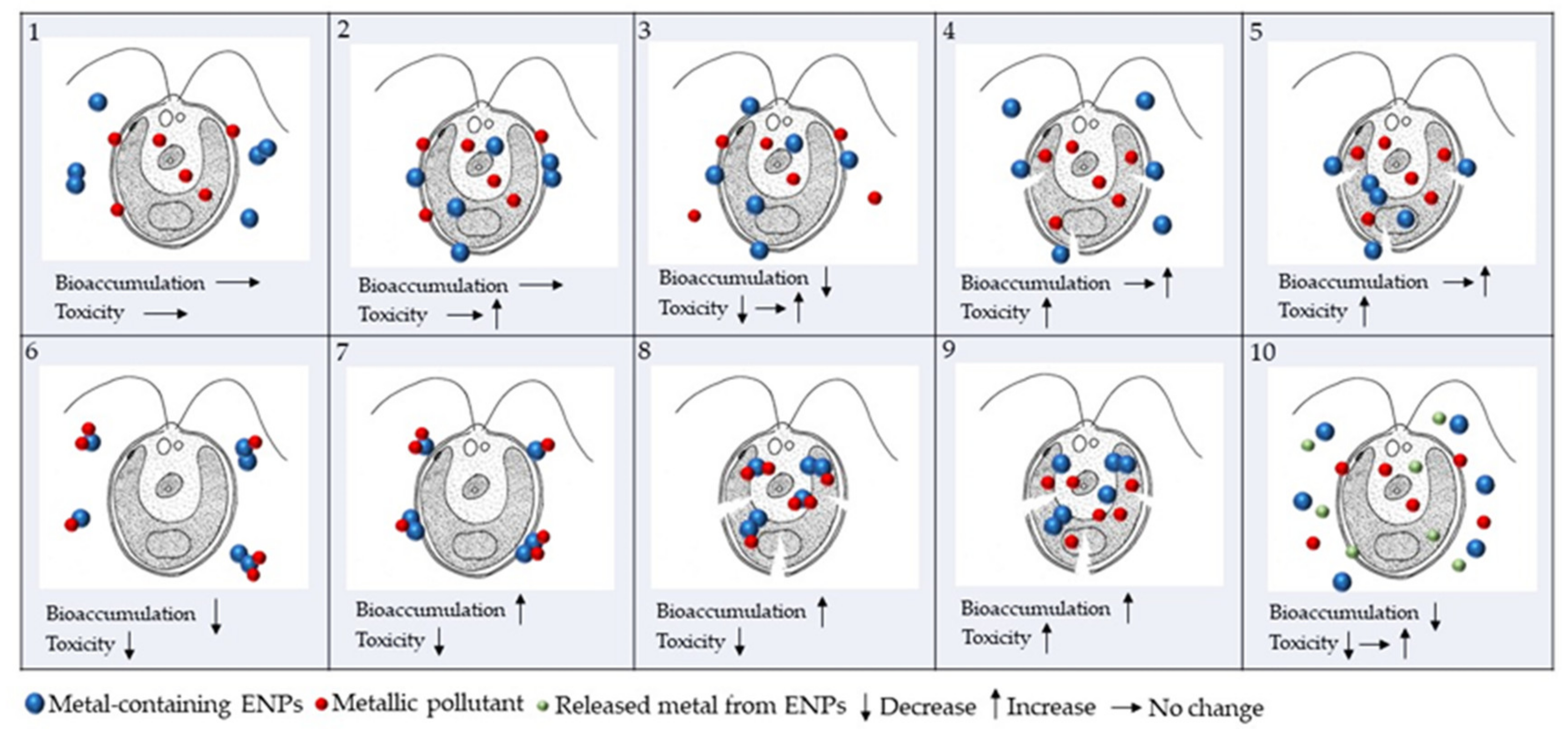
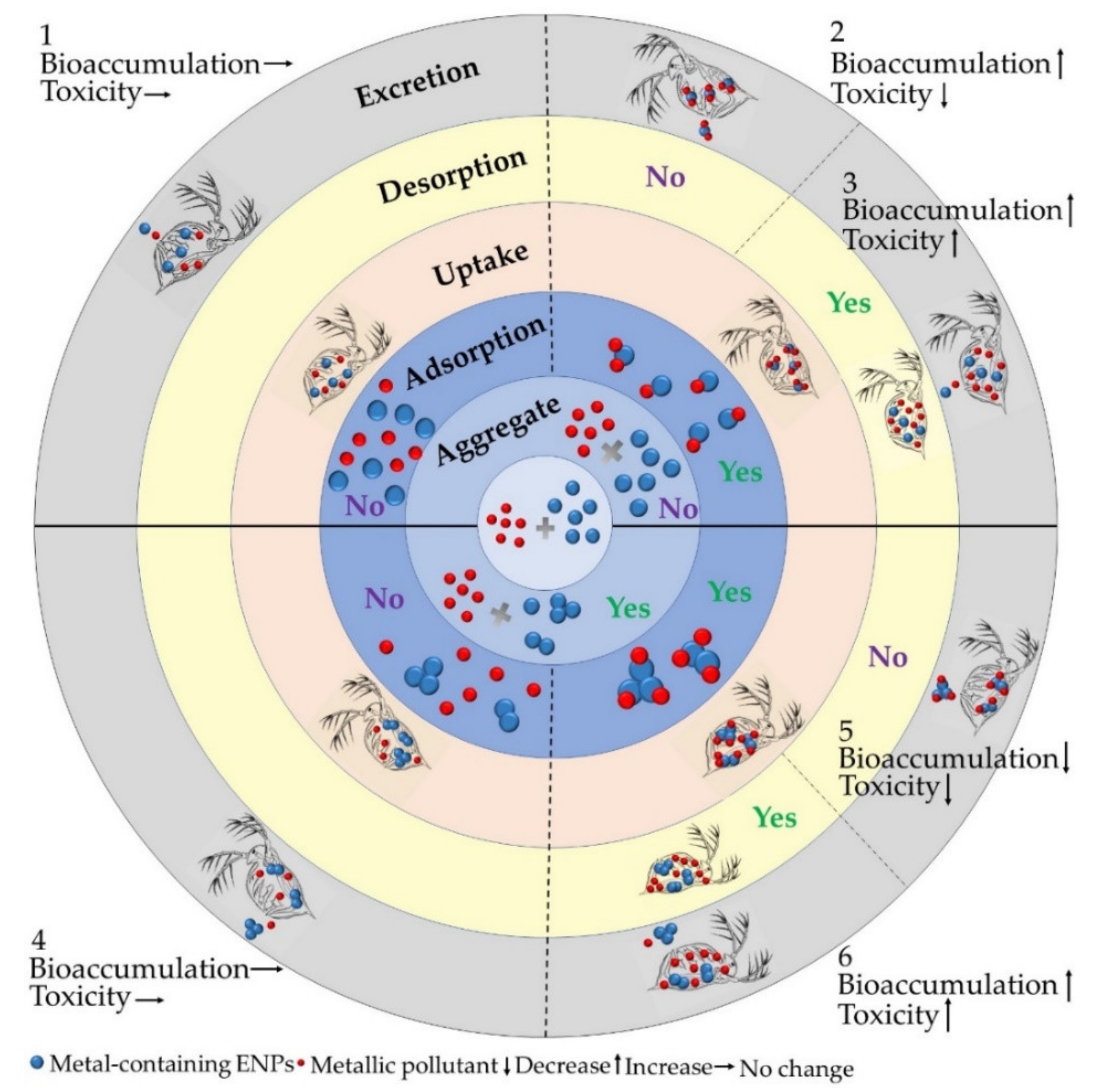
Table 1.
Selected recent examples of studies investigating the combined effects of metals and ENPs on plankton species including algae, bacteria, ciliate and zooplankton.
Table 1.
Selected recent examples of studies investigating the combined effects of metals and ENPs on plankton species including algae, bacteria, ciliate and zooplankton.
| ENPs | Metal | Organisms | Test Conditions | End Point/ Mixture Effect | Ref. |
|---|---|---|---|---|---|
| Al2O3 | Pb(II) | Marine alga Isochrysis galbana | 10–20 nm Al2O3NPs, modified Guillard f2 medium, Exposure time 72 h | Growth inhibition, chlorophyll fluorescence/Synergistic effect in the presence of 10mg L−1 Al2O3NPs | [110] |
| TiO2 | Cu(II) | Bacterium Bacillus thuringiensis | P25, Physiological saline buffer, Exposure time 24 h | Viability, ROS a generation, enzymatic activity, Cu uptake/Increase | [103] |
| TiO2 | Cu(II) | Bacterium Bacillus megaterium | P25, Physiological saline buffer, Exp. time 24 h | Viability, ROS generation, enzymatic activity, Cu uptake/No effect | [103] |
| TiO2 | Cd(II) | Bacterium Escherichia coli | Anatase 5–10 nm, Exposure time 3 h | Growth reduction, Antagonistic interaction | [111] |
| TiO2 | Pb(II) | Zebrafish larvae | 5 nm TiO2NPs, Exposure time 48 h | Mortality, malformation rate, No effect; Locomotion/Decrease; Biouptake and depuration/Increase | [112] |
| TiO2 | Cd(II), As(III), Ni(II) | Nematode Caenorhabditis elegans | Two sizes: 5 and 15 nm; strong aggregation Exposure time 12 h | Cd uptake/Decrease Reproduction and development toxicity/Increase | [113] |
| TiO2 | As(Ⅲ), As(Ⅴ) | Water flea Daphnia magna | 21 nm TiO2, Acute immobilization test, Exposure time 48 h | Immobilization/Decrease | [105] |
| TiO2 | As(Ⅲ), As(Ⅴ) | Water flea Daphnia magna | 21 nm TiO2, Chronic test, Exposure time 21 d | Body length and reproduction/Decrease | [105] |
| TiO2 * | Cu(II) | Water flea Daphnia magna | 29.5 nm TiO2 with surface modified by Al(OH)3, Exposure time 48 h | Bioaccumulation and oxidative stress/Increased | [114] |
| TiO2 * | Cu(II) | Water flea Daphnia magna | 36.7 nm TiO2 with surface modified by Al(OH)3 + stearic acid, 48 h | High intestinal damage | [114] |
| ZnO | Cd(II), Pb(II) | Marine copepod Tigriopus japonicus | 22 nm ZnONPs, artificial seawater Exposure time 96 h | Acute test, mortality/Increase | [104] |
| ZnO | Cd(II), Pb(II) | Marine copepod Tigriopus japonicus | 22 nm ZnONPs, artificial seawater Exposure time 96 h 20 °C, 21 days | Chronic test, Reproduction (spawning rate, time to hatch, Nb of nauplii)/Decrease | [104] |
| ZVI ** | Pb(II), Cd(II), Zn(II) | Bacterium Vibrio fischeri | ZVI 67 nm, Microtox® Test, Exposure time 5 min | Toxicity impact index / Increase (Pb(II)); antagonistic interaction Decrease (Cd(II), Zn(II)) Synergistic interaction | [115] |
| ZVI | Pb(II), Cd(II), Zn(II) | Nematode Caenorhabditis elegans | ZVI 67 nm, Exposure time 96 h | Growth, reproduction, survival Improvement (Cd(II), Zn(II)) No effect (Pb(II)) | [115] |
| ZVI | Pb(II), Cd(II), Zn(II) | Green alga Scenedesmus intermedius | ZVI 67 nm, Exposure time 72h | Growth rate inhibition NOEC b, IC50 c/Increase | [115] |
| ZVI | Pb(II), Cd(II), Zn(II) | Cyanobacterium Microcystis aeruginosa | ZVI 67 nm, Exposure time 72h | Growth rate inhibition NOEC, IC50/Increase | [115] |
*hydrophobic. **ZVI: zero valent iron. aROS: reactive oxygen species. bNOEC: non-observed effect concentration. cIC: mean inhibitory concentration.
© 2020 by the authors. Licensee MDPI, Basel, Switzerland. This article is an open access article distributed under the terms and conditions of the Creative Commons Attribution (CC BY) license (http://creativecommons.org/licenses/by/4.0/).
Share and Cite
MDPI and ACS Style
Li, M.; Liu, W.; Slaveykova, V.I. Effects of Mixtures of Engineered Nanoparticles and Metallic Pollutants on Aquatic Organisms. Environments 2020, 7, 27. https://0-doi-org.brum.beds.ac.uk/10.3390/environments7040027
AMA Style
Li M, Liu W, Slaveykova VI. Effects of Mixtures of Engineered Nanoparticles and Metallic Pollutants on Aquatic Organisms. Environments. 2020; 7(4):27. https://0-doi-org.brum.beds.ac.uk/10.3390/environments7040027
Chicago/Turabian StyleLi, Mengting, Wei Liu, and Vera I. Slaveykova. 2020. "Effects of Mixtures of Engineered Nanoparticles and Metallic Pollutants on Aquatic Organisms" Environments 7, no. 4: 27. https://0-doi-org.brum.beds.ac.uk/10.3390/environments7040027
Note that from the first issue of 2016, this journal uses article numbers instead of page numbers. See further details here.