Article

Light Spectra during Somatic Embryogenesis of Norway Spruce—Impact on Growth, Embryo Productivity, and Embling Survival

Natural Resources Institute Finland (Luke), FI-57200 Savonlinna, Finland
*
Author to whom correspondence should be addressed.
Submission received: 27 January 2021 / Revised: 18 February 2021 / Accepted: 2 March 2021 / Published: 5 March 2021

Abstract

:
For Norway spruce (Picea abies (L.) Karst.) somatic embryogenesis (SE) culture conditions throughout the propagation process affect the final result. Many critical phases can be identified, and all of them cumulatively increase the production costs of SE plants if they cannot be controlled. In order to determine the best lighting protocol for each SE step, Norway spruce embryogenic tissue (ET) was proliferated, and somatic embryos were matured under different light wavelengths, wavelength combinations, and in the dark. Overall, using low-intensity LED lights during proliferation or at the end of maturation had little effect on the growth of ET, embryo productivity, or embryo survival; on the other hand, major negative effects could not be seen. This is beneficial from a practical point of view, indicating no need for lighting or protection of SE cultures from light during their handling in these steps of the propagation process. When somatic embryos were germinated under different spectra, significant differences in embling shoot and root growth, as well as in the survival of the emblings, were found. The best treatment varied between trials, and the genotype of the SE culture was found to have a stronger effect than the light spectrum, indicating that various light spectra and also intensity adjusted using pulse width modulation (PWM) can be successfully applied to the SE germination phase in Norway spruce.

1. Introduction

In the conditions found in the Nordic countries, forest biomass is considered to be the most important raw material in the developing bioeconomy, and the demand for tree biomass has increased due to the growing need for substitutes for fossil fuels, among other reasons. Using the best possible material, i.e., genetically improved stock in forest regeneration of Norway spruce (Picea abies (L.) Karst.), as much as over 30% in stem volume growth can be gained [1]. In order to most efficiently implement the results achieved by tree breeding, material derived via vegetative propagation could be used. Vegetative, i.e., asexual propagation, enables the production of plants of uniform quality and with known, selected characteristics. Somatic embryogenesis (SE) has become the method of choice for vegetative propagation of conifers [2] due to its high multiplication rate and the maintenance of juvenility of cell lines via cryopreservation. Of the Nordic conifers, somatic embryogenesis (SE) is currently the most developed for Norway and white spruce varieties (P. glauca (Moench) Voss) [3,4,5].
Research into SE methods for Norway spruce started over 30 years ago [6,7,8], and since then protocols have been developed in different laboratories [4,9,10]. The production of somatic embryos and emblings involves initiation, proliferation, maturation, optional desiccation, germination, acclimation, and growing in nurseries until planting [3,10]. Culture conditions and handling throughout the propagation process affect the final result of SE production, with many critical phases identified in the process, and all of them cumulatively add to the production costs of SE plants if they cannot be controlled [11]. A good embryo production capacity and embling survival during acclimatization are essential to achieving a desirable propagation method for practical use [12].
Conifer somatic embryo development in vitro differs from the development of seeds and zygotic embryos, for example somatic embryos lack the protective layers of cone and megagametophyte [13]. This has justified carrying out proliferation and maturation in the dark. However, developing seeds get information from their mother plants, and maternal light conditions have been proven to affect aspects such as the seed weight, germination ability, and storability [14].
For the germination of somatic conifer embryos, different light spectra have been used, although an early study by von Arnold and Hakman postulated that the germination of somatic embryos of Norway spruce in darkness would improve the root growth [15]. Kvaalen and Appelgren [16] found that both germination and the root growth of somatic embryos of Norway spruce strongly depend on the light source, with red being the most beneficial spectrum. Red wavelengths during pre-germination and germination steps resulted in higher frequencies of somatic embryo germination, longer tap roots, and more first-order lateral roots than the standard cool white, fluorescent treatments or treatment with blue wavelengths for three pine species [17]. However, studies using Norway spruce seeds have shown that the germination percentage was higher when the combination of blue and red light was 55:45 than when the portion of blue light was lower, or when seeds were germinated under high-pressure sodium lamps [18]. In addition, Norway spruce seedlings grown under light high in far red spectra resulted in taller seedlings and higher shoots and root dry matter production than under light high in red [19].
Only a few studies of the effects of different spectra have been carried out using embryogenic tissue (ET) of conifers during proliferation or maturation. Even then, the growth of Norway spruce ET has been found to be strongly genotype-dependent when proliferated in different lights or in darkness [20]. In some early works, proliferation and maturation have been carried out in dim light [21], but later protocols have deployed darkness.
Light emitting diodes (LED) are favored nowadays over fluorescent or high-pressure sodium (HPS) lights for plant production. LED lights use less electricity to generate light and last longer than comparable light setups and are, thus, more efficient. Especially HPS lights, but also fluorescent lights, generate a lot of heat, which often requires additional ventilation equipment to maintain a proper growing room temperature. Pulse width modulation (PWM) is a popular method for light dimming control in general lighting applications [22]. In PWM the average value of the voltage (and current) fed to the load is controlled by turning the switch between the supply and load on and off at a fast rate. When connected to LEDs containing diodes of different wavelengths it can be used to control the spectra and intensity. Some studies have been made with LED PWM connected and using Arabidopsis thaliana, for example, as plant material [23], but no studies have been made using conifers or SE material.
This study is, to our knowledge, one of the few utilizing different stages of conifer SE material in light studies to this extent. Usability of different LED techniques in Norway spruce SE production instead of fluorescent lamps was evaluated in large-scale experiments. The main objective of the study was to obtain information about the effects of different wavelengths and combination of wavelengths in different steps of SE production. In order to determine the best protocol for each step, the ET growth, embryo production capacity, germination ability of the embryos, and embling growth parameters were measured.

2. Materials and Methods

2.1. Origin of Embryogenic Lines

Continuously subcultured or cryopreserved and thawed embryogenic lines of Norway spruce used in this study were initiated in 2014 and 2015 from immature zygotic embryos originating in the full-sib families of progeny tested plus trees from Southern Finland (Supplementary Tables S1 and S2). Immature green cones were collected for explant excision approximately ten weeks after pollination when the heat sum was around 800 d.d. and the zygotic embryos had reached cotyledonary stage.
The medium and methods developed by Klimaszewska et al. [24], as described by Varis et al. [25], were used for culture establishment. In short, immature zygotic embryos, without megametophytes, were placed on modified Litvay’s medium (mLM) containing half-strength macroelements [26] and 10 μM of 2,4-dichlorophenoxyacetic acid (2,4-D) and 5 μM of 6-benzyladenine (BA). The sucrose concentration of the medium was 1% (w/v), and the pH was adjusted to 5.8 prior to adding gellan gum (Phytagel™, Sigma-Aldrich, Merck KGaA, Darmstadt, Germany, 4 g/L) and autoclaving. The cultures were kept in the dark (24 °C) for 2 to 8 weeks without subculturing, until embryogenic tissue (ET) started to grow. Established ETs were subcultured bi-weekly, on fresh Petri dishes with the same medium.
Cryopreservation and thawing of ETs were done according to Varis et al. [25]. From each genotype one to four samples were cryopreserved right after initiation using an increasing sucrose content in mLM medium (0.1 M and 0.2 M sucrose for 24 h for each concentration) as preconditioning. After that, 200 mg of ET was placed in sterile cryovials containing 400 µL of liquid mLM medium with 0.4 M of sucrose without plant growth regulators (PGR) or glutamine, and 400 µL of preservative mixture containing polyethylene glycol 6000, glucose, and DMSO 10% w/v each was added. Cryovials were cooled at the rate of 0.17 °C/min to −38 °C in a programmable cooling device (Planer, Kryo 10 Series III, Planer Products, Middlesex, UK) followed by immersion in LN. Vials were thawed in a water bath at +37 °C for 2 min and drained by suction on sterilized filter paper (Whatman #2, Whatman International Limited, Kent, UK) placed in a Büchner funnel. The filter papers with samples were placed on a solid mLM medium with a sucrose content of 0.2 M and transferred every 24 h onto media with a decreasing sucrose concentration (0.1 M and 0.03 M).

2.2. SE Development, Germination, and Plant Production

For the maturation in all experiments, from 150 to 200 mg of fresh embryogenic mass was weighed and dried on filter paper (Whatman #2), using a Buchner funnel with suction, as was done by Varis et al. [25]. The maturations were made in three replicates per line using petri dishes filled with 28 mL of mLM maturation medium containing 30 µM of (±)-abscisic acid (ABA) [27]. The maturations were kept at +24 °C in the dark, except for the maturation experiment in Section 2.4. After eight weeks, the number of cotyledonary embryos per gram of embryogenic mass (fresh weight) was counted (E/gFW), after which the embryos were stored on filters on the original maturation medium at +2 °C in the dark until germination.
The germination of the embryos, as well as measuring and growing the germinated embryos, i.e., the emblings, was done according to Tikkinen et al. [28]. For germination, seven to ten embryos were placed on an mLM medium without plant growth regulators and inorganic ammonium nitrate (NH4NO3), supplemented with 20 g/L of sucrose. The media contained 950 mg/L of potassium nitrate (KNO3) and 500 mg/L of L-glutamine as sources of nitrogen. During germination, the petri dishes were kept in a tilted position so that the cotyledons faced up. The embryos were germinated for two weeks in a growth room where the air temperature was set to +20 °C, and an 18:6 day/night light period was applied [28].
Increasing light intensity was used for the germination: 6 µmol/m2/s for the first four days, 60 µmol/m2/s for the three days, and 130 µmol/m2/s for the last seven days, except for fluorescent lights which produced only 120 µmol/m2/s. The intensity was adjusted using either layers of a tissue or cloths or adjustable 3k LED lights. Light intensities were measured using a Li-cor portable photometer (Li-188B, LiCor Inc., Lincoln, NE, USA), and the wavelength distribution was detected using a Gigahertz Optik Luxmeter (BTS256-EF BiTec Sensor, Gigahertz-Optik GmbH, Türkenfeld, Germany). The data were analyzed using S-BTS256 V2015.4 and R software (R Core Team, 2011) together with the packages for photobiology (http://www.r4photobiology.info/, accessed on 4 March 2021) [29]. Emblings were photographed with a Canon Powershot G5, and the length of the shoot (hypocotyl) and root meristems on the embryos were measured using ImageJ software as shown in Figure 1 (version 1.48v; Java 1.8.0_51 [32-bit]) [30].
The following hardware was used to produce light depending on the experimental set up: AP67 spectrum and intensity LED lights (Valoya L14, Helsinki, Finland) (hereafter AP67), fluorescent lights (Phillips TL-D, Amsterdam, The Netherlands), or adjustable LED lights (Electronic 3k Factory, South-Eastern Finland University of Applied Sciences, Savonlinna, Finland) (hereafter adjustable 3k LED). Adjustable 3k LED lights use the pulse width modulation (PWM) dimming technique, which is based on switching the lights on and off at intervals. Adjustable 3k LED lights were installed using blue (peak wavelength of 465), green (peak 530), red (peak 660), and far red (peak 730) diodes. Warm white diodes were also added. The LED lights were placed in shelves in a 10 m2 office room. Different spectra were separated using thick curtains. Fluorescent lights were either in a similar room or in a climatic chamber (Pol-Eko KK350, Wodzisław Śląski, Poland).
The emblings were transplanted into small growing containers (Preforma 126/JIF, ViVi Pak, KH ’s-Gravendeel, The Netherlands), with 126 plugs per container (plug volume: 3.4 mL) and were kept under AP67 lights (130 µmol/m2/s) following the GT-I method described by Tikkinen et al. [28]. After three weeks, the foils of the containers were removed, and the emblings were irrigated for another three weeks, after which the survival of the emblings was noted. As an exception, in Experiment III Trial 1 the growing containers were kept under AP67 spectrum lights and fluorescent lights in a climatic chamber, for four weeks, after which the survival of emblings was noted. Unfortunately, due to a technical problem, adjustable 3k LEDs were not used during the growing, and the spectra inside climatic chamber was not measured. The intensity in the climatic chamber was the same as for the other lights, except that the light came also from the sides. In the climatic chamber the humidity was controlled and maintained between 50% and 60% RH. The humidity of the rooms with the LED lights was also increased using a humidifier to keep the humidity above 50% RH. The temperature in the climatic chamber and rooms was controlled and maintained at 23 and 20 °C in an 18:6 photoperiod. Trial 1 of Experiment III was the first in the timeline, and in later trials the AP67 spectrum was used because of the limited space under the adjustable 3k LED lights or in the climatic chamber.
Because the luminosity of the blue, green, and far red diodes in adjustable 3k LED lights were weaker than the luminosity of the red LEDs, in Experiment I the intensities during proliferation, and in Experiment II Trial 2 intensities during maturation, were adjusted according to the weakest LED far red level, to 6–7 µmol/m2/s. However, germinating plants need a higher intensity than 7 µmol/m2/s to grow properly. Thus, in Experiment III in Trial 3 the intensities during germination were adjusted to the same increasing levels as described above using an additional red diode together with blue, green, or far red diodes.
Experimental design for the present study is summarized in the Table 1. Experiment 1 focused on proliferation phase, Experiment 2 on maturation of somatic embryos, and Experiment 3 on their germination, with the following details.

2.3. Experiment I: Proliferation in Different Spectra

The effect of different light spectra during the proliferation was studied in two trials. In Trial 1, 11 continuously subcultured (no cryopreservation) embryogenic lines of Norway spruce from six families initiated in 2015 were proliferated in December 2016 under adjustable 3k LED lights in blue and red spectra, as well as in the dark. In Trial 2, the same lines were proliferated in February 2017 under adjustable 3k LED lights in green and far red spectra, and in dark. Intensities were adjusted to level 6–7 µmol/m2/s in both trials. Two subcultures were made, and maturations were started in February 2017 in Trial 1 and in April in Trial 2.
Each line was proliferated in one petri dish filled with 21 mL of mLM medium and a weighed amount of ET divided into 15 clumps. After two weeks’ proliferation, the ET was weighed and subcultured in a new medium. Weighing and subculturing was repeated once or twice depending on the condition of the ET. The maturation, germination of the embryos, and growing of the emblings in growth containers were conducted as explained in Section 2.2. Seven well-proliferating lines were maturated in both trials, and five of them produced enough embryos for germination in both trials and for every spectrum tested. For the germination and growing emblings the AP67 spectrum was used.

2.4. Experiment II: Maturation of Embryos in Different Spectra

The effect of different light spectra at the end of the maturation process was studied in two different trials. In Trial 1, nine continuously subcultured embryogenic lines of Norway spruce from seven families initiated in 2015 were maturated in January 2017. After 32 days of maturation in the dark, the petri dishes were placed under lights with the following light spectra: (a) blue:green:red:far red in the ratio of 30:15:40:15, and (b) blue:red:far red in the ratio of 15:80:5, (c) AP67, and (d) in the dark (Figure 2). In Trial 2, the same lines were maturated in February 2017, and after 31 days of maturation in the dark, the petri dishes were placed under lights with the following spectra: (a) blue, (b) green, (c) red, or (d) in the dark. In both trials the maturating embryos were placed for 26 days in different spectra. The intensity was 6–7 µmol/m2/s. Six lines produced enough cotyledonary embryos (with at least four cotyledons) for germination in both trials and for each spectrum. The measurements were taken, and the germination of the embryos was controlled as described in Section 2.2. Light in the AP67 spectrum was used for the germination.

2.5. Experiment III: Germination of Embryos in Different Spectra

The effect of different light spectra during the germination of embryos was studied in three different trials. At first, to test the suitability of the LED lights and the PWM technique, seven continuously subcultured and three cryopreserved lines from seven families initiated in 2014 and 2015 were matured in May 2016. Trial 1 was conducted in two lots in August and September 2016. Two petri dishes per line filled with seven to ten mature embryos were placed under the following spectra: (a) AP67; (b) fluorescent lights; and (c) blue:green:red:far red at a ratio of ratio 30:15:40:15. An increasing light intensity was used as follows: 6 µmol/m2/s for the first five days; 60 µmol/m2/s for the next three days; and 130 µmol/m2/s (fluorescent lights 120 µmol/m2/s) for the last six days. After two weeks of germination, the lengths of the roots and shoots were measured from pictures. All the emblings germinated under fluorescent lights were grown in fluorescent lights in a climatic chamber. Half of the emblings germinated in AP67 or blue:green:red:far red were grown in the spectrum AP67, half of them were first placed in blue:green:red:far red light, but due to technical problems they were moved to a climate chamber after seven days. In exception to the other trials, the survival of the emblings was noted after four weeks instead of six weeks.
In Trial 2 different spectra and intensities were tested using the following spectra: (a) AP67; (b) blue:green:red:far red at a ratio of 30:15:40:15; and (c) blue:red:far red at a ratio of 15:80:5. The intervals of the intensity periods were shortened for spectra b and c in order to raise the accumulating intensity to 6 µmol/m2/s for the first three days, 60 µmol/m2/s for the two days, and 130 µmol/m2/s for the last nine days. The intensity level was not raised at the end to avoid heating of the air inside the petri dishes. In February 2017 embryos from six lines initiated and cryopreserved in 2014 and thawed and matured in autumn 2016 were placed on petri dishes. Nine embryos were placed in one petri dish, and three petri dishes per line were placed under every light spectrum.
In Trial 3 the effect was tested of the individual colors of the light using adjustable 3k LED lights. The lights used were in the blue, green, red, and far red spectra. The intensities were adjusted to the same level using an additional red diode together with blue, green, or far red. The highest intensity gained with only blue and green LEDs was 60 µmol/m2/s, and with far red LEDs it was 6 µmol/m2/s. Valoya AP67 lights were used as controls. Trial 3 was conducted in three lots; in the first lot embryos from five continuously subcultured lines initiated in 2014 and 2015 and matured in October 2017 were germinated in January 2018. In the second lot embryos from five cryopreserved lines initiated in 2014 were thawed and matured in spring 2018 and germinated in August 2018. In the last lot, five cryopreserved lines initiated in 2014 and thawed and matured in spring 2018 were germinated in September 2018. In Trials 2 and 3 the measurements and the growth of the emblings were carried out as described in Section 2.2.

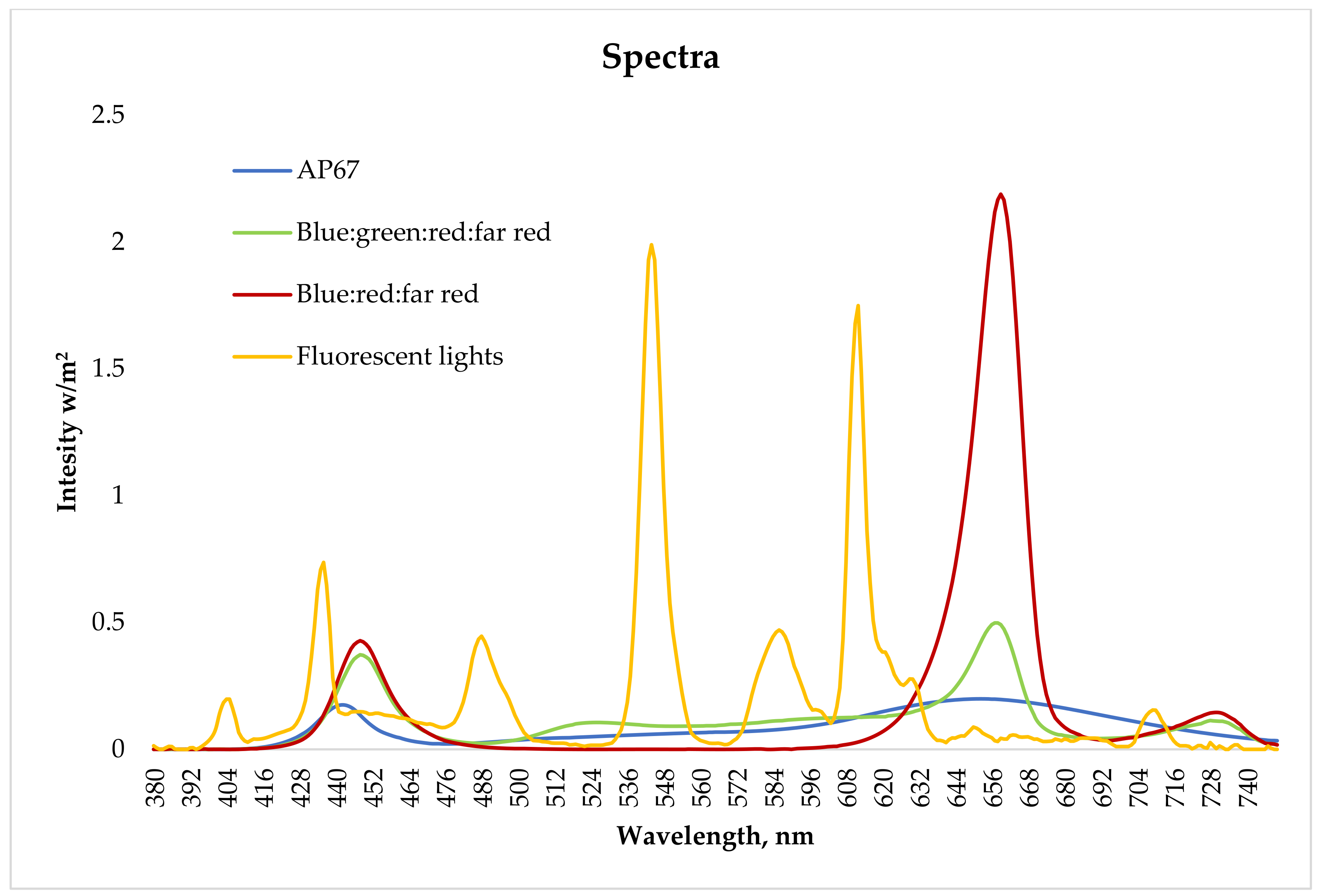
2.6. Measurements and Data Analysis

The ET growth percentage was counted as (weight at subculture 1−weight at the beginning)/weight at the beginning *100. Cotyledonary embryos were counted, and the embryo production capacity was calculated per one gram of fresh cell mass (E/gFW). The mean values are presented with the standard error (±). For the mean ratios of ET growth between the groups of each line or spectrum, and the root length, shoot length, or root-to-shoot ratio between groups of dead or alive emblings, Levene’s test was used to analyze the equality of error variances. For equal error variances, the differences in means were analyzed using a one-way ANOVA with a Tukey test for post hoc comparisons. If the variances of the groups were unequal, an analysis was done using a Kruskall–Wallis test (K-W test) or Mann–Whitney U test with Tamhane’s T2 test for post hoc comparisons. Considering means ratios of E/gFW, the root length, shoot length, or root-to-shoot ratio a univariate generalizer linear model (GLM) was used with the line and spectrum as fixed factors. Descriptions of which statistical methods were used in the different experiments together with p-values are provided in Supplementary Tables S4, S5, S7, S8, and S10. Differences in embryo production, root and shoot length, and survival % between SE lines are illustrated in Supplementary Figures S1–S6.
A logistic regression was used to examine the differences in survival of the emblings (0 = dead, 1 = alive), while the genotype and spectrum were used as covariates. Survival percentages are presented to illustrate the levels of survival in different spectra or SE lines. The effect of the root and shoot length, root-to-shoot ratio, and location inside the growth containers was studied in Experiment III in Trials 1, 2, and 3. The effects were not significant and decreased or increased the percentage of correctly predicted cases by less than 0.5%; hence, those covariates were excluded from the final models in all experiments. Descriptions of the models used are provided in Tables S11–S14. All measurements and inventory results were analyzed using IBM SPSS Statistics 26. The level of confidence used was 95%.

3. Results

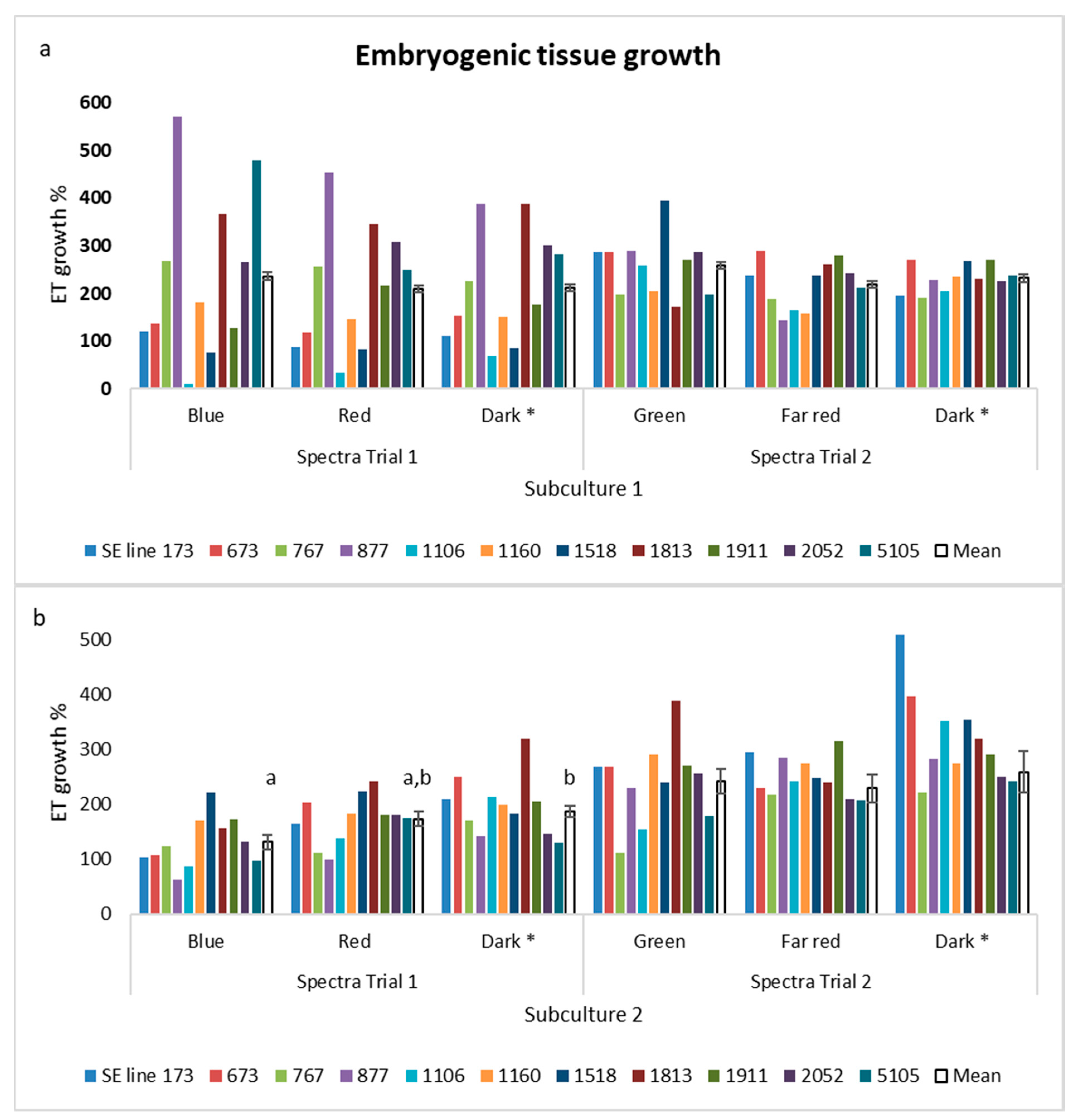
3.1. Experiment I: Proliferation in Different Spectra

The mean growth percentages varied between genotypes (from 10 to 570%), but not between spectra for the first subculture (Figure 3a, Supplementary Tables S3 and S4). For the second subculture the ET growth percentage was higher in the dark (187%) than under blue light (131%) (Figure 3b). The ET growth percentage was better in the dark than under any light also in Trial 2, but the differences were not significant. The ET growth percentage in the dark was higher in Trial 2 than in Trial 1, and in the second subculture it was significantly higher.
More differences were found in the embryo yield. In both trials, the embryo yield differed according to the light used and the SE lines (Figure 4, Figure S1a, Table S4). Similarly, as for the ET growth, the lowest embryo yield was for proliferation in blue light (123 E/g FW) and was the best in the dark (214 E/g FW). In Trial 2, the lowest embryo yield, 42 E/g FW, was when ET had been proliferated under far red light, while under green light it was 73 E/g FW, and in the dark it was 66 E/g FW. Opposite to the ET growth percentage, more embryos were produced from ET proliferated in the dark in Trial 1 than in Trial 2.
In Trial 1, the root length and root-to-shoot ratio differed significantly between the SE lines, and the differences were the same across the light spectra (Figure 5, Figure S2a, Table S5). In Trial 2, the roots of the germinated embryos matured from ET grown in the dark were longer (11 mm) than the roots of embryos matured from ET grown under green light (8 mm). Additionally, the shoot length varied: the longest shoots were for emblings germinated from ET proliferated under far red light, while the shortest shoots were from emblings that had been proliferated under green light. The root-to-shoot ratio was the highest for the embryos that had been grown in the dark.
Differences in embryos matured in the dark in Trials 1 and 2 were contradictory: the roots were longer in Trial 2, while the shoots were longer in Trial 1, and the root-to-shoot ratio was higher in Trial 2.
When the survival of the emblings (dead/alive = 0/1) was modeled using SE lines and spectra as covariates, the effects were not significant, and they decreased or increased the correctly predicted cases by less than 0.5%. Survival percentages are presented to illustrate the levels of survival in different spectra (Figure 6) or SE line (Figures S5 and S6).

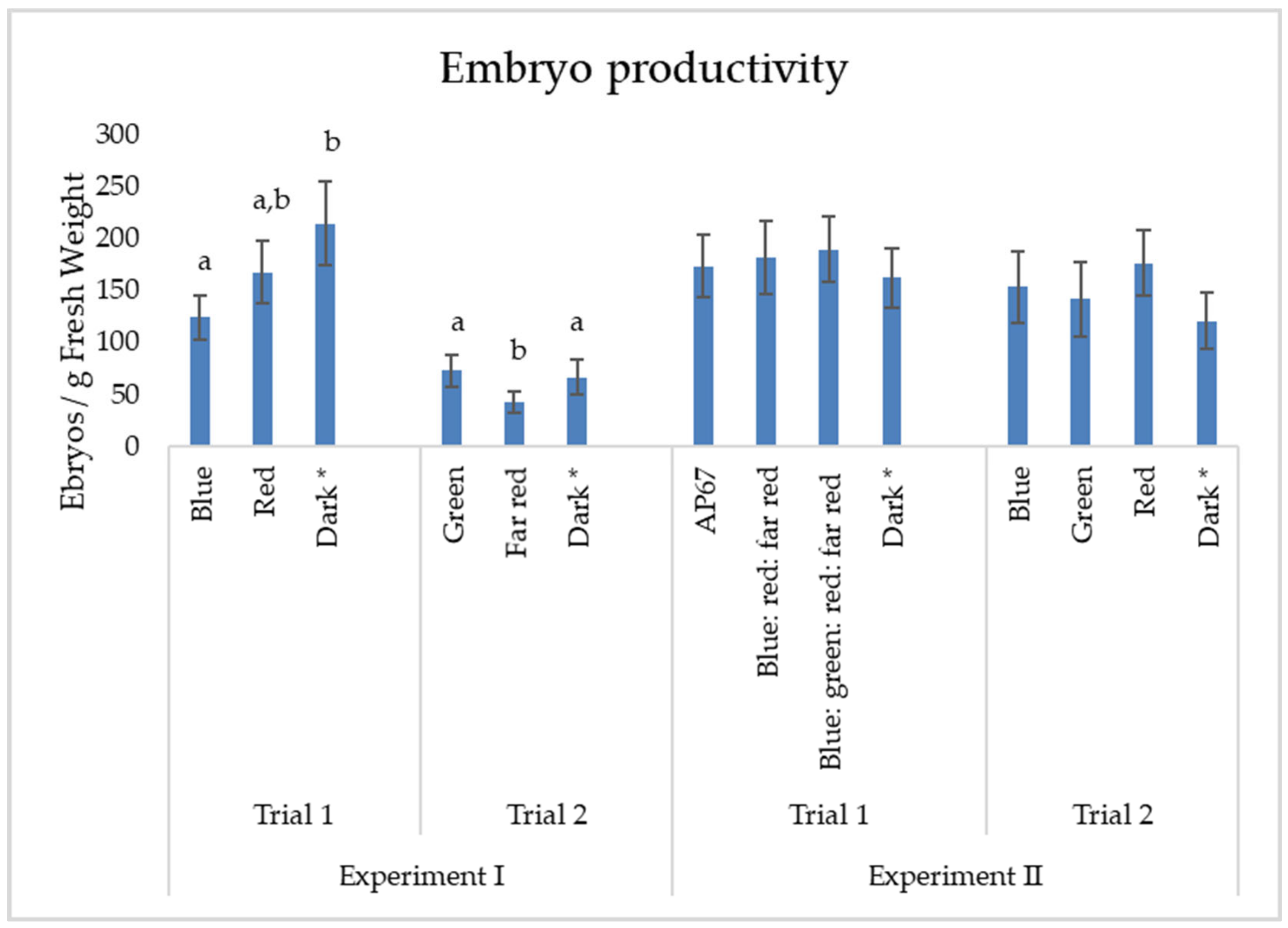
3.2. Experiment II: Maturation of Embryos in Different Light Spectra

Maturing the embryos under different light spectra or in the dark had no effect on the embryo yield. Variation occurred only between the SE lines (Figure 4, Figure S1b, Tables S6–S8). More embryos were gained from the ET proliferated in the dark in Trial 1 (162 E/g FW) than in Trial 2 (121 E/g FW), while some variation was found also between the SE lines.
In all trials the genotype affected the root length, shoot length, and root-to-shoot ratio more than the light spectrum (Figure S2b). In Trial 1, when the embryos were maturated in the dark, the shoots of the emblings were longer than for emblings from maturations in any light (Figure 5). The mean length of the roots did not differ for the different spectra; thus, the root-to-shoot ratio was the lowest when embryos were matured in the dark.
In Trial 2, emblings from maturations under green had the longest roots and shoots and the highest root-to-shoot ratio. The roots of the emblings that had undergone maturation in the dark were longer in Trial 1 than in Trial 2. The root-to-shoot ratio was higher in Trial 1.
In Trial 1 the effects of the SE line or spectrum on survival of the emblings were not significant and increased the correctly predicted cases by less than 0.5%. In Trial 2 the survival rates varied between SE lines but not among tested spectra.

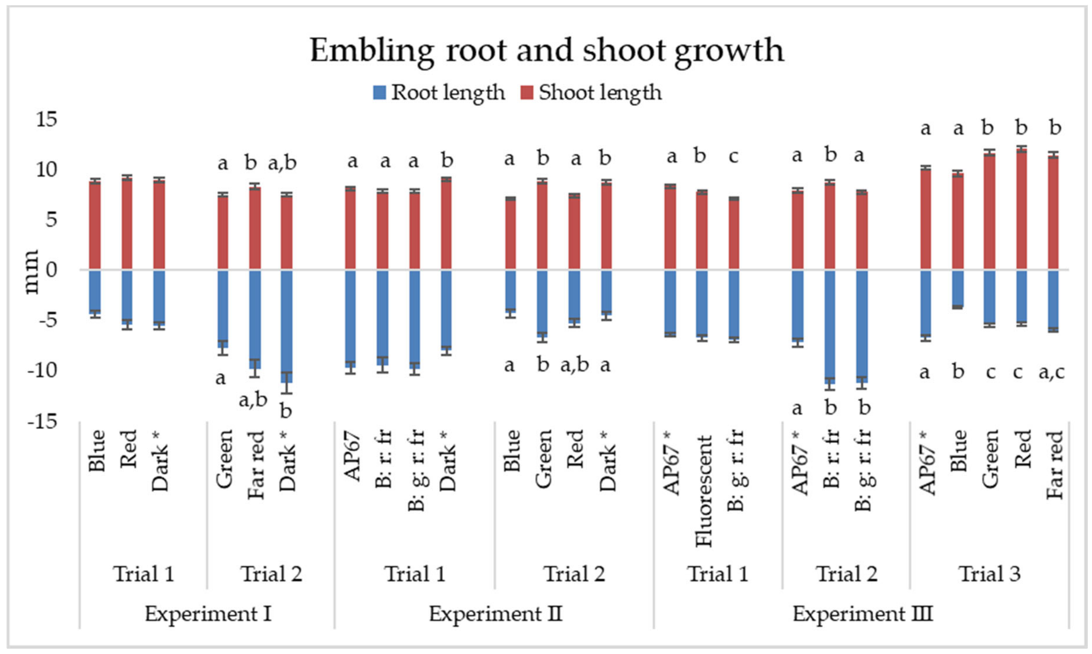
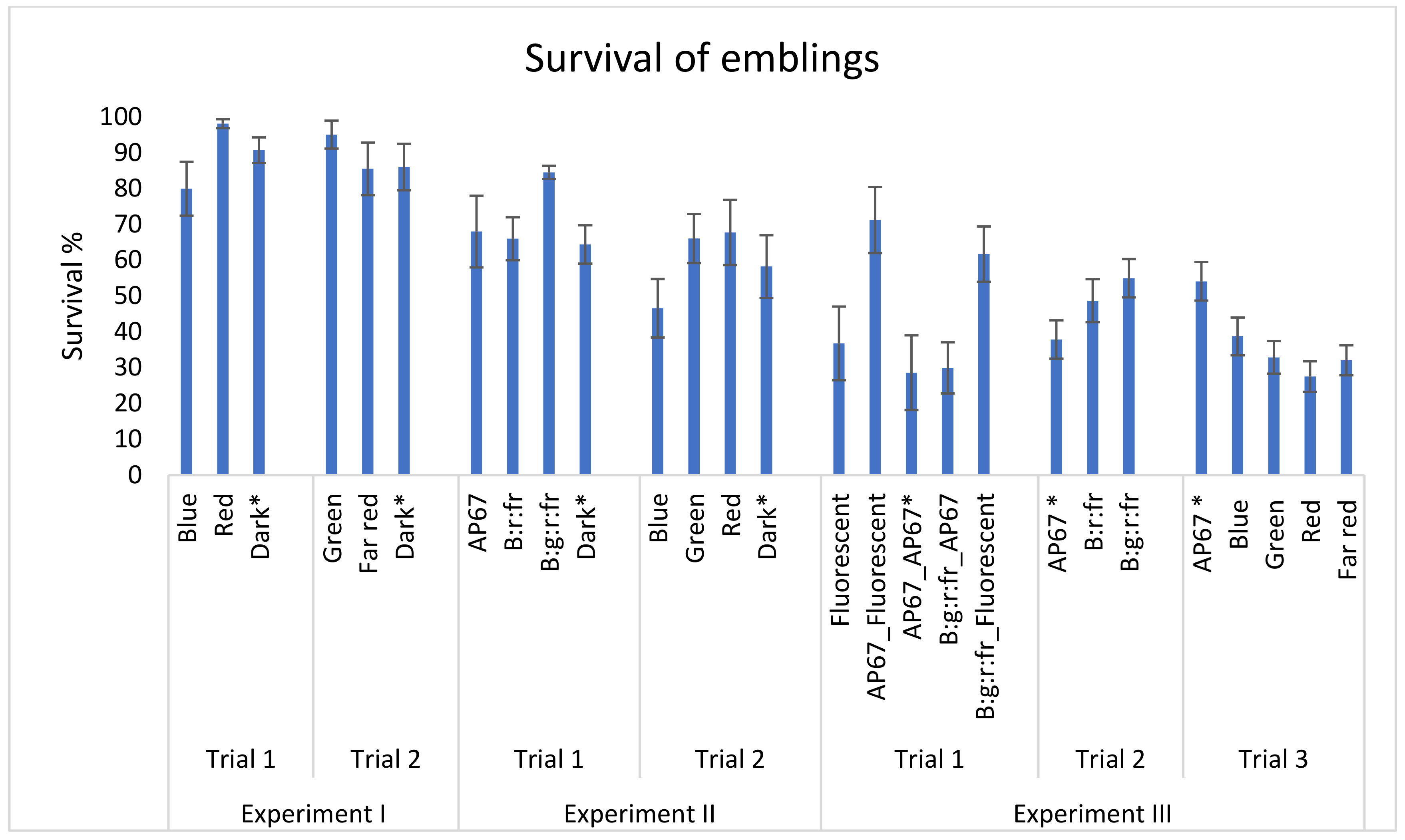
3.3. Experiment III: Germination of Embryos in Different Light Spectra

The effect of different light spectra was more significant in Experiment III than the previous experiments; however, the SE line also affected the germination more than the spectra in this experiment (Tables S9 and S10).
In Trial 1 when the cotyledonary embryos were germinated under spectrum AP67 light, the shoots grew longer than under fluorescent lights or under blue:green:red:far red spectra (Figure 5). Because the root lengths did not differ according to the spectra, the root-to-shoot ratio was lower for the embryos germinated under spectrum AP67 light. In Trial 2, the roots of the embryos were shorter for those germinated under AP67 light than under the blue:red:far red or blue:green:red:far red spectra, and shoots were longest for those germinated under the blue:red:far red spectra; thus, the root-to-shoot ratio was lowest under the AP67 light. In Trial 3 the AP67 and far red spectra were the most beneficial spectra for root growth and green, red, and far red were the most beneficial spectra for shoot growth. The root-to-shoot ratio was highest when the embryos were germinated under AP67 and far red light.
The survival ratio varied between the SE lines in Trial 1, and the spectrum during embling growth had an effect on embling survival; however, the spectra during embryo germination had only a small effect. The highest survival % was when the embryos were germinated under AP67 light and the emblings were grown in a climate chamber under fluorescent lights (71%). The lowest survival % was when both germination and growth were under AP67 lights (29%). In Trial 2, the survival ratio varied between the SE lines and spectra. The best survival percentage was when the embryos were germinated under blue:green:red:far red, light. In Trial 3, the survival rate also varied between the SE lines and spectra. The best survival percentage of the emblings was when the embryos were germinated under light in the AP67 spectrum.

4. Discussion

4.1. The Effect of Light on the Proliferation of ET and Maturation of Somatic Embryos

Genotypic differences explain most of the differences in every experiment and trial, and the differences between SE lines were in some cases remarkable. However, for practical applications SE protocols must be suitable for a wide range of genotypes. Therefore, a larger number of genotypes, from 6 to 13 depending on the trial, were tested in the present study.
Our study showed that low-intensity lighting during the proliferation of Norway spruce ET and during the maturation of SE embryos had some disadvantages but also advantages. Blue light during proliferation inhibited the growth of ET, but also reduced the number of mature embryos (E/g FW). On the other hand, when maturing embryos were exposed to blue light no effect was found on the number of good quality mature embryos (E/g FW). Green light during proliferation enhanced embryo productivity, but the roots and shoots of the germinated embryos were shorter than embryos from ET proliferated under far red light or in the dark. In addition, maturation under green light enhanced the growth of roots and shoots.
The effect of blue light is in line with the study by Latkowska et al. [20], in which blue light inhibited the growth of two Norway spruce ET genotypes out of three studied. In that study the light intensity was 30 µmol/m2/s, which was higher than in our proliferation and maturation experiments (6–7 µmol/m2/s). In practice, when ET is subcultured or mature embryos are counted for research purposes, the lights may be more intense than in our study, but the time of exposure to the lights is usually short, and the intensity of blue light is minor in fluorescent or LED lamps used for lightning in laboratory rooms or with stereo microscopes. When von Aderkas exposed maturing hybrid larch (Larix x marschlinsii) for seven weeks to cool white lights at an intensity of 20 µmol/m2/s the number of mature embryos was same as in the dark [31].
Besides more intensive fluorescent and LED lights, the effect of green light on proliferation and maturation should be further examined. According to Folta and Maruhnich [32], green light has discrete effects on plants via phytochrome and cryptochrome receptors. There are no studies of photoreceptors in ET or maturing somatic embryos as far as we know. We do know that development of Norway spruce seeds and zygotic embryos are controlled by the photoperiod together with temperature, and the memory effect can be expressed even in the second growth season [33]. However, this may be due to methylation processes during the development of zygotic embryos [34]. It seems that the memory effect may not have affected the growth of emblings in our study because the light treatment during proliferation or embryo maturation had no effect on the embling survival, or the light intensity was too low to have an effect.
In proliferation experiments, notable differences were found between the trials in the ET growth and embryo productivity in controls (dark); although the growth of ET was better in the second trial than in the first, the number of mature embryos was much lower. The embryo productivity was lower in Trial 2, also in the maturation experiment. Over time, ET cultures lose their ability to regenerate [35,36]; however, in Experiment II there were only two weeks between the trials. It is likely that the ET from which the material for both trials was taken was still recovering from the subculturing for Trial 1 when Trial 2 began, and there were not enough good quality ET. Abscisic acid (ABA) in the culture medium allows embryo development to proceed [37]. ABA is a photosensitive growth hormone, and possible differences in the ABA lot or in the preparation of the ABA stock or medium lots may have also affected the outcome of maturation.

4.2. Embryo Germination and Embling Growth under Different Lights

The series of experiments started from germinating mature embryos and growing emblings under fluorescent lights, AP67 spectra LEDs, and adjustable 3k LED lights. The aim was to both compare fluorescent lights and LEDs and test the usability of the PWM dimming technique on plant growth. Unfortunately, due to technical problems, the PWM technique could be compared only in germination. Even though using LEDs with PWM technique led to shorter shoots, the embryos germinated successfully, and the PWM dimming technique was adopted also in the other trials. This is, to our knowledge, the first report showing the applicability of the PWM technique to conifers ET. Shimada and Taniguchi used LEDs with a PWM technique to successfully grow Arabidopsis thaliana [23], and carnation (Dianthus caryophyllus) plants made flowers in a study by Senol et al. [38].
Based on the survival % in Trial 1, fluorescent lights should have been the obvious choice for embling growth lights. However, the growth conditions inside the climatic chamber were more controlled than in the room where the LEDs were. For example, the relative humidity was under better control, which may be the real reason for the higher survival %. Unfortunately, the climatic chamber also had technical problems, and it was too small for practical use. Space under the adjustable 3k LEDs was also limited; thus, LEDs with AP67 spectra were adopted for the embling growth lights in the next trials.
In the second trial in Experiment III, the intensity of the adjustable 3k LED lights was increased, and a new combination of spectra was added: green diodes were switched off and the intensity of red was increased. It had a positive effect on shoot growth; however, the balance between the roots and shoots was the best when the embryos were germinated under AP67 light, and the survival of the emblings was the highest when green was still on and the ratios of red and blue light were the same.
Increasing the intensity clearly benefitted the root and shoot growth and the survival of the emblings. However, Norway spruce is a shade-tolerant species, and too much light may be harmful; in addition, it increases the consumption of electricity, and produces excess heat which leads to the need for cooling. For Norway spruce seedlings Hernandez Velasco and Mattsson [39] supposed that an optimum intensity would be between 100 and 200 μmol/m−2/s−1 for 35-day seed germination, but also higher intensities have been recommended for seedling production [40,41]. In our study the intensity was 130 μmol/m−2/s−1 at the end of 14 days of germination.
Differences in the root and shoot growth were more abundant in the germination experiments than in the proliferation or maturation experiments. The shoots grew longest when germinated under red spectrum light or when red was the main component in the lights. The intensities during proliferation and maturation may have been too low to have an impact on the later development of the embryos. Blue, green, and far red diodes in adjustable 3k LEDs had remarkably less luminosity than red diodes, and the results especially from green and far red lights in Trial 3 are connected to the red lights. In Trial 3 the shortest roots and shoots were for those embryos germinated under blue light. The germination and root growth of Norway spruce somatic embryos were inhibited by blue light in a study by Kvaalen and Appelgren [16]. In their study the hypocotyl length was approximately 8 mm in blue light and approximately 13 mm in red or yellow light. The roots were also 50% longer when grown under red rather than blue light. In our study all embryos were determined as germinated because all had either root growth, or their hypocotyls turned to green and had become elongated.
The root or shoot length, or their ratio, had no effect on the embling survival in the germination experiments, in which the development of each individual embling was followed. The survival of an embling was dependent on the genotype, and in Trials 2 and 3 also on the germination spectra. The survival % in the germination experiments was quite low, especially in Trial 3. It seems that in that trial the root-to-shoot ratio was lowest when survival was the lowest, but as mentioned before, the relationship was not significant. One possible explanation is that the relative humidity in the growth room was too low, and the watering of the growth chambers was unbalanced that time round.

5. Conclusions

LED lights connected with a PWM dimming technique can be used in Norway spruce SE production as well as fluorescent lights. Keeping ET and maturing embryos in the dark is already routine in protocols, and that recipe can be followed also in the future. However, there is no harm in exposing ET or maturing embryos to small-intensity lights if a high proportion of blue light is avoided. Green light during proliferation may be beneficial to embryo productivity and during the maturation phase for the later growth of the embryos. It seems that the best survival in emblings was achieved when different wavelengths are in balance and the intensity is high enough during germination. In addition, the spectra and intensity together with other conditions during embling growth have great importance on their ability to survive.

Supplementary Materials

The following are available online at https://0-www-mdpi-com.brum.beds.ac.uk/1999-4907/12/3/301/s1. Tables S1–S2: Information about SE lines used in the experiments. Tables S3–S14: Average and standard error results from the experiments in different spectra, and information about the statistical methods used in the analysis. Figures S1–S6: Bar charts of the genotypic differences in different spectra.

Author Contributions

S.V. (Saila Varis) planned and carried out the experimental set up for the study (Experiments I, II, and III), measured and analyzed data, and wrote the manuscript. M.T. planned and carried out the experimental set up, participated in data analysis and the writing of the manuscript. S.V. (Sakari Välimäki) participated in writing the manuscript. T.A. participated in planning the experimental set up for the study in all experiments (including data measurements and analyses) and in writing the manuscript. All authors have read and agreed to the published version of the manuscript.

Funding

This research was funded by European Regional Development Fund, South Savo Regional Council and Savonlinna municipality (A72814).

Institutional Review Board Statement

Not applicable.

Informed Consent Statement

Not applicable.

Data Availability Statement

The data is available upon request to Saila Varis ([email protected])

Acknowledgments

We would like to thank Elmar Bernhardt and Henri Montonen for technical advice in LED’s, and the Luke laboratory personnel for the SE work.

Conflicts of Interest

The authors declare that they have no conflicts of interest.

References

  1. Haapanen, M. Performance of Genetically Improved Norway Spruce in One-Third Rotation-Aged Progeny Trials in Southern Finland. Scand. J. For. Res. 2020, 35, 1–6. [Google Scholar] [CrossRef]
  2. Sutton, B. Commercial Delivery of Genetic Improvement to Conifer Plantations using Somatic Embryogenesis. Ann. For. Sci. 2002, 59, 657–661. [Google Scholar] [CrossRef]
  3. Lelu-Walter, M.; Thompson, D.; Harvengt, L.; Sanchez, L.; Toribio, M.; Pâques, L.E. Somatic Embryogenesis in Forestry with a Focus on Europe: State-of-the-Art, Benefits, Challenges and Future Direction. Tree Genet. Genomes 2013, 9, 883–899. [Google Scholar] [CrossRef]
  4. Högberg, K.; Varis, S. Vegetative Propagation of Norway Spruce: Experiences and Present Situation in Sweden and Finland. In Vegetative Propagation of Forest Trees; Park, Y.-S., Bonga, J.M., Moon, H.-K., Eds.; National Institute of Forest Science: Seoul, Korea, 2016; pp. 538–550. [Google Scholar]
  5. Adams, G.W.; Kunze, H.A.; McCartney, A.; Millican, S.; Park, Y.S. An Industrial Perspective on the use of Advanced Reforestation Stock Technologies. In Vegetative Propagation of Forest Trees; Park, Y.-S., Bonga, J.M., Moon, H.-K., Eds.; National Institute of Forest Science: Seoul, Korea, 2016; pp. 323–334. [Google Scholar]
  6. Chalupa, V. Somatic Embryogenesis and Plantlet Regeneration from Cultured Immature and Mature Embryos of Picea Abies (L.). Karst. Commun. Inst. For. Cech. 1985, 14, 57–63. [Google Scholar]
  7. Hakman, I.; Von Arnold, S. Plantlet Regeneration through Somatic Embryogenesis in Picea Abies (Norway Spruce). J. Plant Physiol. 1985, 121, 149–158. [Google Scholar] [CrossRef]
  8. Jain, S.M.; Newton, R.J.; Soltes, E.J. Enhancement of Somatic Embryogenesis in Norway Spruce (Picea Abies L.). Theor. Appl. Genet. 1988, 76, 501–506. [Google Scholar] [CrossRef] [PubMed]
  9. Vagner, M.; Fischerova, L.; Spackova, J.; Vondrakova, Z. Somatic embryogenesis in Norway spruce. In Protocol for Somatic Embryogenesis in Woody Plant; Jain, S.M., Gupta, P., Eds.; Springer: Dordrecht, The Netherlands, 2005; Volume 77, pp. 141–155. [Google Scholar]
  10. Varis, S. Norway spruce Picea abies (L.) Karst. In Step Wise Protocols for Somatic Embryogenesis of Important Woody Plants; Jain, S.M., Gupta, P., Eds.; Springer: Dordrecht, The Netherlands, 2018; Volume 84, pp. 255–268. [Google Scholar] [CrossRef]
  11. Tikkinen, M. Improved propagation efficiency in a laboratory–nursery interface for somatic embryogenesis in Norway spruce. Diss. For. 2018, 265, 35. [Google Scholar] [CrossRef]
  12. Thompson, D. Challenges for the Large-Scale Propagation of Forest Trees by Somatic Embryogenesis–a Review. In Proceedings of the Third International Conference of the IUFRO Unit 2.09.02 on Woody Plant Production Integrating Genetic and Vegetative Propagation Technologies, Vitoria-Gasteiz, Spain, 18–12 September 2014; pp. 81–91. [Google Scholar]
  13. Singh, H. Embryology of Gymnosperms; Gebrüder Borntraeger: Berlin, Germany, 1978. [Google Scholar]
  14. Contreras, S.; Bennett, M.A.; Metzger, J.D.; Tay, D. Maternal Light Environment during Seed Development Affects Lettuce Seed Weight, Germinability, and Storability. HortScience 2008, 43, 845–852. [Google Scholar] [CrossRef] [Green Version]
  15. von Arnold, S.; Hakman, I. Plantlet regeneration in vitro via adventitious buds and somatic embryos in Norway spruce (Picea abies). In Genetic Manipulation of Woody Plants; Hanover, J.W., Keatly, D.E., Eds.; Springer: Boston, MA, USA, 1988; Volume 44, pp. 199–215. [Google Scholar]
  16. Kvaalen, H.; Appelgren, M. Light Quality Influences Germination, Root Growth and Hypocotyl Elongation in Somatic Embryos but Not in Seedlings of Norway Spruce. Vitr. Cell. Dev. Biol. Plant 1999, 35, 437–441. [Google Scholar] [CrossRef]
  17. Merkle, S.A.; Montello, P.M.; Xia, X.; Upchurch, B.L.; Smith, D.R. Light Quality Treatments Enhance Somatic Seedling Production in Three Southern Pine Species. Tree Physiol. 2005, 26, 187–194. [Google Scholar] [CrossRef] [PubMed] [Green Version]
  18. Riikonen, J.; Kettunen, N.; Gritsevich, M.; Hakala, T.; Särkkä, L.; Tahvonen, R. Growth and Development of Norway Spruce and Scots Pine Seedlings Under Different Light Spectra. Environ. Exp. Bot. 2016, 121, 112–120. [Google Scholar] [CrossRef]
  19. Fløistad, I.S.; Patil, G.G. Growth and Terminal Bud Formation in Picea Abies Seedlings Grown with Alternating Diurnal Temperature and Different Light Qualities. Scand. J. For. Res. 2002, 17, 15–27. [Google Scholar] [CrossRef]
  20. Latkowska, M.J.; Kvaalen, H.; Appelgren, M. Genotype Dependent Blue and Red Light Inhibition of the Proliferation of the Embryogenic Tissue of Norway Spruce. Vitr. Cell. Dev. Biol. Plant 2000, 36, 57–60. [Google Scholar] [CrossRef]
  21. Hakman, I.; Fowke, L.C.; Von Arnold, S.; Eriksson, T. The Development of Somatic Embryos in Tissue Cultures Initiated from Immature Embryos of Picea abies (Norway Spruce). Plant Sci. 1985, 38, 53–59. [Google Scholar] [CrossRef]
  22. Loo, K.H.; Lun, W.; Tan, S.; Lai, Y.M.; Chi, K.T. On Driving Techniques for LEDs: Toward a Generalized Methodology. IEEE Trans. Power Electron. 2009, 24, 2967–2976. [Google Scholar] [CrossRef]
  23. Shimada, A.; Taniguchi, Y. Red and Blue Pulse Timing Control for Pulse Width Modulation Light Dimming of Light Emitting Diodes for Plant Cultivation. J. Photochem. Photobiol. B Biol. 2011, 104, 399–404. [Google Scholar] [CrossRef] [PubMed]
  24. Klimaszewska, K.; Lachance, D.; Pelletier, G.; Lelu, M.; Séguin, A. Regeneration of Transgenic Picea Glauca, P. Mariana, and P. Abies After Cocultivation of Embryogenic Tissue with Agrobacterium Tumefaciens. Vitr. Cell. Dev. Biol. Plant 2001, 37, 748–755. [Google Scholar] [CrossRef]
  25. Varis, S.; Ahola, S.; Jaakola, L.; Aronen, T. Reliable and Practical Methods for Cryopreservation of Embryogenic Cultures and Cold Storage of Somatic Embryos of Norway Spruce. Cryobiology 2017, 76, 8–17. [Google Scholar] [CrossRef] [PubMed]
  26. Litvay, J.D.; Verma, D.C.; Johnson, M.A. Influence of a Loblolly Pine (Pinus Taeda L.). Culture Medium and its Components on Growth and Somatic Embryogenesis of the Wild Carrot (Daucus Carota L.). Plant Cell Rep. 1985, 4, 325–328. [Google Scholar] [CrossRef]
  27. Tikkinen, M.; Varis, S.; Aronen, T. Development of Somatic Embryo Maturation and Growing Techniques of Norway Spruce Emblings Towards Large-Scale Field Testing. Forests 2018, 9, 325. [Google Scholar] [CrossRef] [Green Version]
  28. Tikkinen, M.; Varis, S.; Peltola, H.; Aronen, T. Improved Germination Conditions for Norway Spruce Somatic Cotyledonary Embryos Increased Survival and Height Growth of Emblings. Trees 2018, 32, 1489–1504. [Google Scholar] [CrossRef]
  29. Aphalo, P.J. R Packages for Photobiology. 2015. Available online: http://www.r4photobiology.info/ (accessed on 4 March 2021).
  30. Svobodová, H.; Albrechtová, J.; Kumstýřová, L.; Lipavská, H.; Vágner, M.; Vondráková, Z. Somatic Embryogenesis in Norway Spruce: Anatomical Study of Embryo Development and Influence of Polyethylene Glycol on Maturation Process. Plant Physiol. Biochem. 1999, 37, 209–221. [Google Scholar] [CrossRef]
  31. Von Aderkas, P.; Teyssier, C.; Charpentier, J.; Gutmann, M.; Pâques, L.; Le Metté, C.; Ader, K.; Label, P.; Kong, L.; Lelu-Walter, M. Effect of Light Conditions on Anatomical and Biochemical Aspects of Somatic and Zygotic Embryos of Hybrid Larch (Larix× Marschlinsii). Ann. Bot. 2015, 115, 605–615. [Google Scholar] [CrossRef]
  32. Folta, K.M.; Maruhnich, S.A. Green Light: A Signal to Slow Down Or Stop. J. Exp. Bot. 2007, 58, 3099–3111. [Google Scholar] [CrossRef] [PubMed]
  33. Johnsen, Ø.; Dæhlen, O.G.; Østreng, G.; Skrøppa, T. Daylength and Temperature during Seed Production Interactively Affect Adaptive Performance of Picea Abies Progenies. New Phytol. 2005, 168, 589–596. [Google Scholar] [CrossRef] [PubMed]
  34. Amaral, J.; Ribeyre, Z.; Vigneaud, J.; Sow, M.D.; Fichot, R.; Messier, C.; Pinto, G.; Nolet, P.; Maury, S. Advances and promises of epigenetics for forest trees. Forests 2020, 11, 976. [Google Scholar] [CrossRef]
  35. Breton, D.; Harvengt, L.; Trontin, J.-F.; Bouvet, A.; Favre, J.-M. Long-term subculture randomly affects morphology and subsequent maturation of early somatic embryos in maritime pine. Plant Cell Tiss. Organ Cult. 2006, 87, 95–108. [Google Scholar] [CrossRef]
  36. Klimaszewska, K.; Noceda, C.; Pelletier, G.; Label, P.; Rodriguez, R.; Lelu-Walter, M.A. Biological characterization of young and aged embryogenic cultures of Pinus pinaster (Ait.). Vitr. Cell. Dev. Biol. Plant 2009, 45, 20–33. [Google Scholar] [CrossRef]
  37. von Arnold, S.; Hakman, I. Regulation of Somatic Embryo Development in Picea Abies by Abscisic Acid (ABA). J. Plant Physiol. 1988, 132, 164–169. [Google Scholar] [CrossRef]
  38. Senol, R.; Kilic, S.; Tasdelen, K. Pulse Timing Control for LED Plant Growth Unit and Effects on Carnation. Comput. Electron. Agric. 2016, 123, 125–134. [Google Scholar] [CrossRef]
  39. Hernandez Velasco, M.; Mattsson, A. Light Quality and Intensity of Light-Emitting Diodes during Pre-Cultivation of Picea Abies (L.) Karst. and Pinus Sylvestris L. Seedlings–impact on Growth Performance, Seedling Quality and Energy Consumption. Scand. J. For. Res. 2019, 34, 159–177. [Google Scholar] [CrossRef] [Green Version]
  40. Landis, T.; Dumroese, R.; Haase, D. The Container Tree Nursery Manual: Seedling Processing, Storage, and Outplanting; U.S. Department of Agriculture, Forest Service: Washington, DC, USA, 2010; Volume 7, p. 200.
  41. Rikala, R. Metsäpuiden Paakkutaimien Kasvatusopas (Manual for Container Seedlings of Forest Trees); Metsäntutkimuslaitos (Finnish Forest Research Institute): Suonenjoki, Finland, 2012; p. 247. [Google Scholar]
Figure 1. Norway spruce embryos after two weeks’ germination and the illustration of the measurement of the shoot (a) and root meristems (b). The ruler was for calibration of the ImageJ software, distance of the short lines is mm.
Forests 12 00301 g001
Figure 2. The spectra and intensities of the fluorescent and led lights used in the germination experiments (Experiment III). In the maturation experiment (Experiment II) AP67, blue:green:red:far red, and blue:red:far red spectra were used, but the intensities were lower.
Forests 12 00301 g002
Figure 3. (a,b). Experiment I: the growth percentages of Norway spruce embryogenic tissue (ET) when 11 somatic embryogenesis (SE) lines were proliferated under different spectra. In the experiment one sample from each SE line was placed in each spectrum. The asterisk (*) represents the same control treatment in which the ET was proliferated, and somatic embryos were matured in the dark. The mean values of the SE lines combined (Total) are presented with standard error bars. Significant differences between the mean values within each trial are marked with differing letters. The 95% confidence level was used.
Forests 12 00301 g003
Figure 4. Norway spruce somatic embryo production capacity in Experiment I, in which ET was proliferated under different light spectra, and then the embryos were matured in the dark. In Experiment II, developing embryos from ET proliferated in the dark were kept under different light spectra for 29 days at the end of maturation. The mean values are presented with standard error bars. Significant differences within each trial are marked with differing letters. The asterisk (*) in different experiments represents the same control treatment in which the ET was proliferated, and somatic embryos were matured in the dark. The 95% confidence level was used. The differences in embryo productivity between SE lines are illustrated in Supplementary Figure S1.
Forests 12 00301 g004
Figure 5. The root and shoot length of germinated embryos from Experiment I, in which ET was grown under different light spectra, the embryos were matured in dark and germinated under AP67 light; Experiment II in which embryos from ETs grown in dark were matured for the last 26 days under different spectra and then germinated under AP67 light; and from Experiment III in which embryos from ET grown and matured in the dark were germinated under different spectra. The mean values are presented with standard error bars. The asterisks (*) in the different experiments represent the same control treatment in which ET was proliferated, and somatic embryos were matured in the dark, and the embryos were germinated under light in the AP67 spectrum. Significant differences within each trial are marked with differing letters. The 95% confidence level was used. The differences in root and shoot lengths between SE lines are illustrated in Supplementary Figures S2–S4.
Forests 12 00301 g005
Figure 6. Survival percentages of emblings after six weeks’ growth (or four weeks in Experiment III Trial 1) in growth containers. In Experiment I ET was grown under the light spectra shown in the figure, embryos were matured in the dark and germinated under AP67 spectrum light. In Experiment II embryos from ET grown in the dark were matured for the last 26 days under the light spectra shown in the figure, and then germinated under AP67 spectrum light. In Experiment III embryos from ET grown and matured in the dark were germinated in the spectra shown in the figure. The emblings were kept in growth containers under AP67 spectrum light except in Experiment III Trial 1 where they were kept under AP67 light and fluorescent lights in a climatic chamber. The treatments marked with an asterisk (*) in the different experiments represent the same control treatment in which the ET was proliferated, and the somatic embryos matured in the dark, while the embryos germinated and grew under AP67 spectrum light. The differences in survival of emblings between SE lines are illustrated in Supplementary Figures S5 and S6. The results from logistic regression analysis used to examine the differences in survival of the emblings (0 = dead, 1 = alive) are in Supplementary Tables S11–S14.
Forests 12 00301 g006
Table 1. Schematic description of experiments and treatments used in different phases of SE production in Experiments I to III. An 18:6 day/night light period was applied in every experiment.
ExperimentTrialProliferation
Spectrum
Maturation
Spectrum
Germination
Spectrum
Growth
Spectrum
Experiment I1BlueDarkAP67 1AP67
RedDarkAP67 1AP67
DarkDarkAP67 1AP67
2GreenDarkAP67 1AP67
Far redDarkAP67 1AP67
DarkDarkAP67 1AP67
Experiment II1DarkAP67AP67 1AP67
DarkBlue, red, far redAP67 1AP67
DarkBlue, green, red, far redAP67 1AP67
DarkDarkAP67 1AP67
2DarkBlueAP67 1AP67
DarkGreenAP67 1AP67
DarkRedAP67 1AP67
DarkDarkAP67 1AP67
Experiment III1DarkDarkFluorescent 2Fluorescent
DarkDarkAP67 3Fluorescent
DarkDarkAP67 3AP67
DarkDarkBlue, green, red, far red 3Fluorescent
DarkDarkBlue, green, red, far red 3AP67
2DarkDarkAP67 4AP67
DarkDarkBlue, red, far red 4AP67
DarkDarkBlue, green, red, far red 4AP67
3DarkDarkAP67 4AP67
DarkDarkBlue 4AP67
DarkDarkGreen 4AP67
DarkDarkRed 4AP67
DarkDarkFar red 4AP67
1 Four days in 6 µmol/m2/s, 3 days in 60 µmol/m2/s, and 7 days in 130 µmol/m2/s; 2 5 days in 6 µmol/m2/s, 3 days in 60 µmol/m2/s, and 6 days in 120 µmol/m2/s; 3 5 days in 6 µmol/m2/s, 3 days in 60 µmol/m2/s, and 6 days in 130 µmol/m2/s; 4 3 days in 6 µmol/m2/s, 2 days in 60 µmol/m2/s, and 9 days in 130 µmol/m2/s.
Publisher’s Note: MDPI stays neutral with regard to jurisdictional claims in published maps and institutional affiliations.

