Article

Induction of Defense Responses in Pinus sylvestris Seedlings by Methyl Jasmonate and Response to Heterobasidion annosum and Lophodermium seditiosum Inoculation

1
Genetic Resource Centre, Latvian State Forest Research Institute “Silava”, 111 Rīgas Street, LV-2169 Salaspils, Latvia
2
Forest Phytopathology and Mycology, Latvian State Forest Research Institute “Silava”, 111 Rīgas Street, LV-2169 Salaspils, Latvia
*
Author to whom correspondence should be addressed.
Submission received: 12 April 2021 / Revised: 7 May 2021 / Accepted: 10 May 2021 / Published: 14 May 2021
(This article belongs to the Section Genetics and Molecular Biology)

Abstract

:
The induction of defense responses in Pinus sylvestris L. seedlings by methyl jasmonate (MeJA) was investigated in three experiments. Two different MeJA application methods were tested, and induction of defense responses was assayed by seedling inoculation with Heterobasidion annosum (Fr.) Bref. and Lophodermium seditiosum Minter, Staley and Millar. In the first experiment, five-year-old P. sylvestris ramets of one clone were directly treated with MeJA, followed by inoculation with H. annosum. In the second experiment, open-pollinated Scots pine seedlings were treated with MeJA by direct spraying and vaporization, and inoculation with H. annosum was done using a slightly modified protocol. In the third experiment, open-pollinated Scots pine seedlings were treated with MeJA by vaporization and inoculated with L. seditiosum. Direct application of MeJA induced seedling mortality, and in some cases, decreased resistance to inoculation with H. annosum. Application of MeJA by vaporization was less stressful for seedlings, and resulted in increased resistance to both H. annosum and L. seditiosum. In addition, an unforeseen Neodiprion sertifer (Geoffroy) and Hylobius abietis L. infestation provided anecdotal evidence of the efficacy of MeJA in inducing resistance to insect pests as well. Further studies are required on the induction of resistance to additional diseases and pests. Induced resistance could be used as a possible protective mechanism for Scots pine seedlings prior to planting during reforestation of stands to increase vitality and survival.

1. Introduction

Methyl jasmonate (MeJA), a methyl ester of jasmonic acid, is a plant hormone involved in plant defense signaling, and can stimulate induced resistance, a natural defense mechanism in plants against various biotic and abiotic factors. Induced resistance results in an increase in the synthesis of pathogenesis related (PR) proteins, terpenoid, phenolic, and alkaloid compounds [1] or the formation of traumatic resin ducts [2,3]. Application of MeJA enhances tree resistance against several insects and pathogens, e.g., resistance of Pinus sylvestris L. and Pinus pinaster Aiton to Hylobius abietis L. [1,4], and Neodiprion sertifer (Geoffroy) [5] or Picea abies (L.) H. Karst. to pathogenic fungus Ceratocystis polonica (Siemaszko) C. Moreau and Pythium ultimum Trow [6,7]. One of the most important defense mechanisms in plants is the biosynthesis of PR proteins. Members of the PR-5 group are called thaumatin-like proteins (TLPs) and under normal conditions, TLPs are expressed at low levels in certain organs at specific developmental stages [8]. Many PR-5 proteins are induced in response to infection by pathogens, wounding, treatment with abscisic acid, ethylene, salicylic acid, methyl jasmonate, and osmotic stress [9,10,11,12,13]. Exogenous application of MeJA stimulates the synthesis of PR proteins, including TLPs [14,15,16]. Some plant TLPs exhibit a strong antifungal activity, but inhibitory effects of TLPs can differ depending on the fungal species tested [17,18,19,20,21]. A previous study in our laboratory identified copy number variations of the P. sylvestris TLP gene [22], which can influence comparative gene expression levels between individuals. The in vitro protein expression of the P. sylvestris TLP gene indicated that it inhibits the growth of 12 fungal species commonly found in Latvian forest ecosystems [23], and transcriptome sequencing indicated that TLP gene expression increased after MeJA treatment of two-year-old P. sylvestris ramets [24].
Plant innate defense mechanisms could be utilized to protect against pathogens in greenhouse and field conditions. There are many overviews of induced resistance in herbaceous plants [25,26,27], but the tree species have not been as widely studied. Induced resistance has been studied in many agricultural systems and chemical and microbial activators, e.g., probenazole (registered as Oryzemate® (Meiji Seika Kaisha Ltd., Tokyo, Japan)), acibenzolar-S-methyl (ASM, registered as Bion® (Novartis, Basel, Switzerland) or Actigard® (Syngenta, Basel, Switzerland), chitosan (Elexa® (Glycogenesys Inc., Boston, MA, USA), harpin protein (Messenger® (Eden Bioscience, Bothel, WA, USA), and Reynoutria sachalinensis extract (Milsana® (BIOFA GmbH, Münsingen, Germany) have been developed. Considering the phylogenetic, developmental, and growth habit differences, trees may use different modes of protection and results from herbaceous model species may not always apply to forest tree species. Protection strategies based on induced resistance in trees have not been developed, and induced resistance in conifers requires additional research and investigation.
Root and butt rot caused by Heterobasidion sensu lato is the most economically important disease of coniferous forests in the Northern Hemisphere, causing significant losses in forestry and considerable economic damage [28,29]. Heterobasidion annosum (Fr.) Bref. is a pathogenic basidiomycete which attacks the sapwood and kills the vascular cambium of pine, leading to mortality. H. annosum spreads to new sites by basidiospore infections to fresh stumps, from which the mycelium grows to neighboring trees via root contact. Root rot primarily affects mature forest stands, and the fungus can remain viable in roots in soil, thus infecting seedlings during forest regeneration. Resistance or relative tolerance of trees to root rot is genetically determined [30,31,32,33]. Furthermore, artificial inoculation methods are well developed [31,34].
Needle cast (caused by Lophodermium spp.) can affect pine trees of all ages, but is particularly damaging, and can be fatal, to young P. sylvestris trees and seedlings. Scots pine trees are most vulnerable to Lophodermium seditiosum Minter, Staley and Millar up to 25 years of age ([35], and references therein), and in stands older than 25–30 years, L. seditiosum is very rare and does not cause visible damage [36]. L. seditiosum infection was considered to be tissue specific, with seedlings being infected shortly after germination via contaminated needles [37,38]. However, a recent study reported that L. seditiosum can also be found in 73.3% of surface-disinfected seeds and in 80% of non-disinfected seeds of P. sylvestris [39]. However, it is not known if the DNA found was from living fungal tissue. Development of needle cast is favored by warm and rainy conditions in autumn and mild winters. Temperature and precipitation are the main factors affecting the spread of needle cast [40,41]. In young, planted forest stands in Latvia, serious damage from needle cast is rare, but this might change in the future, because of changes in climatic conditions and a tendency to reduce seedling age when planting to reduce forest regeneration costs [42]. Recent studies have demonstrated that L. seditiosum is widespread in P. sylvestris stands in Latvia. Lophodermium needle cast was assessed in 44 Scots pine stands in Latvia and was found to be present in 92% of cases [42,43].
The aim of this study was to determine if the MeJA-induced resistance responses are effective against H. annosum and L. seditiosum infection in P. sylvestris seedlings. This required development of a MeJA treatment protocol for young pine trees and seedlings, identifying appropriate dosages and application methods. MeJA treatments can have a negative impact on plants [23,44]; therefore, the appropriate application method and amount of plant hormone were both defined. Induced resistance was assessed by H. annosum inoculation, as inoculation protocols are better developed, and infection rates and progress could be more accurately measured. In addition, TLP gene expression was quantified to determine the temporal expression patterns of this gene and the potential duration of resistance induced by MeJA application. Based on these results, MeJA-induced resistance to L. seditiosum in Scots pine seedlings was investigated. Needle cast infection was assessed both visually and with polymerase chain reaction (PCR) assays to detect fungal DNA in needles without typical signs of infection. The objectives of this study were to optimize MeJA treatment and H. annosum and L. seditiosum inoculation protocols in young (<five years old) Scots pine individuals, and to assess the effectiveness of MeJA treatment in inducing defense responses to H. annosum and L. seditiosum. The obtained knowledge is of fundamental interest, forming the basis for further studies on resistance mechanisms, as well as of practical use, providing the basis for increasing the resilience of germplasm used for forest stand renewal.

2. Materials and Methods

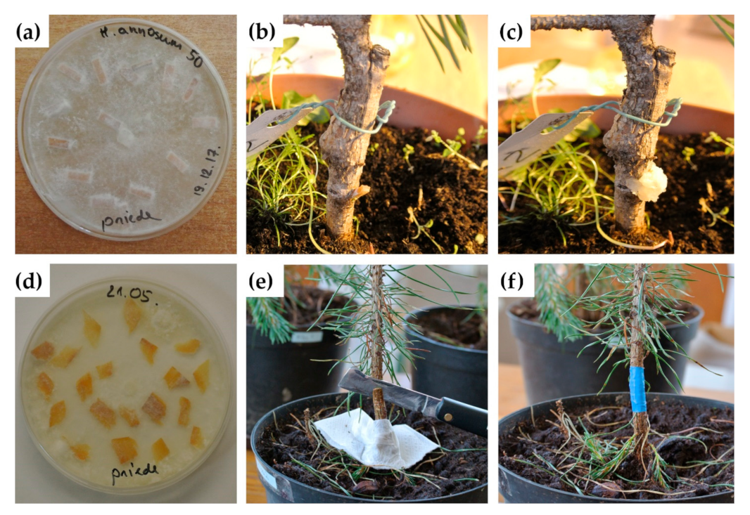
2.1. Plant Material, Exogenous Application of MeJA, Inoculation with H. annosum and L. seditiosum, and Sampling

MeJA application and H. annosum inoculation were done in two parts (experiments 1 and 2), and H. annosum inoculations were done using two different methods—stem drilling and cutting (Figure 1).
Experiment 1: In the first experiment, 30 five-year-old P. sylvestris ramets of a single clone (Sm9-III-2) were used. Previously, this clone was determined to have two TLP gene copies [45]. Ramets were obtained from the Latvian Scots pine breeding program, individually repotted in a fertilized peat substrate (N—100 mg/L, P—60 mg/L, K—200 mg/L; ph = 4.5) in 0.4 L pots. Plants were grown under indoor conditions with green light (Philips Master green plus power 1000 W), 14 h light and 10 h dark, 18–20 °C. Then, 15 ramets were each treated with 12 mL of 10 mM MeJA dissolved in 0.1% Tween 80 solution in deionized water and applied to needles with a hand sprayer. The 15 control ramets were treated with 0.1% Tween 80 solution in deionized water. All ramets were kept in plastic bags for 5 days after treatment to allow for the volatilization of the excess MeJA. The MeJA-treated ramets and control ramets were kept in separate rooms. Needles were harvested prior to and 6 h, 30 h, 50 h, 8 days, and 14 days after treatment and immediately frozen in liquid nitrogen prior to RNA extraction to quantify TLP gene expression.
Heterobasidion annosum (Fr.) Bref. isolate 50 was provided by the Latvian State Forest Research Institute “Silava” Forest phytopathology and mycology laboratory. To initiate fungal mycelial growth, an agar plug containing fungal mycelia was placed at the center of a Petri dish containing 20 mL of 2% malt extract agar and incubated at 23 °C. The first method utilized a protocol described in Swedjemark and Karlsson [31] and Zaluma et al. [34]. Sterile (three times autoclaved) pine sapwood pieces (0.3 × 0.8 cm) were incubated on H. annosum culture for 4 weeks at 23 °C (Figure 1a). Inoculations were done on all MeJA-treated and control ramets one week after treatment. Ramet stems (diameters at soil level were approx. 0.85 cm) were wiped with 70% ethanol, and a circular wound (0.3 cm diam.; 0.4 cm deep) was made 2 cm above the soil surface using a drill bit positioned at 45° angle to the stem (Figure 1b). Colonized sapwood pieces were inserted, and the wound was covered with gardening wax (Figure 1c). Stem discs from ramets were collected after 6 weeks.
Experiment 2: The second experiment was done using 80 three-year-old open-pollinated P. sylvestris seedlings. Seedlings were obtained from the Latvian State Forests department “Seeds and Plants”, Jaunkalsnava, Latvia and individually repotted and grown as described in experiment 1. The MeJA treatment was done in two different ways—by spraying MeJA solution onto needles or by allowing the MeJA to evaporate from cotton wads. Each of the 28 seedlings were sprayed with 6 mL of 10 mM MeJA dissolved in 0.1% Tween 80 solution in deionized water. Additionally, 30 seedlings were treated using cotton wads soaked with 6 mL of 10 mM MeJA dissolved in 96% ethanol and placed on a Petri dish on the soil surface in each pot. Further, 22 control seedlings were sprayed with 0.1% Tween 80 solution. All seedlings were kept in plastic bags for 1 day after treatment. Two weeks after exogenous application (spraying) of MeJA, nine seedlings were wilted and dead, and were therefore excluded from the inoculation experiment.
In experiment 2, an alternative method was used to inoculate all MeJA and control seedlings (stem diameters at soil level were approximately 0.5 cm) with H. annosum two days after MeJA treatment. Inoculum was prepared by incubating sterile thin pine sapwood chips (0.05 × 0.5 cm) on H. annosum culture for 4 weeks at 23 °C (Figure 1d). Inoculation points were located approximately 2 cm above the base of stem. Each stem was wiped with 70% ethanol and cuts made with grafting knife at an angle of 45° to the stem (Figure 1e). Colonized inoculum was inserted, and the wound tightly covered with PVC self-adhesive electrical insulation tape, to avoid excessive excretion of resin that could form a barrier between the inoculated fungi and pine tissues (Figure 1f). In addition, the tightly covered wounding point could reduce additional infections from other soil fungi that could infect seedlings, particularly during watering. Seedlings were maintained under indoor conditions and watered regularly (1–2 times per week). Samples were collected from seedlings after 19 weeks.
All seedlings and ramets from experiment 1 and experiment 2 were cut and processed similarly—branches were removed, and stem surfaces were flame sterilized. Cut stem lengths were approximately 14 cm (10 cm above and 4 cm below the inoculation point to the root collar). Stems were cut into 0.4- to 1-cm-thick discs using secateurs, with cutting surfaces disinfected with 70% ethanol between each cut. The inoculation point was designated as the central disc. Each disc was flame sterilized and incubated for 7 days at room temperature on sterile filter paper in Petri dishes. Filter paper was regularly moistened with sterile deionized water. Discs were examined under a low-power stereo microscope (Leica) for Heterobasidion spp. conidiophores. Infection frequency was determined by the proportion of inoculated individuals with evidence of H. annosum in the stem, detected by observed conidiophores or PCR analyses. The extent of Heterobasidion spread in the stem was determined by observed conidiophores (in experiment 1 and initially in experiment 2) or positive PCR results (used as the main method in experiment 2), and the distance from the inoculation site was measured.
Experiment 3: For MeJA application and L. seditiosum inoculation, 6 seedling cassettes (240 seedlings) of two-year-old open-pollinated P. sylvestris seedlings were obtained from the Latvian State Forests department “Seeds and Plants”, Jaunkalsnava, Latvia. The experiment was done in October 2019. Next, 3 seedling cassettes (120 seedlings) were treated with MeJA using cotton wads soaked with 10 mL of 10 mM MeJA dissolved in 96% ethanol, placed on a Petri dish (2 cotton wads/Petri dishes per 40 seedlings/one cassette) between the seedlings near the soil. Control seedlings were treated with 10 mL of 96% ethanol. All cassettes were kept in plastic bags for 1 day after treatment. The MeJA-treated and control seedlings were kept in separate Binders climate chambers KMF under 10 h light/60% humidity/24 °C and 14 h dark/70% humidity/18 °C.
Inoculations with L. seditiosum were performed two days after MeJA treatment. L. seditiosum was isolated from P. sylvestris needles collected from infected Scots pine trees in the Madona district (57°00′43′ N 26°26′06′ E), Latvia. The ITS region of the DNA extracted from the fungal isolate was sequenced using the ITS1F and ITS4R primers [46], and the sequence was compared with the GenBank database to confirm that the isolate was L. seditiosum. The propagation in liquid MEB medium and inoculation method was developed at the Institute of Horticulture, Dobele, Latvia (I. Moročko-Bičevska, pers. comm.). To initiate fungal mycelial growth, an agar plug containing fungal mycelia was placed at the center of a Petri dish containing 20 mL of 2% malt extract agar and incubated at 23 °C for 2 weeks. An agar plug containing fungal mycelia was placed in 200 mL MEB medium for 3 weeks at 23 °C and 120 rpm in an OS-20 orbital shaker (Biosan, Riga, Latvia). The agar plugs with mycelium were removed and the culture was centrifuged and rinsed three times with deionized water. The culture was homogenized in 1.5-mL deionized water using a Silent Crusher S (Heidolph Instruments, Schwabach, Germany) homogenizer. Next, 5 mL of the concentrated culture suspension was diluted to 50 mL with deionized H2O and all seedlings were sprayed equally (including no-MeJA controls). After inoculation, all seedlings were covered with plastic bags for three days and additionally sprayed daily with deionized H2O to maintain high humidity within the bags. After inoculation, the MeJA-treated and control ramets were kept in separate Binders climate chambers KMF under 10 h light/60% humidity/24 °C and 14 h dark/70% humidity/18 °C. In total, 60 needles (10 needles from a selection of each cassette) from the seedlings were sampled once a month and extracted DNA was analyzed with L. seditiosum specific primers. The first positive PCR results were obtained after three months, and subsequently all seedlings were analyzed. The damage caused by L. seditiosum infection were assessed visually for all trees using a 5-grade scale based on the proportion of damaged needles: 1 (0–5%), 2 (6–35%), 3 (36–65%), 4 (66–95%), and 5 (96–100%) [47]. Needles (approximately 5–6) from the top of seedlings were collected and the proportion of individuals containing L. seditiosum was determined by PCR using species-specific primers [48].
A Mann–Whitney test was used to compare H. annosum and L. seditiosum infection frequency within each experiment and t-tests were used to compare mean longitudinal H. annosum growth. Both tests were performed using R software (v.3.6.2.).

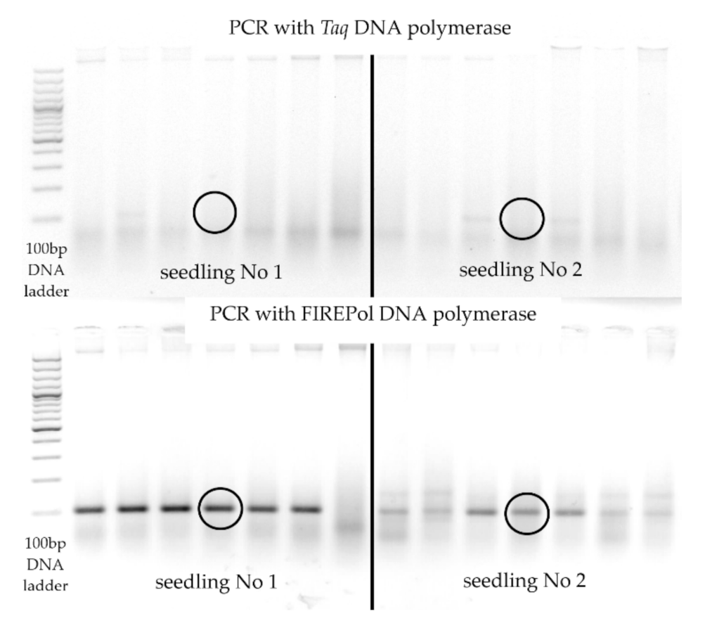
2.2. DNA Extraction, PCR Conditions

Total DNA was extracted from 71 individuals (770 stem fragments) for PCR assessment of the presence of H. annosum, and from needles collected from 162 seedlings (5–6 needles from each individual), for PCR assessment of the presence of L. seditiosum. DNA was extracted using a CTAB method [49]. The width of stem discs was measured and then cut into small chips, placed into liquid nitrogen, and homogenized in a ball mill model MM400 (Retsch, Haan, Germany) at 30 Hz for 40 s. Needles were sterilized in sodium hypochlorite (bleach) and washed in 70% ethanol prior to DNA extraction. DNA was extracted from H. annosum and L. seditiosum culture for use as positive controls. Primers MJ-F (GGTCCTGTCTGGCTTTGC) and MJ-R (CTGAAGCACACCTTGCCA) were used to amplify an approximately 100 bp fragment from H. annosum [50]. Initially, PCR reactions were performed with Taq DNA polymerase (Thermo Fisher Scientific, Waltham, MA, USA), using the following PCR conditions: initial denaturation 10 min, 95 °C; 40 cycles of amplification (30 s denaturation, 95 °C; 35 s annealing, 67 °C; 1 min extension, 72 °C); final extension 7 min, 72 °C. The PCR mixture contained 0.2 mM of (each) dNTP, 0.5 µM of each primer, 0.25U of Taq polymerase, 2 mM of MgCl2, 1× PCR buffer with KCl, and 2 µL of DNA. DNA concentration in samples was not measured as it would not give information about the quantity of fungal DNA in the sample. Subsequently, for more accurate results, PCRs were performed with 5× Hot FirePol Multiplex Mix (Solis Biodyne, Tartu, Estonia), following the manufacturer’s protocol and an annealing temperature of 67 °C. LS11 (CAC CCT TTG TTT ACC ACA CTC A) and LS12 (CGC CAC CTG CTG TCC TTC) were used to amplify a 381 bp fragment from L. seditiosum [48]. PCR reactions were performed using 5× Hot FIREPol MultiPlex Mix (Solis Biodyne, Tartu, Estonia), following the manufacturer’s protocol and an annealing temperature of 64 °C. All PCR products were separated in TAE buffer by 2% agarose gel electrophoresis and visualized using ethidium bromide staining.

2.3. RNA Extraction and Real-Time PCR

RNA was extracted from seven MeJA-treated ramets and two control ramets using a CTAB buffer-based method [51] modified by Rubio-Pina and Zapata-Petrez [52]. Two pine needles were sampled from each ramet for RNA extraction. DNA was removed by incubation with DNase I (Thermo Fisher Scientific, Waltham, MA, USA) and purified using phenol/chloroform extraction. Presence of DNA was assessed using a standard PCR protocol with genomic primers and visualization of PCR products on a 2% agarose gel. RNA concentration was measured with the Qubit system (Invitrogen, Carlsbad, CA, USA) using the Quant-iT RNA BR Assay kit (Invitrogen, Carlsbad, CA, USA) following the manufacturer’s instructions. cDNA was synthesized using Applied Biosystems TaqMan reverse transcription reagents (Thermo Fisher Scientific, Waltham, MA, USA), following the manufacturer’s protocol. 5× Hot FIREPol ®EvaGreen® qPCR Mix Plus/Rox (Solis Biodyne, Tartu, Estonia) was used to perform quantitative real-time PCR using 30 ng of cDNA. Gene expression was determined using the comparative ΔΔCt method using THN-F and THN-R primers, as well as three endogenous control genes as references for normalization: GAPDH (glyceraldehyde-3-phosphate dehydrogenase; [45]), TUB (tubulin), and ACT (actin; [24]): GAPDH-F (ACGGTTTTGGTCGAATTGGA), GPDH-R (CCCCACGAGCTCGATATCAT); Tub5-F (CCACATTGGACAGGCCGGTATCC), Tub5-R (AATGCCGTGCTCGAGGCAGTA); Act5-F (TCATGGTTGGCATGGGACAG), Act5-R (CTCCATGTCATCCCAGTTGC). The following qPCR program was used: 95 °C for 10 min., 40 cycles of 95 °C for 15 s, 60 °C for 40 s, and 72 °C for 30 s. The dissociation step for melting curve analysis was performed at 95 °C for 15 s and 60 °C for 1 min, followed by gradual temperature increase (0.3 °C each step until reaching 95 °C). Experiments were run on a StepOnePlus real-time PCR system (Thermo Fisher Scientific, Waltham, MA, USA), and data were analyzed using the StepOne software v2.2.

3. Results

3.1. Experiment 1: Clonal Ramet Treatment with MeJA, Inoculation with H. annosum, and TLP Gene Expression

One week after MeJA treatment, at the time of inoculation with H. annosum, all seedlings appeared healthy. After 6 weeks, 13 of the 15 MeJA-treated ramets were partly wilted, and all control ramets appeared healthy. After incubation for 6 weeks, all stem discs were examined under a microscope. While discs with conidiophores were observed in all inoculated individuals, discs that were close to the inoculation site were saturated with resin, especially from MeJA-treated ramets (Figure 2a,b) and, therefore, Heterobasidion spp. conidiophores (Figure 2c) were not observed in all discs. In four of the MeJA-treated ramets, conidiophores were only observed in the sixth or seventh disc from the central inoculation disc. Analyzing ramets with observed conidiophores, there were no significant differences in the proportion of infected individuals between the control (71%) and MeJA-treated (64%) ramets (p > 0.05). Mean longitudinal growth of H. annosum in control ramets was 9.22 ± 1.84 mm (as determined by total length of all discs with detected conidiophores), but in MeJA-treated ramets, it was 54.57 ± 9.8 mm. The longer longitudinal growth of H. annosum in the MeJA-treated individuals could be a result of a too high MeJA dosage combined with an inappropriate (harsh) inoculation method that resulted in ramet wilting and reduced resistance to H. annosum.
TLP gene expression was quantified in seven MeJA-treated ramets and two control ramets at 0, 6, 30, and 50 h, and 8 and 14 days after MeJA or Tween 80 treatment. In the first four time points (0, 6, 30, 50 h) of MeJA treatment, only small increases of TLP gene expression were observed (less than 20-fold). However, 14 days after MeJA treatment (and, respectively, one week after inoculation with H. annosum) large increases in TLP gene expression were observed in needles, even up to a 193-fold increase compared to expression before treatment (p < 0.05) (Supplementary File S1). The expression of the TLP gene in MeJA-treated ramets after 14 days increased from 108- up to 193-fold compared with expression before treatment. The highest expression of TLP in more than half of the samples was at 14 days after MeJA treatment, and the highest expression in the other ramets was after 8 days from 31- up to 34-fold, and in one ramet, the highest expression was after 50 h (up to 12-fold). No differences in gene expression were observed in non-MeJA-treated plants (treated with Tween 80) used as controls (expression increase up to 2-fold after 8 days and up to 4-fold after 50 h), even after controls were inoculated with H. annosum. TLP gene expression levels in most of the ramets increased over time; however, expression levels peaked earlier in two ramets (4 and 5). This could be due differential stress on the ramets prior to treatment or during inoculation via the drilling process (drill depth in stem; variation of the stem diameter), or by differences in MeJA treatment efficacy. The results indicate that expression of the P. sylvestris TLP gene was not increased during the first hours after MeJA application, but was substantially increased only after 8 days.
In summary, in experiment 1, the MeJA dosage was too high, as ramets treated with 12 mL of 10 mM MeJA were wilted. No significant differences in H. annosum infection frequency were observed between MeJA-treated and control ramets, but longitudinal growth of H. annosum was significantly longer in MeJA-treated ramets. TLP gene expression levels were low in control ramets, even after inoculation with H. annosum. In MeJA-treated ramets, expression of the TLP gene was initially low, and was highest at the last sampling time point of 14 days, indicating the relatively long-term effects of MeJA treatment on defense mechanisms in Scots pine.

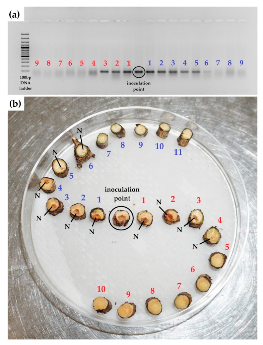
3.2. Experiment 2: P. sylvestris Seedling Treatment with MeJA, PCR Results of Inoculation with H. annosum

In the second experiment, seedlings were treated with lower dosages and two different MeJA application methods (exogenous application on needles and MeJA vaporization from cotton wads to avoid direct contact and phytotoxicity of MeJA). Inoculation with H. annosum was done similarly by cutting the stem and inserting the inoculum; however, in this experiment, thin sapwood chips were used, as the diameter of the stems was smaller; therefore, the initially used drilling method was not possible. After MeJA treatment, nine plants (32%) treated directly with exogenous application of hormone on needles died; therefore, these seedlings were excluded from subsequent inoculation experiments.
Samples from three-year-old seedlings were collected after 19 weeks. Similar to results obtained in the first experiment, discs with conidiophores were observed in all infected individuals, but discs that were close to the inoculation site were saturated with resin, especially from the MeJA-treated seedling stems, which presumably did not allow for the development of Heterobasidion spp. conidiophores. Subsequently, discs were analyzed by PCR with H. annosum-specific primers. During the initial PCR experiments using Taq polymerase, a discrepancy between PCR results and visually detectable discoloration in stem fragments was observed. In samples with noticeable discoloration in the xylem (presumably caused by H. annosum), PCR analysis did not detect the presence of H. annosum. In addition, negative PCR results were observed in the discs at the inoculation point, while discs above and below the inoculation point were positive (Figure 3).
Presumably, the failure to detect H. annosum by PCR at the inoculation site, while discs above and below the inoculation point were positive, was due to the presence of PCR-inhibiting substances. We compared the PCR performance of Taq DNA polymerase against 5× Hot FirePol Multiplex Mix (Solis Biodyne, Tartu, Estonia), containing FirePol polymerase with 5′→ 3′ and 5′ flap endonuclease activity. Comparing the PCR results of the same samples with both polymerases, the detection of H. annosum (positive results) increased using the 5× Hot FirePol Multiplex Mix (Figure 3).
The presence of H. annosum was detected by PCR (Figure 4a) in stem discs with visible discoloration (Figure 4b).
All 30 seedlings treated with MeJA using the vaporization method survived, but only 19 of the 28 seedlings (68%) survived when using the direct application method (Table 1). After inoculation with H. annosum, all seedlings treated by direct application of MeJA were infected with H. annosum, but using the vaporization method, 19 seedlings (63%) were infected. Of the surviving and tested 71 individuals (770 stem fragments), including the controls and MeJA-treated seedlings, H. annosum DNA was detected in 60 trees.
In control seedlings five months after inoculation, the mean longitudinal fungal growth in stems was 58.32 ± 6.50 mm (infection frequency was 100%), and in direct application treated seedlings it was 24.58 ± 4.86 mm (infection frequency was 100%), but in infected seedlings treated by MeJA vaporization from cotton buds (without direct contact), the mean longitudinal growth was 13.34 ± 1.71 mm (infection frequency was 63%). The maximum longitudinal fungal growth in controls was 111 mm, and in directly applied MeJA-treated seedlings it was 75 mm, but in seedlings treated by the vaporization method, it was 33 mm (Table 1). The difference in mean fungal growth between controls and MeJA-treated (both application methods) seedlings were significant (p < 0.05), but no significant differences (p > 0.05) were found when comparing both MeJA application methods (only infected seedlings). Five seedlings, positive for H. annosum DNA, had stem discs with negative PCR results that were between discs with positive PCR amplification results. This indicated that possibly the distribution of H. annosum DNA within infected trees was not homogenous or that some inhibition of PCR occurred. At the end of experiment, eight trees were completely wilted and dead (five trees had direct application method of MeJA, three seedlings were controls), and all were positive for H. annosum infection.

3.3. Experiment 3: Seedling Treatment with MeJA and Inoculation with L. seditiosum

In this experiment, 240 two-year-old open-pollinated seedlings were used. Prior to the experiment, all seedlings were kept outdoors, but two weeks prior to, and for the entire period after the hormone treatment, the MeJA-treated and control seedlings were kept in separate Binders climate chambers. MeJA treatment was done only by the vaporization from the cotton buds method. At end of November, after MeJA treatment and L. seditiosum inoculation, European pine sawfly (Neodiprion sertifer) larvae development was observed on control seedlings (Figure 5), and large pine weevil (Hylobius abietis) caused needle damage. However, while eggs were deposited on MeJA-treated trees, their development was delayed or suspended, and no needle damage was observed.
After inoculation with L seditiosum, 60 needles from a selection of the 162 surviving seedlings (77 control and 85 MeJA-treated seedlings), were sampled once a month and analyzed with L. seditiosum-specific primers. No brown lesions (caused by L. seditiosum) or positive PCR results were observed until 3 months post inoculation. After the first positive PCR results, needles from all seedlings were tested. Lophodermium seditiosum was detected by PCR in 62 individuals of the analyzed needle samples of all control seedlings and in 30 individuals of the analyzed needles samples of all MeJA-treated seedlings (Table 2). Visual assessment of infection level was done using a 5-grade scale, depending on the proportion of damaged (brown) needles. All control seedlings and only 28% of MeJA-treated seedlings had yellow or brown spots on needles. Comparing visual and PCR-based L. seditiosum detection methods, results were similar. Visually, L. seditiosum was detected in 23 (27%) MeJA-treated seedlings and L. seditiosum was detected by PCR in 30 individuals (35%) of seedlings, indicating that L. seditiosum was detected by PCR in some individuals with no visual symptoms. L. seditiosum was detected by PCR in 62 (80%) of control seedlings, but all control seedlings had needle spots, which could be due to other fungi or bacteria that can cause spots on the needles of Scots pine. L. seditiosum was detected by PCR in all seedlings assessed with the 5th grade of damage, both control and MeJA-treated seedlings.

4. Discussion

Numerous studies have shown that it is possible to stimulate plant resistance by applying jasmonate and its derivatives to plants. In this study we compared two different MeJA treatment methods (direct application and vaporization) and characterized the effect of MeJA treatments on resistance of P. sylvestris to H. annosum and L. seditiosum infection. MeJA treatment of seedlings using the vaporization method was less stressful compared to direct application of MeJA to needles. In experiment 1, direct MeJA application both increased TLP expression and had a phytotoxic effect on ramets (all MeJA-treated ramets wilted after six weeks). In subsequent experiments, the vaporization method was less stressful, and did not cause seedling mortality. In addition, while MeJA dosage using the vaporization method was lower, only 63.3% of seedlings were infected with H. annosum, while with direct application, all seedlings were infected (experiment 2). Phytotoxicity of MeJA in high concentrations has been previously reported [1,4,44]. Vivas et al. [44] considered that MeJA is effective in plants older than one year because positive results of application have been previously reported for one-year-old or older P. sylvestris and P. pinaster seedlings [1,4]. MeJA can be lethal to younger seedlings and may not induce resistance because plants are too young for the physiological mechanisms responsible for resistance to be operative, i.e., age-related resistance [53]. However, there are several studies reporting a positive effect of MeJA treatment on resistance in younger conifer seedlings [54,55,56]. Gould et al. [55] mentions that there is an extremely fine margin between efficacy and phytotoxicity at the utilized MeJA concentrations, which again emphasizes the importance of application method and appropriate amount of MeJA. The most popular MeJA application method is exogenous application on needles; however, indirect treatment by evaporation from cotton wads was more effective in our studies. Although seedlings treated by the vaporization method had a reduced mean and maximum fungal growth, statistically there were no significant differences (p > 0.05) with seedlings directly treated with MeJA, which might be due to the small number of tested individuals (only 19 seedlings treated by direct MeJA application were inoculated with H. annosum, as 9 died due to MeJA treatment). Some other studies have utilized this approach, for example, this method has been used with 10-day-old wheat (Triticum aestivum L.) plants [57], 7-day-old P. abies seedlings [6], and 4-week-old Arabidopsis thaliana (L.) Heynh. plants [58]. The length of fungal growth after inoculation, detected by microscopic observation of conidiophores, or by PCR with H. annosum-specific primers, was used as an indicator of susceptibility after inoculation. In some cases, conidiophore growth was not observed on some stem discs, even at or close to the inoculation site. In experiment 1, we observed resin deposits on cut stem disc surfaces, especially from MeJA-treated seedlings. Resin formation and subsequent suppression of conidiophore growth could be promoted by the small disc surfaces and small stem diameters, which is an important limiting factor to inoculation using the drill method. When inoculating trees with larger stem diameters, resin formation still occurs, but conidiophore growth may occur on stem discs due to the larger disc surface. In addition, MeJA treatment induces resin formation [1,2,59], and genetic variation in resin production and its role in conifer defense reactions have been reported previously [60]. In addition, negative results could be due to strong flame sterilization that could kill H. annosum, particularly for discs with small diameters. Based on the negative inoculation results in experiment 1, in experiment 2, we utilized a different inoculation method using thin sapwood chips and DNA detection of H. annosum using PCR. PCR detection of the inoculated fungus can overcome the problems mentioned previously of inconsistent results obtained by observation of conidiophore growth. In the second experiment, the spread of H. annosum in the sapwood of P. sylvestris was significantly greater in the controls compared to the MeJA-treated plants. In our control seedlings 5 months after inoculation, the mean pathogen longitudinal growth was 5.83 cm (infection frequency was 100%). A similar inoculation method was used with similar aged P. sylvestris seedlings where H. annosum growth rates varied from 5 to 6.5 cm after 6 weeks [61], 1.8 cm after 5 months [34], and from 2.4 to 5.3 cm after 10 months [33]. The differences could be related with different seedling susceptibility to the pathogen, differences in virulence of pathogen isolates, and the seedling stem diameter. In addition, Zaļuma et al. [34] drew attention to the importance of small trees in the spread of disease, which is supported by the results reported by Piri and Korhonen [62], where P. abies trees as small as 0.6 cm in diameter can harbor Heterobasidion infection. Our results indicated that H. annosum was able to infect all P. sylvestris seedlings of approximately 0.5 cm diameter at the root collar level. Comparing H. annosum inoculation results from both experiments where the seedlings were directly treated with MeJA, all directly treated and control seedlings were infected, but differences in mean longitudinal H. annosum growth were significant (p < 0.05), with growth in controls being more than twice as large than in the MeJA-treated seedlings. Even though half the amount of MeJA was applied to seedlings in experiment 2 compared to experiment 1, direct MeJA treatment increased seedling mortality in experiment 2, and one-third of seedlings were excluded from the subsequent inoculation stage.
Comparing the effect of MeJA treatment on resistance of P. sylvestris seedlings to H. annosum and L. seditiosum inoculation, in both cases, MeJA reduced the frequency and severity of infection. Neimane et al. [43] reported that one–year-old progenies of Scots pine individuals with intensive resin production were not significantly less affected by needle cast compared to progenies of other individuals selected for growth and stem quality. This could be explained by the report that relative resin production in Pinus nigra J.F. Arnold was much lower in one-year-old than in two-year-old seedlings [63], suggesting that the younger trees allocated a lower proportion of the carbon budget to resin synthesis. Our observation when cutting the stems of three-year-old seedlings and five-year-old ramets was that the MeJA-treated trees produced more resin than the control trees (this was one of the reasons why conidiophores were not observed by microscopy due to resin deposits on disc surfaces that inhibited fungal growth). In addition, recent studies of MeJA application in conifers have shown that the major defense responses occur in the stem rather in the needles [1,3]; therefore, it is possible that progenies of pine individuals with intensive resin production would be more resistant against stem and root diseases rather than needle pathogens. In the H. annosum inoculation experiment, mean fungal growth in MeJA-treated seedlings was more than four times less than in control seedlings, while in the L. seditiosum inoculation experiment, infection frequency, based on PCR assays, was half of that detected in control seedlings. In addition, in the L. seditiosum experiment, needles from the upper part of seedlings had more yellow or brown spots than needles from the basal part. Needles from the upper part were sampled for PCR detection of L seditiosum. Differences in the location of spots could be related with the inoculation method. Growing in liquid medium, L. seditiosum forms colonies, and even after homogenization, might not disperse evenly on seedlings; therefore, some needles might be treated with more hyphae. In addition, after inoculation, seedlings were covered with plastic bags that could make contact with the upper part of the seedlings, providing closer connection with the applied fungi. This could result in differences in fungal growth on different needles. Moreira et al. [4,59] found that the concentration of resins and total phenolics were significantly greater in the upper part of stems in two-year-old Pinus radiata D. Don seedlings after MeJA application, whereas in needles, the concentrations of these compounds was greater in the basal part of the plants. This supports the optimal defense theory—seedlings in stress conditions preferentially protect the most valuable tissues and parts, i.e., the upper parts of stem, which support primary growth, as well as older needles, for example, one-year-old needles in the lower part of the plant may provide the majority of photosynthetic capacity compared to newly developed needles [64].
At the end of experiment 3, where seedlings were treated with MeJA by vaporization, and subsequently inoculated with L. seditiosum, a similar number of control and MeJA-treated seedlings survived. Visually assessing the seedlings, all controls had yellow or brown spots and the infection frequency was 80%, based on PCR results, while MeJA-treated seedlings appeared healthy and the infection frequency was less than half than in the control seedlings (only 35%). In natural conditions, L. seditiosum infection occurs via ascospores. Minter and Millar [65] reported that brown spots are expected to be visible approximately 5 months after infection. However, several studies reported that first symptoms of the disease may be visible on pines as small yellow spots on needles [66,67]. Results from experiment 3 indicate that L. seditiosum was detectable with species-specific PCR primers and visual signs (brown spots) were observed three months after inoculation, which corresponds with our previous experiments [68]. In our experiment, we used inoculation with hyphae fragments (fungi were grown in liquid medium). Possibly hyphal penetration of needles occurs faster than typical ascospore growth despite that L. seditiosum is a slow-growing fungus even on malt extract agar media [69]. We also observed brown spots that were very similar to L. seditiosum ascocarps, but the PCR assays for L. seditiosum were negative. These symptoms could result from other, undetected pathogens. Polmanis et al. [41] reported that not all needles displaying needle cast symptoms returned positive results for PCR assays of L. seditiosum.
The time period during which MeJA treatment induces defense responses in conifers has been reported to be from a few hours to several weeks [1,4,5,14]. In one-year-old Pinus monticola Douglas ex D. Don seedlings, both wounding and MeJA treatment induced expression of the TLP protein after two days [14]. The results from our previous study indicated that the MeJA induction of TLP expression was relatively long term (1–2 weeks after treatment) [23]. In a previous study, we utilized one-year-old P. sylvestris seedlings directly treated with 5 mL of 10 mM MeJA, but with no subsequent inoculation, and TLP gene expression increased up to 10-fold with no phytotoxic effect. As treatment with MeJA concentration above 10 mM MeJA resulted in chlorotic and dead needles, in this study (experiment 1), five-year-old ramets were treated with 12 mL of 10 mM MeJA, as ramets were larger in size and needles were larger compared with the one-year-old seedlings. Then, 14 days after MeJA treatment (and, respectively, one week after inoculation with H. annosum), up to a 193-fold increase in TLP expression was observed. The large difference compared to the previous results could be related to the combined abiotic and biotic stress (hormone treatment and inoculation with pathogen) in a short period of time. The results from this study indicate that expression of the P. sylvestris TLP gene increased slowly during the first hours after MeJA application, but substantially increased after eight days. This expression pattern was similar to that found in our previous study [23]. Comparing results from control ramets that had no MeJA treatment but were inoculated with H. annosum, TLP expression increased before inoculation (2- and 4-fold) but decreased after inoculation. This could be related to wounding stress (needles were harvested from the ramets several times, which could affect gene expression). Additionally, wilted needles indicated that dose of MeJA applied exogenously on ramets was too high, which had a phytotoxic effect. Expression of the TLP gene was analyzed only in experiment 1 using ramets of one clone. We concluded that the utilized MeJA dosage and direct application method in combination with the stem drilling inoculation method increased ramet mortality. Further studies are needed to compare TLP expression in clonal ramets using both application methods—direct application on needles and evaporation from the cotton wad. In this study, in experiments 2 and 3, open-pollinated seedlings were utilized, and therefore, TLP gene expression was not determined, as the genetic background of the seedlings may affect TLP gene expression and complicate direct comparisons of MeJA application method on gene expression.
In experiment 3, before MeJA treatment and L. seditiosum inoculation, all seedlings were growing outside. After moving seedlings into growth chambers at the end of November, N. sertifer larval development and large pine weevil (Hylobius abietis)-induced needle damage was observed on control seedlings, while MeJA-treated trees were undamaged. On MeJA-treated trees, needles with N. sertifer eggs started browning and development of larvae was delayed or suspended. As induced resistance is non-specific, it could be effective against a broad range of pathogens. Heijari [5] reported that exogenous application of MeJA on 14-year-old P. sylvestris reduced growth rates of N. sertifer, increasing the mono-, di-, and sesquiterpenes amount in needles. MeJA also induced resin duct formation [2,3] and enhanced resistance against H. abietis [1,4]. In some cases, an effective defense tactic is to initiate all available defense mechanisms so that at least some may be effective against a particular pathogen [70]; however, this broad induction of defense mechanisms may have associated costs, such as a reduction in growth parameters [1,71,72,73].
This study demonstrated that MeJA induced defense responses in P. sylvestris seedlings ranging from 2 to 5 years old. It also showed the phytotoxic effect of MeJA, and that direct application to needles increases seedling mortality. MeJA application by the vaporization method did not result in seedling mortality and had a larger inhibitory effect on H. annosum growth in stems after inoculation. MeJA application had a broad range effect on defense mechanisms in P. sylvestris. MeJA application increased resistance to both H. annosum and L. seditiosum, and this study also showed that MeJA application had an effect on reducing the impact of insect pests, although this was an unplanned stress factor, and results should be replicated in controlled experiments. Further directions of study include a more detailed investigation of the efficacy of different compounds in stimulating defense responses in Scots pine, including fungal metabolites, or use of fungal species as biocontrol agents [74]. Anatomical parameters, e.g., vessel size and properties, could also have an effect on differential resistance between individuals. In addition, the duration of priming (after induction of defense responses, but prior to pathogen infection), as well as duration of defense responses after inoculation should be examined in more detail by using different pathogen species or isolates, as well as further analysis of expression of additional resistance marker genes.

5. Conclusions

It is predicted that the negative impact on Scots pine caused by L. seditiosum and H. annosum will increase in the future due to climate changes. In this report, we investigated induced resistance ability in P. sylvestris seedlings. We utilized H. annosum-specific PCR primers to assess the infection level of individual seedlings and determine the effect of MeJA treatment on resistance. Two different inoculation approaches and MeJA application methods were used in the experiments with H. annosum. The lesions formed in response to inoculation were significantly larger in control seedlings than treated seedlings. Our results show that MeJA-treated seedlings have greater resistance to L. seditiosum than control seedlings and induced resistance had a long-term effect (at least three months). MeJA application induces broad spectrum resistance against stem and needle fungal pathogens, and even insect pests, as shown by unplanned additional observations in experiment 3. Further studies are needed on the effect of MeJA treatment of P. sylvestris seedlings on gene expression and induction of resistance to additional diseases and pests. Induced resistance could be used as a possible protective mechanism on seedlings in forest nurseries prior to planting in areas where use of fungicides is forbidden.

Supplementary Materials

The following are available online at https://0-www-mdpi-com.brum.beds.ac.uk/article/10.3390/f12050628/s1, Supplementary File S1. Quantification of TLP gene expression by real time PCR.

Author Contributions

Conceptualization, D.E.R. and I.Š.; methodology, I.Š. and A.Z.; laboratory analyses, I.Š., B.K., V.Š., and A.Z.; initial analysis of results. I.Š., B.K., V.Š., and I.Š.; writing—original draft preparation, I.Š.; writing—review and editing, I.Š., B.K., V.Š., A.Z., and D.E.R.; supervision, project administration, funding acquisition, D.E.R. All authors have read and agreed to the published version of the manuscript.

Funding

This research was funded by the Latvian Council of Science, grant number 284/2012, Investigation of molecular defense mechanisms in Scots pine (Pinus sylvestris L.).

Acknowledgments

The L. sedtiosum inoculation method was developed by I. Moročko-Bičevska at the Institute of Horticulture, Dobele, Latvia. The forest phytopathology laboratory at the LSFRI Silava provided fungal cultures and consultation on inoculation methods.

Conflicts of Interest

The funders had no role in the design of the study; in the collection, analyses, or interpretation of data; in the writing of the manuscript, or in the decision to publish the results.

References

  1. Heijari, J.; Nerg, A.M.; Kainulainen, P.; Viiri, H.; Vuorinen, M.; Holopainen, J.K. Application of methyl jasmonate reduces growth but increases chemical defence and resistance against Hylobius abietis in Scots pine seedlings. Entomol. Exp. Appl. 2005, 115, 117–124. [Google Scholar] [CrossRef]
  2. Martin, D.; Tholl, D.; Gershenzon, J.; Bohlmann, J. Methyl Jasmonate Induces Traumatic Resin Ducts, Terpenoid Resin Biosynthesis, and Terpenoid Accumulation in Developing Xylem of Norway Spruce Stems. Plant Physiol. 2002, 129, 1003–1018. [Google Scholar] [CrossRef] [PubMed] [Green Version]
  3. Hudgins, J.W.; Christiansen, E.; Franceschi, V.R. Induction of anatomically based defense responses in stems of diverse conifers by methyl jasmonate: A phylogenetic perspective. Tree Physiol. 2004, 24, 251–264. [Google Scholar] [CrossRef] [PubMed] [Green Version]
  4. Moreira, X.; Sampedro, L.; Zas, R. Defensive responses of Pinus pinaster seedlings to exogenous application of methyl jasmonate: Concentration effect and systemic response. Environ. Exp. Bot. 2009, 67, 94–100. [Google Scholar] [CrossRef] [Green Version]
  5. Heijari, J.; Nerg, A.-M.; Kainulainen, P.; Vuorinen, M.; Holopainen, J.K. Long-term effects of exogenous methyl jasmonate application on Scots pine (Pinus sylvestris) needle chemical defence and diprionid sawfly performance. Entomol. Exp. Appl. 2008, 128, 162–171. [Google Scholar] [CrossRef]
  6. Kozlowski, G.; Buchala, A.; Métraux, J.-P. Methyl jasmonate protects Norway spruce [Picea abies (L.) Karst.] seedlings against Pythium ultimum Trow. Physiol. Mol. Plant Pathol. 1999, 55, 53–58. [Google Scholar] [CrossRef]
  7. Krokene, P.; Nagy, N.E.; Solheim, H. Methyl jasmonate and oxalic acid treatment of Norway spruce: Anatomically based defense responses and increased resistance against fungal infection. Tree Physiol. 2008, 28, 29–35. [Google Scholar] [CrossRef] [Green Version]
  8. Liu, J.-J.; Zamani, A.; Ekramoddoullah, A.K.M. Expression profiling of a complex thaumatin-like protein family in western white pine. Planta 2009, 231, 637–651. [Google Scholar] [CrossRef]
  9. Singh, N.K.; Bracker, C.A.; Hasegawa, P.M.; Handa, A.K.; Buckel, S.; Hermodson, M.A.; Pfankoch, E.; Regnier, F.E.; Bressan, R.A. Characterization of Osmotin. A thaumatin-like protein associated with osmotic adaptation in plant cells. Plant Physiol. 1987, 85, 529–536. [Google Scholar] [CrossRef] [Green Version]
  10. Singh, N.K.; Nelson, D.E.; Kuhn, D.; Hasegawa, P.M.; Bressan, R.A. Molecular Cloning of Osmotin and Regulation of Its Expression by ABA and Adaptation to Low Water Potential. Plant Physiol. 1989, 90, 1096–1101. [Google Scholar] [CrossRef] [Green Version]
  11. Neale, A.D.; Wahleithner, J.A.; Lund, M.; Bonnett, H.T.; Kelly, A.; Meeks-Wagner, D.R.; Peacock, W.J.; Dennis, E.S. Chitinase, beta-1,3-glucanase, osmotin, and extensin are expressed in tobacco explants during flower formation. Plant Cell 1990, 2, 673–684. [Google Scholar] [CrossRef] [PubMed] [Green Version]
  12. Koiwa, H.; Sato, F.; Yamada, Y. Characterization of Accumulation of Tobacco PR-5 Proteins by IEF-Immunoblot Analysis. Plant Cell Physiol. 1994, 35, 821–827. [Google Scholar] [CrossRef] [PubMed]
  13. Reymond, P.; Farmer, E.E. Jasmonate and salicylate as global signals for defense gene expression. Curr. Opin. Plant Biol. 1998, 1, 404–411. [Google Scholar] [CrossRef]
  14. Piggott, N.; Ekramoddoullah, A.K.M.; Liu, J.-J.; Yu, X. Gene cloning of a thaumatin-like (PR-5) protein of western white pine (Pinus monticola D. Don) and expression studies of members of the PR-5 group. Physiol. Mol. Plant Pathol. 2004, 64, 1–8. [Google Scholar] [CrossRef]
  15. Pervieux, I.; Bourassa, M.; Laurans, F.; Hamelin, R.; Séguin, A. A spruce defensin showing strong antifungal activity and increased transcript accumulation after wounding and jasmonate treatments. Physiol. Mol. Plant Pathol. 2004, 64, 331–341. [Google Scholar] [CrossRef]
  16. Liu, J.-J.; Ekramoddoullah, A.K.M.; Zamani, A. A Class IV Chitinase Is Up-Regulated by Fungal Infection and Abiotic Stresses and Associated with Slow-Canker-Growth Resistance to Cronartium ribicola in Western White Pine (Pinus monticola). Phytopathology 2005, 95, 284–291. [Google Scholar] [CrossRef] [Green Version]
  17. Wang, H.; Ng, T.B. Isolation of an antifungal thaumatin-like protein from kiwi fruits. Phytochemistry 2002, 61, 1–6. [Google Scholar] [CrossRef]
  18. Krebitz, M.; Wagner, B.; Ferreira, F.; Peterbauer, C.; Campillo, N.; Witty, M.; Kolarich, D.; Steinkellner, H.; Scheiner, O.; Breiteneder, H. Plant-based Heterologous Expression of Mal d 2, a Thaumatin-like Protein and Allergen of Apple (Malus domestica), and its Characterization as an Antifungal Protein. J. Mol. Biol. 2003, 329, 721–730. [Google Scholar] [CrossRef]
  19. Jayaraj, J.; Velazhahan, R.; Fu, D.; Liang, G.H.; Muthukrishnan, S. Bacterially produced rice thaumatin-like protein shows in vitro antifungal activity. J. Plant Dis. Prot. 2004, 111, 334–344. [Google Scholar]
  20. Vitali, A.; Pacini, L.; Bordi, E.; de Mori, P.; Pucillo, L.; Maras, B.; Botta, B.; Brancaccio, A.; Giardina, B. Purification and characterization of an antifungal thaumatin-like protein from Cassia didymobotrya cell culture. Plant Physiol. Biochem. 2006, 44, 604–610. [Google Scholar] [CrossRef]
  21. Mohamed, M.S.; El-Soud, W.A.; Mohamed, M.; Fervat, S.M.; Walid, A.E.S.; Magda, F.M. Cloning and expression of the recombinant NP24I protein from tomato fruit and study of its antimicrobial activity. Afr. J. Biotechnol. 2011, 10, 14276–14285. [Google Scholar] [CrossRef]
  22. Šķipars, V.; Rauda, E.; Snepste, I.; Krivmane, B.; Rungis, D. Assessment of gene copy number variation of Scots pine thaumatin-like protein gene using real-time PCR based methods. Tree Genet. Genomes 2017, 13, 127. [Google Scholar] [CrossRef]
  23. Šņepste, I.; Šķipars, V.; Krivmane, B.; Bruna, L.; Ruņģis, D. Characterization of a Pinus sylvestris thaumatin-like protein gene and determination of antimicrobial activity of the in vitro expressed protein. Tree Genet. Genomes 2018, 14, 58. [Google Scholar] [CrossRef]
  24. Kanberga-Siliņa, K.; Rauda, E.; Šķipars, V.; Vivian-Smith, A.; Yakovlev, I.; Krivmane, B.; Šņepste, I.; Ruņģis, D. Transcriptomic response to methyl jasmonate treatment of Scots pine (Pinus sylvestris) seedlings. Environ. Exp. Biol. 2017, 15, 257–274. [Google Scholar] [CrossRef]
  25. Gatehouse, J.A. Plant resistance towards insect herbivores: A dynamic interaction. New Phytol. 2002, 156, 145–169. [Google Scholar] [CrossRef] [Green Version]
  26. Thakur, M.; Sohal, B.S. Role of Elicitors in Inducing Resistance in Plants against Pathogen Infection: A Review. ISRN Biochem. 2013, 2013, 762412. [Google Scholar] [CrossRef] [Green Version]
  27. Nejat, N.; Mantri, N. Plant Immune System: Crosstalk Between Responses to Biotic and Abiotic Stresses the Missing Link in Understanding Plant Defence. Curr. Issues Mol. Biol. 2017, 23, 1–16. [Google Scholar] [CrossRef] [Green Version]
  28. Arhipova, N.; Gaitnieks, T.; Donis, J.; Stenlid, J.; Vasaitis, R. Butt rot incidence, causal fungi, and related yield loss in Picea abies stands of Latvia. Can. J. For. Res. 2011, 41, 2337–2345. [Google Scholar] [CrossRef]
  29. Garbelotto, M.; Gonthier, P. Biology, Epidemiology, and Control of Heterobasidion Species Worldwide. Annu. Rev. Phytopathol. 2013, 51, 39–59. [Google Scholar] [CrossRef] [Green Version]
  30. Swedjmark, G.; Stenlid, J. Between-tree and between-isolate variation for growth of S-group Heterobasidion annosum in sapwood of Picea abies cuttings. Can. J. For. Res. 1997, 27, 711–715. [Google Scholar] [CrossRef]
  31. Swedjemark, G.; Karlsson, B. Mycelial growth and exclusion of Heterobasidion parviporum inoculated in branches of 15-year-old Picea abies clones. For. Pathol. 2006, 36, 209–214. [Google Scholar] [CrossRef]
  32. Skrøppa, T.; Solheim, H.; Steffenrem, A. Genetic variation, inheritance patterns and parent–offspring relationships after artificial inoculations with Heterobasidion parviporum and Ceratosystis polonica in Norway spruce seed orchards and progeny tests. Silva Fenn. 2015, 49, 49. [Google Scholar] [CrossRef] [Green Version]
  33. Marčiulynas, A.; Sirgedaitė-Šėžienė, V.; Žemaitis, P.; Baliuckas, V. The Resistance of Scots Pine (Pinus sylvestris L.) Half-sib Families to Heterobasidion annosum. Forests 2019, 10, 287. [Google Scholar] [CrossRef] [Green Version]
  34. Zaluma, A.; Gaitnieks, T.; Arhipova, N.; Vasaitis, R. Growth rates of Heterobasidion annosum ss and H. aparviporum in functional sapwood of Pinus sylvestris and Picea abies. For. Pathol. 2015, 45, 437–439. [Google Scholar] [CrossRef]
  35. Hanso, M.; Drenkhan, R. Lophodermium needle cast, insect defoliation and growth responses of young Scots pines in Estonia. For. Pathol. 2011, 42, 124–135. [Google Scholar] [CrossRef]
  36. Kowalski, T. Interrelationships among Lophodermium seditiosum, L. pinastri and Cyclaneusma minus in pine plantations (Pinus sylvestris L.) in Poland. In Proceedings of the Conference: Recent Research on Foliage Diseases, Carlisle, PE, USA, 29 May–2 June 1989; pp. 13–18. [Google Scholar]
  37. Diwani, S.A.; Millar, C.S. Sources of inoculum of Lophodermium seditiosum on Pinus sylvestris. For. Pathol. 1990, 20, 1–7. [Google Scholar] [CrossRef]
  38. Ganley, R.J.; Newcombe, G. Fungal endophytes in seeds and needles of Pinus monticola. Mycol. Res. 2006, 110, 318–327. [Google Scholar] [CrossRef]
  39. Bentele, M.; Morgenstern, K.; Krabel, D. Lophodermium seditiosum Minter, Staley & Millar seed-borne on Pinus sylvestris. J. For. Landsc. Res. 2014, 1, 1–8. [Google Scholar] [CrossRef]
  40. Rajkovic, S.; Markovic, M.; Rakonjac, L. Incubation Methods for Forecasting the Occurrence and Development of Lophodermium seditiosum Minter, Staley & Millar on Pine. In Fungicides—Showcases of Integrated Plant Disease Management from around the World; Mizuho, N., Ed.; INTECH: London, UK, 2013; Chapter 8; pp. 153–183. ISBN 978-953-51-1130-6. [Google Scholar]
  41. Polmanis, K.; Gaitnieks, T.; Beļeviča, V.; Ruņģis, D.; Baumane, A. Occurrence of Lophodermium spp. in young scots pine stands in Latvia. In Proceedings of the Annual 23rd International Scientific Conference, the Research for Rural Development, Jelgava, Latvia, 17–19 May 2017; Latvia University of Life Sciences and Technologies: Jelgava, Latvia, 2017; pp. 14–20. [Google Scholar]
  42. Jansons, A.; Neimane, U.; Baumanis, I. Needlecast resistance of Scots pine and possibilities of its improvement. Mežzinātne 2008, 18, 3–18. (In Latvian) [Google Scholar]
  43. Neimane, U.; Polmanis, K.; Zaluma, A.; Klaviņa, D.; Gaitnieks, T.; Jansons, A. Damage caused by Lophodermium needle cast in open-pollinated and control-crossed progeny trials of Scots pine (Pinus sylvestris L.). For. Chron. 2018, 94, 155–161. [Google Scholar] [CrossRef]
  44. Vivas, M.; Martin, J.A.; Gil, L.; Solla, A. Evaluating methyl jasmonate for induction of resistance to Fusarium oxysporum, F. circinatum and Ophiostoma novoulmi. For. Syst. 2012, 21, 289–299. [Google Scholar] [CrossRef] [Green Version]
  45. Šķipars, V.; Krivmane, B.; Runģis, D. Thaumatin-like protein gene copy number variation in Scots pine (Pinus sylvestris). Environ. Exp. Biol. 2011, 9, 75–81. [Google Scholar]
  46. White, T.J.; Bruns, T.D.; Lee, S.; Taylor, J. Analysis of phylogenetic relationships by amplification and direct sequencing of ribosomal RNA genes. In PCR Protocols: A Guide to Methods and Applications; Innis, M.A., Gelfand, D.H., Sninsky, J.J., White, T.J., Eds.; Academic Press: New York, NY, USA, 1990; Chapter 38; pp. 315–322. ISBN 978-0-12-372180-8. [Google Scholar]
  47. Polmanis, K.; Klavina, D.; Gaitnieks, T.; Baumanis, I.; Lazdins, A. Genetic differences in needle cast damage of Scots pine (Pinus sylvestris L.). In Proceedings of the Annual 22nd International Scientific Conference Proceedings, Research for Rural Development, Jelgava, Latvia, 18–20 May 2016; pp. 34–40. [Google Scholar]
  48. Stenstrom, E.; Ihrmark, K. Identification of Lophodermium seditiosum and L. pinastri in Swedish forest nurseries using species-specific PCR primers from the ribosomal ITS region. For. Pathol. 2005, 35, 163–172. [Google Scholar] [CrossRef]
  49. Murray, M.G.; Thompson, W.F. Rapid isolation of high molecular weight plant DNA. Nucleic Acids Res. 1980, 8, 4321–4326. [Google Scholar] [CrossRef] [Green Version]
  50. Hantula, J.; Vainio, E. Specific primers for the differentiation of Heterobasidion annosum (s.str.) and H. parviporum infected stumps in northern Europe. Silva Fenn. 2003, 37, 181–187. [Google Scholar] [CrossRef] [Green Version]
  51. Yang, G.; Zhou, R.; Tang, T.; Shi, S. Preparative biochemistry and biotechnology simple and Efficient Isolation of High-Quality Total RNA from Hibiscus tiliaceus, a Mangrove Associate and Its Relatives. Prep. Biochem. Biotechnol. 2008, 38, 257–264. [Google Scholar] [CrossRef] [PubMed]
  52. Zapata-Pérez, O.; Rubio-Piña, J.A. Isolation of total RNA from tissues rich in polyphenols and polysaccharides of mangrove plants. Electron. J. Biotechnol. 2011, 14, 11. [Google Scholar] [CrossRef]
  53. Panter, S.; Jones, D. Age-related resistance to plant pathogens. Adv. Bot. Res. 2002, 38, 251–280. [Google Scholar] [CrossRef]
  54. Gould, N.; Reglinski, T.; Spiers, M.; Taylor, J.T. Physiological trade-offs associated with methyl jasmonate—Induced resistance in Pinus radiata. Can. J. For. Res. 2008, 38, 677–684. [Google Scholar] [CrossRef]
  55. Gould, N.; Reglinski, T.; Northcott, G.L.; Spiers, M.; Taylor, J.T. Physiological and biochemical responses in Pinus radiata seedlings associated with methyl jasmonate-induced resistance to Diplodia pinea. Physiol. Mol. Plant Pathol. 2009, 74, 121–128. [Google Scholar] [CrossRef]
  56. Sampedro, L.; Moreira, X.; Zas, R. Costs of constitutive and herbivore-induced chemical defences in pine trees emerge only under low nutrient availability. J. Ecol. 2011, 99, 818–827. [Google Scholar] [CrossRef] [Green Version]
  57. Liu, H.; Carvalhais, L.C.; Schenk, P.M.; Paul, D.G. Effects of jasmonic acid signalling on the wheat microbiome differ between body sites. Sci. Rep. 2017, 7, 19. [Google Scholar] [CrossRef]
  58. Makandar, R.; Nalam, V.; Chaturvedi, R.; Jeannotte, R.; Sparks, A.A.; Shah, J. Involvement of Salicylate and Jasmonate Signaling Pathways in Arabidopsis Interaction with Fusarium graminearum. Mol. Plant Microbe Interact. 2010, 23, 861–870. [Google Scholar] [CrossRef] [PubMed] [Green Version]
  59. Moreira, X.; Zas, R.; Sampedro, L. Differential Allocation of Constitutive and Induced Chemical Defenses in Pine Tree Juveniles: A Test of the Optimal Defense Theory. PLoS ONE 2012, 7, e34006. [Google Scholar] [CrossRef] [Green Version]
  60. Bridgen, M.R.; Hanover, J.W. Indirect selection for pest resistance using terpenoid compounds. In Proceedings of the Third International Workshop on the Genetics of Host-Parasite Interactions in Forestry, Wageningen, The Netherlands, 14–21 September 1980; pp. 161–168. [Google Scholar]
  61. Stenlid, J.; Swedjemark, G. Differential growth of S- and P-isolates of Heterobasidion annosum in Picea abies and Pinus sylvestris. Trans. Br. Mycol. Soc. 1988, 90, 209–213. [Google Scholar] [CrossRef]
  62. Piri, T.; Korhonen, K. Infection of advance regeneration of Norway spruce by Heterobasidion parviporum. Can. J. For. Res. 2001, 31, 937–942. [Google Scholar] [CrossRef]
  63. Wainhouse, D.; Staley, J.T.; Jinks, R.; Morgan, G. Growth and defence in young pine and spruce and the expression of resistance to a stem-feeding weevil. Oecologia 2008, 158, 641–650. [Google Scholar] [CrossRef] [PubMed]
  64. Pavia, H.; Toth, G.B.; Åberg, P. Optimal Defense Theory: Elasticity Analysis as a Tool to Predict Intraplant Variation in Defenses. Ecology 2002, 83, 891–897. [Google Scholar] [CrossRef]
  65. Minter, D.W.; Millar, C.S. Ecology and biology of three Lophodermium species on secondary needles of Pinus sylvestris. Eur. J. For. Pathol. 1980, 10, 169–181. [Google Scholar] [CrossRef]
  66. Diwani, S.A.; Millar, C.S. Infection processes of three Lophodermium species on Pinus sylvestris L. In Proceedings of the Conference on Recent Research on Conifer Needle Disease, Gulfport, MI, USA, 14–18 October 1984; pp. 22–27. [Google Scholar]
  67. Diwani, S.A.; Millar, C.S. Pathogenicity of three Lophodermium species on Pinus sylvestris L. Eur. J. For. Pathol. 1987, 17, 53–58. [Google Scholar] [CrossRef]
  68. Snepste, I.; Skipars, V.; Belevica, V.; Krivmane, B.; Voronova, A.; Rauda, E.; Veinberga, I.; Rungis, D. Induced resistance re-sponses in Scots pine (Pinus sylvestris L.). In Proceedings of the International Scientific Conference, Knowledge based Forest Sector, Riga, Latvia, 4–6 November 2015; pp. 130–135. [Google Scholar]
  69. Minter, D.W.; Millar, C.S. A study of three pine inhabiting Lophodermium species in culture. Nova Hedzvigia 1980, 32, 361–368. [Google Scholar]
  70. Katagiri, F. A global view of defense gene expression regulation—A highly interconnected signaling network. Curr. Opin. Plant Biol. 2004, 7, 506–511. [Google Scholar] [CrossRef] [PubMed]
  71. Bowling, S.A.; Clarke, J.D.; Liu, Y.; Klessig, D.F.; Dong, X. The cpr5 Mutant of Arabidopsis Expresses Both NPR1-Dependent and NPR1-Independent Resistance. Plant Cell 1997, 9, 1573–1584. [Google Scholar] [CrossRef] [Green Version]
  72. Mauch, F.; Mauch-Mani, B.; Gaille, C.; Kull, B.; Haas, D.; Reimmann, C. Manipulation of salicylate content in Arabidopsis thaliana by the expression of an engineered bacterial salicylate synthase. Plant J. 2001, 25, 67–77. [Google Scholar] [CrossRef] [Green Version]
  73. Heil, M.; Bostock, R.M. Induced Systemic Resistance (ISR) Against Pathogens in the Context of Induced Plant Defences. Ann. Bot. 2002, 89, 503–512. [Google Scholar] [CrossRef] [Green Version]
  74. Oszako, T.; Voitka, D.; Stocki, M.; Stocka, N.; Nowakowska, J.A.; Linkiewicz, A.; Hsiang, T.; Belbahri, L.; Berezovska, D.; Malewski, T. Trichoderma asperellum efficiently protects Quercus robur leaves against Erysiphe alphitoides. Eur. J. Plant Pathol. 2021, 159, 295–308. [Google Scholar] [CrossRef]
Figure 1. Two different inoculation techniques utilized: (ac) Inoculation of P. sylvestris ramets with H. annosum in experiment 1. (a) Pine sapwood pieces (0.3 × 0.8 cm) with H. annosum mycelium; (b) circular drill wound and sapwood piece inserted into stem; (c) inoculation point covered with gardening wax. (df) Inoculation experiment of P. sylvestris seedlings in experiment 2. (d) Pine sapwood chips (0.05 × 0.5 cm) with H. annosum mycelium; (e) cut in the stem made by grafting knife for inoculation; (f) inoculation point covered with PVC electrical insulation tape.
Forests 12 00628 g001
Figure 2. (a,b) Stem discs of MeJA-treated P. sylvestris ramets with resin on their surface; (c) Stem disc of a control ramet with Heterobasidion spp. conidiophores.
Forests 12 00628 g002
Figure 3. Comparison of the sensitivity of Taq and FIREPol DNA polymerases in detecting H. annosum in DNA extracted from stem discs of two seedlings. Circles indicate DNA samples from stem discs at the inoculation points, lanes to the left and right contain PCR products from DNA samples extracted from discs below and above the inoculation point, respectively.
Forests 12 00628 g003
Figure 4. (a) PCR products from the control seedling; (b) cut stem disks from the same seedling. N—necrosis, circle—inoculation point, blue numbers—discs above inoculation point (sequential direction to the stem apex), red numbers—discs below inoculation point (sequential direction to the roots).
Forests 12 00628 g004
Figure 5. European pine sawfly (N. sertifer) larvae and their caused damage on the control seedlings.
Forests 12 00628 g005
Table 1. Mean longitudinal H. annosum growth in sapwood of P. sylvestris and infection frequency, based on positive PCR results.
Control SeedlingsDirect MeJA
Application
MeJA Vapor
Treatment
Total no. of seedlings / no. of seedlings utilized in inoculation experiment22/2228/1930/30
Mortality of MeJA-treated seedlings (no.)-90
Infection frequency (%) detected by PCR10010063
Mean longitudinal H. annosum growth ± standard error, mm58.32 ± 6.5024.58 ± 4.8613.34 ± 1.71 *
Minimum and maximum longitudinal H. annosum fungal growth, mm (SD)13.00–111.00 (30.49)3.50–75.00 (21.17)4.00–33.00 (7.45) *
Mortality of H. annosum inoculated seedlings, no. (%)3 (13)5(26)0 *
* only 19 seedlings with positive H. annosum inoculation results were analyzed.
Table 2. Needle cast damage in 5-grade scale, analyzed visually, and infection frequency based on positive PCR results 3 months post inoculation.
Control
Seedlings
MeJA-Treated
Seedlings
Total number of seedlings inoculated with L. seditiosum7785
Needle cast damage grade
1 (0–5%)
41 13
2 (6–35%)313
3 (36–65%)-2
4 (66–95%)--
5 (96–100%)55
No yellow or brown spots-62
Number of individuals where L. seditiosum was detected using PCR analyses62 30
Publisher’s Note: MDPI stays neutral with regard to jurisdictional claims in published maps and institutional affiliations.

