Article

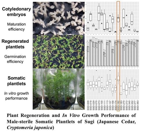
Plant Regeneration and In Vitro Growth Performance of Male-Sterile Somatic Plantlets of Sugi (Japanese Cedar, Cryptomeria japonica) Derived from Different Embryogenic Cell Lines

by
Momi Tsuruta
1,†,
Tsuyoshi E. Maruyama
1,*,†,
Saneyoshi Ueno
1,
Takumi Kaneeda
2 and
Yoshinari Moriguchi
2
1
Department of Forest Molecular Genetics and Biotechnology, Forestry and Forest Products Research Institute, Matsunosato 1, Tsukuba 305-8687, Japan
2
Graduate School of Science and Technology, Niigata University, Ikarashi 8050, Niigata 950-2181, Japan
*
Author to whom correspondence should be addressed.
Co-first authors contributed equally to this work.
Submission received: 7 September 2021 / Revised: 9 November 2021 / Accepted: 16 November 2021 / Published: 18 November 2021
(This article belongs to the Special Issue Application of Biotechnology Techniques on Tree Species)

Abstract

:
With the spread of pollinosis caused by sugi (Japanese cedar, Cryptomeria japonica) pollen, the use of pollen-free somatic seedlings of sugi is expected in Japan. However, there is a lack of knowledge on the relationship between the abilities during somatic embryogenesis, initial in vitro growth traits, and subsequent growth of somatic seedlings. In the present study, we provide the first basic information on somatic embryo maturation efficiency, somatic embryo germination, and plantlet conversion frequencies, as well as on in vitro growth performance of pollen-free somatic plantlets derived from different embryogenic cell lines (ECLs). Somatic embryo maturation efficiency varied from 34 to 514 cotyledonary embryos per plate and the average for the 19 ECLs tested was 244 embryos per plate. Subsequently, the overall average rates of somatic embryo germination and conversion among ECLs were 87.8% and 85.3%, respectively. The results of in vitro growth performance of pollen-free somatic plantlets showed significant differences in growth rate among ECLs.

Graphical Abstract

1. Introduction

Among the Organisation for Economic Co-operation and Development (OECD) member countries, Japan is in the top three regarding forest rate (68.4%), surpassed only by Sweden (68.7%) and Finland (73.3%) [1]. In Japanese artificial forests, the planted area of sugi (Japanese cedar, Cryptomeria japonica (Thunb. ex L.f.) D. Don, Cupressaceae) covers approximately 4.5 million ha, representing 44% of the total reforested area [2]. Since large amounts of pollen are released from sugi forests each spring, allergic reactions caused by exposure to pollen are a serious social and public health problem in Japan. It is estimated that almost 40% of people living in Japan suffer from allergic rhinitis caused by sugi pollen [3]. The use of male-sterile plants (MSPs; pollen-free plants) for reforestation has been proposed. Against this background, the application of biotechnology to produce superior male-sterile trees in a short period of time represents a powerful tool to speed up the process.
Recently, efficient protocols to propagate MSPs combining marker-assisted selection (MAS) and micropropagation techniques have been established in sugi [4]. Among the micropropagation techniques, owing to its efficiency in both the large-scale propagation and the ease of long-term conservation in liquid nitrogen, somatic embryogenesis (SE) has become an indispensable tool for accelerating tree breeding programs [5,6]. However, for many coniferous species, SE protocols still require optimization [7]. SE is a complex multistage process in which each stage represents different approaches, and these are strongly dependent on the results of the previous stage [8]. Bottlenecks related to a low frequency of SE initiation, low production of somatic embryos, and low frequencies of germination and conversion are the main impediments that must be resolved for practical applications [9]. Since plant regeneration through SE was first reported for sugi [10], several studies on SE initiation [11,12,13], somatic embryo maturation [14,15,16,17], and plantlet conversion [4,18] were reported, showing progress in enhancing SE protocols. In recent years, our group reported an improved methodology for SE initiation from the analysis of results on the influence of plant material, explant collection time, explant genotype, and culture conditions [19]. Subsequently, an optimized protocol for somatic embryo maturation from studies on the effect of basal salts contained in maturation medium, polyethylene glycol (PEG) concentration, abscisic acid (ABA) concentration, additional supplementation of potassium chloride, amino acid concentration, and proliferation culture medium, as the main culture-related factors affecting somatic embryo maturation in sugi, was reported [20].
In this context, to further improve plant regeneration protocols, in the present study, we aimed to obtain basic information on somatic embryo maturation efficiency, somatic embryo germination, and plantlet conversion frequencies, as well as on in vitro growth performance of pollen-free somatic plantlets derived from different embryogenic cell lines (ECLs). Different morphological variables involved in the initial growth of C. japonica were analyzed in order to apply them to the improvement of protocols for producing somatic plants and selecting potential fast-growing lines. However, to evaluate the growth and wood quality characteristics, many years of field monitoring are required. If the initial growth performance of somatic plantlets in vitro can be regarded as an indicator of the ex vitro growth tendency, then early selection of lines with good traits could be achieved. In fact, there is a case where the early selection of SE-derived plants of Norway spruce (Picea abies (L.) H. Karst.) upregulated growth values and reduced clonal differences [21]. Although it is essential to compare the traits in the early stage of culture and after several subsequent growth periods to test this hypothesis, to our knowledge, there are no reports of growth in SE-derived sugi lines, not even from in vitro plantlets. Therefore, in the present study, we provide the first basic information on the in vitro growth performance of pollen-free somatic Japanese cedar plantlets, which may serve as scientific evidence to support the application of efficient propagation protocols for practical uses.

2. Materials and Methods

2.1. Plant Materials, SE Initiation, and Maintenance and Proliferation of ECLs

Embryogenic cells (ECs) were induced by Maruyama et al. [19] from entire megagametophytes of immature seeds collected from a full sib seed family (‘Shindai 3’ × ‘Suzu 2’) carrying the male sterility allele ms1, in July 2016. Of induced ECLs, 19 male sterile lines from different megagametophytes were used for experiments. Tissues of induced ECLs were maintained and proliferated by regular subcultures at 2- to 3-week intervals on maintenance/proliferation medium containing basal salts reduced to half concentration from the standard EM medium [10] and supplemented with 3 μM 2,4-dichlorophenoxyacetic acid (2,4-D), 1 μM 6-benzylaminopurine (BA), 30 g L−1 sucrose, 1.5 g L−1 glutamine, and 3 g L1 gellan gum. Clumps of ECs (12 per plate) were cultured in the dark at 25 °C.

2.2. Selection of Male-Sterile ECLs

The major cause of male sterility in Japanese cedar is due to a recessive mutation (ms1) at a single locus, MALE-STERILITY 1 [22]. Discrimination of male-sterile ECLs (namely, homozygotes of ms1) was performed in accordance with the methodology described by Ueno et al. [23]. Briefly, from one spoonful of ECs (30–40 mg fresh weight), DNA was extracted by the CTAB method [22]. Using DNA solution of each sample, an SNP site (dD_Contig_3995-165) closely linked to the male sterility was amplified as an allele-specific PCR marker. A total of 10 μL of PCR reaction mixture containing 3 μL of 2 × Multiplex (Qiagen, Hilden, Germany), 0.2 μM of each of the forward and reverse primers, and 1 μL of DNA template was reacted with the following thermal conditions: initial denaturation at 95 °C for 15 min; 4 cycles of 95 °C for 30 s, 64 °C for 90 s with −1 °C per cycle, and 72 °C for 30 s; and 34 cycles of 95 °C for 30 s, 60 °C for 90 s, and 72 °C for 30 s. The PCR products were electrophoresed on 2% agarose gel and visualized by ethidium bromide staining. Finally, 19 male-sterile ECLs were selected based on the genotype determined from the presence of male-sterile- and/or wild-type-specific amplified fragments [4]. The genotypes and male-sterile discrimination of these lines were subsequently confirmed with a more accurate genetic marker, which was developed on the causable mutation itself [24].

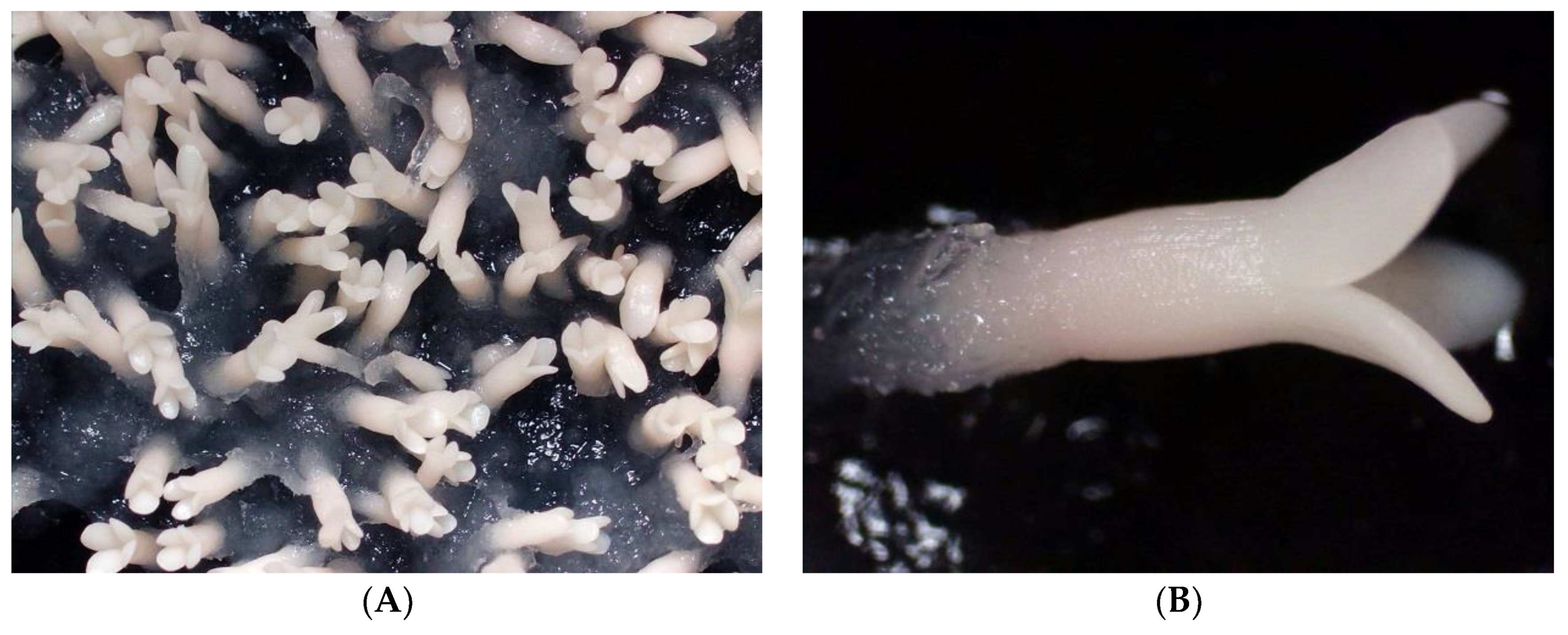
2.3. Maturation of Somatic Embryos

Somatic embryo maturation was carried out in accordance with the methodology described by Maruyama et al. [20]. ECs from 19 ECLs that had proliferated for 2 weeks were cultured in clumps (5 masses per 90 × 20 mm plate, 100 mg each) on maturation medium containing the basal salt concentration of standard EM medium [10] and supplemented with 30 g L−1 maltose, 2 g L−1 activated charcoal (AC), 100 µM ABA, amino acids (in g L−1: glutamine 2, asparagine 1, arginine 0.5, citrulline 0.079, ornithine 0.076, lysine 0.055, alanine 0.04, and proline 0.035), 175 g L−1 PEG (Av. Mol. Wt.: 7300–9300; Wako Pure Chemical, Osaka, Japan), and solidified with 3.3 g L−1 gellan gum. The plates were sealed with Parafilm® and kept in the dark at 25 °C for 8 weeks. The number of matured cotyledonary embryos was counted by plate (5 replicates for each line).

2.4. Somatic Embryo Germination and Plantlet Conversion

Cotyledonary embryos derived from 19 ECLs, picked up one-by-one from maturation medium, were laid horizontally onto germination medium (maintenance/proliferation medium supplemented with 20 g L−1 sucrose, 2 g L−1 AC, and 10 g L−1 agar, but without added plant hormones) and cultured at 25 °C under photon flux density of 45–65 µmol m−2 s−1 provided by white fluorescent lamps (100 V, 40 W) for 16 h daily. Somatic embryo germination, as emergence of root, and plantlet conversion, as emergence of both root and epicotyl, were recorded after 4 and 8 weeks of culture, respectively. The germination rate and the plantlet conversion rate were calculated for each plate as the numbers of germinants and the converted plantlets per number of tested cotyledonary embryos, respectively. The average value of the plates was used as the germination rate and conversion rate of each line (5 to 10 replicates).

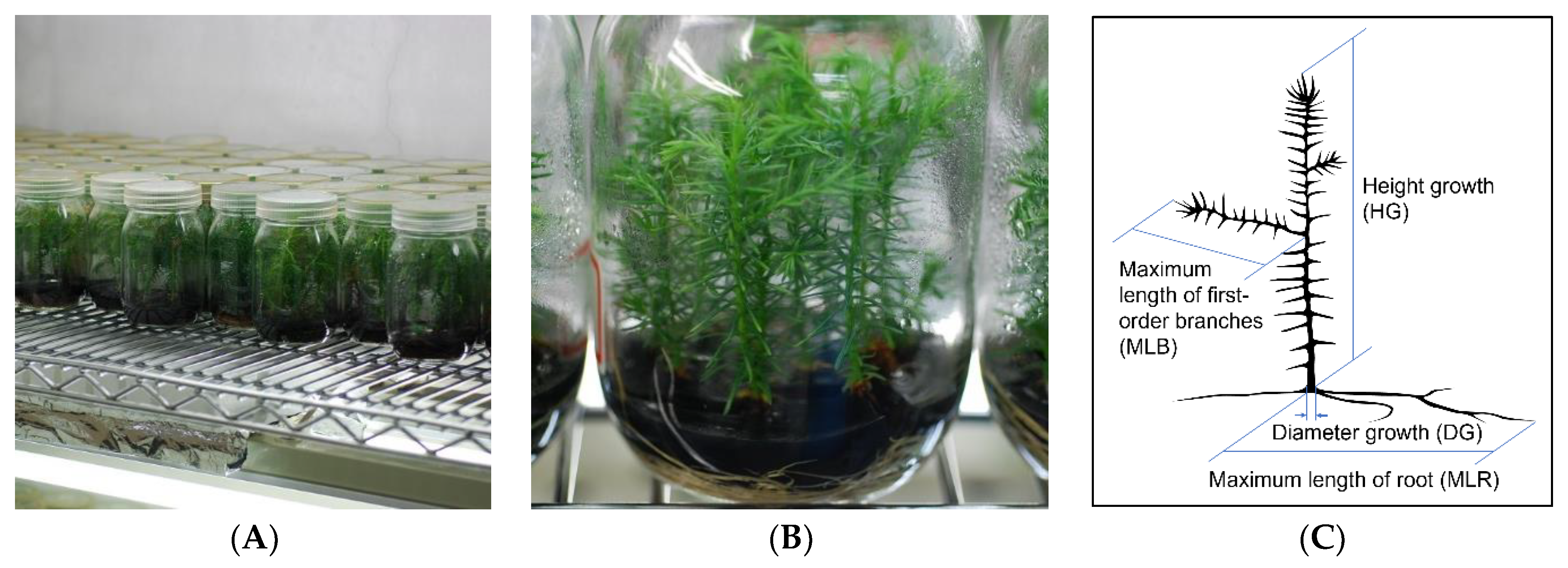
2.5. In Vitro Growth of Somatic Plantlets

Plantlet-converted somatic embryos derived from 19 ECLs were transferred to 450 mL culture bottles (https://axel.as-1.co.jp/asone/d/2-085-04/, accessed on 17 November 2021) containing 100 mL of growth medium (germination medium supplemented with 30 g L−1 sucrose and 5 g L−1 AC) and cultured under the same conditions as described above for somatic embryo germination and plantlet conversion. At the end of the 6-month growth period, height growth (HG), diameter growth (DG), developed number of first-order branches (DNB), maximum length of first-order branches (MLB), developed number of roots (DNR), and maximum length of root (MLR) were measured on five to ten somatic plantlets from each ECL tested. The HG was measured from the root collar to the apex of the main stem. The DG was measured at the root collar. The DNB and DNR represent the total numbers of developed branches and roots in each somatic plantlet, respectively. The MLB and MLR represent the length of the largest developed branch and root in each somatic plantlet, respectively.
Additionally, mature seeds that were collected from the same seed family in October 2016 and stored at 4 °C until examination were germinated on 1% agar medium (N = 25 × 6 replicates). Two months after germination in vitro, ten randomly selected seedlings were transferred into 450 mL culture bottles and, after 6 months of growth, traits were measured as experimental controls.

2.6. Statistical Analysis

The data for somatic embryo maturation efficiency, somatic embryo germination, and plantlet conversion from different ECLs were analyzed using one-way analysis of variance following model fittings of the generalized linear models (GLMs) with the lines as the explanatory variable. Here, Poisson and binomial distributions were used for the error distributions of maturation efficiency, germination, and plantlet conversion rate, respectively. The significance of variance components by cell lines was determined from the difference of Akaike’s Information Criterion (ΔAIC > 2) with the null model (i.e., intercept only model). The percent deviance explained was calculated as {1–(residual deviance)/(null deviance)} × 100. Comparisons among ECLs for the morphological characteristics (HG, DG, DNB, MLB, DNR, MLR) of in vitro growth performance in pollen-free somatic plantlets of sugi were analyzed using GLMs with Poisson and binomial error distributions for HG, DG, DNB, and MLB, and DNR and MLR, respectively. Correlations among the mean values of these objective variables, number of matured cotyledonary embryos, germination rate, and conversion rate were analyzed by pairwise Spearman’s correlation coefficient. The post hoc analysis in comparisons between the cultured seedlings (SSD-Seed) and each ECL were tested using the R package “multicomp” [25] with Dunnett adjustment. All statistical analyses were conducted in the R software [26].

3. Results

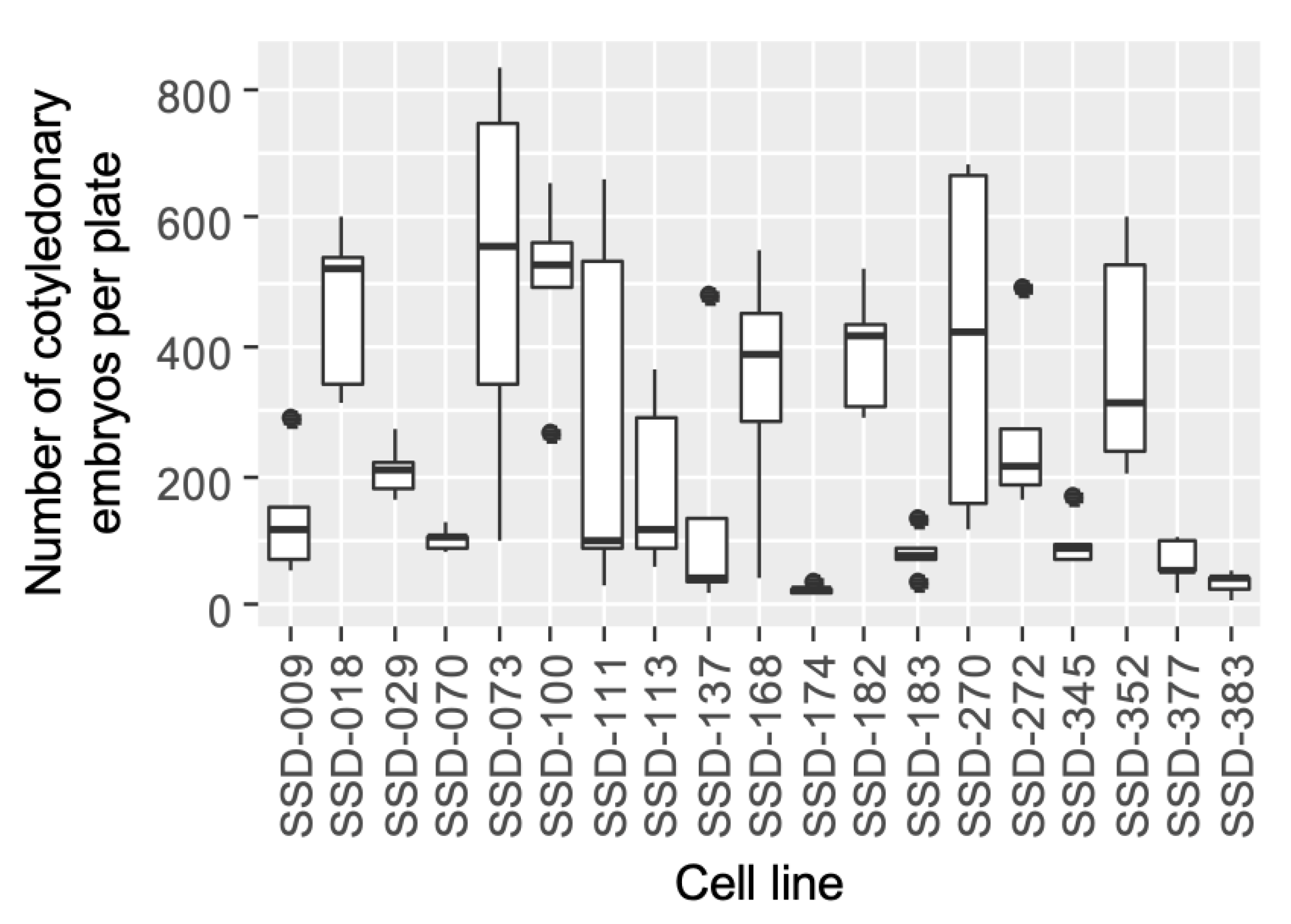
3.1. Somatic Embryo Maturation

Initial extrusion of ECs from megagametophyte explants was first observed about 2 weeks after the start of culture (Figure 1A) and led to evident proliferation most frequently after 4–6 weeks of culture (Figure 1B). Maintenance/proliferation medium was able to support the growth of initiated ECLs by subculture routines at 2–3-week intervals (Figure 1C).
Development of cotyledonary embryos from most of the ECLs tested was observed 6–8 weeks after the transfer of the ECs to maturation medium (Figure 2A,B). As shown in Figure 3, the SSD-073 line produced the highest average number of somatic embryos per plate (513.8 cotyledonary embryos), whereas the lowest number (25.0 embryos) was recorded for the SSD-174 line. Data analysis of our experimental results indicated that the maturation efficiency significantly differed among the ECLs (deviance explained = 63.2%, df = 18, Figure 3 and Supplementary Table S1). Although the number of mature embryos produced varied among the ECLs, the induction of cotyledonary embryos was confirmed in all lines with an average of 243.6 embryos per plate; and 14 out of 19 ECLs tested showed somatic embryo maturation efficiency greater than 100 cotyledonary embryos per plate (equivalent to more than 200 embryos per gram).

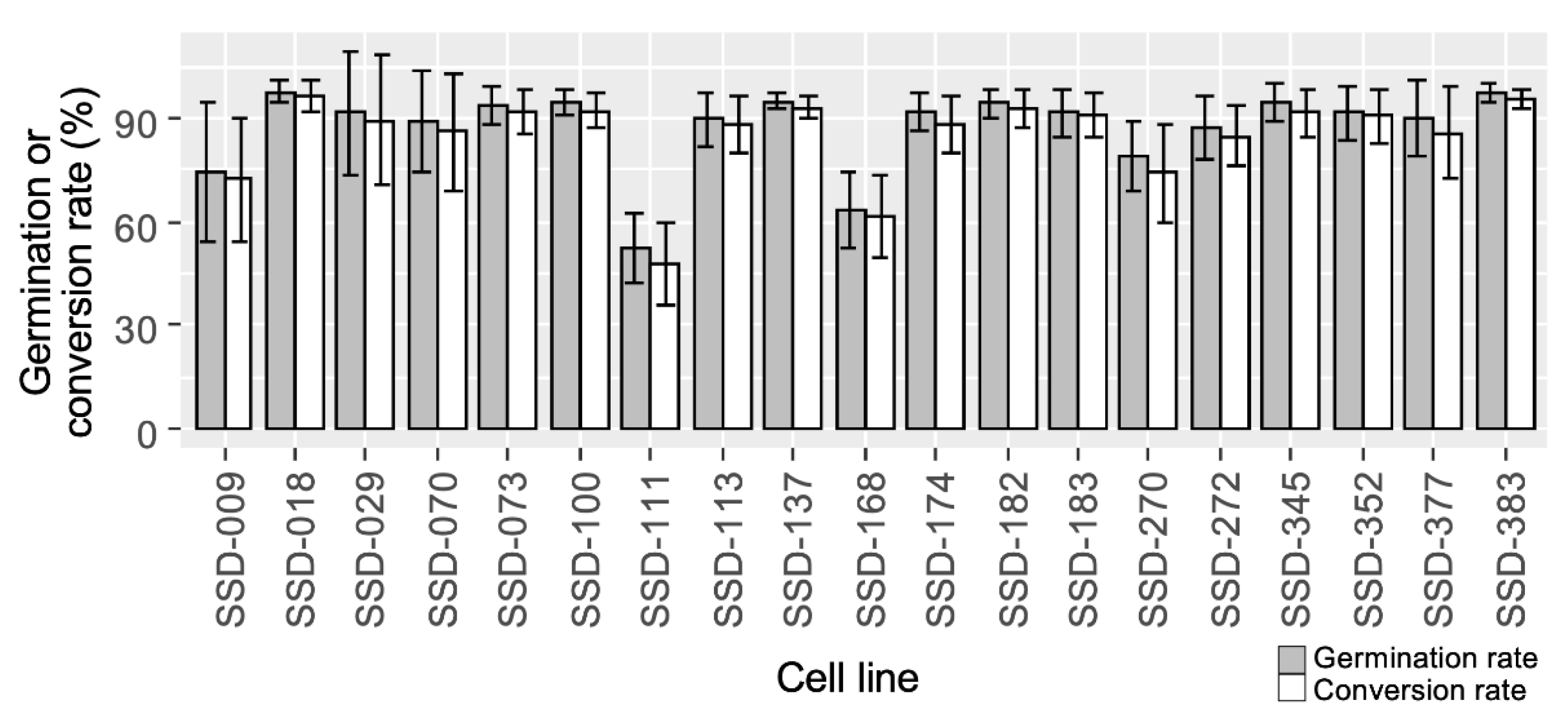
3.2. Somatic Embryo Germination and Plantlet Conversion

The start of germination of the developed cotyledonary embryos was observed at 1–2 weeks after transfer to germination medium (Figure 4A), and the subsequent plantlet conversion in most of the tested lines was achieved 3–6 weeks after (Figure 4B). The frequency of successful germination among ECLs varied from 52.1% to 97.5% with significant differences among the ECLs (deviance explained = 46.8%, df = 18, Figure 5 and Supplementary Table S2). On the other hand, almost all (average 97.3%) germinated cotyledonary embryos successfully converted to the plantlet. There were no significant differences between lines in efficiency from germination to conversion (deviance explained = 18.7%, df = 18, ΔAIC < 2, Supplementary Table S3). The final plantlet conversion rate was 47.7–96.5%. Among ECLs, the best germination and conversion results were achieved with somatic embryos derived from the SSD-18 line, whereas the lowest result was recorded for the SSD-111 line (Figure 5).
The germination rate of seeds of the same family (SSD-Seed) investigated for control comparison was 58.0% (SD ± 7.04), and the success rate of conversion to plantlets was 55.3% (SD ± 7.76).

3.3. In Vitro Growth of Somatic Plantlets and Seedlings

After the end of the 6-month growth period in the culture room (Figure 6), the growth of somatic plantlets derived from different ECLs was evaluated. The overall average HG and DG of somatic plantlets were 6.6 cm and 1.6 mm, whereas those of the control seedlings (SSD-Seed) were 8.1 cm and 1.9 mm, respectively (Figure 7A,B). Among somatic plantlets, the HG and DG varied significantly from 4.6 to 8.6 cm and 1.3 to 1.6 mm, respectively (Supplementary Table S4). Here, the somatic line SSD-182 exceeded the HG of the seedlings, while in contrast, the lowest HG was recorded for the line SSD-183. Although the HG and DG averages of somatic plantlets were lower than those of the control, some of the somatic lines showed similar growth to the control (no significant differences, p > 0.05, GLM and Dunnett test, Figure 7A,B). Regarding DNB and DNR, the averages for somatic plantlets were 3.0 branches and 4.7 roots, whereas for seedlings they were 6.0 branches and 6.3 roots, respectively. DNB was significantly lower than in the control in many cell lines (11/19), while DNR was only significantly lower in SSD-272 (p < 0.05, Figure 7C,D). Although the DNB and DNR varied by cell line from 1.6 to 4.6 branches and from 3.1 to 6.0 roots per plantlet, respectively, cell line was not selected as a significant explanatory variable (ΔAIC < 2, Supplementary Table S4). Regarding the MLB of somatic plantlets, the highest (5.2 cm) and lowest (1.9 cm) values were achieved for the lines SSD-182 and SSD-183, respectively. The overall average for the MLB of somatic plantlets was 3.7 cm, while for the seedlings it was 5.7 cm. MLB varied significantly among strains and was lower than in the control in 11 of 19 strains (Figure 7E, Supplementary Table S4). On the other hand, the highest (24.2 cm) and lowest (7.7 cm) values for the MLR were observed in lines SSD-352 and SSD-270, respectively, where most cell lines showed similar growth to the control (p > 0.05, Figure 7F).

4. Discussion

In the present study, using a previously established culture protocol [4], we obtained efficient cotyledonary embryo development of over 100 embryos per plate (average 243.6), a high level of embryo germination (average 88.9%), and an extremely high rate of plantlet conversion of germinants (average 97.3%) in most of the lines tested. Stable plantlet regeneration from megagametophytes of immature seeds was achieved in the MSPs of sugi. Although it is not possible to make a simple comparison, considering that the percentage of seedlings established from seeds was 55.3%, the plantlet yield from SE is impressive enough. In general, somatic embryo maturation, germination, and plantlet conversion efficiency values achieved in this study were comparable with those reported for other conifers [9,27,28,29,30]. However, the bottleneck was the low initiation rate of ECs (17.2% of the total [19]), which has been similarly reported in many studies of coniferous species including MSP or non-MSP sugi [11,12,13,31,32]. Thus, low induction rates are not a feature characteristic of MSP. If the total yield of seedlings obtained through SE, from the initiation of ECs to plantlet conversion, became higher than that produced from seeds, SE can be expected to have a wide range of applications, not only in mass clonal propagation, but also for example in shortening the breeding period and the conservation of endangered or recalcitrant lines with poor seed germination rates (e.g., [29,33,34]).
Six months after the plantlet conversion, the growth of 19 somatic lines was evaluated, making it—to the best of our knowledge—the first study to evaluate in vitro growth traits of SE-derived MSPs in sugi. In other conifers, growth traits of somatic plants have been evaluated for comparison with seedlings (seed-derived plants), and some of these studies reported lower growth performance of somatic plants compared with seedlings [21,35,36,37]. The reduction in initial growth may have been due to several culture stresses during the whole SE process, such as osmotic, drought, and altered endogenous hormone balance stress. Similar to these reports, the mean values of growth traits of sugi somatic plantlets were lower than those of seedlings. However, there were several lines that showed similar growth to seedlings, and since the traits of seedlings were within the range of variation among that of the somatic cell lines (Figure 7), this difference may indicate variation among siblings rather than an effect of the SE process. Although most of the deviance in maturation and germination was explained by inter-cell-line variation (63.2% and 46.8%, respectively), the cell line variables explained only 19.2–39.7% of the variance components of growth traits (Supplementary Table S4). As some studies comparing growth between male-sterile and fertile sugi cultivars showed no clear differences in growth traits between them [38,39], the male-sterile genotype is also not related to seedling growth. The MSPs propagated via SE would be useful as plant material for afforestation.
As shown in other conifer species (e.g., [40,41,42,43]), our statistical analysis showed that HG, DG, MLB, and MLR were significantly different among the cell lines. Correlation analysis also showed no significant relationships between performance in SE (i.e., the efficiency of cotyledonary embryo maturation, germination rate, and plantlet conversion rate) and initial in vitro plantlet growth traits (p > 0.05, Supplementary Figure S1). This indicates that the difference in the performance of the initial growth of the somatic sugi plantlet is a cell-line-specific characteristic rather than a culture-induced variation. The initial growth traits of the lines are considered to be genetically fixed [21,42]. The cell line SSD-182 was comparable to SSD-seed in all growth traits. Our group has also evaluated the growth of SE-derived MSPs after ex vitro transfer and found that the in vitro growth results were reflected in initial sapling growth, where the SSD-182 line continuously showed better growth among the tested lines, even better than seedlings, not only in plant containers, but also after transfer to the field [44]. The efficiency of initial selection may be achieved by using a strain with good growth performance like SSD-182. Similarly, Högberg et al. [21] reported that the early selection at the time of ex vitro transfer improved clonal performance and reduced intraclonal variation of Norway spruce somatic plants. Although some studies reported that the further growth patterns of seedlings were fixed by each strain in Japanese cedar [45,46], it has also been reported that growth trait heritability is not constant after plantation [46]. To confirm how long the initial in vitro growth trait will be maintained for later growth, continued monitoring of the growth of SE-derived plants for several years and even decades must be performed.

5. Conclusions

In the present study, an efficient and stable method for propagating pollen-free somatic plantlets through SE after early discrimination of ECLs with MAS was demonstrated in Japanese cedar, sugi. Highly efficient embryo maturation, germination, and plantlet conversion from different lines carrying the male sterility allele ms1 were confirmed. In vitro growth performance of the developed somatic plantlets, analyzing different morphological variables involved in the initial growth of sugi, showed significant differences among ECLs. To the best of our knowledge, this is the first report to describe the in vitro growth performance of C. japonica. Additionally, the information from this study may be considered as a first step for evaluating in vitro growth traits of SE-derived MSPs.

Supplementary Materials

The following are available online at https://0-www-mdpi-com.brum.beds.ac.uk/article/10.3390/f12111592/s1, Table S1: Analysis of deviance table of maturation efficiency by cell lines of sugi (Japanese cedar, C. japonica). Residual deviance was estimated by generalized linear models using the cell line as the explanatory variable, Table S2: Analysis of deviance table of germination rate by cell lines of sugi (Japanese cedar, C. japonica). The number of germinants per number of installed cotyledonary embryos was applied to the GLMs with a binomial distribution, Table S3: Analysis of deviance table of efficiency from germination to plantlet conversion of sugi (Japanese cedar, C. japonica). The number of converted plantlets per number of germinants was applied to the GLMs with a binomial distribution, Table S4: Analysis of deviance table for somatic plantlet growth of sugi (Japanese cedar, C. japonica) in height (HG), diameter (DG), developed number of first-order branches (DNB), developed number of roots (DNR), maximum length of first-order branches (MLB), and maximum length of roots (MLR), Figure S1: Pairwise correlations among mean value of the number of matured cotyledonary embryos, average rates of germination and plantlet conversion, and growth traits of 19 somatic sugi lines. NCE: number of matured cotyledonary embryos, GR: germination rate, CR: plantlet conversion rate, HG: height growth, DG: diameter growth, DNB: developed number of first-order branches, DNR: developed number of roots, MLB: maximum length of first-order branches, and MLR: maximum length of roots. Spearman’s coefficient values and their significances are presented in the upper right.

Author Contributions

Conceptualization and methodology, M.T., T.E.M., S.U., T.K. and Y.M.; funding acquisition and project administration, Y.M.; plant material preparation, T.E.M. and S.U.; data curation, M.T. and T.E.M.; experiments and data analysis, T.E.M., M.T., T.K. and S.U.; writing—original draft, M.T. and T.E.M.; writing—review and editing, M.T., T.E.M., S.U., T.K. and Y.M. All authors have read and agreed to the published version of the manuscript.

Funding

This research was supported by grants from the Ministry of Agriculture, Forestry and Fisheries of Japan (MAFF) and Bio-oriented Technology Research Advancement Institution (BRAIN) (the science and technology research promotion program for agriculture, forestry, fisheries and food industry (No. 28013B)) and grants from BRAIN (research program on development of innovative technology (No. 28013BC)).

Institutional Review Board Statement

Not applicable.

Informed Consent Statement

Not applicable.

Data Availability Statement

Original data presented in the study are included in the main text, further inquiries can be directed to the corresponding author.

Acknowledgments

The authors would like to thank Niigata Prefectural Forestry Research Institute for support in the seed material preparation.

Conflicts of Interest

The authors declare no conflict of interest.

References

  1. FAO. Global Forest Resources Assessment 2020 Main Report; Food and Agriculture Organization of the United Nations: Rome, Italy, 2020; 186p. [Google Scholar]
  2. Forestry Agency. Statistical Handbook of Forest and Forestry; Forestry Agency, Ministry of Agriculture, Forestry and Fisheries: Tokyo, Japan, 2020; pp. 8–9. (In Japanese) [Google Scholar]
  3. Matsubara, A.; Sakashita, M.; Gotoh, M.; Kawashima, K.; Matsuoka, T.; Kondo, S.; Yamada, T.; Takeno, S.; Takeuchi, K.; Urashima, M.; et al. Epidemiological Survey of Allergic Rhinitis in Japan 2019. Nippon. Jibiinkoka Gakkai Kaiho 2020, 123, 485–490, (In Japanese with English abstract). [Google Scholar] [CrossRef]
  4. Maruyama, T.E.; Ueno, S.; Hirayama, S.; Kaneeda, T.; Moriguchi, Y. Somatic embryogenesis and plant regeneration from sugi (Japanese Cedar, Cryptomeria japonica D. Don, Cupressaceae) seed families by marker assisted selection for the male sterility allele ms1. Plants 2020, 9, 1029. [Google Scholar] [CrossRef] [PubMed]
  5. Park, J.S.; Barret, J.D.; Bonga, J.M. Application of somatic embryogenesis in high-value clonal forestry: Deployment, genetic control, and stability of cryopreserves clones. In Vitro Cell. Dev. Biol. Plant 1998, 34, 231–239. [Google Scholar] [CrossRef]
  6. Klimaszewska, K.; Trontin, J.F.; Becwar, M.R.; Devillard, C.; Park, Y.S.; Lelu-Walter, M.A. Recent progress in somatic embryogenesis of four Pinus spp. Tree For. Sci. Biotechol. 2007, 1, 11–25. [Google Scholar]
  7. Bonga, J.M.; Klimaszewska, K.; von Aderkas, P. Recalcitrance in clonal propagation, in particular of conifers. Plant Cell Tissue Organ Cult. 2010, 100, 241–254. [Google Scholar] [CrossRef]
  8. Klimaszewska, K.; Cyr, D.R. Conifer somatic embryogenesis: I. Development. Dendrobiology 2002, 48, 31–39. [Google Scholar]
  9. Maruyama, T.E.; Hosoi, Y. Progress in somatic embryogenesis of Japanese pines. Front. Plant Sci. 2019, 10, 31. [Google Scholar] [CrossRef]
  10. Maruyama, E.; Tanaka, T.; Hosoi, Y.; Ishii, K.; Morohoshi, N. Embryogenic cell culture, protoplast regeneration, cryopreservation, biolistic gene transfer and plant regeneration in Japanese cedar (Cryptomeria japonica D. Don). Plant Biotechnol. 2000, 17, 281–296. [Google Scholar] [CrossRef]
  11. Maruyama, T.E.; Hosoi, Y.; Futamura, N.; Saito, M. Initiation of embryogenic cultures from immature seeds of pollen-free sugi (Cryptomeria japonica). Kanto Shinrin Kenkyu 2014, 65, 107–110, (In Japanese with English Abstract). [Google Scholar]
  12. Maruyama, T.E.; Miyazawa, S.; Ueno, S.; Onishi, N.; Totsuka, S.; Iwai, J.; Moriguchi, Y. Differences among families on embryogenic cell induction from seed of pollen-free sugi (Cryptomeria japonica) produced at the Niigata prefecture. Kanto Shinrin Kenkyu 2018, 69, 1–2, (In Japanese with English Abstract). [Google Scholar]
  13. Taniguchi, T.; Konagaya, K.; Nanasato, Y. Somatic embryogenesis in artificially pollinated seed families of 2nd generation plus trees and cryopreservation of embryogenic tissue in Cryptomeria japonica D. Don (sugi). Plant Biotechnol. 2020, 37, 239–245. [Google Scholar] [CrossRef]
  14. Igasaki, T.; Sato, T.; Akashi, N.; Mohri, T.; Maruyama, E.; Kinoshita, I.; Walter, C.; Shinohara, K. Somatic embryogenesis and plant regeneration from immature zygotic embryos of Cryptomeria japonica D. Don. Plant Cell Rep. 2003, 22, 239–243. [Google Scholar] [CrossRef]
  15. Igasaki, T.; Akashi, N.; Ujino-Ihara, T.; Matsubayashi, Y.; Sakagami, Y.; Shinohara, K. Phytosulfokine stimulates somatic embryogenesis in Cryptomeria japonica. Plant Cell Physiol. 2003, 44, 1412–1416. [Google Scholar] [CrossRef] [Green Version]
  16. Nakagawa, R.; Ogita, S.; Kubo, T.; Funada, R. Effect of polyamines and L-ornithine on the development of proembryogenic masses of Cryptomeria japonica. Plant Cell Tissue Organ Cult. 2006, 85, 229–234. [Google Scholar] [CrossRef]
  17. Maruyama, E.; Hosoi, Y. Polyethylene glycol enhance somatic embryo production in Japanese cedar (Cryptomeria japonica D. Don). Propag. Ornam. Plants 2007, 7, 57–61. [Google Scholar]
  18. Maruyama, T.E.; Hosoi, Y.; Miyazawa, S.; Ueno, S.; Onishi, N.; Totsuka, S.; Iwai, J.; Moriguchi, Y. Pollen-free plant regeneration from embryogenic cells derived from sugi (Cryptomeria japonica). Kanto Shinrin Kenkyu 2019, 70, 37–40, (In Japanese with English Abstract). [Google Scholar]
  19. Maruyama, T.E.; Ueno, S.; Hosoi, Y.; Miyazawa, S.I.; Mori, H.; Kaneeda, T.; Bamba, Y.; Itoh, Y.; Hirayama, S.; Kawakami, K.; et al. Somatic embryogenesis initiation in sugi (Japanese Cedar, Cryptomeria japonica D. Don): Responses from male-fertile, male-sterile, and polycoss-pollinated-derived seed explants. Plants 2021, 10, 398. [Google Scholar] [CrossRef] [PubMed]
  20. Maruyama, T.E.; Ueno, S.; Mori, H.; Kaneeda, T.; Moriguchi, Y. Factors influencing somatic embryo maturation in sugi (Japanese Cedar, Cryptomeria japonica (Thunb. ex L.f.) D. Don). Plants 2021, 10, 874. [Google Scholar] [CrossRef]
  21. Högberg, K.-A.; Bozhkov, P.V.; Von Arnold, S. Early selection improves clonal performance and reduces intraclonal variation of Norway spruce plants propagated by somatic embryogenesis. Tree Physiol. 2003, 23, 211–216. [Google Scholar] [CrossRef] [Green Version]
  22. Hasegawa, Y.; Ueno, S.; Wei, F.J.; Matsumoto, A.; Ujino-Ihara, T.; Uchiyama, K.; Moriguchi, Y.; Kasahara, M.; Fujino, T.; Shigenobu, S.; et al. Development of diagnostic PCR and LAMP markers for MALE STERILITY 1 (MS1) in Cryptomeria japonica D. Don. BMC Res. Notes 2020, 13, 457. [Google Scholar] [CrossRef]
  23. Ueno, S.; Uchiyama, K.; Moriguchi, Y.; Ujino-Ihara, T.; Matsumoto, A.; Wei, F.J.; Saito, M.; Higuchi, Y.; Futamura, N.; Kanamori, H.; et al. Scanning RNA-Seq and RAD-Seq approach to develop SNP markers closely linked to MALE STERILITY 1 (MS1) in Cryptomeria japonica D. Don. Breed. Sci. 2019, 69, 19–29. [Google Scholar] [CrossRef] [PubMed] [Green Version]
  24. Tsuruta, M.; Maruyama, T.E.; Ueno, S.; Hasegawa, Y.; Moriguchi, Y. Marker-assisted selection for pollen-free somatic plants of sugi (Japanese Cedar, Cryptomeria japonica): A simple and effective methodology for selecting male-sterile mutants with ms1-1 and ms1-2. Front. Plant Sci. 2021, 12, 748110. [Google Scholar] [CrossRef]
  25. Bretz, F.; Hothorn, T.; Westfall, P. Multiple Comparisons Using R; CRC Press: Boca Raton, FL, USA, 2010. [Google Scholar]
  26. R Core Team. R: A Language and Environment for Statistical Computing; R Foundation for Statistical Computing: Vienna, Austria, 2020; Available online: https://www.R-project.org/ (accessed on 17 November 2021).
  27. Maruyama, E.; Hosoi, Y.; Ishii, K. Somatic embryogenesis in Sawara cypress (Chamaecyparis pisifera Sieb. et Zucc.) for stable and efficient plant regeneration, propagation and protoplast culture. J. For. Res. 2002, 7, 23–34. [Google Scholar] [CrossRef]
  28. Maruyama, E.; Ishii, K.; Hosoi, Y. Efficient plant regeneration of Hinoki cypress (Chamaecyparis obtusa Sieb. et Zucc.) via somatic embryogenesis. J. For. Res. 2005, 10, 73–77. [Google Scholar] [CrossRef]
  29. Maruyama, T.E.; Hosoi, Y.; Katsuki, T. Plant regeneration of Yatsugataketouhi (Picea koyamae) through somatic embryogenesis. Kanto Shinrin Kenkyu 2011, 62, 127–130. (In Japanese) [Google Scholar]
  30. Maruyama, T.E.; Hosoi, Y.; Katsuki, T. Somatic embryo induction from embryogenic cells of Himebaramomi (Picea maximowiczii Regel ex Mast.). Kanto Shinrin Kenkyu 2012, 63, 67–71, (In Japanese with English abstract). [Google Scholar]
  31. Garin, E.; Isabel, N.; Plourde, A. Screening of large numbers of seed families of Pinus strobus L. for somatic embryogenesis from immature and mature zygotic embryos. Plant Cell Rep. 1998, 18, 37–43. [Google Scholar] [CrossRef]
  32. Miguel, C.; Goncalves, S.; Tereso, S.; Marum, L.; Maroco, J.; Oliveira, M. Somatic embryogenesis from 20 open-pollinated families of Portuguese plus trees of maritime pine. Plant Cell Tissue Organ Cult. 2004, 76, 121–130. [Google Scholar] [CrossRef]
  33. Ma, X.; Bucalo, K.; Determann, R.O.; Cruse-Sanders, J.M.; Pullman, G.S. Somatic embryogenesis, plant regeneration, and cryopreservation for Torreya taxifolia, a highly endangered coniferous species. In Vitro Cell. Dev. Biol. Plant 2012, 48, 324–334. [Google Scholar] [CrossRef]
  34. Ahn, C.H.; Tull, A.R.; Montello, P.M.; Merkle, S.A. A clonal propagation system for Atlantic white cedar (Chamaecyparis thyoides) via somatic embryogenesis without the use of plant growth regulators. Plant Cell Tissue Organ Cult. 2017, 130, 91–101. [Google Scholar] [CrossRef]
  35. Grossnickle, S.C.; Major, J.E.; Folk, R.S. Interior spruce seedlings compared with emblings produced from somatic embryogenesis. I. Nursery development, fall acclimation, and over-winter storage. Can. J. For. Res. 1994, 24, 1376–1384. [Google Scholar] [CrossRef]
  36. Dean, C.; Welty, D.; Herold, G. Performance and genetic parameters of somatic and zygotic progenies of coastal Douglas-fir at 7 ½-years across Washington and Oregon, USA. Silvae Genet. 2009, 58, 212–219. [Google Scholar] [CrossRef] [Green Version]
  37. Hosoi, Y.; Kuramoto, N.; Maruyama, T.E. Screening RAPD primers to assess clonal fidelity in somatic embryos of Sawara cypress (Chamaecyparis pisifera Sieb. et Zucc.) and field performance of somatic embryo-derived trees. Plant Biotechnol. 2015, 32, 149–155. [Google Scholar] [CrossRef] [Green Version]
  38. Miura, S.; Moriguchi, Y.; Taira, H. Comparison of traits between male sterile and fertile Cryptomeria japonica D. Don trees in selected stands. J. Jpn. For. Soc. 2009, 91, 290–294, (In Japanese with English Abstract). [Google Scholar] [CrossRef]
  39. Hakamata, T.; Kondo, A.; Yamamoto, S.; Saito, M. Growth of containerized Japanese ceder seedlings crossing with mother trees having male sterile gene. Chubu For. Res. 2017, 65, 3–4. (In Japanese) [Google Scholar]
  40. Lamhamedi, M.S.; Chamberland, H.; Bernier, P.Y.; Tremblay, F.M. Clonal variation in morphology, growth, physiology, anatomy and ultrastructure of container-grown white spruce somatic plants. Tree Physiol. 2000, 20, 869–880. [Google Scholar] [CrossRef] [PubMed] [Green Version]
  41. Grossnickle, S.C.; Folk, R.S. Field performance potential of a somatic interior spruce seedlot. New For. 2007, 34, 51–72. [Google Scholar] [CrossRef]
  42. Wahid, N.; Rainville, A.; Lamhamedi, M.; Margolis, H.; Beaulieu, J.; Deblois, J. Genetic parameters and performance stability of white spruce somatic seedlings in clonal tests. For. Ecol. Manag. 2012, 270, 45–53. [Google Scholar] [CrossRef]
  43. Puentes, A.; Högberg, K.A.; Björklund, N.; Nordlander, G. Novel avenues for plant protection: Plant propagation by somatic embryogenesis enhances resistance to insect feeding. Front. Plant Sci. 2018, 9, 1553. [Google Scholar] [CrossRef]
  44. Kaneeda, T.; Moriguchi, Y.; Maruyama, T.E.; Tsuruta, M.; Ueno, S.; (Niigata University, Niigata, Japan). Unpublished work. 2021.
  45. Kurinobu, S.; Toda, T. Genetic variation in height growth curves observed in three clonal tests of Sugi (Cryptomeria japonica D. Don) in Kyushu. J. For. Res. 2000, 5, 95–98. [Google Scholar] [CrossRef]
  46. Sato, Y.; Fukatsu, E.; Hiraoka, Y.; Watanabe, A.; Takahashi, M. The effect of genotype and planting density on the growth patterns and selection of local varieties of Sugi (Cryptomeria japonica). J. Jpn. For. Soc. 2016, 98, 45–52, (In Japanese with English Abstract). [Google Scholar] [CrossRef] [Green Version]
Figure 1. SE initiation in sugi (Japanese cedar, C. japonica). (A) Extrusion of ECs from megagametophyte explant 2 weeks after the start of culture; (B) proliferation from extruded ECs after 6 weeks of culture; (C) maintenance/proliferation of ECs by 2–3-week interval subculture routines. Bars: 5 mm (A,B), 1 cm (C).
Forests 12 01592 g001
Figure 2. Somatic embryo maturation in sugi (Japanese cedar, C. japonica). (A) Maturation of somatic embryos 8 weeks after the transfer of ECs to maturation medium; (B) a close-up of developed cotyledonary embryo. Bars: 5 mm (A), 1 mm (B).
Forests 12 01592 g002
Figure 3. Box plot of the efficiency of cotyledonary embryo maturation by each line of sugi (Japanese cedar, C. japonica). The black dots indicate outliers.
Forests 12 01592 g003
Figure 4. Somatic embryo germination and plantlet conversion in sugi (Japanese cedar, C. japonica). (A) Germination of somatic embryos about 2 weeks after the transfer to germination medium; (B) plantlet conversion about 6 weeks after somatic embryo germination. Bars: 1 cm.
Forests 12 01592 g004
Figure 5. Cell line differences in germination and plantlet conversion of sugi (Japanese cedar, C. japonica). The germination rate of somatic embryos (gray bars) and plantlet conversion rate (white bars) per each plate are presented as mean and standard deviation.
Forests 12 01592 g005
Figure 6. In vitro growth of sugi (Japanese cedar, C. japonica) somatic plantlets. (A) Somatic plantlets growing in the culture room; (B) growth of somatic plantlets about 6 months after transfer to culture bottles; (C) schematic diagram of each measurement of growth trait. Bars: 5 cm.
Forests 12 01592 g006
Figure 7. Box plots for the growth traits in somatic plantlets and control seedlings (SSD-Seed) of sugi (Japanese cedar, C. japonica) for height growth (A), diameter growth (B), developed number of first-order branches (C) and roots (D), and maximum length of branches (E) and roots (F). Significant differences from control seedlings at the 5%, 1%, and 0.1% levels are indicated by *, **, and ***, respectively (Dunnett test). The black dots indicate outliers.
Forests 12 01592 g007
Publisher’s Note: MDPI stays neutral with regard to jurisdictional claims in published maps and institutional affiliations.

