Article

LC-ESI-QTOF-MS/MS Characterisation of Phenolics in Herbal Tea Infusion and Their Antioxidant Potential

1
School of Agriculture and Food, Faculty of Veterinary and Agricultural Sciences, The University of Melbourne, Parkville, VIC 3010, Australia
2
Centre for Chemistry and Biotechnology, School of Life and Environmental Sciences, Deakin University, Waurn Ponds, VIC 3217, Australia
3
Faculty of Biological Sciences, The University of Leeds, Leeds LS2 9JT, UK
*
Author to whom correspondence should be addressed.
Submission received: 4 April 2021 / Revised: 22 April 2021 / Accepted: 6 May 2021 / Published: 10 May 2021
(This article belongs to the Special Issue Implementation of Digital Technologies on Beverage Fermentation)

Abstract

:
Ginger (Zingiber officinale R.), lemon (Citrus limon L.) and mint (Mentha sp.) are commonly consumed medicinal plants that have been of interest due to their health benefits and purported antioxidant capacities. This study was conducted on the premise that no previous study has been performed to elucidate the antioxidant and phenolic profile of the ginger, lemon and mint herbal tea infusion (GLMT). The aim of the study was to investigate and characterise the phenolic contents of ginger, lemon, mint and GLMT, as well as determine their antioxidant potential. Mint recorded the highest total phenolic content, TPC (14.35 ± 0.19 mg gallic acid equivalent/g) and 2,2′-azino-bis(3-e-thylbenzothiazoline-6-sulfonic acid), ABTS (24.25 ± 2.18 mg ascorbic acid equivalent/g) antioxidant activity. GLMT recorded the highest antioxidant activity in the reducing power assay, RPA (1.01 ± 0.04 mg ascorbic acid equivalent/g) and hydroxyl radical scavenging assay, OH-RSA (0.77 ± 0.08 mg ascorbic acid equivalent/g). Correlation analysis showed that phenolic content positively correlated with the antioxidant activity. Venn diagram analysis revealed that mint contained a high proportion of exclusive phenolic compounds. Liquid chromatography coupled with electrospray ionisation and quadrupole time of flight tandem mass spectrometry (LC-ESI-QTOF-MS/MS) characterised a total of 73 phenolic compounds, out of which 11, 31 and 49 were found in ginger, lemon and mint respectively. These characterised phenolic compounds include phenolic acids (24), flavonoids (35), other phenolic compounds (9), lignans (4) and stilbene (1). High-performance liquid chromatography photometric diode array (HPLC-PDA) quantification showed that GLMT does contain a relatively high concentration of phenolic compounds. This study presented the phenolic profile and antioxidant potential of GLMT and its ingredients, which may increase the confidence in developing GLMT into functional food products or nutraceuticals.

1. Introduction

Herbal teas made from various medicinal plants are rich sources of phenolic compounds and are consumed by many cultures all around the world. Medicinal plants prepared for tea infusions have been studied for their antioxidant, anti-inflammatory and other properties [1]. Herbal teas have also been reported to exhibit synergistic antioxidant effects, which increases their value as beverages for potential health benefits [2,3]. Common medicinal plants such as ginger, lemon and mint can be fused together as ginger lemon mint tea (GLMT) and be consumed as herbal tea to improve health being.
Gingers (Zingiber officinale Roscoe) from the family Zingiberaceae [4,5,6], are native to Southeast Asia and have been incorporated into a diverse array of cuisines. In China and India, gingers are regarded as medicine [5]. Ginger is reported to have various health benefits as it demonstrates antioxidant, antimicrobial and anti-inflammatory activities [7]. A gingerol is a group of phenols commonly found in the rhizomes of Zingiberaceae plants, which have been extensively studied for their cytotoxic potential [8]. Lemon (Citrus limon L.) is from the citrus family Rutaceae [9], and has been demonstrated to contain phenolic compounds which are beneficial to health [10]. Eriocitrin, a phenolic compound in lemons was found to increase antioxidant activities in plasma after ingestion [11]. Lemon has potent antioxidant activities in its peel and zest and its protective properties against DNA and cell damage have prompted the research on lemon as a cancer preventative [12]. Other studies have also shown that lemon’s essential oil has antioxidant and antimicrobial effects [13]. Mint (Mentha sp.) is from the Lamiaceae plant family [14,15]. The essential oils of plants within the Mentha genus exhibit antioxidant and antimicrobial activities [16,17]. Notable antioxidants in mints were found to be rosmarinic acid, caffeic acid, α-terpinene, luteolin and eriocitrin and these compounds are believed to be the major antioxidants particularly found in peppermint [15].
Phenolic compounds are a large group of secondary plant metabolites commonly found in fruits, vegetables and spices, which are thought to play prominent roles in human nutrition and health [18]. Some of these metabolites produced by plants are classified as antioxidants [19]. Antioxidants are molecules that can reduce the damaging effects of free radicals, which are chemicals with one or more unpaired electrons produced in an organism via natural metabolism or environmental factors [20]. Free radicals can turn non-free radical molecules in the body into radicals, cascading a chain reaction that causes destructive effects within cells and tissues [21]. Thus, medicinal plants rich in antioxidants have been lauded and advised to be part of the human diet to promote health and wellbeing [22,23].
Various assays and equipment have been developed that have been useful for the estimation and analysis of phenolic and antioxidant content. For the estimation of phenolic compounds, common assays include Total Phenolic Content (TPC) Assay with Folin-Ciocalteu reagent [24], Total Flavonoid Content (TFC) Assay by the aluminium chloride method [25] and Total Tannins Content (TTC) using vanillin method [26]. 2,2′-diphenyl-1-picrylhydrazyl (DPPH) assay [27], Ferric reducing antioxidant power (FRAP) assay [28], 2,2′-azino-bis(3e-thylbenzothiazoline-6-sulfonic acid) (ABTS) radical scavenging assay [29], reducing power assay (RPA) [30], hydroxyl radical scavenging activity (OH-RSA) [31] and ferrous ion chelating activity (FICA) [32] assays are common assays utilised to determine the antioxidant activities of samples. In addition, the phosphomolybdate method has been used in the past for the estimation of total antioxidant content (TAC). Statistical analysis was performed on the phenolic content and antioxidant activity of the samples to investigate whether the phenolic content correlated with the antioxidant activity. Liquid chromatographic-coupled mass spectrometry was useful as a separation, identification and quantification technique, enabling the characterisation of phenolic compounds. This experiment deployed HPLC-PDA and LC-ESI-QTOF-MS/MS, which enabled the quantification and characterisation of phenolic compounds from ginger, lemon, mint and GLMT. Previous experiments have also successfully utilised both HPLC and LC-MS/MS techniques to quantify and characterise phenolic compounds in various herbal teas and medicines [33,34,35]. The comprehensive understanding of the phenolic profile of ginger, lemon and mint, GLMT has not been extensively studied and there is a research gap that requires investigation into GLMT’s antioxidant effect and its phenolic profile. Thus, the aims of this study were to: (1) investigate and compare antioxidant activities of ginger, lemon, mint and GLMT herbal tea infusion; (2) characterise and quantify their phenolic compounds through LC-ESI-QTOF-MS/MS and HPLC-PDA respectively. Results indicating high phenolic content and potent antioxidant activity from the samples could be used to inform the benefits of dietary uptake of ginger, lemon, mint and GLMT, which may also encourage commercialisation of GLMT infusions.

2. Materials and Methods

2.1. Chemicals and Reagents

Most of the chemicals utilised were analytical grade and purchased from Sigma-Aldrich (St. Louis, MO, USA). Reagents for the antioxidant assays include 2,2′-diphenyl-1-picrylhydrazyl (DPPH), 2,2′-azino-bis(3-ethylbenzothiazoline-6-sulfonic acid) (ABTS), 2,4,6-tripyridyl-s-triazine (TPTZ), Folin and Ciocalteu’s phenol, hexahydrate aluminium chloride, potassium persulfate and vanillin were purchased from Sigma-Aldrich (St. Louis, MO, USA). The phenolic acid and flavonoid reference standards used such as caffeic acid, caftaric acid, catechin, chlorogenic acid, coumaric acid, epicatechin gallate, gallic acid, kaempferol, kaempferol-3-O-glucoside, L-ascorbic acid, p-hydroxybenzoic acid, protocatechuic acid, quercetin, quercetin-3-galactose, quercetin-3-O-glucuronide and syringic acid were also bought from Sigma-Aldrich (St Louis, MO, USA). Other chemicals including acetic acid, ethanol, ferric (III) chloride (Fe [III] Cl3·6H2O), sodium acetate and sodium carbonate were purchased from Sigma-Aldrich (St. Louis, MO, USA). The HPLC grade acetic acid, methanol and acetonitrile used were bought from Sigma-Aldrich (St. Louis, MO, USA). The 98% sulphuric acid used in this study was bought from RCI Labscan (Rongmuang, Thailand). Anhydrous sodium carbonate, glacial acetic acid, hydrochloric acid and methanol were purchased from Thermo Fisher Scientific Inc. (Waltham, MA, USA).

2.2. Sample Preparation

Ginger rhizomes, lemon and mint were bought from local supermarkets. Each sample was blended into slurries separately using a 1.5 L blender (Russell Hobbs Classic, model DZ-1613, Melbourne, VIC, Australia). Samples were stored at −20 °C for further analysis.
The herbal infusion ratio sampled the available commercial ginger, lemon, mint green tea from Tetley (Tata Global Beverages Pty. Ltd., Richmond VIC, Australia) in which its flavouring had a ratio of 50% lemon flavouring, 33% ginger flavouring and 17% mint flavouring. Ginger rhizome, lemon and mint were sliced thin and steeped in 100 mL of boiling Milli-Q water. After 10 min, debris in the herbal infusion was filtered out. The herbal tea infusion was then stored at −20 °C until required for further analysis.

2.3. Extraction of Phenolic Compounds

Sample extracts of 1 mL (herbal infusions and herbal tea infusion) were homogenised with the Ultra-Turrax T25 Homogenizer IKA, Staufen, Germany) in 10 mL of 70% ethanol at 10,000 rpm for 30 s. The samples were then incubated in a shaking incubator (ZWYR-240, Labwit, Ashwood, VIC, Australia) at 4 °C, 120 rpm for 12 h. Samples were then centrifuged for 15 min at 5000 rpm (ROTINA 380 R centrifuge, Hettich, Victoria, Australia). The supernatant was collected and then stored at −20 °C [36]. For HPLC and LC-MS analysis, the extracts were filtered through a 0.45 μm sterile syringe filter (hydrophilic polyvinylidene fluoride—PVDF) purchased from Millipore, Merck (KGaA, Darmstadt, Germany).

2.4. Phenolic Compound Estimation and Antioxidant Assays

The following assays were performed based on the previously published methodologies with some alterations [37,38,39]. The absorbance of samples was measured by the Multiskan® Go microplate spectrophotometer (Thermo Fisher Scientific, Waltham, MA, USA) in triplicates. Standard curves were generated with R2 > 0.995. Phenolic compound estimation was assayed through TPC, TFC and TTC while antioxidant capacity was measured by 4 antioxidant assays, TAC, ABTS, DPPH and FRAP.

2.4.1. Total Phenolic Content (TPC) Assay

TPC was measured by following a modified version of a previous method [40]. A total of 25 µL of Folin-Ciocalteu reagent (1:3 diluted with water) was mixed with 25 µL sample and 200 µL of Milli-Q water and incubated in the dark for 5 min at 25 °C. A sodium carbonate solution (10%, w/w) of 25 µL was then added and the mixtures were incubated at 25 °C for 60 min before absorbance was measured at 765 nm. Gallic acid was dissolved in 70% ethanol and was used to construct the standard curve with concentrations ranging from 0 to 200 µg/mL. Results were expressed as mg of gallic acid equivalents (GAE) per gram of fresh weight (mg GAE/gf.w).

2.4.2. Total Flavonoid Content (TFC) Assay

The flavonoid content of the samples was estimated by a previous aluminium chloride (AlCl3) colourimetric-based method with some modification [26]. A total of 80 µL of the extract and 80 µL of 2% AlCl3 was mixed (dilution aided by 70% ethanol). An amount of 120 µL of 50 g/L sodium acetate was added to a 96-well plate and incubated at 25 °C for 2.5 h. Sample absorbance was measured at 440 nm as triplicates. TFC of the samples was expressed as mg of quercetin per gram of fresh weight of the sample (mg QE/gf.w). The calibration curve of quercetin was constructed using various concentrations between 0 to 50 μg/mL.

2.4.3. Total Tannins Content (TTC) Assay

The following TTC is a modified iteration of a previous colourimetric method [26]. A total of 150 µL of 4% vanillin was mixed with 25 µL of sample extract and diluted with methanol. An amount of 25 µL of 32% sulphuric acid was also added to each well in the 96-well plate. The well plate was incubated in the dark at 25 °C for 15 min, and then the absorbance was measured at 500 nm. Catechin was dissolved in 70% ethanol to make varying concentrations from 0 to 1000 µg/mL, which was then used to construct the standard curve. Results were expressed as mg catechin equivalents per gram of fresh weight (mg CE/gf.w).

2.4.4. DPPH Radical Scavenging Assay

DPPH assay was used to measure the radical scavenging activity of each extract and GLMT mixture and the assay method was sourced from previous research with some modifications for this research [41]. In total, 40 μL of extract and 40 μL of 0.1 mM DPPH methanolic solution were mixed in a 96-well plate and incubated at 25 °C within a shaker for 30 min. Then the absorbance was measured at 517 nm as triplicates. The standard curve was generated results were expressed as mg AAE/gf.w with ascorbic acid’s concentration ranging from 0 to 50 μg/mL. This was then used to determine the sample’s scavenging activity against DPPH free radicals.

2.4.5. Ferric Reducing Antioxidant Power (FRAP) Assay

FRAP assay assesses the ability of the sample to reduce iron in the Fe3+-TPTZ complex (ferric-2,4,6-tripyridyl-s-Triazine) to Fe2+-TPTZ complex [42]. The performed FRAP assay adapted a previous method with some modifications [41]. Fresh FRAP reagent was made for each FRAP assay. A sodium acetate buffer (pH = 3.6) of 300 mM, 20 mM FeCl3 and 10 mM TPTZ were mixed together in a volume ratio of 10:1:1. The mixture of 1 mL of the FRAP reagent and 400 μL of the sample was incubated for 5 min at 37 °C. Then the absorbance of each sample was measured at 593 nm as triplicates. Varying concentrations between 0 to 50 μg/mL of ascorbic acid were prepared for the construction of the standard curve. The results were expressed as mg AAE/gf.w.

2.4.6. ABTS Radical Scavenging Assay

The free radical scavenging activity of the sample was also determined through the ABTS+ radical cation decolourisation assay, with a slight modification to a previous methodology [41]. A total of 5 mL of 7 mmol/L ABTS solution was mixed with 88 μL of 140 mM potassium persulfate, which allowed for the generation of ABTS+ for the scavenging assay. The mixture was left in the dark for 16 h before being retrieved and diluted with 70% ethanol. A total of 10 μL of sample and 290 μL of the ABTS solution were added to the 96-well plate, then the mixture was incubated at 25 °C for 6 min in the dark. Following that, the sample absorbance was read at 734 nm as triplicates. The calibration curve was generated from ascorbic acid with concentration ranging from 0 to 2000 μg/mL, in which the results were expressed as mg AAE/gf.w.

2.4.7. Reducing Power Assay (RPA)

The reducing power assay was a modified version of Ferreira, et al. [30]. A total of 10 μL of the sample, 25 μL of 0.2 M phosphate buffer (pH 6.6) and 25 μL of K3[Fe(CN)6] were mixed sequentially and the solution was then incubated at 25 °C for 20 min. An amount of 25 μL of 10% TCA solution was added to stop the reaction, which was followed by the addition of 85 μL of water and 8.5 μL of FeCl3. The solution was further incubated for 15 min at 25 °C, and then the absorbance was measured at 750 nm. Ascorbic acid with concentrations ranging from 0 to 500 μg/mL was used to construct a standard curve and results were expressed as mg AAE/g.fw.

2.4.8. Hydroxyl Radical Scavenging Activity (OH-RSA)

A modified Fenton-type reaction method of Smirnoff and Cumbes [31] was conducted to determine OH-RSA of samples. In total, 50 μL of the sample was mixed with 50 μL of 6 mM H2O2 (30%) and with 50 μL of 6 mM FeSO4.7H2O, and the solution was incubated at 25 °C for 10 min. Then, 50 μL of 6 mM 3-hydroxybenzoic acid was added and the absorbance was measured at 510 nm. Ascorbic acid concentrations ranging from 0 to 300 μg/mL were used to create a standard curve and data were expressed as mg AAE/g.fw.

2.4.9. Ferrous Ion Chelating Activity (FICA)

The ferrous ion chelating activity of samples was measured using a modified method of Dinis, et al. [32]. A total of 15 μL of the sample was mixed with 85 μL of water, 50 μL of 1:15 water-diluted 2 mM ferrous chloride and 50 μL of 1:6 water-diluted 5 mM ferrozine. The mixture was then incubated at 25 °C for 10 min and then the absorbance was measured at 562 nm. Ethylenediaminetetraacetic acid (EDTA) with concentrations ranging from 0 to 50 μg/mL was used to construct a standard curve and data was expressed as mg EDTA/g.fw.

2.4.10. Total Antioxidant Content (TAC) Assay

TAC in the sample was determined by a modified phosphomolybdate method [43]. The phosphomolybdate reagent was a combination of 0.004 M ammonium molybdate, 0.028 M sodium phosphate and 0.6 M sulfuric acid. An amount of 260 µL phosphomolybdate reagent was mixed with 40 µL extracts and was then incubated at 95 °C for 10 min. Absorbance was then measured at 695 nm at 25 °C and ascorbic acid was quantified by linear regression plotting the absorbance against standard concentration (0–200 µg/mL). The amount of TAC was expressed in ascorbic acid equivalent per gram of fresh weight (mg AAE/gf.w).

2.5. LC-ESI-QTOF-MS/MS Analysis

Phenolic compounds were characterised following a previous protocol [44], which was performed with Agilent 1200 series HPLC (Agilent Technologies, Santa Clara, CA, USA) coupled with Agilent 6520 Accurate-Mass Q-TOF LC/MS (Agilent Technologies, Santa Clara, CA, USA). Synergi Hydro-RP 80A, with LC Column of 250 mm × 4.6 mm internal diameter and 4 μm particle size (Phenomenex, Torrance, CA, USA), was used for the separation of compounds with a column temperature of 25 °C and sample temperature of 10 °C. Mobile phase A was a mixture of water/acetic acid at a ratio of 98:2 (v/v). Mobile phase B was a mixture of acetonitrile/water/acetic acid at a ratio of 100:99:1 (v/v/v). Both mobile phases were degassed at 21 °C for 15 min. The injection volume for each infusion sample was 6 µL, the linear gradient elution of water contained 1% acetic acid and the flow rate was set at 0.8 mL/min. The gradient elution ran for 85 min with a specific mixture of mobile phase A and B following the gradient as described in a past method [45]. The gradient reset to the initial gradient after the end of the program and the column was equilibrated for 3 min before the next injection. Mass spectrometry conditions were set with nitrogen gas temperature of 300 °C with a flow rate of 5 L/min, sheath gas temperature of 250 °C with a flow rate of 11 L/min and nebuliser gas pressure of 45 psi. The nozzle voltage was set at 500 V and the capillary voltage was set at 3.5 kV. A complete mass scan ranging from m/z 50 to 1300 was used, MS/MS analyses were carried out in automatic mode with collision energy (10, 15 and 30 eV) for fragmentation. Peak identification was performed in both positive and negative modes while the instrument control, data acquisition and processing were performed using MassHunter workstation software (Qualitative Analysis, version B.03.01) (Agilent Technologies, Santa Clara, CA, USA).

2.6. HPLC-PDA Analysis

Quantification of the targeted phenolic compounds was performed following a previous methodology [45] using Agilent 1200 series HPLC (Agilent Technologies, Santa Clara, CA, USA), which was equipped with Waters Model 2998 photodiode array (PDA) detector. Settings for the column conditions were maintained as per the LC-ESI-QTOF-MS/MS method above, with only one difference in sample injection at 20 µL. Detection was set at wavelengths of 280 nm, 320 nm and 370 nm, which were selected for the identification of hydroxybenzoic acids, hydroxycinnamic acids and flavonols respectively. The spectral acquisition rate was set at 1.25 scan/second with a peak width of 0.2 min. Standards for each phenolic compound sample were diluted into 7 different concentrations to generate calibration standard curves for the quantification. Data acquisition and analysis were performed using Agilent LC-ESI-QTOF-MS/MS MassHunter Workstation Software Version B.03.01 (Agilent Technologies, Santa Clara, CA, USA).

2.7. Statistical Analysis

The one-way analysis of variance (ANOVA) via Minitab Version 19.0 (Minitab, LLC, State College, PA, USA) was calculated to differentiate the mean values between samples, using the setting Tukey’s honestly significant differences (HSD) at p ≤ 0.05. The result yielded were portrayed as mean ± standard deviation (SD). Pearson’s pairwise correlation test was also performed on the same software to elucidate the relationship between the phenolic content assays and the antioxidant assays.

3. Results and Discussion

3.1. Phenolic Compound Estimation (TFC, TPC and TTC)

Ginger, lemon and mint are all considered to be medicinal plants with diverse phenolic contents. The phenolic content of ginger, lemon, mint and GLMT were measured by TFC, TPC and TTC assays with the results expressed as quercetin, gallic acid and catechin equivalent respectively. Table 1 shows that mint has the highest total phenolic content, followed by GLMT. Our ginger sample yielded 2.93 ± 0.07 mg GAE/g of TPC, which is higher than a previous study’s fresh ginger (control group) TPC yield of 0.44 mg GAE/gf.w [46]. However, when compared to the same study’s oven-dried ginger samples, the TPC of the oven-dried ginger sample yielded 9.19 mg GAE/gf.w, nearly 4 times more yield than our sample. This suggests that sample drying may help increase phenolic compound yield. The phenolic content of lemon obtained in this study was considerably lower compared to a previous study on dried Beldi lemon flesh, which reported 105.55 mg GAE/gf.w of TPC, 56.16 mg QE/gf.w of TFC and 26.66 mg CE/gf.w of condensed tannins, respectively [12]. The mint samples in this experiment extracted less TPC and TFC compared to a previous study, in which they were able to obtain 19.9 mg GAE/gf.w of TPC and 13.1 mg QE/gf.w of TFC respectively [47]. There was also another study of 6 different mint species in northeast Algeria, which found that Mentha x piperita produced 31.4 mg GAE/gd.w for TPC assay and 6.50 mg CE/gd.w for TTC assay, both of which are higher than the results obtained in this study [48].
The variation of phenolic content difference with the previous study may be due to differences in variety or growing condition. Using lemon as an example, the variety in the previous study was identified to be Beldi, whereas the variety of the lemon used within this study could be any of the common lemon varieties in Australia such as Eureka or Lisbon [49]. Different varieties of the same fruit may exhibit different phenotypic traits and, thus, this could be a contributing factor to the observed difference in phenolic content. Growing conditions can also alter the phenolic content in plants such as a change in metabolism when dealing with environmental stress [50]. Finally, as seen in the ginger sample comparison with a previous study, sample treatment such as drying may increase phenolic content dramatically. Similarly, sample extraction and preparation will directly affect the yield of phenolic compounds in the sample.

3.2. Antioxidant Activities (DPPH, FRAP, ABTS, RPA, OH-RSA, FICA and TAC)

Antioxidant radical scavenging activities of samples were measured through DPPH, FRAP, ABTS, RPA, OH-RSA, FICA and TAC assays. These are common assays used for determining the antioxidant capacity of plant materials. Table 1 shows that ginger, lemon, mint and GLMT’s antioxidant activities significantly differ from each other according to Tukey’s HSD Test. Across most assays, the highest antioxidant activity has been recorded in mint samples, followed by GLMT sample, then ginger and lastly lemon. This pattern matches that of the phenolic compound content discussed above (Section 3.1), with mint recording the highest phenolic content. Previous studies have demonstrated that there is a significant positive correlation between antioxidant activity and phenolic content [51]. This correlation may explain why mint exhibited the highest antioxidant activity out of all samples. However, other non-phenolic phytochemicals may also contribute to the antioxidant assays and mint does contain other phytochemicals that can also act as antioxidant such as α-terpinene [15].
Contrary to past research, lemon did not produce high antioxidant activity or a high phenolic compound content. For antioxidant assays that are predicated on the antioxidants donating hydrogen to reduce the free radical, the amount of phenolic antioxidants will be correlated with the amount of antioxidant activity, thus, the lower the phenolic content, the more likely it is that the antioxidant capacity of that sample will be lower [52]. Since mint displayed the highest antioxidant activity for most assays, the antioxidant activity in GLMT may be contributed predominantly by mint’s polyphenols or other phytochemicals.
DPPH radical scavenging assay measures antioxidant activity, which is indicated by the colourimetric change of the DPPH radical solution from the oxidised violet form to an antioxidant-reduced yellow form [53]. A previous study’s peppermint DPPH assay yielded 147.5 mg AAE/g [54], which is drastically higher than our result. Possible reasons for the dramatic difference may be that the previous study dried all leaf samples before extracting the sample whereas this study did not dry the sample. A previous characterisation study of phenolic compounds from a variety of Citrus fruits found that sun dried and grounded Eureka lemon exhibited 8.26 mg TEAC/gd.w [55], which is much higher than this study’s lemon DPPH result. As discussed, dried samples may result in increased phenolic compound measured in a sample and thus increased antioxidant activity of that sample per gram of fresh weight. This study’s ginger result was slightly higher compared to a previous study on phenolic compound-rich vegetables, which found that ginger yielded 0.21 ± 0.00 mg AAE/g for the DPPH assay [56]. The size and age of the sampled ginger rhizome may also be a factor in the differences.
FRAP assay is a colourimetric assay that measures the absorbance of the developed blue solution from the reduction of Fe(III)-TPTZ complex to Fe(II)-TPTZ by antioxidants in acidic conditions, in which the amount of absorption provides an estimate of the antioxidant activity [28]. In a previous phenolic compound-rich vegetable antioxidant study with similar methods, the FRAP assay yielded 0.04 mg AAE/g [56], which is very similar to the result obtained in this study. A study on fruit peels found that lemon peels exhibited 14.03 mg AAE/g from the FRAP assay [57], much higher than this study’s lemon results. Many studies have consistently reported that fruit peels yield more polyphenols and thus more antioxidant activity. A study on tropical and temperate tea drinks found that mint and peppermint samples produced 31 mg GAE/g and 37 mg GAE/g, respectively, from the FRAP assay [58]. These results are much higher than the mint result from this study, which may potentially be attributed to the source of mint and also the aqueous extraction method performed.
ABTS assay determines the sample’s antioxidant radical scavenging activity by measuring the colourimetric change of the sample solution from blueish green to colourless as ABTS free radicals become scavenged and reduced by the sample’s antioxidants in the presence of persulfate salt [59]. In a previous study on vegetable phenolic content and antioxidants, the ginger samples yielded 1.09 mg AAE/g from ABTS assay, roughly 10 times higher than the results obtained in this study [56]. A study on spices and food condiments in South Korea found that ginger rhizome and mint recorded 7.49 mg AAE/gd.w and 19.89 mg AAE/gd.w, respectively for the ABTS assay [60]. A previous Singaporean study found that blended lemon flesh produced approximately 0.93 mg AAE/g from ABTS assay [61], which is higher than this study’s lemon ABTS result. The difference may be due to a different variety of lemon studied and regional variance of the fruit.
RPA is an assay that measures the sample’s ability to reduce the ferric ion in potassium ferricyanide into ferrous ion in the form of potassium ferrocyanide [62]. GLMT recorded the highest RPA at 1.01 ± 0.04 mg AAE/g and markedly contrasts with the RPA of other samples (ranging from 0.02 to 0.08 mg AAE/g). A past study observed that ginger roots using methanolic and ethanolic extraction produced the best RPA results [63]. Additionally, they also reported that their RPA values correlated with the total tannin content of the sample. A study on spearmint recorded RPA activities in a spearmint sample [64].OH-RSA determines the sample’s ability to scavenge hydroxyl radicals. GLMT recorded the highest value of 0.77 ± 0.08 mg AAE/g, followed by 0.53 ± 0.03 mg AAE/g from ginger sample. Previous research showed that ginger displayed the ability to scavenge hydroxyl radicals [65].
FICA measures the sample’s ability to compete with ferrozine to chelate ferrous ions. Our mint sample produced the highest FICA at 0.09 ± 0.02 mg EDTA/g, followed by GLMT at 0.07 ± 0.04 mg EDTA/g. In a past study, mint has demonstrated the ability to chelate ferrous ion and its ability to chelate ferrous ion was stated to be proportional to its polyphenol content [66]. Our lemon sample did not appear to have chelated ferrous ions. A previous study also did not observe ferrous ion chelation in their lemon fruit sample [67].
Phosphomolybdate assay was chosen to measure the TAC of samples. The presence of antioxidants turns the greenish solution blue upon reduction of the phosphomolybdenum complex’s molybdenum ion from Mo6+ to Mo5+ or Mo4+ [43]. A study on culinary spices found that ginger yielded 1.5 μg AAE/mL and peppermint yielded 1 μg AAE/mL [68]. A study on lemon’s total antioxidant activity using phosphomolybdenum assay reported 1.75 mg of butylated hydroxytoluene equivalent per gram of sample (BHT/g) when using 70% methanol as solvent [69].

3.3. Phenolic Content and Antioxidant Activity Correlation Analysis

Pairwise Pearson’s correlation test was performed to determine whether the phenolic compound content from the samples contributed to their corresponding antioxidant activities. The correlation test results are as shown in Table 2. The r values for the pairwise correlations between TTC and the antioxidant assays were the most significant, with TTC significantly correlated with the FRAP, ABTS and TAC assays. TTC’s high correlation with the antioxidant assays suggests that the tannins are likely the compounds that are primarily responsible for the antioxidant activities measured. TFC did not correlate well with the antioxidant assays except for DPPH and FICA. TPC was also significantly correlated with DPPH, FRAP, ABTS, FICA and TAC assays, which indicates that the observed antioxidant activities were likely caused primarily by the phenolic compounds extracted and not by other non-phenolic compounds.
FRAP was significantly correlated with ABTS and TAC assays and TAC was significantly correlated with ABTS assay. The correlation between DPPH with TPC and TTC was low compared to other antioxidant assays. This could mean that the phenolic compounds found in the samples were not as capable of scavenging DPPH radicals. Although DPPH assays are used widely to determine the antioxidant capacity of bioactive compounds, there have been reports of its limitations. The sensitivity of DPPH radicals to some compounds including oxygen and their reactivity with other radicals in the solution are among some of the drawbacks of the DPPH assay [53]. Interestingly, both RPA and OH-RSA did not significantly correlate with any other assays, except for their correlation with each other at r = 0.886.
Mint is known to contain various phenolic compounds such as rosmarinic acid, ferulic acid, caffeic acid, apigenin and more [70]. Mint’s phenolic content has been shown to be correlated with its antioxidant activity [48]. A previous study reported that ginger’s phenolic content in the leaves and rhizome correlated with its antioxidant activity [71]. A study on Sudanese wasted Citrus fruits (including wasted lemons) showed that these wasted peels had higher phenolic content and was significantly correlated with antioxidant activity [72]. These findings in the literature further increase our confidence that the recorded antioxidant activities were due to the samples’ phenolic contents.

3.4. LC-MS/MS Analysis

LC-MS/MS is a commonly utilised technique to characterise phenolic compounds from a diverse source of organic samples. In tandem mass spectrometry, product ions are generated, in which the product ion pattern is characteristic of a particular molecule, and this feature allows for the characterisation of compounds of a particular sample [73]. Since its conception, LC-MS/MS has been widely used for sample analysis in food, biomedical and pharmaceutical industries. For polyphenol characterisation, LC-MS/MS has been regarded as a powerful and accurate technique to identify and quantify polyphenols due to its versatility [74]. Out of all the screened phenolic compounds, 73 were characterised in this study.

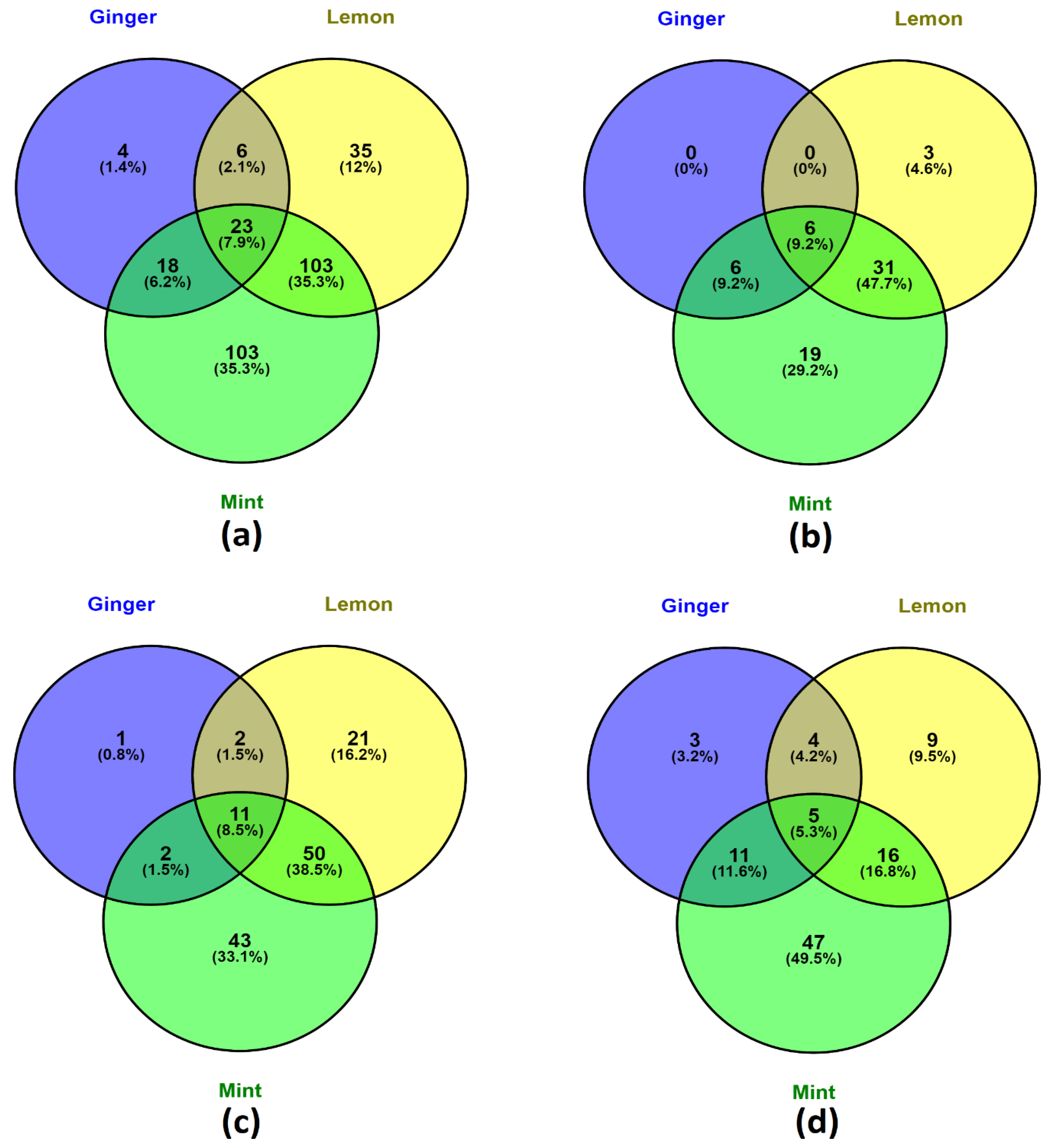
3.4.1. Phenolic Distribution—Venn Diagram Analysis

A total of 292 phenolic compounds were screened from ginger (51), lemon (167) and mint (247) using LC-MS analysis. It was found that 23 phenolic compounds were common in all three samples as shown in Figure 1. The majority of phenolic acids were identified in mint, and mint shared a relatively large proportion of its phenolic acid profile with ginger and lemon. However, the phenolic acid profiles of ginger and lemon were relatively dissimilar when compared to the similarity of their phenolic acid profiles with mint. For flavonoids, ginger shared a large portion of its flavonoids profile with both lemon and mint. Despite sharing a significant portion of its flavonoids profile with mint, lemon did contain a relatively large portion of flavonoids that were found only in lemon. Mint had the highest number of other polyphenols identified. Ginger and lemon both share a large portion of their other polyphenol profile with mint. Despite ginger’s seemingly low phenolic compound profile, ginger is still considered to be a medicinal plant that offers beneficial phytochemicals with potent antioxidant and anti-inflammatory activities such as gingerol and shogaol [75]. Thus, it is still considered worthwhile to infuse ginger into herbal teas to incorporate ginger’s other potentially healthy and useful phytochemicals.

3.4.2. LC-ESI-QTOF-MS/MS Characterization

The untargeted shotgun qualitative analysis of ginger, lemon and mint was performed via LC-ESI-QTOF-MS/MS in both positive and negative ionisation modes ([M − H]/[M + H]+). The phenolic compounds from the samples were identified based on their m/z value and MS spectra in both ionisation modes using Agilent’s LC-MS Qualitative Software and Personal Compound Database and Library (PCDL). Mass error less than ± 5 ppm and a PCDL library score of more than 80 were used as a benchmark to select further MS/MS identification, m/z characterisation and to verify the compounds (Supplementary Materials, Figures S1 and S2). In this research, through LC-MS/MS, 73 compounds were identified and characterised out of ginger (11 compounds), lemon (31 compounds) and mint (49 compounds), which include phenolic acids (24), flavonoids (35), other phenolic compounds (9), lignans (4) and stilbene (1) as listed in Table 3. It is possible that the relatively high diversity of phenolic compounds found in mint samples may explain its relatively higher antioxidant activities compared with ginger and lemon samples.

Phenolic Acids

Phenolic acids are a subclass of phenolic compounds with a carboxyl group. Phenolic acids mainly comprise hydroxybenzoic acids and hydroxycinnamic acids and they have been extensively studied for their antioxidant, antimicrobial and anti-inflammatory effects [76]. Phenolic acid species can be found in ginger, lemon and mint samples [77,78,79]. In this study, five subclasses of phenolic acid were identified, which include hydroxybenzoic acids (6), hydroxycinnamic acids (15), hydroxyphenylacetic acids (2) and hydroxyphenylpropanoic acids (1).
Compound 8 was identified to be cinnamic acid based on the product ion formed at m/z 103, which represents the loss of carbon dioxide (44 Da) from the precursor ion. Cinnamic acids are found in a variety of dietary plant materials such as Citrus fruits, tea, Brassica vegetables, cereals and more [80]. Cinnamic acids and their derivatives possess antioxidant properties, especially cinnamic acid derivatives with cinnamoyl and hydroxyl moieties in which these moieties reportedly increase cinnamic acid derivatives’ antioxidant activities [81]. Compound 10 found in lemon and mint samples, had a precursor ion at [M − H] m/z 193.0515 and was assigned to be ferulic acid based on the MS/MS fragmentation product ions produced. The product ions of ferulic acid were at m/z 178, m/z 149 and m/z 134, which coincide with the loss of methyl group (15 Da), carbon dioxide (44 Da) and both methyl group and carbon dioxide (59 Da total) from the precursor ion respectively. A previous study on phenolic compounds found in Phoenix dactylifera’s male flowers also reported product ions at m/z 178 and m/z 134 for ferulic acid [82]. Previously, as ferulic acid has been reported in mint, it is likely that ferulic acid contributed to the antioxidant activity observed in this study’s mint sample.
Two hydroxyphenylacetic acids were identified. Compound 22 ([M − H] m/z at 167.0354) was found in both ginger and mint samples and was identified as 3,4-dihydroxyphenylacetic acid (DOPAC). The result was two daughter ions, which were m/z 149 and m/z 123, corresponding to the loss of hydroxide group and carbon dioxide, respectively. To the best of our knowledge, this is the first time that DOPAC has been found in both ginger and mint. A previous study on the bioactive compounds of buckwheat found that DOPAC exhibits radical scavenging antioxidant activity [83]. DOPAC at micromolar concentrations was shown to display antioxidative activity against lipid peroxidation in vitro rat plasma model [84]. Although some other polyphenols such as quercetin may be more potent antioxidants, DOPAC has been suggested as a suitable additive or supplement alternative due to its relatively lowered cytotoxicity as it is a naturally occurring metabolite of dopamine within the human body [85].

Flavonoids

Flavonoids are a diverse group of phenolic compounds that are present in many dietary plant foods. Flavonoids have attracted interest due to their antioxidant, anti-inflammatory effects and their ability to modulate certain enzymatic functions [86]. Flavonoids are divided into further subgroups based on the composition and structure of their B and C rings [86]. In this experiment, compounds from seven flavonoid subclasses were identified, which include flavanols (4), flavones (9), flavanones (3), flavonols (8), dihydroflavonols (1), anthocyanins (1) and isoflavonoids (9).
Compound 39 was found in both lemon and mint samples. And had a precursor ion at [M − H] m/z 595.1658 and was identified to be neoeriocitrin as the precursor ion generated product ions at m/z 431 (loss of rhamnose, 164 Da) and m/z 287 (loss of rhamnose and glucose moieties, total 308 Da) in MS/MS fragmentation. A similar fragmentation pattern of neoeriocitrin was also reported in a study on Exocarpium Citri grandis flavonoid metabolites in human urine after oral administration [87]. Neoeriocitrin was previously shown to have high antioxidant activity through the superoxide radical scavenging assay and low-density lipoprotein oxidation assay [88]. Compound 40 was present in both ionisation modes and had a precursor ion at [M + H]+ m/z 611.1956. Compound 40 was identified to be hesperidin based on the MS/MS fragment ion peaks at m/z 593, m/z 465, m/z 449 and m/z 303. Hesperidin has been reported in both peppermint and lemon previously [89,90]. Studies have demonstrated hesperidin as a potent radical scavenger, with beneficial in vitro effects such as antimicrobial and anticancer effects and has been suggested for the management of cutaneous functions [89]. Hesperidin is thought to be one of the main contributors to lemon peel’s antioxidant properties [90]. Hesperidin was highly likely to have contributed to the observed antioxidant activity of this study’s mint sample. The presence of neoeriocitrin and hesperidin is consistent with the literature, as these are two of the main flavanones found in Citrus fruits such as lemon [60].
Compound 41 was found in lemon and mint samples in both negative and positive modes. The precursor ion at [M − H] m/z 477.067 generated a product ion at m/z 301 through MS/MS fragmentation, which was the loss of glucuronide (176 Da) from the precursor ion. This confirmed the identity of Compound 41 as quercetin 3′-O-glucuronide and, to the best of our knowledge, this is the first time quercetin 3′-O-glucuronide was characterised in lemon and mint. Quercetin 3′-O-glucuronide has been demonstrated to exhibit antioxidant activities [91,92]. Additionally, it has been suggested that quercetin 3′-O-glucuronide can protect cell membranes from lipid peroxidation through its catechol structure [93]. As one of the metabolites of dietary quercetin with health benefits, this further reaffirms the value of adopting a polyphenol-rich diet.

Other Phenolic Compounds

Compounds identified as other phenolic compound were further divided into hydroxycoumarins (1), hydroxybenzaldehydes (1), hydroxybenzoketones (1), phenolic terpenes (1), tyrosols (3) and other phenolic compounds (2). A total of 9 phenolic compounds classified as other phenolic compounds were found in the samples.
Compound 60 was the only hydroxycoumarin identified and was found in mint samples in positive mode, with a precursor ion at [M + H]+ m/z 147.0436. The product ion generated from the MS/MS fragmentation peaked at m/z 103 [M + H − CO2] and m/z 91 [M + H − 2CO], which identified Compound 60 as coumarin. A previous study also observed the same fragmentation pattern at m/z 103 and m/z 91 [94]. Previously coumarin and its derivatives have been reported to be associated with beneficial health effects such as reducing inflammation and risk of cancer, which is thought to be due to their antioxidant properties [95]. Compound 61 is a hydroxybenzaldehyde, which was identified in mint samples in negative mode. Compound 61 was identified as 4-hydroxybenzaldehyde because the precursor ion at [M − H] m/z 121.0299 produced a product ion at m/z 77, representing the loss of carbon dioxide (44 Da) from the parent ion. A previous vanilla extract study showed that 4-hydroxybenzaldehyde exhibited little DPPH scavenging activity [96]. Referring to the literature, it is postulated that 4-hydroxybenzaldehyde contributed little to mint’s observed antioxidant activity.
Three tyrosols were identified, of which one was present in all samples and the other two were exclusively present only in mint samples. Present in the negative mode, Compound 65 had a precursor ion at [M − H] m/z 403.1262 and was identified as oleoside 11-methylester based on the product ions produced at m/z 223 and m/z 165. A previous study on olive polyphenols also characterised oleoside 11-methylester, with product ion at m/z 223 [97]. These product ions correspond to the loss of glycoside (180 Da) and the loss of glycoside moiety and methyl ester (238 Da) from the precursor ion, respectively. To the best of our knowledge, this is the first time that oleoside 11-methylester has been characterised in mint.

Lignans and Stilbenes

Only one stilbene was identified which was only present in ginger samples. Four lignans were identified across ginger, lemon and mint samples. Compounds 71 and 72 were identified as pinoresinol and schisandrin C respectively from the product ions they produced. Compound 71 was identified in negative mode in mint samples, while Compound 72 was identified in both positive and negative modes and was present in ginger and lemon samples. Compound 71 precursor ion [M − H] at m/z 357.1364 produced 4 product ions at m/z 342, m/z 327, m/z 313 and m/z 221. A study on fringe tree found that pinoresinol possesses considerable antioxidant activity [98]. This is the first time that pinoresinol has been characterised in mint. Compound 72 precursor ion in positive mode [M + H]+ at m/z 385.1646 was identified to be schisandrin C based on the product ions produced at m/z 370, m/z 315 and m/z 300. A study on Schisandra chinensis fruit lignans using tandem mass spectrometry also reported schisandrin C’s product ions at m/z 370, m/z 315 and m/z 300, as well as other fragments [99]. Previously, it was reported that schisandrin C has potential anti-inflammatory effects, was capable of inducing autophagy and enhanced C2C12 skeletal muscle cells’ ability to deal with oxidative stress [100]. To the best of our knowledge, this is the first time schisandrin C has been characterised in both ginger rhizome and lemon fruit.

3.5. HPLC Quantification Analysis

From the characterised and identified phenolic compounds, 10 compounds were selected to be quantified using HPLC-PDA. The results in Table 4 were generated through calculation from the calibration curve. The lemon samples had a relatively lower amount of the selected phenolic compounds compared to other samples. Ginger samples recorded the greatest amount of certain molecules, such as quercetin, kaempferol and p-hydroxybenzoic acid. This is consistent with the literature as kaempferol and certain flavonoids comprise a significant portion of the phenolic compounds found within ginger [101]. This is unlike the TPC assay result, clearly demonstrating HPLC’s ability to generate higher-quality data. GLMT consistently scored relatively well in terms of quantity of the selected phenolic compounds, and had relatively higher levels of chlorogenic acid, caffeic acid and catechin compared to other samples. Therefore, there is still an advantage to ingesting herbal tea infusions because the combination of different plant parts results in the incorporation of more of a variety of phenolic compounds and other phytochemicals. This reinforces the importance of herbal tea infusions and combinations of diverse sources of phenolic compounds with antioxidant properties for consumption. The relatively high quantity of phenolic content observed in GLMT may be viewed as a promising attribute for GLMT to be considered as a healthy herbal tea infusion, which could be exploited for commercialisation.
A previous study on Malaysian ginger varieties found that 16-week old Halia Bentong rhizomes were found to contain 0.803 mg/gd.w of quercetin, 0.360 mg/gd.w of catechin and 0.045 mg/gd.w of kaempferol [102]. Compared to that study, our study’s samples produced significantly higher yields of those flavonoids from HPLC quantification. The differences in flavonoids observed may be attributed to many factors such as different variety, age, growth condition, harvesting method and more. For lemon, the content of certain phenolic acids and flavonoids demonstrated in this study was higher in comparison with previous studies on lemon phenolic quantity. A previous study on flavonoids in common plants showed that lemon yielded the equivalent of 11 μg/g of quercetin [103], and another study showed that Eureka lemons contained no quercetin nor kaempferol, but was high in eriocitrin and hesperidin [104]. In a different study on the phenolic content of five varieties of lemon, the juices contained a range of 0.38 µg/g to 7.62 µg/g of gallic acid and a range of 2.70 µg/g to 22.08 µg/g of chlorogenic acid [105]. In the same study, it was revealed that the peel contained the most phenolic acid and flavonoids compared to the pulp and juice of lemon. Thus, methods that increase the infusion of phenolic compounds from the lemon peel into the herbal tea would reap higher phenolic content and, subsequently, higher antioxidant activity. All ten compounds in Table 4 were present and quantified in mint and were all found in relatively high quantities. In a previous study, Mentha x piperita (mint) crude extract yielded 1.86 mg/g of caffeic acid, 0.73 mg/g of chlorogenic acid and 0.84 mg/g of quercetin but no kaempferol was observed [106]. Another study was also unable to quantify any kaempferol in mint but was able to quantify small amounts of caffeic acid and catechin (0.027 mg/g and 0.147 mg/g, respectively). However, previous studies revealed that mint and other related species do contain conjugated kaempferol and kaempferol derivatives [107,108].

4. Conclusions

GLMT is a herbal infusion composed of ingredients with marked antioxidant properties and with unique polyphenols. To the best of our knowledge, our examination of GLMT’s phenolic and antioxidant properties was unprecedented. The samples do exhibit antioxidant activities and the activities were likely attributed to the variety of phenolic compounds and possibly other phytochemicals found within the samples. Correlation analysis suggested that the antioxidant activities recorded were significantly correlated with the phenolic content from the samples. Furthermore, the LC-ESI-QTOF-MS/MS characterisation aided the identification of the phenolic compounds within the samples, in which a few were identified in the samples for the first time. Most of the phenolic compounds identified through LC-MS/MS were found in mint (49). Ten compounds were selected for HPLC quantification in which some phenolic compounds were abundant in ginger and GLMT samples. As discussed, although ginger did not appear to contain a diverse array of phenolic compounds from the analysis of this study, a wealth of literature has reported other phytochemicals in ginger that are also considered to be beneficial to health. With an understanding of the phenolic content in each sample from the characterisation analysis, further research should consider the optimal ingredient ratio for the maximum antioxidant activity or in vitro studies. Follow-up research could increase the acceptance of functional foods like GLMT, which may encourage commercialisation and promote a health-conscious society.

Supplementary Materials

The following are available online at https://0-www-mdpi-com.brum.beds.ac.uk/article/10.3390/fermentation7020073/s1, Figure S1: LC-ESI-QTOF-MS/MS basic peak chromatograph (BPC) for characterisation of phenolic compounds of herbal tea. Figure S2: Extracted ion chromatogram and their mass spectrum of characterised compounds in herbal tea.

Author Contributions

Conceptualization, methodology, formal analysis, validation and investigation, O.C., A.A., V.S. and H.A.R.S.; resources, H.A.R.S., C.J.B. and F.R.D.; writing—original draft preparation, O.C.; writing—review and editing, O.C., V.S., C.J.B., H.A.R.S. and F.R.D.; supervision, H.A.R.S.; A.A. and F.R.D. ideas sharing, H.A.R.S.; A.A.; C.J.B. and F.R.D.; funding acquisition, H.A.R.S., F.R.D. and C.J.B. All authors have read and agreed to the published version of the manuscript.

Funding

This research was funded by the University of Melbourne under the “McKenzie Fellowship Scheme” (Grant No. UoM-18/21), the “Richard WS Nicholas Agricultural Science Scholarship” and the “Faculty Research Initiative Funds” funded by the Faculty of Veterinary and Agricultural Sciences, The University of Melbourne, Australia and “The Alfred Deakin Research Fellowship” funded by Deakin University, Australia.

Institutional Review Board Statement

Not applicable.

Informed Consent Statement

Not applicable.

Data Availability Statement

Not applicable.

Acknowledgments

We would like to thank Michael Leeming, Nicholas Williamson and Shuai Nie from the Mass Spectrometry and Proteomics Facility, Bio21 Molecular Science and Biotechnology Institute, the University of Melbourne, VIC, Australia for providing access and support for the use of HPLC-PDA and LC-ESI-QTOF-MS/MS and data analysis.

Conflicts of Interest

The authors declare no conflict of interest.

References

  1. Buyukbalci, A.; El, S.N. Determination of in vitro antidiabetic effects, antioxidant activities and phenol contents of some herbal teas. Plant. Foods Hum. Nutr. 2008, 63, 27–33. [Google Scholar] [CrossRef] [PubMed]
  2. Gupta, R.K.; Chawla, P.; Tripathi, M.; Shukla, A.K.; Pandey, A.J.I.J.O.P.; Sciences, P. Synergistic antioxidant activity of tea with ginger, black pepper and tulsi. Int. J. Pharm. Pharm. Sci. 2014, 6, 477–479. [Google Scholar]
  3. Malongane, F.; McGaw, L.J.; Mudau, F.N. The synergistic potential of various teas, herbs and therapeutic drugs in health improvement: A review. J. Sci. Food Agric. 2017, 97, 4679–4689. [Google Scholar] [CrossRef] [PubMed]
  4. Bekkouch, O.; Harnafi, M.; Touiss, I.; Khatib, S.; Harnafi, H.; Alem, C.; Amrani, S. In vitro antioxidant and in vivo lipid-lowering properties of zingiber officinale crude aqueous extract and methanolic fraction: A follow-up study. Evid. Based Complement Altern. Med. 2019, 2019, 9734390. [Google Scholar] [CrossRef] [Green Version]
  5. Nair, K.P. Turmeric (Curcuma Longa l.) and Ginger (Zingiber Officinale Rosc.)-World’s Invaluable Medicinal Spices: The Agronomy and Economy of Turmeric and Ginger; Springer Nature: Berlin/Heidelberg, Germany, 2019. [Google Scholar]
  6. Yeh, H.-Y.; Chuang, C.-H.; Chen, H.-C.; Wan, C.-J.; Chen, T.-L.; Lin, L.-Y. Bioactive components analysis of two various gingers (zingiber officinale roscoe) and antioxidant effect of ginger extracts. Lwt Food Sci. Technol. 2014, 55, 329–334. [Google Scholar] [CrossRef]
  7. Mao, Q.Q.; Xu, X.Y.; Cao, S.Y.; Gan, R.Y.; Corke, H.; Beta, T.; Li, H.B. Bioactive compounds and bioactivities of ginger (zingiber officinale roscoe). Foods 2019, 8, 185. [Google Scholar] [CrossRef] [Green Version]
  8. Jitoe, A.; Masuda, T.; Tengah, I.G.P.; Suprapta, D.N.; Gara, I.W.; Nakatani, N. Antioxidant activity of tropical ginger extracts and analysis of the contained curcuminoids. J. Agric. Food Chem. 1992, 40, 1337–1340. [Google Scholar] [CrossRef]
  9. Curk, F.; Ollitrault, F.; Garcia-Lor, A.; Luro, F.; Navarro, L.; Ollitrault, P. Phylogenetic origin of limes and lemons revealed by cytoplasmic and nuclear markers. Ann. Bot. 2016, 117, 565–583. [Google Scholar] [CrossRef] [Green Version]
  10. Shimizu, C.; Wakita, Y.; Inoue, T.; Hiramitsu, M.; Okada, M.; Mitani, Y.; Segawa, S.; Tsuchiya, Y.; Nabeshima, T. Effects of lifelong intake of lemon polyphenols on aging and intestinal microbiome in the senescence-accelerated mouse prone 1 (samp1). Sci. Rep. 2019, 9, 3671. [Google Scholar] [CrossRef] [PubMed] [Green Version]
  11. Miyake, Y.; Shimoi, K.; Kumazawa, S.; Yamamoto, K.; Kinae, N.; Osawa, T.J. Identification and antioxidant activity of flavonoid metabolites in plasma and urine of eriocitrin-treated rats. J. Agric. Food Chem. 2000, 48, 3217–3224. [Google Scholar] [CrossRef] [PubMed]
  12. Makni, M.; Jemai, R.; Kriaa, W.; Chtourou, Y.; Fetoui, H. Citrus limon from tunisia: Phytochemical and physicochemical properties and biological activities. Biomed. Res. Int. 2018, 2018, 6251546. [Google Scholar] [CrossRef] [PubMed] [Green Version]
  13. Moosavy, M.; Hassanzadeh, P.; Mohammadzadeh, E.; Mahmoudi, R.; Khatibi, S.; Mardani, K.J.J.o.f.q. Antioxidant and antimicrobial activities of essential oil of lemon (citrus limon) peel in vitro and in a food model. J. Food Qual. Hazards Control 2017, 4, 42–48. [Google Scholar]
  14. Bunsawat, J.; Elliott, N.E.; Hertweck, K.L.; Sproles, E.; Alice, L.A.J.S.B. Phylogenetics of mentha (lamiaceae): Evidence from chloroplast DNA sequences. Syst. Bot. 2004, 29, 959–964. [Google Scholar] [CrossRef]
  15. Riachi, L.G.; De Maria, C.A. Peppermint antioxidants revisited. Food Chem. 2015, 176, 72–81. [Google Scholar] [CrossRef]
  16. Singh, R.; Shushni, M.A.M.; Belkheir, A. Antibacterial and antioxidant activities of mentha piperita L. Arab. J. Chem. 2015, 8, 322–328. [Google Scholar] [CrossRef] [Green Version]
  17. Wu, Z.; Tan, B.; Liu, Y.; Dunn, J.; Martorell Guerola, P.; Tortajada, M.; Cao, Z.; Ji, P. Chemical composition and antioxidant properties of essential oils from peppermint, native spearmint and scotch spearmint. Molecules 2019, 24, 2825. [Google Scholar] [CrossRef] [Green Version]
  18. Peng, D.; Zahid, H.F.; Ajlouni, S.; Dunshea, F.R.; Suleria, H.A.R. LC-ESI-QTOF/MS Profiling of Australian Mango Peel By-Product Polyphenols and Their Potential Antioxidant Activities. Processes 2019, 7, 764. [Google Scholar] [CrossRef] [Green Version]
  19. Imran, M.; Butt, M.S.; Akhtar, S.; Riaz, M.; Iqbal, M.J.; Suleria, H.A.R. Mangiferin from Mango Peel. J. Food Process. Preserv. 2016, 40, 760–769. [Google Scholar] [CrossRef]
  20. Halliwell, B.; Murcia, M.A.; Chirico, S.; Aruoma, O.I. Free radicals and antioxidants in food and in vivo: What they do and how they work. Crit. Rev. Food Sci. Nutr. 1995, 35, 7–20. [Google Scholar] [CrossRef] [PubMed]
  21. Riley, P.A. Free radicals in biology: Oxidative stress and the effects of ionizing radiation. Int. J. Radiat. Biol. 1994, 65, 27–33. [Google Scholar] [CrossRef]
  22. Briskin, D.P.J.P.p. Medicinal plants and phytomedicines. Linking plant biochemistry and physiology to human health. Plant Physiol. 2000, 124, 507–514. [Google Scholar] [CrossRef] [PubMed] [Green Version]
  23. Awan, K.A.; Butt, M.S.; Haq, I.U.; Ashfaq, F.; Suleria, H.A.R. Storage Stability of Garlic Fortified Chicken Bites. J. Food Chem. Nanotechnol. 2017, 3, 80–85. [Google Scholar] [CrossRef]
  24. Feng, Y.; Dunshea, F.R.; Suleria, H.A.R. LC-ESI-QTOF/MS characterization of bioactive compounds from black spices and their potential antioxidant activities. J. Food Sci. Technol. 2020, 57, 4671–4687. [Google Scholar] [CrossRef] [PubMed]
  25. Chang, C.-C.; Yang, M.-H.; Wen, H.-M.; Chern, J.-C. Estimation of total flavonoid content in propolis by two complementary colorimetric methods. J. Food Drug Anal. 2002, 10, 178–182. [Google Scholar]
  26. Stavrou, I.J.; Christou, A.; Kapnissi-Christodoulou, C.P. Polyphenols in carobs: A review on their composition, antioxidant capacity and cytotoxic effects, and health impact. Food Chem. 2018, 269, 355–374. [Google Scholar] [CrossRef]
  27. Shotorbani, N.Y.; Jamei, R.; Heidari, R.J.A.J.o.P. Antioxidant activities of two sweet pepper capsicum annuum l. Varieties phenolic extracts and the effects of thermal treatment. Avicenna J. Phytomed. 2013, 3, 25. [Google Scholar]
  28. Benzie, I.F.; Strain, J.J.J.A.b. The ferric reducing ability of plasma (frap) as a measure of “antioxidant power”: The frap assay. Anal. Biochem. 1996, 239, 70–76. [Google Scholar] [CrossRef] [PubMed] [Green Version]
  29. Re, R.; Pellegrini, N.; Proteggente, A.; Pannala, A.; Yang, M.; Rice-Evans, C.J.F. Antioxidant activity applying an improved abts radical cation decolorization assay. Free Radic. Biol. Med. 1999, 26, 1231–1237. [Google Scholar] [CrossRef]
  30. Ferreira, I.C.F.R.; Baptista, P.; Vilas-Boas, M.; Barros, L. Free-radical scavenging capacity and reducing power of wild edible mushrooms from northeast portugal: Individual cap and stipe activity. Food Chem. 2007, 100, 1511–1516. [Google Scholar] [CrossRef]
  31. Smirnoff, N.; Cumbes, Q.J. Hydroxyl radical scavenging activity of compatible solutes. Phytochemistry 1989, 28, 1057–1060. [Google Scholar] [CrossRef]
  32. Dinis, T.C.P.; Madeira, V.M.C.; Almeida, L.M. Action of phenolic derivatives (acetaminophen, salicylate, and 5-aminosalicylate) as inhibitors of membrane lipid peroxidation and as peroxyl radical scavengers. Arch. Biochem. Biophys. 1994, 315, 161–169. [Google Scholar] [CrossRef]
  33. Kadam, D.; Palamthodi, S.; Lele, S.S. Lc-esi-q-tof-ms/ms profiling and antioxidant activity of phenolics from l. Sativum seedcake. J. Food Sci. Technol. 2018, 55, 1154–1163. [Google Scholar] [CrossRef]
  34. Pekal, A.; Drozdz, P.; Biesaga, M.; Pyrzynska, K. Screening of the antioxidant properties and polyphenol composition of aromatised green tea infusions. J. Sci. Food Agric. 2012, 92, 2244–2249. [Google Scholar] [CrossRef]
  35. Seo, C.-S.; Lee, M.-Y. Hplc–pda and lc–ms/ms analysis for the simultaneous quantification of the 14 marker components in sojadodamgangki-tang. Appl. Sci. 2020, 10, 2804. [Google Scholar] [CrossRef] [Green Version]
  36. Yang, D.; Dunshea, F.R.; Suleria, H.A.R. Lc-esi-qtof/ms characterization of australian herb and spices (garlic, ginger, and onion) and potential antioxidant activity. J. Food Process. Preserv. 2020, 44, e14497. [Google Scholar] [CrossRef]
  37. Tang, J.; Dunshea, F.R.; Suleria, H.A.R. Lc-esi-qtof/ms characterization of phenolic compounds from medicinal plants (hops and juniper berries) and their antioxidant activity. Foods 2019, 9, 7. [Google Scholar] [CrossRef] [Green Version]
  38. Subbiah, V.; Zhong, B.; Nawaz, M.A.; Barrow, C.J.; Dunshea, F.R.; Suleria, H.A. Screening of phenolic compounds in australian grown berries by lc-esi-qtof-ms/ms and determination of their antioxidant potential. Antioxidants 2021, 10, 26. [Google Scholar] [CrossRef] [PubMed]
  39. Wang, Z.; Barrow, C.J.; Dunshea, F.R.; Suleria, H.A.R. A comparative investigation on phenolic composition, characterization and antioxidant potentials of five different australian grown pear varieties. Antioxidants 2021, 10, 151. [Google Scholar] [CrossRef] [PubMed]
  40. Kılıç, C.; Can, Z.; Yılmaz, A.; Yıldız, S.; Turna, H. Antioxidant properties of some herbal teas (green tea, senna, corn silk, rosemary) brewed at different temperatures. Int. J. Second. Metab. 2017, 148–154. [Google Scholar] [CrossRef]
  41. Sogi, D.S.; Siddiq, M.; Greiby, I.; Dolan, K.D. Total phenolics, antioxidant activity, and functional properties of ‘tommy atkins’ mango peel and kernel as affected by drying methods. Food Chem. 2013, 141, 2649–2655. [Google Scholar] [CrossRef]
  42. Alam, M.N.; Bristi, N.J.; Rafiquzzaman, M. Review on in vivo and in vitro methods evaluation of antioxidant activity. Saudi Pharm. J. Spj Off. Publ. Saudi Pharm. Soc. 2013, 21, 143–152. [Google Scholar] [CrossRef] [Green Version]
  43. Prieto, P.; Pineda, M.; Aguilar, M.J.A.b. Spectrophotometric quantitation of antioxidant capacity through the formation of a phosphomolybdenum complex: Specific application to the determination of vitamin E. Anal. Biochem. 1999, 269, 337–341. [Google Scholar] [CrossRef] [PubMed]
  44. Suleria, H.A.; Barrow, C.J.; Dunshea, F.R.J.F. Screening and characterization of phenolic compounds and their antioxidant capacity in different fruit peels. Foods 2020, 9, 1206. [Google Scholar] [CrossRef]
  45. Zhong, B.; Robinson, N.A.; Warner, R.D.; Barrow, C.J.; Dunshea, F.R.; Suleria, H.A.R. Lc-esi-qtof-ms/ms characterization of seaweed phenolics and their antioxidant potential. Mar. Drugs 2020, 18, 331. [Google Scholar] [CrossRef] [PubMed]
  46. Ghafoor, K.; Al Juhaimi, F.; Özcan, M.M.; Uslu, N.; Babiker, E.E.; Mohamed Ahmed, I.A. Total phenolics, total carotenoids, individual phenolics and antioxidant activity of ginger (zingiber officinale) rhizome as affected by drying methods. Lwt 2020, 126, 109354. [Google Scholar] [CrossRef]
  47. Farnad, N.; Heidari, R.; Aslanipour, B. Phenolic composition and comparison of antioxidant activity of alcoholic extracts of peppermint (mentha piperita). J. Food Meas. Charact. 2014, 8, 113–121. [Google Scholar] [CrossRef]
  48. Benabdallah, A.; Rahmoune, C.; Boumendjel, M.; Aissi, O.; Messaoud, C. Total phenolic content and antioxidant activity of six wild mentha species (lamiaceae) from northeast of algeria. Asian Pac. J. Trop. Biomed. 2016, 6, 760–766. [Google Scholar] [CrossRef] [Green Version]
  49. Sarooshi, R.; Broadbent, P.J.A.J.o.E.A. Evaluation of rootstocks for eureka and lisbon lemons in replant ground in new south wales. Aust. J. Exp. Agric. 1992, 32, 205–209. [Google Scholar] [CrossRef]
  50. Rahmati, M.; Vercambre, G.; Davarynejad, G.; Bannayan, M.; Azizi, M.; Genard, M. Water scarcity conditions affect peach fruit size and polyphenol contents more severely than other fruit quality traits. J. Sci. Food Agric. 2015, 95, 1055–1065. [Google Scholar] [CrossRef]
  51. Velioglu, Y.; Mazza, G.; Gao, L.; Oomah, B.D. Antioxidant activity and total phenolics in selected fruits, vegetables, and grain products. J. Agric. Food Chem. 1998, 46, 4113–4117. [Google Scholar] [CrossRef]
  52. Contreras-López, E.; Castañeda-Ovando, A.; Jaimez-Ordaz, J.; Cruz-Cansino, N.d.S.; González-Olivares, L.G.; Rodríguez-Martínez, J.S.; Ramírez-Godínez, J.J.A.S. Release of antioxidant compounds of zingiber officinale by ultrasound-assisted aqueous extraction and evaluation of their in vitro bioaccessibility. Appl. Sci. 2020, 10, 4987. [Google Scholar] [CrossRef]
  53. Kedare, S.B.; Singh, R.P. Genesis and development of dpph method of antioxidant assay. J. Food Sci. Technol 2011, 48, 412–422. [Google Scholar] [CrossRef] [Green Version]
  54. Chrpova, D.; Kouřimská, L.; Gordon, M.H.; Heřmanová, V.; Roubíčková, I.; Panek, J.J.C.J.o.F.S. Antioxidant activity of selected phenols and herbs used in diets for medical conditions. Czech J. Food Sci. 2010, 28, 317–325. [Google Scholar] [CrossRef] [Green Version]
  55. Sun, Y.; Qiao, L.; Shen, Y.; Jiang, P.; Chen, J.; Ye, X. Phytochemical profile and antioxidant activity of physiological drop of citrus fruits. J. Food Sci. 2013, 78, C37–C42. [Google Scholar] [CrossRef]
  56. Gu, C.; Howell, K.; Dunshea, F.R.; Suleria, H.A.R. Lc-esi-qtof/ms characterisation of phenolic acids and flavonoids in polyphenol-rich fruits and vegetables and their potential antioxidant activities. Antioxidants 2019, 8, 405. [Google Scholar] [CrossRef] [Green Version]
  57. Mittal, N. Estimation of antioxidant levels in pomegranate, banana, orange, lemon, sweet lime. Stud. Ethno-Med. 2019, 13. [Google Scholar] [CrossRef]
  58. Chan, E.W.C.; Lim, Y.Y.; Chong, K.L.; Tan, J.B.L.; Wong, S.K. Antioxidant properties of tropical and temperate herbal teas. J. Food Compos. Anal. 2010, 23, 185–189. [Google Scholar] [CrossRef]
  59. Ilyasov, I.R.; Beloborodov, V.L.; Selivanova, I.A.; Terekhov, R.P. Abts/pp decolorization assay of antioxidant capacity reaction pathways. Int J. Mol Sci. 2020, 21, 1131. [Google Scholar] [CrossRef] [Green Version]
  60. Assefa, A.D.; Keum, Y.-S.; Saini, R.K. A comprehensive study of polyphenols contents and antioxidant potential of 39 widely used spices and food condiments. J. Food Meas. Charact. 2018, 12, 1548–1555. [Google Scholar] [CrossRef]
  61. Leong, L.; Shui, G.J.F.c. An investigation of antioxidant capacity of fruits in singapore markets. Food Chem. 2002, 76, 69–75. [Google Scholar] [CrossRef]
  62. Bhalodia, N.R.; Nariya, P.B.; Acharya, R.N.; Shukla, V.J. In vitro antioxidant activity of hydro alcoholic extract from the fruit pulp of cassia fistula linn. Ayu 2013, 34, 209–214. [Google Scholar] [CrossRef] [Green Version]
  63. Prakash, J.J.J.o.M.P.R. Chemical composition and antioxidant properties of ginger root (zingiber officinale). J. Med. Plants Res. 2010, 4, 2674–2679. [Google Scholar]
  64. Kanatt, S.R.; Chander, R.; Sharma, A.J.F.C. Antioxidant potential of mint (mentha spicata L.) in radiation-processed lamb meat. Food Chem. 2007, 100, 451–458. [Google Scholar] [CrossRef]
  65. Stoilova, I.; Krastanov, A.; Stoyanova, A.; Denev, P.; Gargova, S. Antioxidant activity of a ginger extract (zingiber officinale). Food Chem. 2007, 102, 764–770. [Google Scholar] [CrossRef]
  66. Dorman, H.D.; Koşar, M.; Kahlos, K.; Holm, Y.; Hiltunen, R.J.J. Antioxidant properties and composition of aqueous extracts from mentha species, hybrids, varieties, and cultivars. J. Agric. Food Chem. 2003, 51, 4563–4569. [Google Scholar] [CrossRef]
  67. Muthiah, P.; Umamaheswari, M.; Asokkumar, K.J.I.J.o.P. In vitro antioxidant activities of leaves, fruits and peel extracts of citrus. Int. J. Phytopharm. 2012, 2, 13–20. [Google Scholar]
  68. Ramkissoon, J.S.; Mahomoodally, M.F.; Ahmed, N.; Subratty, A.H. Therapeutic potential of common culinary herbs and spices of mauritius. In Chemistry: The Key to Our Sustainable Future; Springer: Berlin, Germany, 2014; pp. 147–162. [Google Scholar]
  69. Agarwal, M.; Kumar, A.; Gupta, R.; Upadhyaya, S.J.O.J.o.C. Extraction of polyphenol, flavonoid from emblica officinalis, citrus limon, cucumis sativus and evaluation of their antioxidant activity. Orient. J. Chem. 2012, 28, 993. [Google Scholar] [CrossRef] [Green Version]
  70. Brown, N.; John, J.A.; Shahidi, F. Polyphenol composition and antioxidant potential of mint leaves. Food Prod. Process. Nutr. 2019, 1, 1–14. [Google Scholar] [CrossRef] [Green Version]
  71. Ghasemzadeh, A.; Jaafar, H.Z.; Rahmat, A. Antioxidant activities, total phenolics and flavonoids content in two varieties of malaysia young ginger (zingiber officinale roscoe). Molecules 2010, 15, 4324–4333. [Google Scholar] [CrossRef] [Green Version]
  72. Sir Elkhatim, K.A.; Elagib, R.A.A.; Hassan, A.B. Content of phenolic compounds and vitamin c and antioxidant activity in wasted parts of sudanese citrus fruits. Food Sci. Nutr. 2018, 6, 1214–1219. [Google Scholar] [CrossRef]
  73. Vogeser, M.; Parhofer, K.G. Liquid chromatography tandem-mass spectrometry (lc-ms/ms)—technique and applications in endocrinology. Exp. Clin. Endocrinol. Diabetes 2007, 115, 559–570. [Google Scholar] [CrossRef] [PubMed]
  74. Ali, A.; Wu, H.; Ponnampalam, E.N.; Cottrell, J.J.; Dunshea, F.R.; Suleria, H.A.R. Comprehensive Profiling of Most Widely Used Spices for Their Phenolic Compounds through LC-ESI-QTOF-MS2 and Their Antioxidant Potential. Antioxidants 2021, 10, 721. [Google Scholar] [CrossRef]
  75. Dugasani, S.; Pichika, M.R.; Nadarajah, V.D.; Balijepalli, M.K.; Tandra, S.; Korlakunta, J.N. Comparative antioxidant and anti-inflammatory effects of [6]-gingerol, [8]-gingerol, [10]-gingerol and [6]-shogaol. J. Ethnopharmacol. 2010, 127, 515–520. [Google Scholar] [CrossRef]
  76. Kumar, N.; Goel, N. Phenolic acids: Natural versatile molecules with promising therapeutic applications. Biotechnol. Rep. (Amst) 2019, 24, e00370. [Google Scholar] [CrossRef]
  77. Klimek-Szczykutowicz, M.; Szopa, A.; Ekiert, H. Citrus limon (lemon) phenomenon-a review of the chemistry, pharmacological properties, applications in the modern pharmaceutical, food, and cosmetics industries, and biotechnological studies. Plants 2020, 9, 119. [Google Scholar] [CrossRef] [PubMed] [Green Version]
  78. Tahira, R.; Naeemullah, M.; Akbar, F.; Masood, M.S.J.P.J.B. Major phenolic acids of local and exotic mint germplasm grown in islamabad. Pak. J. Bot. 2011, 43, 151–154. [Google Scholar]
  79. Tohma, H.; Gülçin, İ.; Bursal, E.; Gören, A.C.; Alwasel, S.H.; Köksal, E. Antioxidant activity and phenolic compounds of ginger (zingiber officinale rosc.) determined by hplc-ms/ms. J. Food Meas. Charact. 2016, 11, 556–566. [Google Scholar] [CrossRef]
  80. Guzman, J.D. Natural cinnamic acids, synthetic derivatives and hybrids with antimicrobial activity. Molecules 2014, 19, 19292–19349. [Google Scholar] [CrossRef]
  81. Pontiki, E.; Hadjipavlou-Litina, D.; Litinas, K.; Geromichalos, G. Novel cinnamic acid derivatives as antioxidant and anticancer agents: Design, synthesis and modeling studies. Molecules 2014, 19, 9655–9674. [Google Scholar] [CrossRef]
  82. Ben Said, R.; Hamed, A.I.; Mahalel, U.A.; Al-Ayed, A.S.; Kowalczyk, M.; Moldoch, J.; Oleszek, W.; Stochmal, A. Tentative characterization of polyphenolic compounds in the male flowers of phoenix dactylifera by liquid chromatography coupled with mass spectrometry and dft. Int. J. Mol. Sci. 2017, 18, 512. [Google Scholar] [CrossRef]
  83. Gimenez-Bastida, J.A.; Zielinski, H.; Piskula, M.; Zielinska, D.; Szawara-Nowak, D. Buckwheat bioactive compounds, their derived phenolic metabolites and their health benefits. Mol. Nutr. Food Res. 2017, 61. [Google Scholar] [CrossRef]
  84. Raneva, V.; Shimasaki, H.; Ishida, Y.; Ueta, N.; Niki, E.J.L. Antioxidative activity of 3, 4-dihydroxyphenylacetic acid and caffeic acid in rat plasma. Lipids 2001, 36, 1111. [Google Scholar] [CrossRef] [PubMed]
  85. Tang, Y.; Nakashima, S.; Saiki, S.; Myoi, Y.; Abe, N.; Kuwazuru, S.; Zhu, B.; Ashida, H.; Murata, Y.; Nakamura, Y. 3,4-dihydroxyphenylacetic acid is a predominant biologically-active catabolite of quercetin glycosides. Food Res. Int. 2016, 89, 716–723. [Google Scholar] [CrossRef]
  86. Panche, A.N.; Diwan, A.D.; Chandra, S.R. Flavonoids: An overview. J. Nutr. Sci. 2016, 5, e47. [Google Scholar] [CrossRef] [PubMed] [Green Version]
  87. Zeng, X.; Su, W.; Zheng, Y.; Liu, H.; Li, P.; Zhang, W.; Liang, Y.; Bai, Y.; Peng, W.; Yao, H. Uflc-q-tof-ms/ms-based screening and identification of flavonoids and derived metabolites in human urine after oral administration of exocarpium citri grandis extract. Molecules 2018, 23, 895. [Google Scholar] [CrossRef] [Green Version]
  88. Yu, J.; Wang, L.; Walzem, R.L.; Miller, E.G.; Pike, L.M.; Patil, B.S. Antioxidant activity of citrus limonoids, flavonoids, and coumarins. J. Agric. Food Chem. 2005, 53, 2009–2014. [Google Scholar] [CrossRef]
  89. Man, M.Q.; Yang, B.; Elias, P.M. Benefits of hesperidin for cutaneous functions. Evid. Based Complement. Altern. Med. 2019, 2019, 2676307. [Google Scholar] [CrossRef] [Green Version]
  90. Mcharek, N.; Hanchi, B.J.J.A.B.F.Q. Maturational effects on phenolic constituents, antioxidant activities and lc-ms/ms profiles of lemon (citrus limon) peels. J. Appl. Bot. Food Qual. 2017, 90, 1–9. [Google Scholar]
  91. Csepregi, K.; Neugart, S.; Schreiner, M.; Hideg, E. Comparative evaluation of total antioxidant capacities of plant polyphenols. Molecules 2016, 21, 208. [Google Scholar] [CrossRef] [PubMed] [Green Version]
  92. Zhou, M.; Lin, Y.; Fang, S.; Liu, Y.; Shang, X. Phytochemical content and antioxidant activity in aqueous extracts of cyclocarya paliurus leaves collected from different populations. PeerJ 2019, 7, e6492. [Google Scholar] [CrossRef] [Green Version]
  93. Shirai, M.; Moon, J.-H.; Tsushida, T.; Terao, J.J.J.o.A.; Chemistry, F. Inhibitory effect of a quercetin metabolite, quercetin 3-o-β-d-glucuronide, on lipid peroxidation in liposomal membranes. J. Agric. Food Chem. 2001, 49, 5602–5608. [Google Scholar] [CrossRef] [PubMed]
  94. Ren, Z.; Nie, B.; Liu, T.; Yuan, F.; Feng, F.; Zhang, Y.; Zhou, W.; Xu, X.; Yao, M.; Zhang, F. Simultaneous determination of coumarin and its derivatives in tobacco products by liquid chromatography-tandem mass spectrometry. Molecules 2016, 21, 1511. [Google Scholar] [CrossRef] [Green Version]
  95. Al-Majedy, Y.; Al-Amiery, A.; Kadhum, A.A.; BakarMohamad, A. Antioxidant activity of coumarins. Syst. Rev. Pharm. 2016, 8, 24–30. [Google Scholar] [CrossRef]
  96. Shyamala, B.; Naidu, M.M.; Sulochanamma, G.; Srinivas, P. Studies on the antioxidant activities of natural vanilla extract and its constituent compounds through in vitro models. J. Agric. Food Chem. 2007, 55, 7738–7743. [Google Scholar] [CrossRef]
  97. Melliou, E.; Zweigenbaum, J.A.; Mitchell, A.E. Ultrahigh-pressure liquid chromatography triple-quadrupole tandem mass spectrometry quantitation of polyphenols and secoiridoids in california-style black ripe olives and dry salt-cured olives. J. Agric. Food Chem. 2015, 63, 2400–2405. [Google Scholar] [CrossRef]
  98. Gülçin, İ.; Elias, R.; Gepdiremen, A.; Boyer, L. Antioxidant activity of lignans from fringe tree (chionanthus virginicus L.). Eur. Food Res. Technol. 2006, 223, 759–767. [Google Scholar] [CrossRef]
  99. Huang, X.; Song, F.; Liu, Z.; Liu, S. Studies on lignan constituents from schisandra chinensis (turcz.) baill. Fruits using high-performance liquid chromatography/electrospray ionization multiple-stage tandem mass spectrometry. J. Mass Spectrom 2007, 42, 1148–1161. [Google Scholar] [CrossRef] [PubMed]
  100. Kim, J.S.; Yi, H.K. Schisandrin c enhances mitochondrial biogenesis and autophagy in c2c12 skeletal muscle cells: Potential involvement of anti-oxidative mechanisms. Naunyn Schmiedebergs Arch. Pharm. 2018, 391, 197–206. [Google Scholar] [CrossRef] [PubMed]
  101. Tanweer, S.; Mehmood, T.; Zainab, S.; Ahmad, Z.; Shehzad, A. Comparison and hplc quantification of antioxidant profiling of ginger rhizome, leaves and flower extracts. Clin. Phytosci. 2020, 6. [Google Scholar] [CrossRef]
  102. Ghasemzadeh, A.; Jaafar, H.Z.; Rahmat, A. Variation of the phytochemical constituents and antioxidant activities of zingiber officinale var. Rubrum theilade associated with different drying methods and polyphenol oxidase activity. Molecules 2016, 21, 780. [Google Scholar] [CrossRef] [PubMed] [Green Version]
  103. Mattila, P.; Astola, J.; Kumpulainen, J. Determination of flavonoids in plant material by hplc with diode-array and electro-array detections. J. Agric. Food Chem. 2000, 48, 5834–5841. [Google Scholar] [CrossRef]
  104. Kawaii, S.; Tomono, Y.; Katase, E.; Ogawa, K.; Yano, M.J. Quantitation of flavonoid constituents in citrus fruits. J. Agric. Food Chem. 1999, 47, 3565–3571. [Google Scholar] [CrossRef] [PubMed]
  105. Xi, W.; Lu, J.; Qun, J.; Jiao, B. Characterization of phenolic profile and antioxidant capacity of different fruit part from lemon (citrus limon burm.) cultivars. J. Food Sci. Technol. 2017, 54, 1108–1118. [Google Scholar] [CrossRef] [Green Version]
  106. Mišan, A.; Mimica-Dukić, N.; Mandić, A.; Sakač, M.; Milovanović, I.; Sedej, I. Development of a rapid resolution hplc method for the separation and determination of 17 phenolic compounds in crude plant extracts. Open Chem. 2011, 9, 133–142. [Google Scholar] [CrossRef]
  107. Dolzhenko, Y.; Bertea, C.M.; Occhipinti, A.; Bossi, S.; Maffei, M.E. Uv-b modulates the interplay between terpenoids and flavonoids in peppermint (mentha x piperita L.). J. Photochem. Photobiol. B 2010, 100, 67–75. [Google Scholar] [CrossRef] [PubMed]
  108. Kapp, K. Polyphenolic and Essential Oil Composition of Mentha and Their Antimicrobial Effect; Helsingin Yliopisto: Helsinki, Finland, 2015. [Google Scholar]
Figure 1. Venn Diagram of the distribution of screened phenolic compounds. (a) Total phenolic compounds found in ginger, lemon and mint; (b) Phenolic acids in ginger, lemon and mint; (c) Flavonoids in ginger, lemon and mint; (d) Other polyphenols (including lignans and stilbenes) in ginger, lemon and mint.
Fermentation 07 00073 g001
Table 1. Phenolic compound content (TPC, TFC and TTC) and antioxidant activities (DPPH, FRAP, ABTS, RPA, OH-RSA, FICA and TAC) of ginger, lemon, mint and GLMT per gram of fresh weight.
AssaysGingerLemonMintGLMT
TPC (mg GAE/g)2.93 ± 0.07 c0.12 ± 0.04 d14.35 ± 0.19 a5.85 ± 0.08 b
TFC (mg QE/g)0.98 ± 0.02 c0.07 ± 0.03 d1.29 ± 0.07 a1.25 ± 0.04 b
TTC (mg CE/g)0.02 ± 0.04 d0.04 ± 0.01 c2.13 ± 0.08 a0.45 ± 0.09 b
DPPH (mg AAE/g)1.24 ± 0.09 c0.09 ± 0.04 d3.15 ± 0.12 a2.24 ± 0.01 b
FRAP (mg AAE/g)0.83 ± 0.03 c0.08 ± 0.01 d7.15 ± 0.14 a1.91 ± 0.07 b
ABTS (mg AAE/g)0.11 ± 0.01 c0.07 ± 0.03 d24.25 ± 2.18 a5.48 ± 0.21 b
RPA (mg AAE/g)0.08 ± 0.01 a0.02 ± 0.03 c0.04 ± 0.02 b1.01 ± 0.04 b
OH-RSA (mg AAE/g)0.53 ± 0.03 b0.39 ± 0.07 a0.27± 0.01 c0.77± 0.08 c
FICA (mg EDTA/g)0.04 ± 0.03 b-0.09 ± 0.02 a0.07 ± 0.04 a
TAC (mg AAE/g)0.73 ± 0.08 c0.03 ± 0.04 d6.74 ± 0.58 a1.35 ± 0.41 b
Data expressed as mean ± SD of three replicates; Different letters a,b,c,d indicate that the data is significantly different from the other data of the same row (p ≤ 0.05), in which a is assigned to the largest value, then b assigned to second largest and so forth. The significant difference was calculated through one-way analysis of variance (ANOVA) and Tukey’s HSD Test.
Table 2. Pearson’s correlation coefficient (r) for the pairwise correlation between the phenolic content assays (TPC, TFC, TTC) and the antioxidant assays (DPPH, FRAP, ABTS, RPA, OH-RSA, FICA and TAC).
AssaysTPCTFCTTCDPPHFRAPABTSRAPOH-RSAFICA
TFC0.747
TTC0.971 *0.565
DPPH0.942 *0.915 *0.839
FRAP0.988 *0.6390.994 **0.879
ABTS0.976 *0.5831.000 **0.8520.996 **
RAP0.0040.440−0.1500.293−0.126−0.126
OH-RSA−0.3570.269−0.537−0.030−0.492−0.5150.886 *
FICA0.907 *0.948 *0.7830.995 **0.831 *0.7980.3530.055
TAC0.977 *0.6040.994 **0.8500.998 **0.993 **−0.192−0.5460.799
* indicates significant correlation with p ≤ 0.1, while ** indicates highly significant correlation with p ≤ 0.01.
Table 3. LC-ESI-QTOF-MS/MS characterisation of phenolic compounds in ginger, lemon and mint samples.
No.Proposed CompoundsMolecular FormulaRT
(min)
Ionisation (ESI+/ESI)Molecular WeightTheoretical (m/z)Observed (m/z)Error
(ppm)
MS2
Productions
Samples
Phenolic acids
Hydroxybenzoic Acids
1Gallic acid 4-O-glucosideC13H16O106.580[M − H]332.0743331.067331.06844.2169, 125M
24-Hydroxybenzoic acid 4-O-glucosideC13H16O811.898[M − H]300.0845299.0772299.077−0.7255, 137M
32,3-Dihydroxybenzoic acidC7H6O412.378[M − H]154.0266153.0193153.01940.7109M
4Protocatechuic acid 4-O-glucosideC13H16O913.256[M − H]316.0794315.0721315.07230.6153M
52-Hydroxybenzoic acidC7H6O319.932** [M − H]138.0317137.0244137.02450.793M
6PaeoniflorinC23H28O1158.033[M − H]480.1632479.1559479.15773.8449, 357, 327L
Hydroxycinnamic Acids
7p-Coumaroyl tartaric acidC13H12O88.232** [M − H]296.0532295.0459295.04662.4115*G, L
8Cinnamic acidC9H8O29.166[M − H]148.0524147.0451147.0448−2.0103L
9Caffeoyl tartaric acidC13H12O913.438[M − H]312.0481311.0408311.04131.6161M
10Ferulic acidC10H10O415.708** [M − H]194.0579193.0506193.05154.7178, 149,134L, *M
11Caffeic acid 3-O-glucuronideC15H16O1016.354** [M − H]356.0743355.067355.06854.2179M
123-p-Coumaroylquinic acidC16H18O817.695** [M − H]338.1002337.0929337.09434.2265, 173, 162M
13p-Coumaric acid 4-O-glucosideC15H18O819.137[M − H]326.1002325.0929325.0930.3163M
14m-Coumaric acidC9H8O319.153[M − H]164.0473163.04163.0395−3.1119M
15Caffeic acid 4-sulfateC9H8O7S19.248[M + H]+259.9991261.0064261.0057−2.7179, 135M
163-Caffeoylquinic acidC16H18O919.766** [M − H]354.0951353.0878353.08780.0253, 190, 144L, *M
17Feruloyl tartaric acidC14H14O922.185[M − H]326.0638325.0565325.05732.5193, 149M
18Ferulic acid 4-O-glucosideC16H20O925.779** [M − H]356.1107355.1034355.1019−4.2193, 178, 149, 134M
19Chicoric acidC22H18O1235.138[M − H]474.0798473.0725473.07535.0293, 311M
201-Sinapoyl-2-feruloylgentiobioseC33H40O1835.768[M − H]724.2215723.2142723.21844.1529, 499M
211,5-Dicaffeoylquinic acidC25H24O1248.341** [M − H]516.1268515.1195515.1224.9353, 335, 191, 179M
Hydroxyphenylacetic Acids
223,4-Dihydroxyphenylacetic acidC8H8O410.059** [M − H]168.0423167.035167.03542.4149, 123G, *M
232-Hydroxy-2-phenylacetic acidC8H8O315.310** [M − H]152.0473151.04151.04083.8136, 92M
Hydroxyphenylpropanoic Acids
24Dihydroferulic acid 4-O-glucuronideC16H20O106.978** [M − H]372.1056371.0983371.09994.3195M
Flavonoids
Flavanols
254’-O-MethylepigallocatechinC16H16O733.560[M + H]+320.0896321.0969321.0958−3.4302L, *M
264’-O-Methyl-(-)-epigallocatechin 7-O-glucuronideC22H24O1348.622[M − H]496.1217495.1144495.1151.2451, 313M
27(+)-Catechin 3-O-gallateC22H18O1050.445** [M − H]442.09441.0827441.08331.4289, 169, 125M
28(+)-Gallocatechin 3-O-gallateC22H18O1163.448** [M − H]458.0849457.0776457.07943.9305, 169M
Flavones
29Apigenin 6,8-di-C-glucosideC27H30O1526.791** [M − H]594.1585593.1512593.15564.7503, 473G, *L, M-T
30Apigenin 7-O-apiosyl-glucosideC26H28O1431.067[M + H]+564.1479565.1552565.1545−1.2296*G, L, M-T
317,4’-DihydroxyflavoneC15H10O437.337[M + H]+254.0579255.0652255.06592.7227, 199, 171M
326-Hydroxyluteolin 7-O-rhamnosideC21H20O1139.131** [M − H]448.1006447.0933447.0953.8301L, *M
33RhoifolinC27H30O1443.471** [M − H]578.1636577.1563577.15823.3413, 269L, *M
34CirsilineolC18H16O745.338[M + H]+344.0896345.0969345.0966−0.9330, 312, 297, 284L, *M
35Apigenin 7-O-glucuronideC21H18O1147.673[M + H]+446.0849447.0922447.0901−4.7271, 253M
36Chrysoeriol 7-O-glucosideC22H22O1154.565[M + H]+462.1162463.1235463.1234−0.2445, 427, 409, 381L, *M
37DiosminC28H32O1559.17** [M + H]+608.1741609.1814609.18190.8301, 286L, *M
Flavanones
38Naringin 4’-O-glucosideC33H42O1925.233[M − H]-742.232741.2247741.22794.3433, 271M
39NeoeriocitrinC27H32O1536.946** [M − H]596.1741595.1668595.1658−1.7431, 287*L, M
40HesperidinC28H34O1542.745** [M + H]+610.1898611.1971611.1956−2.5593, 465, 449, 303*L, M
Flavonols
41Quercetin 3’-O-glucuronideC21H18O1312.512** [M − H]478.0747477.0674477.067−0.8301*L, M
42Quercetin 3-O-glucosyl-xylosideC26H28O1615.395[M − H]596.1377595.1304595.1299−0.8265, 138, 116L
43Kaempferol 3,7-O-diglucosideC27H30O1623.162** [M − H]610.1534609.1461609.14864.1447, 285L, *M
44Kaempferol 3-O-(2’’-rhamnosyl-galactoside) 7-O-rhamnosideC33H40O1934.660** [M − H]740.2164739.2091739.21143.1593, 447, 285G, *L, M-T
45Kaempferol 3-O-glucosyl-rhamnosyl-galactosideC33H40O2037.254** [M − H]756.2113755.204755.2037−0.4285*G, L, M-T
46Myricetin 3-O-rhamnosideC21H20O1239.479** [M − H]464.0955463.0882463.0874−1.7317L, *M
473-MethoxysinensetinC21H22O861.671[M + H]+402.1315403.1388403.13880.0388, 373, 355, 327M
48Myricetin 3-O-rutinosideC27H30O1781.239** [M − H]626.1483625.141625.1404−1.0301L, *M
Dihydroflavonols
49Dihydromyricetin 3-O-rhamnosideC21H22O1239.926[M − H]466.1111465.1038465.10512.8301M
Anthocyanins
50Isopeonidin 3-O-arabinosideC21H21O1029.965[M + H]+433.1135434.1208434.12131.2271, 253, 243M
Isoflavonoids
513’-O-MethylviolanoneC18H18O612.494** [M − H]330.1103329.103329.10413.3314, 299, 284, 256G, *M
52SativanoneC17H16O514.051[M − H]300.0998299.0925299.0919−2.0284, 269, 225M
532’-HydroxyformononetinC16H12O528.896[M + H]+284.0685285.0758285.0760.7270, 229*L, M
545,6,7,3’,4’-PentahydroxyisoflavoneC15H10O731.563** [M + H]+302.0427303.05303.05010.3285, 257*L, M
553’-HydroxygenisteinC15H10O639.466** [M + H]+286.0477287.055287.0543−2.4269, 259*G, L, M-T
562’,7-Dihydroxy-4’,5’-dimethoxyisoflavoneC17H14O641.246[M + H]+314.079315.0863315.085−4.1300, 282M
576’’-O-AcetylglycitinC24H24O1145.345** [M + H]+488.1319489.1392489.1378−2.9285, 270*L, M
583’-HydroxydaidzeinC15H10O546.895[M + H]+270.0528271.0601271.06030.7253, 241, 225L, *M
59GlycitinC22H22O1070.633[M + H]+446.1213447.1286447.1276−2.2285M
Other Phenolic Compounds
Hydroxycoumarins
60CoumarinC9H6O260.230[M + H]+146.0368147.0441147.0436−3.4103, 91M
Hydroxybenzaldehydes
614-HydroxybenzaldehydeC7H6O219.932[M − H]122.0368121.0295121.02993.377M
Hydroxybenzoketones
622,3-Dihydroxy-1-guaiacylpropanoneC10H12O513.157** [M − H]212.0685211.0612211.0623.8167, 123, 105, 93M
Phenolic Terpenes
63Carnosic acidC20H28O480.86[M − H]332.1988331.1915331.19222.1287, 269L
Tyrosols
64Hydroxytyrosol 4-O-glucosideC14H20O810.49[M − H]316.1158315.1085315.1091.6153, 123M
65Oleoside 11-methylesterC17H24O1114.217[M − H]404.1319403.1246403.12624.0223, 165M
663,4-DHPEA-ACC10H12O433.080** [M − H]196.0736195.0663195.06714.1135*G, L, M-T
Other Phenolic Compounds
67Lithospermic acidC27H22O1249.119** [M − H]538.1111537.1038537.10543.0493, 339, 295M
68Salvianolic acid BC36H30O1676.568** [M − H]718.1534717.1461717.14914.2519, 339, 321, 295M
Lignans
697-OxomatairesinolC20H20O730.089[M + H]+372.1209373.1282373.12974.0358, 343, 328, 325L
70ConidendrinC20H20O645.653[M + H]+356.126357.1333357.1325−2.2339, 221, 206M
71PinoresinolC20H22O650.544[M − H]358.1416357.1343357.13641.3342, 327, 313, 221M
72Schisandrin CC22H24O680.132** [M + H]+384.1573385.1646385.163−4.2370, 315, 300G, *L
Stilbenes
734-Hydroxy-3,5,4’-trimethoxystilbeneC17H18O476.456[M + H]+286.1205287.1278287.1266−4.2271, 241, 225G
RT is short for “retention time”. * Signals the sample in which the displayed data was obtained from. ** Indicates that the compound was detected in both negative [M − H] and positive [M + H]+ mode of ionisation, with only one ionisation mode presented in the table. Samples G, L and M are abbreviations for ginger, lemon and mint respectively while—T represents that the compound was also characterised in the herbal tea infusion.
Table 4. Quantification of phenolic compounds in ginger, lemon, mint and GLMT.
No.Compound NameGinger
(mg/g)
Lemon
(mg/g)
Mint
(mg/g)
GLMT
(mg/g)
1Gallic acid3.21 ± 0.15 d4.42 ± 0.25 c7.21 ± 0.12 a6.85 ± 0.08 b
2Protocatechuic acid2.36 ± 0.14 b-4.27 ± 0.13 a2.16 ± 0.11 c
3p-Hydroxybenzoic acid7.87 ± 0.23 a3.17 ± 0.023 c6.37 ± 0.31 b7.84 ± 0.36 a
4Chlorogenic acid15.78 ± 1.12 d21.45 ± 1.72 b18.79 ± 1.05 c31.47 ± 1.86 a
5Caffeic acid4.39 ± 0.18 b2.16 ± 0.02 d3.47 ± 0.05 c8.73 ± 0.40 a
6Catechin11.95 ± 0.48 c4.56 ± 0.09 d17.87 ± 0.91 b21.56 ± 1.42 a
7Epicatechin2.34 ± 0.03 c-5.43 ± 0.33 a3.71 ± 0.02 b
8Epicatechin gallate-1.25 ± 0.05 b3.42 ± 0.14 a3.32 ± 0.10 a
9Quercetin32.56 ± 1.00 a6.78 ± 0.26 d8.45 ± 0.40 c17.76 ± 0.66 b
10Kaempferol14.37 ± 0.66 a7.98 ± 0.34 d11.43 ± 0.29 b9.74 ± 0.32 c
Data expressed as mean ± SD of three replicates; Different letters a,b,c,d indicates that the data is significantly different from the other data of the same row (p ≤ 0.05), in which a is assigned to the largest value, then b assigned to second largest and so forth. The significant difference was calculated through one-way analysis of variance (ANOVA) and Tukey’s HSD Test.
Publisher’s Note: MDPI stays neutral with regard to jurisdictional claims in published maps and institutional affiliations.

