Article

Effect of Microbial Enzymes on the Changes in the Composition and Microstructure of Hydrolysates from Poultry By-Products

1
Department of Food and Biotechnology, South Ural State University (National Research University), 76 Lenin Avenue, Chelyabinsk 454080, Russia
2
Department of Infectious Diseases and Veterinary-Sanitary Expertise, South Ural State Agrarian University, Gagarin Street 13, Chelyabinsk Region, Troitsk 457100, Russia
3
Department of Morphology, Physiology and Pharmacology, South Ural State Agrarian University, Gagarin Street 13, Chelyabinsk Region, Troitsk 457100, Russia
*
Author to whom correspondence should be addressed.
Submission received: 14 August 2021 / Revised: 9 September 2021 / Accepted: 10 September 2021 / Published: 12 September 2021
(This article belongs to the Special Issue Value-Added Co-products from Industrial Fermentation)

Abstract

:
Poultry by-products are promising for the production of protein hydrolysates by enzymatic hydrolysis. The aim of the study is to research the effect of bacterial concentrates on the changes in the amino acid composition and microstructure of poultry by-products during fermentation. Hydrolysis of the gizzards and combs was carried out with a liquid concentrate of bifidobacteria and propionic acid bacteria. As a result of microstructural study of fermented by-products, a decrease in the perception of histological dyes, poor visualization of the cell elements and blurring of the connective tissue matrix were established. During morphometric analyses, we found a reduction in the specific area of connective tissue, the diameter of collagen fibers and the thickness of muscle fibers. A significant effect of the fermentation on the particle size distribution was noted; samples hydrolyzed by microbial enzymes were characterized by a high uniformity of particle sizes and a large number of small particles. Our research revealed an increase in the concentration of free amino acids in the hydrolysates during the fermentation period. The results of biochemical and microscopic analysis confirm the good hydrolysability of hen combs and gizzards under the action of microbial enzymes.

1. Introduction

Fermentation is a biotechnological process that results in the splitting of complex organic molecules under the action of enzymes into simpler ones. During fermentation, the raw material components are cleaved and then modified by biotransformation reactions [1]. For example, under the action of proteolytic enzymes, native proteins are converted into large protein fragments, polypeptides, peptides and free amino acids [2,3]. The fermentation processes of food systems are used to purposefully change the nutritional and biochemical characteristics. To ensure effective fermentation of raw materials, it is necessary to take into account the specifics of the substrate for certain types of microorganisms, particular environmental conditions (humidity, temperature, pH), the availability of nutrients and oxygen, the presence of competitive microorganisms and the duration of the process [4].
Fermented food systems are complex biosystems that include both their own raw material enzymes and enzymes produced by microorganisms as a result of their metabolic activity. Fermenting microorganisms provide structural and biochemical changes in the collagen of raw materials, increasing the bioavailability of the protein [5]. It is established that under the action of biological substances, proteolysis occurs, in which protein molecules are partially broken, which leads to an increase in the content of free amino acids and a change in the structure of the meat [6,7].
Innovative developments allow creating ingredients with high added value from hen’s by-products, which are generated during slaughtering and poultry processing [8,9]. The collagen-containing raw materials are characterized by a stable, resisting structure and require a complex multi-stage biotechnological treatment. Numerous studies have been confirmed that collagen can be considered as a promising biomaterial for the pharmaceutical, cosmetic, biomedical and food industries [10,11,12]. In addition, it is a precursor of bioactive peptides with antihypertensive, antithrombotic and antioxidant activities [13]. Poultry by-products such as gizzards and combs are promising for the production of protein hydrolysates by enzymatic hydrolysis [12,14]. These by-products contain muscle, adipose and connective tissues, which are modified by the action of enzyme systems of microorganisms in different ways. Furthermore, not only proteolytic enzymes play a crucial role in changing the structure of by-products, but also lactic and propionic acids, which accumulate during the metabolism of propionic acid bacteria and lactic acid bacteria [15]. Processing of meat raw materials with organic acids has a strong effect on muscle fibers and connective tissue and increases tenderness [16]. Sun-Waterhouse et al. [17] proved that connective tissue proteins are hydrolyzed by acids, alkalis or enzymes to polypeptides, peptides and amino acids, which are well absorbed by the organism. During protein hydrolysis, products with physiological effects can be generated, such as bioactive peptides, for applications in the food, pharmaceutical and cosmetics industries [18].
Hydrolysates of protein are complex and multicomponent systems consisting of several fractions: amino acids and lower peptides in solution, polypeptide aggregates and colloidal nanoparticles and insoluble microparticles. The main characteristics of enzymatic hydrolysates are the degree of hydrolysis, the peptide composition and the amount of uncleaved protein. According to the degree of hydrolysis, it is possible to differentiate the partial hydrolysates containing long peptides and a minimum amount of free amino acids, and the deep hydrolysates represented by short-chain peptides and free amino acids [19,20].
The positive effect of fermentation on raw materials can be noted both quantitatively by the accumulation of metabolic products, and qualitatively by observing the changes in the microstructure of individual tissue elements [19]. The results of microstructural studies show the effect of specific microorganisms on the structural components of raw materials.
The aim of the study is to research the effect of bacterial concentrates on the changes in the amino acid composition, dispersed composition and microstructure of poultry by-products during fermentation.

2. Materials and Methods

2.1. Biotechnological Processing of By-Product Samples

Combs and gizzards were obtained by slaughtering 300 hens of the parent stock at the age of 11 months with an average weight of 4 kg (Poultry farm of the Chelyabinsk region, Russia). The combs were manually removed from the heads. The muscular stomach (gizzard) was separated from the glandular stomach. By-products were washed, packaged in plastic bags and frozen at −18 °C. The proximate composition of the combs (%): moisture content—71.82; protein content—14.81; fat content—10.47; ash content—1.26. The proximate composition of the gizzards (%): moisture content—75.08; protein content—17.88; fat content—4.38; ash content—1.76.
The by-products were ground in a meat grinder (Fimar 32/RS Unger, Italy, Rimini), and then defatted with a mixture of chloroform: methanol (2: 1), shaking for 2 h.
The bacterial concentrate of the Bifidobacterium longum B379M strain (BLC) with an activity of 1011–1012 CFU/cm3, and concentrate of the Propionibacterium freudenreichii subsp. strain. Shermanii-KM 186 (Propionix LCSC) with an activity of 1010–1011 CFU/cm3 were used for biotechnological treatment of the hen by-products. Bacterial concentrates were inoculated into the grounded by-products at the level of 1011 CFU/cm3 (test samples).
Before adding bacterial concentrates, pre-sterilized whey was added to the grounded by-products in a ratio of 1:8 and homogenized (Stegler DG-360, Guangzhou, China) at 28,000 rpm for 1 min. By-product homogenates without added bacterial concentrates were taken as controls. All homogenates were fermented at 37 °C for 24 h. Optimization of the hydrolysis process is given in an earlier work [21].
In total, 8 batches of by-products for research were obtained: 4 batches of gizzards: 1. Control samples in the start time (with the addition of whey without bacterial concentrates and not fermented in a thermostat); 2. Fermented control samples (with the addition of whey without bacterial concentrates and fermented in a thermostat); 3. Fermented test samples with BLC (with the addition of whey and bacterial concentrate of the Bifidobacterium longum and fermented in a thermostat); 4. Fermented test samples with Propionix LCSC (with the addition of whey and bacterial concentrate of the Propionibacterium freudenreichii subsp. strain. Shermanii and fermented in a thermostat).
Additionally, 4 batches of combs with the same processing algorithm were also obtained. Each batch of samples was prepared five times. In total, 20 samples of gizzards and 20 samples of combs were examined.

2.2. Preparation of By-Product Hydrolysates

After incubation at 37 °C for 24 h, the resulting fermented by-products were cooled to 20 °C. Subsequently, the homogenates were filtered through membrane filters with 0.45 µm pore diameter, and 0.5 mL aliquots were taken for each analysis. In total, 8 batches of hydrolysates were obtained: 4 batches of gizzard hydrolysates: control samples in the start time; hydrolyzed control samples; hydrolyzed test samples with BLC; hydrolyzed test samples with Propionix LCSC. Additionally, also 4 batches of comb hydrolysates with the same processing algorithm were obtained.

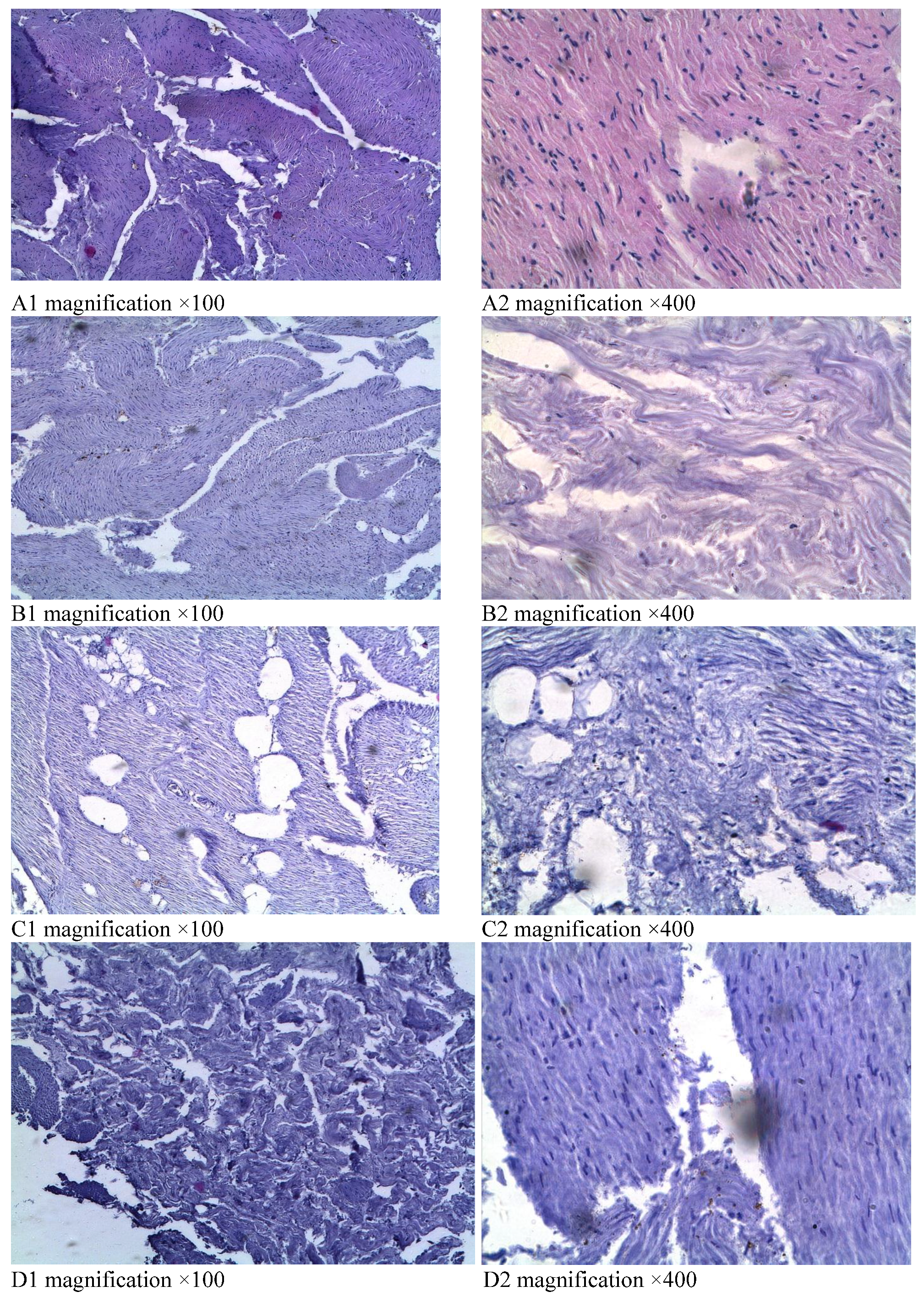
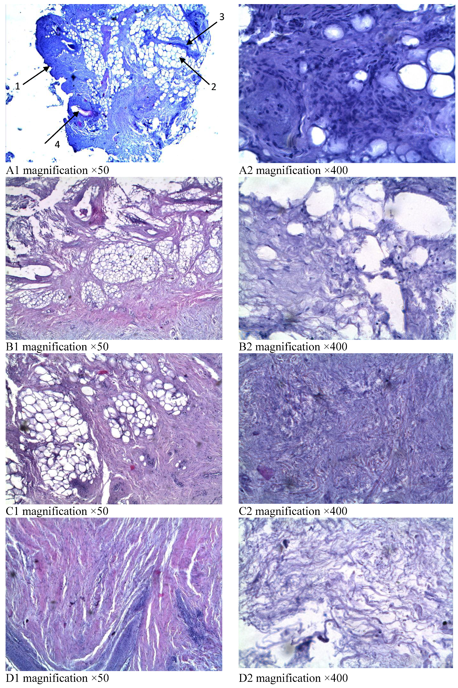
2.3. Research of the Fermented By-Products Microstructure

For microstructural analysis, the fermented by-products were kept in a 40% formalin solution for 72 h to hardening the consistency. The microstructure modification of fermented by-products during biotechnological processing was studied by microscopy of stained histopreparations using a microscope LEICA DMRXA (Wetzlar, Germany). The sections of the samples were stained with hematoxylin-eosin and picrofuxin according to the Van Gieson method. Using a digital video camera LEICA DFC 290 (Wetzlar, Germany), images of micro-preparations were obtained in the format of graphic TIFF files in the RGB color space, which were the objects of morphometric studies.
Using the ImageScope M image analysis program (Wetzlar, Germany), the following indicators were analyzed: the specific area of collagen tissue (%), the average thickness of collagen fibers (microns); the average thickness of myocytes (microns).

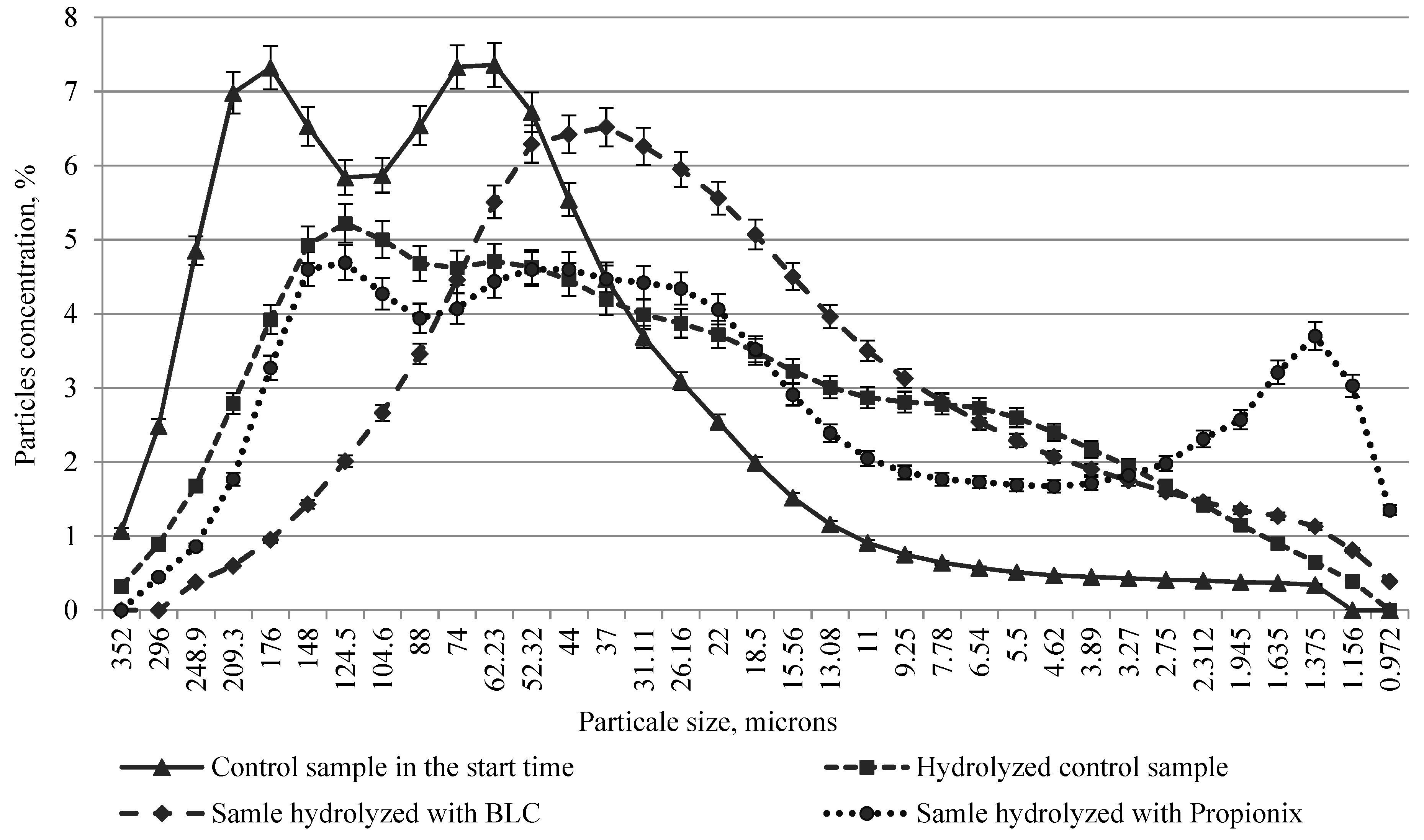
2.4. Determination of the Dispersed Composition of the By-Product Hydrolysates

The study of the dispersed composition and the analysis of the particle sizes of the hydrolysate samples were carried out by the method of laser dynamic light scattering on a laser diffraction analyzer Microtrac S3500.

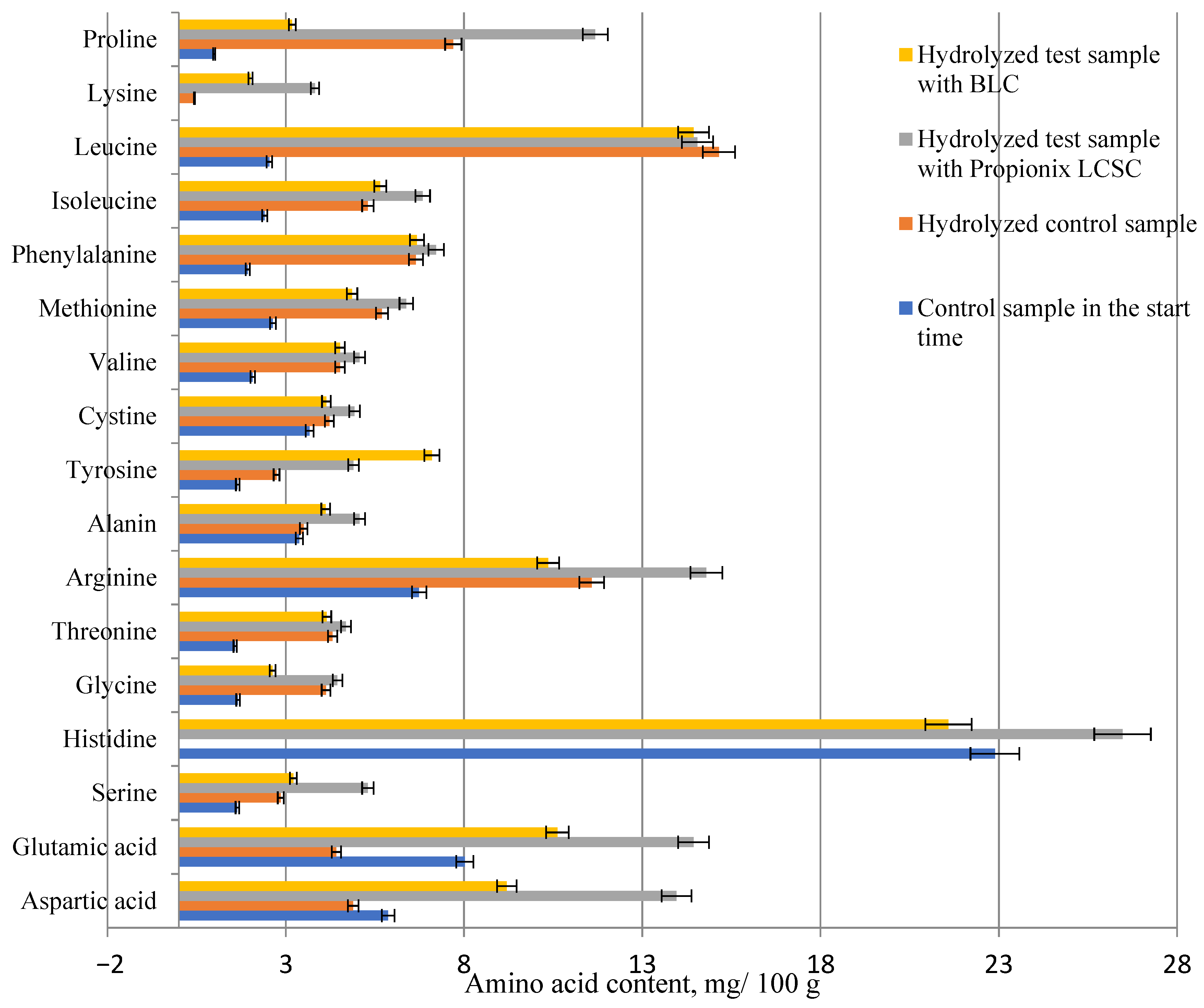
2.5. Determination of Free Amino Acids in By-Product Hydrolysates

The liquid chromatograph Agilent 1260 Infinity LC (Santa-Clara, CA, USA) was used to determine unbound amino acids. Liquid extraction was carried out to prepare the samples. Five grams of the hydrolysate sample was taken into an Eppendorf tube, and 4 mL of 20% trichloroacetic acid was added to precipitate proteins and peptides. The volume was brought to 30 mL with acidified hydrochloric acid buffer with pH 2.2 for 8 min, maintaining the homogenate under refrigerated conditions. The resulting mixture was centrifuged (20 min, 4 °C, 10,000× g) and the supernatant was passed through a syringe filter into a vial.
After that, pre-column derivatization was performed in the HPLC system autosampler using reagents: orthophthalic aldehyde for primary amino acids and 9-fluoromethyl chloroformate for secondary amino acids. The ratio of derivatives to the selected sample volume was 1:10.
Chromatographic separation was carried out on a ZORBAX C18 PA column, 3.5 µm 4.6 mm × 150 mm (Agilent, Santa-Clara, CA, USA) in the gradient elution mode for 25 min. For primary amino acids, measurements were carried out in a UV detector at a wavelength of λ = 338 nm, for secondary amino acids λ = 262 nm.
Mobile phase A was used: Acetonitrile: Methanol: Water (45:45:10); mobile phase B with pH 8.2: Na2HPO4 1.42 g and Na2B4O7 2.1 g pH 8.2. The flow rate was 1 mL/min, and the column temperature was 40 °C.
Standards of amino acids purchased from Sigma-Aldrich (Santa-Clara, CA, USA) were used for quantitative analysis of amino acids. All calibration curves had R2 > 0.9. The content of unbound (free) amino acids was expressed in units: mg AA per 100 g of liquid hydrolysates.

2.6. Statistical Analysis

A total of five replicates per sample were analyzed and measured three times. Results were expressed as the mean values of the five replicates ± the standard deviation. Probability values of p ≤ 0.05 were taken to indicate statistical significance. The data were analysed via one-way ANOVA and the Tukey–Kramer (Tukey’s HSD) test using the free web-based software (https://houssein-assaad.shinyapps.io/TableReport/ (accessed on 8 September 2021)) offered by Assaad et al. [22].

3. Results and Discussion

3.1. Microstructure of the Fermented By-Products

In control samples of hen gizzards not subjected to fermentation, microsections are represented by fragments of the muscle layer with small areas of the mucous membrane (Figure 1(A1)). Bundles of smooth muscle fibers are located in mutually perpendicular directions. The structure of myocytes and their nuclei is clearly visible at high magnification (Figure 1(A2)).
Microslides of fermented control gizzards are represented by muscle layer fragments (Figure 1(B1)). Compared with the non-fermented control group, attention is drawn to the poor perception of nuclear dyes, the fuzzy outlines of muscle cells (Figure 1(B2)). Diffuse tissue changes, as well as morphological manifestations of moderate autolysis, are expressed in all preparations of samples treated with bacterial concentrates (Figure 1(C1,D1)). These changes consist of a weak perception of histological dyes and a partial loss of cell nuclei. Smooth muscle cells are reduced in volume and ungrouped complexity, with fuzzy outlines and poorly stained nuclei (Figure 1(C2,D2)).
The staining of collagen fibers of micro-sections with picrofuchsin in various shades of pink was established. Diffuse tissue staining was observed in all preparations of gizzards (Figure 2). In the samples of the non-fermented control group, it was noted that the collagen fibers of the connective tissue matrix were folded into compact bundles and stained bright pink (Figure 2A).
However, in the control and test samples, after fermentation, a weak perception of histological dyes and a partial loss of cell nuclei were found. Smooth muscle cells are reduced in volume and discomplexed, with unclear borders and poorly stained nuclei (Figure 2B–D). Weak staining of muscle and connective tissue structures after fermentation is associated with a decrease in the adsorption of histological dyes by partially destroyed cellular elements.
In microslides of gizzards treated with bifidobacteria, diffuse fuchsinophilic staining of smooth muscle tissue was found, and collagen fibers against the background of muscle layers were stained with picric acid in yellow (Figure 2E). When processing the gizzards with propionic acid bacteria, the discomplexed, weakly stained collagen fibers were observed, as well as diffuse poor yellow staining of smooth muscle tissue (Figure 2F).
The microslides of the combs of control samples are represented by skin fragments with a thin stratum corneum. The surface of the skin is folded, covered with a thin layer of epidermal cells (Figure 3(A1, arrow 1)). In the subepidermal layer, a large number of lacunae-type blood vessels are visible (Figure 3(A1, arrow 4)). Cells and their nuclei are clearly visible, with good acceptance of histological dyes. The extracellular matrix is represented by randomly located collagen fibers. In the deep layers, stratums of adipose tissue are visible, separated by thick connective tissue septa, consisting mainly of compactly packed collagen fibers (Figure 3(A1, arrows 2, 3)). The cellular composition is dominated by mature fibroblasts, against which tissue macrophages, lymphoid cells and tissue basophils are determined in small quantities (Figure 3(A2)).
Partial loss of tissue structural units, monotony of coloring and poor perception of histological dyes were found in histosections of control fermented samples (Figure 3(B1)), as well as a few weakly stained cell nuclei with fuzzy, blurred borders (Figure 3(B2)).
In the microslides of hen combs fermented with bacterial concentrates, the histological structure is clearly traced, the monotony of the skin color has been established (Figure 3(C1,D1)). Compared with the control non fermented sample, attention is drawn to the reduced perception of histological dyes and blurring of the connective tissue matrix, the boundaries of the cellular elements and their nuclei are poorly visualized. The number of visible cell nuclei in these groups is less than in control samples (Figure 3(C2,D2).
When stained with picrofuchsin according to the Van Gieson method, chaotically located collagen fibers are visible in all samples against the background of a slightly pink staining of the intercellular substance (Figure 4). Collagen fibers are clearly visible in the control combs samples, as well as areas stained with only picric acid in yellow (Figure 4A). In all layers of the skin of the control fermented samples, thin collagen fibers are visible against the background of a slightly pink staining; the fields free of collagen fibers (bundles of smooth muscle fibers) are stained with picric acid (Figure 4B). The appearance of yellow staining is associated with the collagen hydrolysis and the diffusion of hydrolysate through the tissue.
In the samples treated with bacterial concentrates, a diffuse monochrome pinkish-crimson staining is observed in all fields of vision; thin collagen fibers are discomplexed and located in the intercellular substance, slightly colored in shades of pink (Figure 4C,D).
As a result of morphometric studies, regularity was established for a decrease in both the specific area of connective tissue and the diameter of collagen fibers for samples of hen combs and gizzards processed with bacterial enzymes. A reduction in muscle fiber thickness was also found in gizzard samples (Table 1).
The results obtained are due to the proteolytic activity of the enzymes of propionic acid bacteria and bifidobacteria, which are capable of hydrolyzing proteins of muscle and connective tissue. Additionally, lactic, acetic and propionic acids, which are a metabolic product of probiotic microorganisms, affect the change in the structure of muscle and collagen fibers. As noted by Aktas and Kaya [23], lactic acid increased animal tissue breakdown.
Deep proteolysis of meat systems is accompanied by the destruction of the conformation of protein macromolecules, changing the structure and softening the consistency of collagen and muscle fibers. At the same time, the amount of free amino acids rises sharply (up to 20–30% of their total content) [6].
Vlahova-Vangelova et al. [16] found that treatment of mutton with marinade with sodium lactate for 24 h leads to fragmentation in sarcomeres and destruction of Z-lines, and deeper destructive changes occur when acid marinating is extended to 48 h. The authors noted that acidic marinade solutions affect the native structure of connective tissue due to the activation of proteases.
The results of studies of the microstructure of pork treated with salt-fermented shrimp sauce, carried out by electrophoresis in SDS-polyacrylamide gel (SDS-PAGE), showed depolarization of the myosin heavy chain (MHC) and complete disappearance of the Z-line and muscle fibers using electron microscopy [7].

3.2. Dispersed Composition

As a result of the analysis of the fractional composition of gizzard hydrolysates, it was found that they were characterized mainly by a high heterogeneity and a significant proportion of large particles (more than 60 microns—62%) and a low proportion of small particles (less than 10 microns, 3.3%). The samples presented an irregular distribution of particles over the size range, from 30 to 248 microns.
A strong influence of the fermentation on the particle size distribution was noted; samples hydrolyzed by microbial enzymes differ in the uniformity of particle sizes and a large number of small particles. Thus, gizzards hydrolysates processed with propionic acid bacteria contain 59% of medium size particles (6–26 microns), more than 28% of small particles with a size of 1–9 microns. At the same time, hydrolysates treated with bifidobacteria contain 58% of particles with a size of 11–52 microns and about 10% of particles with a size of 1–9 microns. During hydrolysis of gizzards without the addition of bacterial concentrates, the number of medium particles was 54% (11–52 microns), and the amount of small particles was about 15% (Figure 5).
During the study of non-fermented comb hydrolysates, it was found that they were characterized by a high heterogeneity of the dispersed composition. Thus, 51.3% of the total fractional composition is occupied by large particles from 88 to 259 microns. Almost 26% of the particles have a size in the range of 22–53 microns. A small proportion (5.5%) is accounted for particles of 2–9 microns in size.
The test samples hydrolyzed by proteolytic enzymes of microbial concentrates are distinguished by greater uniformity of particle sizes and a significant number of small particles. Thus, comb hydrolysates treated with Propionix LCSC contain about 50% of particles of 15–104 microns in size, more than 30% particles of 2–9 microns in size. Hydrolysates treated with BLC include 56.5% particles with a size of 15–74 microns and 24.5% particles with a size of 2–9 microns.
The amount of the smallest particles, 1–2 microns in size, in hidrolysates was 13.9%—when fermentation with propionic acid bacteria, 5.0%—when treated with bifidobacteria and 3.1%—during fermentation of the control sample, without bacterial concentrates (Figure 6).
Studies of the dispersed composition of gizzard hydrolysates have shown that the particle size in samples treated with bacteria decreases (Table 2). The results of the evaluation of the dispersed composition proved that under the action of proteolytic enzymes produced by bacteria, proteins are broken down into shorter fragments. Hou et al. [24] mentioned that proteins are degraded by enzymes into peptides of various molecular weights and free amino acids. Wilkins et al. [25] found a correlation between the length of the polypeptide chain and the molecular size of various proteins, as determined by the hydrodynamic radius. Vorob’ev and Sinitsyna [26], when studying the proteolytic action of trypsin on β-casein molecules, determined a decrease in the diameter of small micelles as a result of proteolysis.

3.3. Analyses of the Free Amino Acid Composition in By-Product Hydrolysates

Proteolysis occurs in by-products, mainly under the action of intra- and extracellular enzymes of probiotic microorganisms. These bacteria have a complex proteolytic system, represented by proteases, which catalyze the breakdown of protein into oligopeptides and intracellular peptidases, which decompose peptides into shorter peptides and free amino acids [27,28].
Our research revealed an increase in the concentration of free amino acids in the control and the test samples of hydrolysates during the fermentation period.
It should be point out that there was a significant accumulation of essential and nonessential amino acids in the hen gizzard hydrolysates fermented with Propionix LCSC. These findings are due to the effectiveness of exoproteases of propionic acid bacteria in relation to muscle and collagen tissue of the gizzards, as well as the ability of the microorganisms to produce free amino acids in the substrate during metabolism (Figure 7).
Our results proved that the proteolytic enzymes of the selected microorganisms are able to catalyze the hydrolysis of connective tissue (which predominates in the combs), but less effectively than muscle tissue. The findings indicated an increasing in free amino acids in the comb hydrolysates treated with bifidobacteria and prionic acid bacteria concentrates, compared with control samples. Biochemical analysis of the comb hydrolysates revealed a decrease in the concentration of amino acids by 2.5–4 times as compared with gizzards hydrolysates. This is due to the high content of collagen fibers, as well as the more complex structure of the collagen molecules of the hen combs (Figure 8).
Arihara et al. [9] confirmed that collagen lacks certain essential amino acids, and it has less nutritional value than most other food proteins. Unsal and Aktas [29] established the difference in the amino acid composition of by-products and muscle tissue due to differences in the content of connective tissue. At the same time, it was noted that by-products are a good source of essential amino acids, in particular such limiting amino acids as lysine, methionine and tryptophan [11]. Seong et al. [30] explain the differences in the quantitative and qualitative composition of amino acid by-products by differences in protein types. Jayathilakan et al. [31] emphasized that by-products such as ears, legs, lungs and stomach are high in proline, hydroxyproline and glycine, as well as lower levels of tryptophan and tyrosine.

4. Conclusions

Poultry by-products are promising sources for the obtention of protein hydrolysates by enzymatic hydrolysis. As a result of microstructural study of fermented by-products, a decrease in the perception of histological dyes, poor visualization of the cell elements and blurring of the connective tissue matrix were established. Morphometric analysis showed a reduction in the specific area of connective tissue, the diameter of collagen fibers and the thickness of muscle fibers for by-products treated with bacterial concentrates. A strong influence of fermentation on the particle size distribution was established; hydrolysates were characterized by a high uniformity of particle sizes and a large number of small particles. Our research revealed an increase in the concentration of free amino acids in the test samples of hydrolysates during the fermentation period. The results of biochemical and microscopic analysis confirm the good hydrolysability of hen combs and gizzards under the action of microbial enzymes.

Author Contributions

Conceptualization, O.Z.; methodology, O.Z., S.M., I.L., A.K. and T.S.; investigation, O.Z. and S.M.; data curation and analysis, O.Z. and S.M.; supervision, O.Z.; writing—original draft, O.Z. and S.M.; writing—review and editing, O.Z. and S.M. All authors have read and agreed to the published version of the manuscript.

Funding

The research was funded by RFBR and Chelyabinsk Region, project number 20-416-740002.

Data Availability Statement

Data will be made available up on request to the corresponding author.

Acknowledgments

We would like to thank managers of Scientific and Educational Center of Nanotechnology of South Ural State University for their technical support during this work.

Conflicts of Interest

The authors declare no conflict of interest. The funders had no role in the design of the study; in the collection, analyses, or interpretation of data; in the writing of the manuscript, or in the decision to publish the results.

References

  1. Xiang, H.; Sun-Waterhouse, D.; Waterhouse, G.I.N.; Cui, C.; Ruan, Z. Fermentation-enabled wellness foods: A fresh perspective. Food Sci. Human Wellness 2019, 8, 203–243. [Google Scholar] [CrossRef]
  2. Ghosh, D.; Chattoraj, D.K.; Chattopadhyay, P. Studies on Changes in Microstructure and Proteolysis in Cow and Soy Milk Curd During Fermentation Using Lactic Cultures For Improving Protein Bioavailability. J. Food Sci. Technol. 2013, 50, 979–985. [Google Scholar] [CrossRef] [PubMed] [Green Version]
  3. Lorusso, A.; Coda, R.; Montemurro, M.; Rizzello, C.G. Use of Selected Lactic Acid Bacteria and Quinoa Flour for Manufacturing Novel Yogurt-Like Beverages. Foods 2018, 7, 51. [Google Scholar] [CrossRef] [PubMed] [Green Version]
  4. Melini, F.; Melini, V.; Luziatelli, F.; Ficca, A.G.; Ruzzi, M. Health-promoting components in fermented foods: An up-to-date systematic review. Nutrients 2019, 11, 1189. [Google Scholar] [CrossRef] [Green Version]
  5. Sharma, R.; Garg, P.; Kumar, P.; Bhatia, S.K.; Kulshrestha, S. Microbial Fermentation and Its Role in Quality Improvement of Fermented Foods. Fermentation 2020, 6, 106. [Google Scholar] [CrossRef]
  6. Kassymov, S.; Nesterenko, A.; Yunusov, E.; Radchenko, E.; Bychkova, T. Effect of Alkali-Salt Treatment of Meat By-Products on Physical, Chemical and Rheological Properties. Int. J. Innov. Technol. Explor. Eng. 2020, 9, 3221–3224. [Google Scholar] [CrossRef]
  7. Kim, J.-S.; Shahidi, F.; Heu, M.-S. Tenderization of meat by salt-fermented sauce from shrimp processing by-products. Food Chem. 2005, 93, 243–249. [Google Scholar] [CrossRef]
  8. Paulsen Thoresenb, P.; García Álvareza, R.; Risa Vakaa, M.; Rustadb, T.; Sonea, I.; Noriega Fernándeza, E. Potential of innovative pre-treatment technologies for the revalorisation of residual materials from the chicken industry through enzymatic hydrolysis. Innov. Food Sci. Emerg. Technol. 2020, 64, 102377. [Google Scholar] [CrossRef]
  9. Arihara, K.; Yokoyamaa, I.; Ohatab, M. Bioactivities generated from meat proteins by enzymatic hydrolysis and the Maillard reaction. Meat Sci. 2021, 180, 108561. [Google Scholar] [CrossRef] [PubMed]
  10. Santana, J.C.C.; Gardim, R.B.; Almeida, P.F.; Borini, G.B.; Quispe, A.P.B.; Llanos, S.A.V.; Heredia, J.A.; Zamuner, S.; Gamarra, F.M.C.; Farias, T.M.B.; et al. Valorization of Chicken Feet By-Product of the Poultry Industry: High Qualities of Gelatin and Biofilm from Extraction of Collagen. Polymers 2020, 12, 529. [Google Scholar] [CrossRef] [Green Version]
  11. Mullen, A.M.; Álvarez, C.; Zeugolis, D.I.; Henchion, M.; O’Neill, E.; Drummond, L. Alternative uses for co-products: Harnessing the potential of valuable compounds from meat processing chains. Meat Sci. 2017, 132, 90–98. [Google Scholar] [CrossRef]
  12. Lasekan, A.; Abu Bakar, F.; Hashim, D. Potential of hens’ byproducts as sources of useful biological resources. Waste Manag. 2013, 33, 552–565. [Google Scholar] [CrossRef]
  13. Lafarga, T.; Hayes, M. Bioactive peptides from meat muscle and by-products: Generation, functionality and application as functional ingredients. Meat Sci. 2014, 98, 227–239. [Google Scholar] [CrossRef] [PubMed]
  14. Vikman, Y.M.; Siipola, V.; Kanerva, H.; Šližyte, R.; Wikberg, H. Poultry By-products as a Potential Source of Nutrients. Adv. Recycl. Waste Manag. 2017, 2, 142. [Google Scholar] [CrossRef]
  15. Mozuriene, E.; Bartkiene, E.; Krungleviciute, V.; Zadeike, D.; Juodeikiene, G.; Damasius, J.; Baltusnikiene, A. Effect of natural marinade based on lactic acid bacteria on pork meat quality parameters and biogenic amine contents. LWT—Food Sci. Technol. 2016, 69, 319–326. [Google Scholar] [CrossRef]
  16. Vlahova-Vangelova, D.B.; Dragoev, S.G.; Balev, D.K.; Assenova, B.K.; Amirhanov, K.J. Quality, Microstructure, and Technological Properties of Sheep Meat Marinated in Three Different Ways. J. Food Qual. 2017, 2017, 5631532. [Google Scholar] [CrossRef] [Green Version]
  17. Sun-Waterhouse, D.; Zhao, M.; Waterhouse, G. Protein Modification During Ingredient Preparation and Food Processing: Approaches to Improve Food Processability and Nutrition. Food Bioprocess Technol. 2014, 7, 1853–1893. [Google Scholar] [CrossRef]
  18. Toldrá, F.; Reig, M.; Aristoy, M.C.; Mora, L. Generation of bioactive peptides during food processing. Food Chem. 2018, 267, 395–404. [Google Scholar] [CrossRef] [PubMed]
  19. Shen, P.; Zhou, F.; Zhang, Y.; Yuan, D.; Zhao, Q.; Zhao, M. Formation and characterization of soy protein nanoparticles by controlled partial enzymatic hydrolysis. Food Hydrocolloids 2020, 105, 105844. [Google Scholar] [CrossRef]
  20. Halavach, T.N.; Kurchenko, V.P. Milk Protein Hydrolysis with Enzyme Preparation and Proteolytic Systems of Lactic Acid Bacteria; Series: Physiological, biochemical and molecular foundations of the functioning of biosystems; Belarusian State University: Minsk, Belarus, 2012; Volume 7, pp. 106–126. [Google Scholar]
  21. Zinina, O.; Merenkova, S.; Galimov, D. Optimization of Microbial Hydrolysis Parameters of Poultry By-Products Using Probiotic Microorganisms to Obtain Protein Hydrolysates. Fermentation 2021, 7, 122. [Google Scholar] [CrossRef]
  22. Assaad, H.; Zhou, L.; Carroll, R.J.; Wu, G. Rapid Publication-Ready MS-Word Tables for One-Way ANOVA. In SpringerPlus; Springer: Berlin, Germany, 2014; Volume 3, p. 474. Available online: https://houssein-assaad.shinyapps.io/TableReport/ (accessed on 8 September 2021). [CrossRef] [Green Version]
  23. Aktas, N.; Kaya, M. The influence of marinating with weak organic acids and salts on the intramuscular connective tissue and sensory properties of beef. Eur. Food Res. Technol. 2001, 213, 88–94. [Google Scholar] [CrossRef]
  24. Hou, Y.; Wu, Z.; Dai, Z.; Wang, G.; Wu, G. Proteinhydrolysates in animal nutrition: Industrial production, bioactive peptides, and functional significance. J. Anim. Sci. Biotechnol. 2017, 8, 24. [Google Scholar] [CrossRef] [PubMed] [Green Version]
  25. Wilkins, D.K.; Grimshaw, S.B.; Receveur, V.; Dobson, C.M.; Jones, J.A.; Smith, L.J. Hydrodynamic radii of native and denatured proteins measured by pulse field gradient NMR techniques. Biochemistry 1999, 38, 6424–16431. [Google Scholar] [CrossRef] [PubMed]
  26. Vorob’ev, M.M.; Sinitsyna, O.V. Degradation and assembly of β-casein micelles during proteolysis by trypsin. Int. Dairy J. 2020, 104, 104652. [Google Scholar] [CrossRef]
  27. Donkor, O.N.; Henriksson, A.; Vasiljevic, T.; Shah, N.P. Proteolytic activity of dairy lactic acid bacteria and probiotics as determinant of growth and in vitro angiotensin-converting enzyme inhibitory activity in fermented milk. Le Lait 2007, 87, 21–38. [Google Scholar] [CrossRef]
  28. Law, J.; Haandrikman, A. Proteolytic enzymes of lactic acid bacteria. Int. Dairy J. 1997, 7, 1–11. [Google Scholar] [CrossRef]
  29. Unsal, M.; Aktas, N. Fractionation and characterization of edible sheep tail fat. Meat Sci. 2003, 63, 235–239. [Google Scholar] [CrossRef]
  30. Seong, P.N.; Kang, G.H.; Park, K.M.; Cho, S.H.; Kang, S.M.; Park, B.Y. Characterization of Hanwoo Bovine By-products by Means of Yield, Physicochemical and Nutritional Compositions. Korean J. Food Sci. Anim. Resour. 2014, 34, 434–447. [Google Scholar] [CrossRef] [Green Version]
  31. Jayathilakan, K.; Sultana, K.; Radhakrishna, K.; Bawa, A.S. Utilization of byproducts and waste materials from meat, poultry and fish processing industries: A review. J. Food Sci. Technol. 2012, 49, 278–293. [Google Scholar] [CrossRef] [Green Version]
Figure 1. Microstructure of fermented hen gizzards (hematoxylin-eosin staining). (A1,A2) control sample in the start time; (B1,B2) fermented control sample; (C1,C2) fermented test sample with BLC; (D1,D2) fermented test sample with Propionix LCSC.
Fermentation 07 00190 g001
Figure 2. Microstructure of fermented hen gizzards (staining with picrofuxin according to Van Gieson). (A) control sample in the start time; (B) fermented control sample; (C,E) fermented test sample with BLC; (D,F) fermented test sample with Propionix LCSC (magnification ×100).
Fermentation 07 00190 g002
Figure 3. Microstructure of fermented hen combs (hematoxylin-eosin dying). (A1,A2) control sample in the start time (arrows: 1- folded surface of the skin, 2—layers of adipose tissue, 3—connective tissue septa, 4—lacunae-type blood vessels); (B1,B2) fermented control sample; (C1,C2) fermented test sample with BLC; (D1,D2) fermented test sample with Propionix LCSC.
Fermentation 07 00190 g003
Figure 4. Microstructure of fermented hen combs (staining with picrofuxin according to Van Gieson). (A) control sample in the start time; (B) fermented control sample; (C) fermented test sample with BLC; (D) fermented test sample with Propionix LCSC (magnification ×100).
Fermentation 07 00190 g004
Figure 5. Particle size distribution in the samples of hen gizzard hydrolysates.
Fermentation 07 00190 g005
Figure 6. Particles size distribution in the samples of hen comb hydrolysates.
Fermentation 07 00190 g006
Figure 7. The results of the amino acids composition evaluation of hen gizzards hydrolysates (mg AA/100 g of hydrolysate).
Fermentation 07 00190 g007
Figure 8. The results of the amino acids composition evaluation of hen combs hydrolysates (mg AA/100 g of hydrolysate).
Fermentation 07 00190 g008
Table 1. Results of morphometric analysis of fermented hen by-products.
IndicatorsControl Sample in the Start TimeFermented Control SampleFermented Test Sample with BLCFermented Test Sample with Propionix LCSC
hen gizzards
Specific area of connective tissue (%)61.43 ± 1.922 a37.91 ± 1.259 b33.80 ± 1.981 c24.99 ± 0.266 d
Average thickness of collagen fibers (microns)15.36 ± 3.056 a10.37 ± 2.071 b6.51 ± 0.958 c6.65 ± 0.953 c
Average thickness of myocytes (microns)15.59 ± 1.856 a11.18 ± 1.764 b4.54 ± 1.055 c5.53 ± 0.855 c
hen combs
Specific area of connective tissue (%)59.22 ± 3.067 a54.85 ± 1.439 a51.58 ± 3.239 b43.77 ± 1.742 c
Average thickness of collagen fibers (microns)19.90 ± 3.006 a6.58 ± 0.9615 b4.49 ± 1.267 c7.18 ± 2.492 b
Values are means ± SEM, n = 5 per treatment group. Means in a row without a common superscript letter differ statistically (p < 0.05) as analyzed by one-way ANOVA and the TUKEY test.
Table 2. Average hydrodynamic diameter determined from the numerical particle size distribution in hydrolysates.
IndicatorControl Sample in the Start TimeHydrolyzed Control SampleHydrolyzed Test Sample with Propionix LCSCHydrolyzed Test Sample with BLC
hen gizzards
MN (micron)11.83 ± 0.85 a6.60 ± 0.62 b4.69 ± 0.35 c2.73 ± 0.24 d
hen combs
MN (micron)1.93 ± 0.11 a1.91 ± 0.08 a1.28 ± 0.07 b1.53 ± 0.08 c
Values are means ± SEM, n = 5 per treatment group. Means in a row without a common superscript letter differ statistically (p < 0.05) as analyzed by one-way ANOVA and the TUKEY test.
Publisher’s Note: MDPI stays neutral with regard to jurisdictional claims in published maps and institutional affiliations.

