Article

Modulation of the Expression of Immune-related Gene in Atlantic and Coho Salmon during Infestation with the Sea lice Caligus rogercresseyi

by
Juan Pablo Pontigo
1,*,
Julia Saravia
1,2,
Ricardo Oyarzún
1,2,3,
Margarita P. González
2,
Christopher Hawes
4,
Francisco J. Morera
5,
Jorge Pino
4,
Simon Wadsworth
4,
Jose Luis P. Muñoz
6,* and
Luis Vargas-Chacoff
1,3,*
1
Instituto de ciencias Marinas y Limnologicas, Universidad Austral de Chile, 5090000 Valdivia, Chile
2
Programa de Doctorado en Ciencias de la Acuicultura, Universidad Austral de Chile, 5480000 Puerto Montt, Chile
3
Centro Fondap de Investigación de Altas Latitudes (IDEAL), Universidad Austral de Chile, 5090000 Valdivia, Chile
4
Cargill Innovation Center-Colaco, Camino Pargua km 57, Colaco km 5, Calbuco, 5480000 Puerto Montt, Chile
5
Instituto de Farmacología y Morfofisiología, Facultad de Ciencias Veterinarias, Universidad Austral de Chile, 5090000 Valdivia, Chile
6
Centro de Investigación y Desarrollo i~mar, Universidad de los Lagos, 5480000 Puerto Montt, Chile
*
Authors to whom correspondence should be addressed.
Submission received: 29 March 2019 / Revised: 12 July 2019 / Accepted: 12 July 2019 / Published: 25 July 2019

Abstract

:
Caligus rogercresseyi, a marine ectoparasite, causes notable economic losses for the Chilean salmonid industry. Nevertheless, the immunological responses of infected fish remain poorly understood, including proinflammatory cytokine generation and the respective modulatory effects of various cytokine receptors. This study evaluated mRNA expression of the NLRC5, major histocompatibility complex (MHC) class II, I-kappa-B-alpha, a regulatory that inhibits NF-kappa-B, and proinflammatory cytokines (IL-1β and IL-18) in the liver and muscle of Atlantic salmon (Salmo salar) and Coho salmon (Oncorhynchus kisutch) during a time-course C. rogercresseyi infestation trial. All assessed mRNA were strongly regulated during infestation, but S. salar showed up-regulated expression, possibly accounting for the high infestation vulnerability of this salmonid. In conclusion, this work helps to understand the modulation of the expression of different transcripts involved over short periods of C. rogercresseyi infestation in two salmonid species (S. salar and O. kisutch).

Graphical Abstract

1. Introduction

Proinflammatory cytokine generation and the respective modulation of cytokines by different receptor types are two poorly studied immunological mechanisms of fish infected by bacteria, viruses, and ectoparasites [1,2,3]. Some cytokine receptors are also components of the inflammasome complex, specifically acting as innate immune system receptors/sensors that regulate caspase-1 activation and induce inflammation in response to infectious pathogens and molecules derived from host proteins [4]. The inflammasome complex is further composed of a nucleotide-binding domain, leucine-rich repeat (NLR) proteins, such as NLRP1, NLRP3, NLRC4, or NLRC5; HIN-200 family member absent in melanoma 2 (AIM2) protein; cytosolic retinoic-acid-inducible I (RIG-I) RNA; and an ASC/PYCARD adapter molecule attached to caspase-1, which provides the enzymatic activity of the complex [5].
Of the known NLR proteins, NLRC5 is key in forming the inflammasome complex, as described by Beckley et al. (2013) [6]. Furthermore, an NLRC5 gene was recently identified and analyzed within the inflammasome complex of teleost fish, reporting involvement in modulating the inflammatory response [7]. This inflammatory response would be initiated by immune-cell (e.g., leukocytes) recruitment to and differentiation at the site of infection, thereby activating the antimicrobial effector mechanism and stimulating the immune response [8]. In addition, the NF-kappa B/ I-kappa-B-alpha pathway plays an important role in the regulation of the immune response, where I-kappa-B-alpha is an inhibitory molecule that sequesters the NF-kappaB dimer transcription activator in the cytoplasm of unstimulated cells, blocking nuclear translocation and thus the expression of proinflammatory cytokines [9]. It has been reported that NF-kappa B binds to sites of the NLRC5 promoter region, stimulating its expression [10].
Interleukin-1 is an apical pro-inflammatory cytokine, and interleukin-18 (IL-18), a recently described member of the IL-1 cytokine super-family, is now recognized as an important regulator of innate and acquired immune responses, meaning its activity initiates and directs the cascade of inflammatory signals in response to sensing pathogen-associated molecular patterns [11]. Several pattern recognition receptors are involved in the control and elimination of pathogens/microorganisms, including NLR protein receptors [12].
Aquaculture is a growing industry worldwide. Within the wide spectrum of etiological agents that affect fish farming, crustacean copepods are a particular point for concern, especially in salmonid cultures [13,14]. Sea lice, a common ectoparasitic copepod, are distributed globally and cause notable economic losses for salmon farming in Chile and Norway. Furthermore, increased salmon farming along the coastal areas of the Northern Hemisphere has also resulted in increased sea lice abundance, posing a serious threat to wild salmon populations [15]. This scenario is mirrored in the Southern Hemisphere, which has also seen considerable growth in the salmon industry [14].
The most prevalent parasite in the Chilean aquaculture industry is Caligus rogercresseyi [16,17], a Caligidae family copepod [18]. Furthermore, the most cultivated salmonids in Chile (i.e., Salmo salar [Atlantic salmon] and Oncorhynchus mykiss [rainbow trout]) are also the most vulnerable to C. rogercresseyi infestation, whereas Oncorhynchus kisutch (Coho salmon) has a greater infestation resistance [14]. Ectoparasites infest fish species, such as salmonids, and the consequences of infestation can include changes in epidermal morphology and mucus composition [19,20]. Interestingly, coho salmon express pro-inflammatory cytokines such as IL-1β, TNF-α, and MHIIb during L. salmonis infestation [21]. At the same time, there is evidence of changes at the transcriptomic level of genes related to the immune system of Atlantic and coho salmon when infested by C. rogercresseyi [2,3].
The evaluation of 27 genes related to the immune response, antioxidant system, and secretome in Atlantic and coho salmon at 1, 3, and 7 days post-infestation by C. rogercresseyi has been described, as well as the up-regulation of immune-related genes in head kidney and the skin of both salmonid species [22]. Differences in susceptibility levels of C. rogercresseyi have been associated with the regulation of iron as a mechanism to confer immunity during infestation. The regulation of transcripts with iron is modulated in Atlantic salmon by the depletion of cellular iron, which represents a mechanism of nutritional immunity, thus being the liver that is the most important organ in the regulation of iron and muscle is the closest organ during infestation with the parasite, which makes it more susceptible to C. rogercresseyi infestation than coho salmon, and therefore, makes the liver and muscle of both salmonid species an interesting target to study [2,23].
The present study is the first to experimentally evaluate the gene expression of NLRC5, caspase-1, I-kappa-B-alpha (inhibitory NF-kappaB pathway) of the proinflammatory cytokines IL-1β and IL-18 in two tissues (liver and muscle) in Atlantic and coho salmon during temporary infestation by C. rogercresseyi, in order to provide information on the immune response activity that occurs when coming into contact with this parasite. The results indicate that there are differences in gene expression between the Atlantic salmon and coho salmon during sea lice infestation, which could activate the immune system in muscle and liver.

2. Results

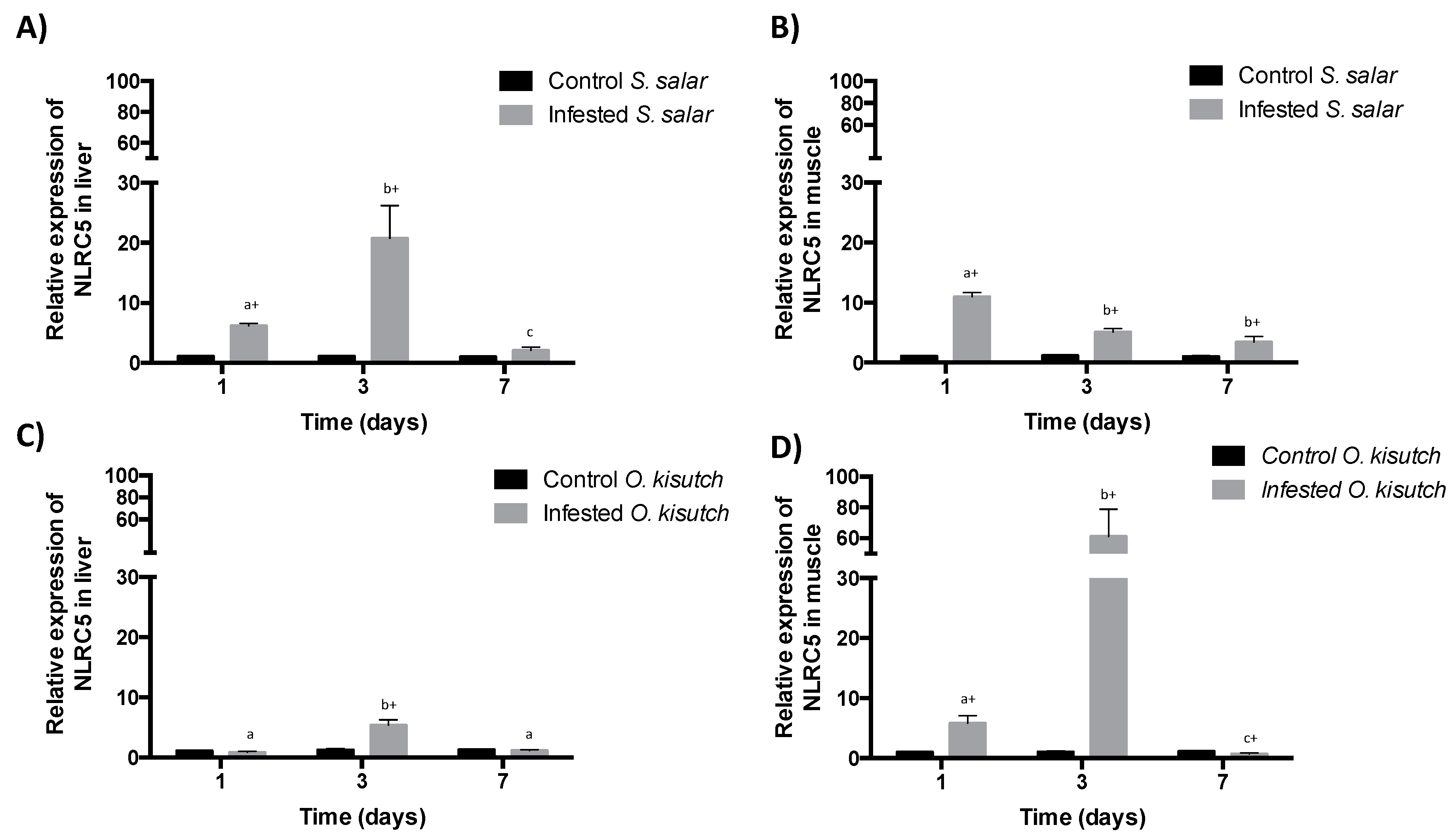
2.1. Analysis of NLRC5 Transcript Expression in the Muscle and Liver of Atlantic and Coho Salmon Infested with C. rogercresseyi

The mRNA levels of NLRC5 in the muscle of Atlantic salmon infested with C. rogercresseyi were statistically significant at all time-points compared to the control group. High expression of the transcript can be observed every day post infestation (dpi) over the trial period (Figure 1B). In Coho salmon, NLRC5 transcript expression significantly increased at 1 and 3 dpi compared to non-infested fish (Figure 1D), with 3 dpi presenting the highest expression. Regarding NLRC5 transcript expression in the liver of Atlantic salmon infested with C. rogercresseyi, levels were statistically significant at 1 and 3 dpi compared to the control group. The highest transcript expression levels in Atlantic salmon liver were recorded at 3 dpi (Figure 1A). In the liver of infested Coho salmon, NLRC5 transcript expression was lower than the control group at 1 and 7 dpi, but was significantly higher than the control group at 3 dpi (Figure 1D).

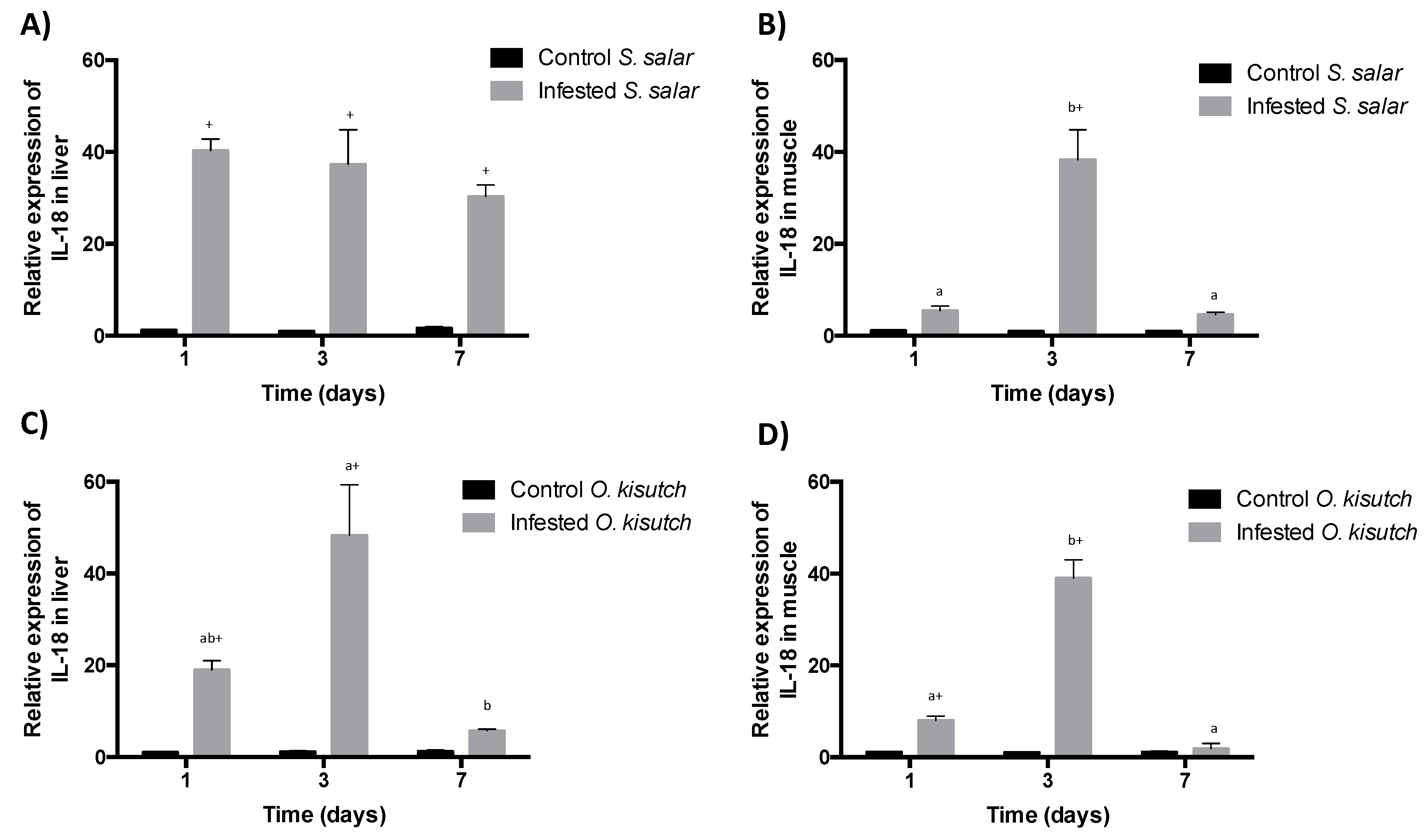
2.2. Analysis of IL-18 Transcript Expression in the Muscle and Liver of Atlantic and Coho Salmon Infested with C. rogercresseyi

Muscle IL-18 mRNA expression in infested Atlantic salmon increased significantly on every post-infestation day compared with the control group, but was mainly at day 3 post-infection. A near identical response was presented by Coho salmon (Figure 2B,D). In turn, the expression of hepatic IL-18 mRNA in Atlantic salmon infested with C. rogercresseyi increased significantly on each of the days of the time-course trial compared to the control group (Figure 2A). Similarly, the expression of IL-18 mRNA in the liver of Coho salmon was significantly increased compared to the control group at 1 and 3 dpi (Figure 2C).

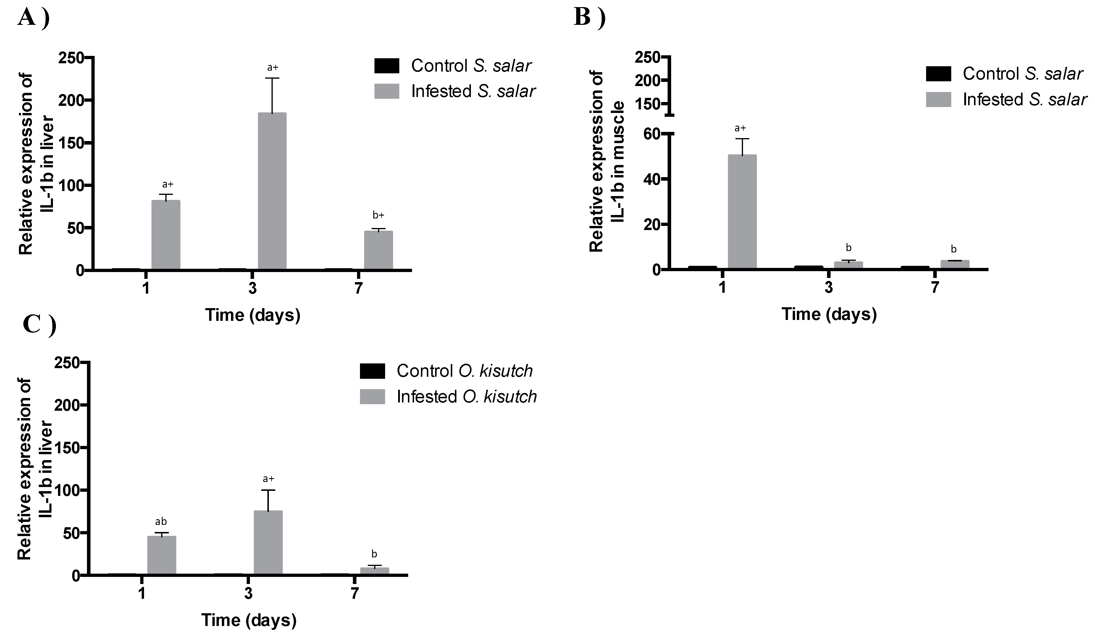
2.3. Analysis of IL-1β Transcript Expression in the Muscle and Liver of Atlantic and Coho Salmon Infested with C. rogercresseyi

Muscle IL-1β mRNA expression in Atlantic salmon infested by C. rogercresseyi significantly increased at 1 dpi compared to the control group. Transcript expression subsequently decreased at 3 and 7 dpi (Figure 3B). In contrast, infested Coho salmon muscle did not present IL-1β mRNA expression (data not shown). In turn, liver IL-1β transcript expression was similar for infested Atlantic and Coho salmon, with both salmonids presenting increased expression levels at 1 and 3 dpi followed by a decrease at 7 dpi (Figure 3A,C). As compared to the control group, both Atlantic and Coho salmon presented significant, and the highest levels of liver IL-1β mRNA expression at 1 and 3 dpi. However, expression levels of the IL-1β transcript were more important in Atlantic salmon on all days of the trial (Figure 3B).

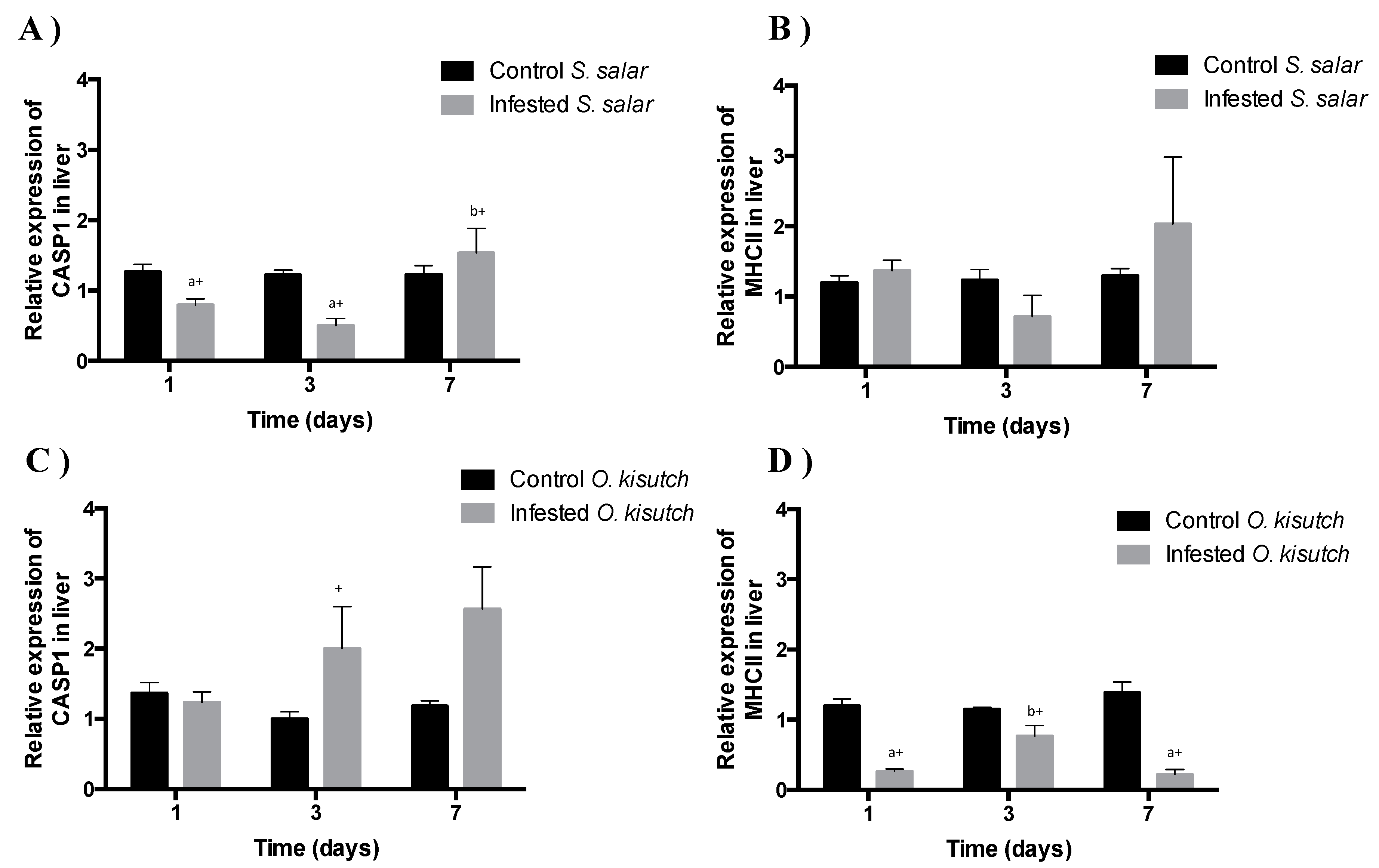
2.4. Analysis of Caspase-1 Transcript Expression in Liver of Atlantic and Coho salmon Infested with C. rogercresseyi

Expression of caspase-1 mRNA in Atlantic salmon liver infested by C. rogercresseyi increased its expression at 7 dpi compared to the control group. However, transcript expression decreased at 1 and 3 dpi (Figure 4A). In turn, the expression of caspase-1 transcript in the liver of infested salmon Coho, increased at 3 and 7 dpi (Figure 4C). In contrast, in infested Atlantic salmon and Coho muscle did not exhibit caspase-1 mRNA expression.

2.5. Analysis of MHCII Transcript Expression in the Muscle and Liver of Atlantic and Coho Salmon Infested with C. rogercresseyi

The expression of major histocompatibility complex (MHC) class II mRNA in Atlantic salmon liver infested with C. rogercresseyi showed no significant differences over time compared to the control. However, in the liver of Coho salmon the expression of the transcript significantly decreased at 1, 3, and 7 dpi (Figure 4B,D). In the muscle of both Atlantic salmon and Coho infested with C. rogercresseyi, the expression of MHC II mRNA was not induced.

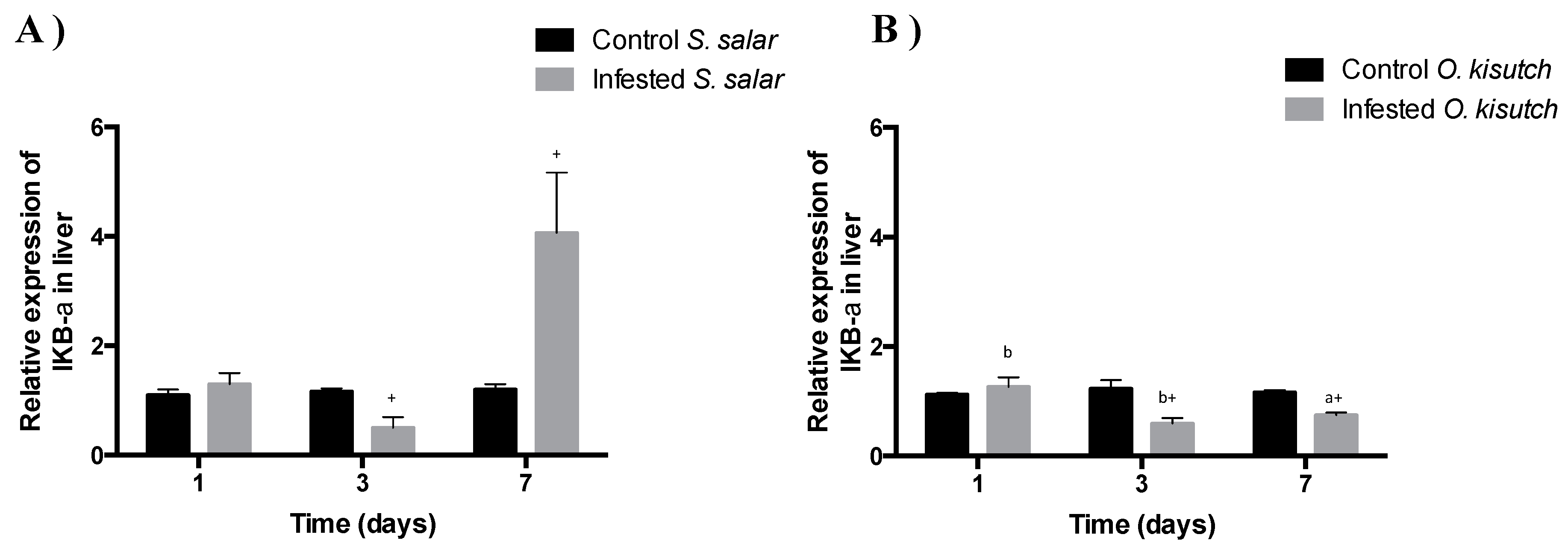
2.6. Analysis of I-kappa-B-alpha Transcript Expression in the Muscle and Liver of Atlantic and Coho Salmon Infested with C. rogercresseyi

Liver I-kappa-B-alpha mRNA expression in infested Atlantic salmon significantly increased compared to the control group only at 7 dpi. At 1 and 3 dpi the mRNA expression response was almost identical (Figure 5A). Liver I-kappa-B-alpha mRNA decreased in the infested Coho salmon at 3 and 7 dpi compared to the control group (Figure 5B). In contrast, the infested Atlantic and Coho salmon did not exhibit I-kappa-B-alpha mRNA expression in the muscle.

3. Discussion

Cytokines are molecules with key roles in regulating the immune response [24] and effector phase in both innate and adaptive immunity [25]. These small-protein mediators produced by immune cells mediate inflammation, immunity, and hematopoiesis [26]. In particular, interleukins are potent proinflammatory cytokines that have been described in teleost fishes, where IL-1β is key in the early response, being secreted when pathogens enter circulation [10], where the receptor IL -1β is expressed in all tissues of Atlantic salmon [27]. In turn, IL-18 has functions in the potent stimulation of interferon-γ production, enhancement of natural killer cell cytotoxicity, and stimulation of T-helper1 cell differentiation [28,29,30,31]. In general, IL-1β and IL-18 are potent proinflammatory cytokines that promote innate immune processes associated with infection, inflammation, and autoimmunity, activating monocytes, macrophages, and neutrophils, as well as inducing Th1 and Th17 cellular adaptive responses [12,32]. Nevertheless, a balance is needed to ensure host defense against viral and bacterial pathogens without resulting in tissue damage due to an excessive inflammatory response. The need for this balance could explain why some species have developed mechanisms for the regulation of these cytokines [27,32]. In mammals, the complex inflammasome caspase-1 is activated by autoproteolitic maturation through cleavage and secretion of inflammatory cytokines such as IL-1β, IL-18, and cell death [33]. While caspase-1 is essential for the release of mature IL-1β, it has also been associated with the processing of many extracellular proteins involved in inflammatory regulation [34] and is known for its pleiotropic role in innate immunity, however, in teleosts, the IL-1β processing site does not exist, which is why its activation system is different [35,36].
The major proinflammatory cytokines, IL-1β and IL-18, were also evaluated in infested Atlantic and Coho salmon. IL-18 mRNA expression increased mostly at 3 dpi in muscle and liver tissues, following the same expression pattern as NLRC5. Meanwhile, IL-1β transcript expression was only observed in Atlantic salmon muscle; the highest levels being observed at 1 dpi. The mRNA expression of IL-1β in the liver was several times greater than the control group for both salmon species. This observation aligns with that obtained for NLRC5 transcript expression (Figure 1).
However, when analyzing the expression of I-kappa-B-alpha mRNA, which is the inhibitor of the NF-kappa B transcription factor, it was observed that it increases in Atlantic salmon at 7 dpi, and decreases in Coho salmon. It is presumed that it represses the NF-kappa B pathway in Atlantic salmon, which may then activate an alternative route that has not yet been identified. While immune mechanisms play an important role in the responses of salmonids to sea lice infestation, the starting point for the regulation of the inflammatory response has not yet been elucidated.
To address this lack of knowledge, the present study evaluated the transcriptional modulations of NLRC5, MHCII, caspase-1, I-kappa-B-alpha, IL-1β, and IL-18 during C. rogercresseyi infestation in Atlantic and Coho salmon muscle and liver tissues. Further analyses evaluated how the aforementioned genes could be related to the inflammatory process through the inflammasome complex. While both salmonids presented high transcript expression in response to infestation, some transcripts were differentially expressed over the experimental period and between species. This is of great importance, mainly due to the characteristics of the tissues used (muscle and liver), since they are not precisely immunological, they presented gene expressions related to the fish immune system. This can be due to the high-energy rate, that is to say lactate, which is used by these organs before infestation with C. rogercresseyi, which could activate the immune system in both species [37], or also by an iron regulation that could affect infestation, conferring a type of nutritional immunity [23].
The mRNA expression of NLRC5 was highest within the inflammasome complex of C. rogercresseyi infested muscle at 1 dpi for Atlantic salmon and 3 dpi for Coho salmon. In turn, infested liver samples from both salmonids showed increased NLRC5 transcript expression at 3 dpi. These findings provide the first description of expressional changes for the NLRC5 transcript in two species with different susceptibilities to C. rogercresseyi infestation. Indeed, prior reports have only described that C. rogercresseyi modifies the main routes of energy metabolism in the liver and muscle [38]. The current results and the latter report indicate that C. rogercresseyi can modulate the expression of genes related to the immune response in both salmonid species and that this is dependent on the infested tissue.
In addition, the expression of MHCII, which is directly regulated by NLRC5 in mammals, has been evaluated by binding sequences that activate the transcription of this gene, through the enhancer of binding sites [39]. No significant differences in MHCII mRNA expression were observed in Atlantic salmon liver, this is probably due to a differential regulation in this organ or because the energy is used for the energetic metabolism during infestation with C. rogercresseyi [37,38]. It was even possible to determine that mRNA expression decreased in Coho salmon liver during the infestation process, which could be possible due to different signaling pathways, and MHCII mRNA was not detected in the muscle of both salmonid species. Ectoparasite-induced inflammatory responses mainly affected Atlantic salmon, which presents a rapidly induced, mixed inflammatory response to the initial infestation [40]. Additionally, immune response modulation in Atlantic salmon can change mRNA expression in the skin, spleen, and head kidney [41]. Skin expression could best explain the defense mechanisms present in the head kidney of Atlantic salmon, specifically when evaluating different parameters during C. rogercresseyi infestation [42].
The presented data indicate increased early-phase mRNA expression of NLRC5, IL-18, and IL-1β in Atlantic salmon muscle, whereas expression were altered in the later infestation phases for Coho salmon muscle and liver tissues. This is consistent with transcriptomic response analyses of Atlantic and Coho salmon when infested with the sea louse C. rogercresseyi, with reported modulation of the TLR/IMD signaling pathway during the early phase of Atlantic salmon infestation and increased transcription during the infestation process in Coho salmon [2]. However, the non-expression of caspase-1 transcripts in the muscle of both salmonids, suggests that the activation of these genes may be involved in other signaling pathways, such as the NF-kappa B pathway, which could be activated in early stages in Atlantic salmon and at later stages in Coho salmon (Figure 5A,B). The mRNA expression profiles of NLRC5 in Atlantic and Coho salmon have also been evaluated in the muscle of both salmonids to determine the relationship of this NLR, and of the NLR family in general, with the fish immune system [43] and with how the inflammasome complex responds to a parasite. A clear difference in gene activity modulation has been reported between both salmonid species, further supporting data obtained in the current study.
Additionally, the susceptibilities of different salmonid species to the Northern Hemisphere sea louse Lepeophtheirus salmonis have been evaluated. Atlantic salmon have the highest degree of L. salmonis infection, while pink salmon (Oncorhynchus gorbuscha) has the lowest, with differences particularly during early-phase infection [44]. It has been described that teleost fishes are exposed to stress by modified parasite energy metabolites that modulate the immune responses against pathogens [45,46,47,48]. In addition, the muscle of salmonids needs more energy to be able to adapt to an infestation [37]. Therefore, the muscles of these two salmonids have a decreased immune reaction against an infestation with C. rogercresseyi. These findings regarding vulnerability align with those presently obtained for S. salar and O. kisutch when infested with C. rogercresseyi in this research.

4. Materials and Methods

All experiments complied with guidelines established by the Comisión Nacional de Ciencias y Tecnología de Chile (CONICYT) and the Universidad Austral de Chile authorization for use in laboratory animals.

4.1. Fish and Experimental Design

The present study was based on the same specimens and experimental procedures described in the study by Vargas-Chacoff et al. (2016) [38]. Briefly, a group of Atlantic salmon (166 ± 17.5 g body weight [mean ± SD], n = 240) and a group of Coho salmon (161 ± 15.8 g body weight [mean ± SD], n = 240) were, respectively, purchased from the Puerto Phillipi Fish Farm (Puerto Montt, Chile) and Chaparano Fish Farm (Puerto Montt, Chile). Prior to acquisition, 30 fish from each center were health-screened by accredited laboratories to verify pathogen-free statuses. All fish were transported to the Fundación Chile Experimental Unit (Lenca, Puerto Montt Municipality, Chile). For each species, fish were equally distributed among eight tanks (n = 30 fish per tank; 500 L tanks with continuous flow, 12:12 h light:dark photoperiod, and 12 ± 2 °C). Fish were acclimatized for two weeks. Once reaching this salinity (35 practical salinity unit [psu]), fish were maintained without changes to conditions for a further three weeks. Fish were fed to satiety during the acclimatization and maintenance stages using EWOS transfer 100 (EWOS, Puerto Montt, Chile).

4.2. Experimental Conditions

The salmonid immune response (O. kisutch and S. salar) to C. rogercresseyi infestation was evaluated through an experimental design that considered two parasite-infested groups (n =10 samples per time-point, per tank, with three replicates) and a non-infested control group (n = 10 samples per time-point, per tank, with two replicates). Three tanks per species were infested with 35 C. rogercresseyi copepodites/fish. Their age was 3 to 5 days after moulting, where 90% of the free-living lice were in the copepod stage. Where the abundance was of 21 parasites per fish (copepodid stage) on coho salmon and Atlantic salmon (meanwhile the other 14 parasites were in free-living lice) at 1 dpi; after 3 dpi the adherence was to coho salmon of 12 parasites per fish (copepodid stage) and 12 parasites per fish in Chalimus stages I-II, meanwhile Atlantic salmon had 7 parasites per fish (copepodid stage) and 28 parasites per fish in Chalimus stages I-II. At 7 dpi the adherence was of 4 parasites per fish at 7 dpi on coho salmon and 35 parasites per fish on Atlantic salmon all parasites were in Chalimus stages I-II. All the calculated values were approximated. The 220 μm labels were used as a filter for the parasites that were detached, the loss or shedding of parasites by the fish, and the accidental ingestion or expulsion by the seawater flow system. The collection of sea lice was less than 24 h, where the fish were placed in tanks in complete darkness without water flow for 2 h, being supplemented with oxygen and breath daily during the period of infestation The applied C. rogercresseyi were obtained from fish maintained at the Fundación Chile Laboratory (Puerto Montt, Chile) according to protocols established in Gonzalez et al. (2015) [47]. The non-infested control tank was subject to the same procedures as the infested tanks, but without the addition of parasites. Samples were taken at 1, 3, and 7 days post-infestation (dpi) [2].

4.3. Sampling Procedure

Fish were netted, euthanized with lethal doses of clove oil (50mg L−1; AQUI-S, Lower Hutt, New Zealand), and subjected to spinal sectioning before tissue removal. Each fish, respective water tray, and tray were inspected for detached parasites, which were counted and classified according to their developmental stage according to González and Carvajal (2003) [48]. The number of parasites per fish was quantified for both species (35 copepodids per fish). Fish were weighed, and then muscle portions (muscle without skin) and the complete liver were dissected aseptically, frozen in liquid nitrogen, and stored at −80 °C.

4.4. Gene Expression Analyses

Total RNA was extracted with TRIzol reagent (Invitrogen, Thermo Fisher Scientific, Carlsbad, CA, USA) following the manufacturer’s instructions, and the obtained samples were treated with amplification-grade DNase I (1 U μg1 RNA; Invitrogen). The SuperScript III RNase H-Reverse Transcriptase platform (Invitrogen) synthesized first-strand cDNA from total RNA (1 μg) using the oligo-dT primer [Integrated DNA Technologies, Inc. (IDT)] at 50 °C for 50 min. Quantitative PCR (qPCR) analysis was carried out with the AriaMx Real-Time PCR System (Agilent Technologies, Santa Clara, CA, USA). Reaction mixtures were incubated for 10 min at 95 °C, followed by 40 cycles of 10 s at 90 °C, 30 s at 60 °C, and, finally, 15 s at 95 °C, 1 min at 60 °C, and 15 s at 95 °C. Melting curve analysis of the amplified products was performed after each PCR to confirm that only one PCR product was amplified and detected. Expression levels were analyzed using the comparative Ct method (2−ΔΔCT) [49]. Data are expressed as the fold-difference in normalized mRNA expression relative to values obtained for un-infested control fish. The primers used are listed in Table 1. In all cases, each qPCR was performed with triplicate samples and repeated with at least two independent samples. The PCR products were visualized on 2% agarose gel, purified using the E.Z.N.A Gel Extraction Kit (Omega Biotek), and sequenced by Macrogen Inc. Sequences were identified through BLAST analysis (http://blast.ncbi.nlm.nih.gov) against sequences in the NCBI GenBank database. All data are given in terms of relative expression and are expressed as the mean ± standard error of the mean (S.E.M.). PCR efficiencies were determined by linear regression analysis (Table 1) of sample data using LinRegPCR [50].

4.5. Statistical Analyses

Assumptions of normality and homogeneity for the variances were tested. Each gene expression was analyzed through two-way analysis of variance. The factors of variance were the infested fish and time. A post-hoc Tukey’s test was used to identify significant differences, as established at p < 0.05.

5. Conclusions

This study reports the effects of C. rogercresseyi sea lice on genes related to the immune system of two salmonids (Atlantic and coho salmon) in poorly studied tissues (liver, muscle), which can play an important role in the type of defense or nutritional immunity to an infestation [50]. Infestation vulnerability has been evaluated in coho and pink salmon, which are more resistant to L. salmonis sea louse infestation, mainly due to an increased expression of pro-inflammatory cytokines [21,45]. Therefore, our results suggest the regulation of transcripts related to the inflammatory complex, such as NLRC5, caspase-1, IL-18, IL-1β, MHCII, and I-kappa-B-alpha during C. rogercresseyi infestation of S. salar and O. kisutch. Furthermore, the differential expression of these genes during early-phase infestation would likely explain the higher vulnerability of S. salar to this ectoparasite. Additionally suggesting the activation of NF-kappa B signaling pathway, mainly in the liver of both species. In contrast, O. kisutch responded with changes in gene-level regulation of the inflammasome complex during the later phase of C. rogercresseyi infestation. Apparently different modifications on gene expression of immune response, among salmonid species are indicating that the expression is tissue-dependent, and this is likely due to a use of energy to the detriment of the immune response against infestation with C. rogercresseyi.

Author Contributions

J.P.P.: He is the principal and corresponding author, the contribution was to help with the figures and tables design, sampling and assays the samples. L.V.-C.: He is the principal and corresponding author, the contribution was design the experiment, writing the manuscript, sampling and assay the samples. J.L.P.M.: The contribution was design the experiment, to help with writing the manuscript, sampling and assay the samples. C.H.: The contribution was design the experiment to help with writing the manuscript, and sampling. R.O.: The contribution was to help with the figures and tables design, sampling and assays the samples. J.S.: The contribution was to help with the figures and tables design, sampling and assays the samples. M.P.G.: The contribution was to help with MS, the figures and tables design, sampling and assays the samples. F.J.M.: The contribution was to help with writing the manuscript, the figures and tables design, sampling and assays the samples. J.P.: The contribution was to help with writing the manuscript. S.W.: The contribution was to help with writing the manuscript.

Funding

This work was supported by INNOVA CORFO [Grant 13IDL2 23565], FONDECYT [Grant 1160877], and FONDAP IDEAL [Grant 15150003].

Acknowledgments

J.P.P. acknowledges reception of the FONDECYT Postdoctoral Scholarship no 3180251. The authors also acknowledge support provided by the Vicerrectoría de Investigación y Creación Artística (VIDCA) of the Universidad Austral de Chile.

Conflicts of Interest

The authors declare that they have no conflicts of interest.

References

  1. Rebl, A.; Goldammer, T.; Seyfert, H.M. Toll-like Receptor Signaling in Bony Fish. Vet. Immunol. Immunopathol. 2010, 134, 139–150. [Google Scholar] [CrossRef] [PubMed]
  2. Vera-Bizama, F.; Valenzuela-Muñoz, V.; Gonçalves, A.T.; Marambio, J.P.; Hawes, C.; Wadsworth, S.; Gallardo-Escárate, C. Transcription expression of immune-related genes from Caligus rogercresseyi evidences host-dependent patterns on Atlantic and coho salmon. Fish Shellfish Immunol. 2015, 47, 725–731. [Google Scholar] [CrossRef] [PubMed]
  3. Valenzuela-Muñoz, V.; Gallardo-Escárate, C. TLR and IMD Signaling Pathways from Caligus rogercresseyi (Crustacea: Copepoda): In Silico Gene Expression and SNPs Discovery. Fish Shellfish Immunol. 2014, 36, 428–434. [Google Scholar] [CrossRef] [PubMed]
  4. Guo, H.; Callaway, J.; Ting, P. Inflammasomes: Mechanism of Action, Role in Disease and Therapeutics. Nat. Med. 2015, 21, 677–687. [Google Scholar] [CrossRef] [PubMed]
  5. Keller, M.; Ruegg, A.; Werner, S.; Beer, H.D. Active caspase-1 is a regulator of unconventional protein secretion. Cell 2008, 132, 818–831. [Google Scholar] [CrossRef]
  6. Beckley, D.; Reid, R.; Huang, M.; Willingham, S.; Brian, J.; Brickey, J.; Barker, B.; Kwan, M.; Taxman, D.; Accavitti-loper, A.; et al. NLRC5-Dependent Activation of the Inflammasome. J. Immunol. 2013, 186, 1333–1337. [Google Scholar]
  7. Pontigo, J.P.; Agüero, M.; Sanchez, P.; Oyarzún, R.; Vargas-Lagos, C.; Mancilla, J.; Kossmann, H.; Morera, F.; Yáñez, A.; Vargas-Chacoff, L. Identification and Expressional Analysis of NLRC5 Inflammasome Gene in Smolting Atlantic Salmon (Salmo salar). Fish Shellfish Immunol. 2016, 58, 259–265. [Google Scholar] [CrossRef]
  8. Medzhitov, R. Toll-like receptors and innate immunity. Nat. Rev. Immunol. 2001, 1, 135–145. [Google Scholar] [CrossRef]
  9. Ghosh, S.; May, M.J.; Kopp, E.B. NF-kappa B and Rel proteins: Evolutionarily conserved mediators of immune responses. Annu. Rev. Immunol. 1998, 16, 225–260. [Google Scholar] [CrossRef]
  10. Angosto, D.; López-Castejón, G.; López-Muñoz, A.; Sepulcre, M.P.; Arizcun, M.; Meseguer, J.; Mulero, V. Evolution of inflammasome functions in vertebrates: Inflammasome and caspase-1 trigger fish macrophage cell death but are dispensable for the processing of IL-1β. Innate Immun. 2012, 18, 815–824. [Google Scholar] [CrossRef]
  11. Ogura, Y.; Sutterwala, F.; Flavell, R. The inflammasome: First line of the immune response to cell stress. Cell 2006, 126, 659–662. [Google Scholar] [CrossRef] [PubMed]
  12. Biswas, G.; Korenaga, H.; Nagamine, R.; Takayama, H.; Kawahara, S.; Takeda, S.; Kikuchi, Y.; Dashnyam, B.; Kono, T.; Sakai, M. Cytokine Responses in the Japanese Puffer Fish (Takifugu rubripes) Head Kidney Cells Induced with Heat-Killed Probiotics Isolated from the Mongolian Dairy Products. Fish Shellfish Immunol. 2013, 34, 1170–1177. [Google Scholar] [CrossRef] [PubMed]
  13. Guo, F.; Woo, P. Selected Parasitosis in Cultured and Wild Fish. Vet. Parasitol. 2009, 163, 207–216. [Google Scholar] [CrossRef] [PubMed]
  14. Hamilton-West, C.; Arriagada, G.; Yatabe, T.; Valdes, P.; Herve-Claude, L.P.; Urcelay, S. Epidemiological description of the sea lice (Caligus rogercresseyi) situation in southern Chile in August 2007. Prev. Vet. Med. 2012, 104, 341–345. [Google Scholar] [CrossRef] [PubMed]
  15. Bjørn, P.A.; Finstad, B.; Kristoffersen, R. Salmon lice infection of wild sea trout and Arctic char in marine and freshwaters: The effects of salmon farms. Aquac. Res. 2001, 32, 947–962. [Google Scholar] [CrossRef]
  16. Bravo, S. The Reproductive Output of Sea Lice Caligus rogercresseyi under Controlled Conditions. Exp. Parasitol. 2010, 125, 51–54. [Google Scholar] [CrossRef] [PubMed]
  17. Costello, M.J. The Global Economic Cost of Sea Lice to the Salmonid Farming Industry. J. Fish Dis. 2009, 32, 115–118. [Google Scholar] [CrossRef] [PubMed]
  18. Fast, M.D. Fish immune responses to parasitic copepod (namely sea lice) infection. Dev. Comp. Immunol. 2013, 43, 300–312. [Google Scholar] [CrossRef]
  19. Fast, M.D.; Ross, N.W.; Mustafa, A.; Sims, D.E.; Johnson, S.C.; Conboy, G.A.; Speare, D.J.; Johnson, G.; Burka, J.F. Vulnerability of rainbow trout Oncorhynchus mykiss, Atlantic salmon Salmo salar and coho salmon Oncorhynchus kisutch to experimental infection with sea lice Lepeophtheirus salmonis. Dis. Aquat. Organ. 2002, 52, 57–68. [Google Scholar] [CrossRef]
  20. Firth, K.; Johnson, S.; Ross, N. Characterization of Proteases in the Skin Mucus of Atlantic Salmon (Salmo salar) Infected with the Salmon Louse (Lepeophtheirus salmonis) and in Whole-Body Louse Homogenate (Salmo salar) Infected with the Salmon Louse (Lepeophtheirus salmonis). J. Parasitol. 2000, 86, 1199–1205. [Google Scholar] [CrossRef]
  21. Braden, L.; Koop, M.B.F.; Simon, R.M.J. Signatures of Resistance to Lepeophtheirus salmonis Include a T H 2-Type Response at the Louse-Salmon Interface. Dev. Comp. Immunol. 2015, 48, 178–191. [Google Scholar] [CrossRef] [PubMed]
  22. Valenzuela-Muñoz, V.; Boltaña, S.; Gallardo-Escárate, C. Comparative immunity of Salmo salar and Oncorhynchus kisutch during infestation with the sea louse Caligus rogercresseyi: An enrichment transcriptome analysis. Fish Shellfish Immunol. 2016, 59, 276–287. [Google Scholar] [CrossRef] [PubMed]
  23. Valenzuela-Muñoz, V.; Boltaña, S.; Gallardo-Escárate, C. Uncovering iron regulation with species-specific transcriptome patterns in Atlantic and coho salmon during a Caligus rogercresseyi infestation. J. Fish Dis. 2017, 40, 1169–1184. [Google Scholar] [CrossRef] [PubMed]
  24. Whyte, S.K. The Innate Immune Response of Finfish -A Review. Fish Shellfish Immunol. 2007, 23, 1127–1151. [Google Scholar] [CrossRef] [PubMed]
  25. Zhu, L.; Nie, L.; Zhu, G.; Xiang, L.; Shao, J. Advances in Research of Fish Immune-Relevant Genes: A Comparative Overview of Innate and Adaptive Immunity in Teleosts. Dev. Comp. Immunol. 2013, 39, 39–62. [Google Scholar] [CrossRef] [PubMed]
  26. Savan, R.; Sakai, M. Genomics of Fish Cytokines. Comp. Biochem. Physiol. D. 2006, 1, 89–101. [Google Scholar] [CrossRef] [PubMed]
  27. Huising, M.; Stet, R.; Savelkoul, H.; Verburg-van Kemenade, B. The molecular evolution of the interleukin-1 family of cytokines; IL-18 in teleost fish. Dev. Comp. Immunol. 2004, 28, 395–413. [Google Scholar] [CrossRef] [PubMed]
  28. Akira, S. The Role of IL-18 in Innate Immunity. Curr. Opin. Immunol. 2000, 12, 59–63. [Google Scholar] [CrossRef]
  29. Biet, F.; Locht, C.; Kremer, L. Immunoregulatory Functions of Interleukin 18 and Its Role in Defense against Bacterial Pathogens. J. Mol. Med. 2002, 80, 147–162. [Google Scholar] [CrossRef]
  30. Gracie, J.A.; Robertson, S.E.; McInnes, I.B. Interleukin-18. J. Leukoc. Biol. 2003, 73, 213–224. [Google Scholar] [CrossRef]
  31. Nakanishi, K.; Yoshimoto, T.; Tsutsui, H.; Okamura, H. Interleukin-18 is a unique cytokine that stimulates both Th1 and Th2 responses depending on its cytokine milieu. Cytokine Growth Factor Rev. 2001, 12, 53–72. [Google Scholar] [CrossRef]
  32. Biswas, G.; Bilen, S.; Kono, T.; Sakai, M.; Hikima, J. Inflammatory Immune Response by Lipopolysaccharide-Responsive Nucleotide Binding Oligomerization Domain (NOD) -like Receptors in the Japanese Puffer Fish (Takifugu rubripes). Dev. Comp. Immunol. 2016, 55, 21–31. [Google Scholar] [CrossRef] [PubMed]
  33. Schroder, K.; Tschopp, J. The inflammasomes. Cell 2010, 140, 821–832. [Google Scholar] [CrossRef] [PubMed]
  34. Álvarez, C.; Ramírez-Cepeda, F.; Santana, P.; Torres, E.; Cortés, J.; Guzmán, F.; Schmitt, P.; Mercado, L. Insights into the diversity of NOD-like receptors: Identification and expression analysis of NLRC3, NLRC5 and NLRX1 in rainbow trout. Mol. Immunol. 2017, 87, 102–113. [Google Scholar] [CrossRef] [PubMed]
  35. Lopez-Castejon, G.; Luheshi, N.M.; Compan, V.; High, S.; Whitehead, R.C.; Flitsch, S.; Kirov, A.; Prudovsky, I.; Swanton, E.; Brough, D. Deubiquitinases regulate the activity of caspase-1 and interleukin-1beta secretion via assembly of the inflammasome. J. Biol. Chem. 2013, 288, 2721–2733. [Google Scholar] [CrossRef] [PubMed]
  36. Ogryzko, N.; Renshaw, S.; Wilson, H. The IL-1 family in fish: Swimming through the muddy waters of inflammasome evolution. Dev. Comp. Immunol. 2014, 46, 53–62. [Google Scholar] [CrossRef] [PubMed]
  37. Vargas-Chacoff, L.; Muñoz, J.L.P.; Hawes, C.; Oyarzún, R.; Pontigo, J.P.; Saravia, J.; González, M.P.; Mardones, O.; Labbé, B.S.; Morera, F.J.; et al. Ectoparasite Caligus rogercresseyi modifies the lactate response in Atlantic salmon (Salmo salar) and Coho salmon (Oncorhynchus kisutch). Vet. Parasitol. 2017, 243, 6–11. [Google Scholar] [CrossRef]
  38. Vargas-Chacoff, L.; Muñoz, J.L.; Hawes, C.; Oyarzún, R.; Pontigo, J.P.; Saravia, J.; Gonzalez, M.P.; Morera, F.; Labbé, B.; Bertran, C.; et al. Atlantic salmon (Salmo salar) and Coho Salmon (Oncorhynchus kisutch) Display Differential Metabolic Changes in Response to Infestation by the Ectoparasite Caligus rogercresseyi. Aquaculture 2016, 464, 469–479. [Google Scholar] [CrossRef]
  39. Neerincx, A.; Rodriguez, G.M.; Steimle, V.; Kufer, T.A. NLRC5 Controls Basal MHC Class I Gene Expression in an MHC Enhanceosome-Dependent Manner. J. Immunol. 2012, 188, 4940–4950. [Google Scholar] [CrossRef]
  40. Skugor, S.; Glover, K.A.; Nilsen, F.; Krasnov, A. Local and Systemic Gene Expression Responses of Atlantic Salmon (Salmo salar L.) to Infection with the Salmon Louse (Lepeophtheirus salmonis). BMC Genom. 2008, 9, 498. [Google Scholar] [CrossRef]
  41. Tadiso, T.M.; Krasnov, A.; Skugor, S.; Afanasyev, S.; Hordvik, I.; Nilsen, F. Gene expression analyses of immune responses in Atlantic salmon during early stages of infection by salmon louse (Lepeophtheirus salmonis) revealed bi-phasic responses coinciding with the copepod-chalimus transition. BMC Genom. 2011, 12, 141. [Google Scholar] [CrossRef] [PubMed]
  42. Núñez-Acuña, G.; Gonçalves, A.T.; Valenzuela-Muñoz, V.; Pino-Marambio, J.; Wadsworth, S.; Gallardo-Escárate, C. Transcriptome immunomodulation of in-feed additives in Atlantic Salmon Salmo salar infested with sea lice Caligus rogercresseyi. Fish Shellfish Immunol. 2015, 47, 450–460. [Google Scholar] [CrossRef] [PubMed]
  43. Li, M.; Wang, Q.; Lu, Y.; Chen, S.; Li, Q.; Sha, Z. Expression Profiles of NODs in Channel Catfish (Ictalurus punctatus) after Infection with Edwardsiella tarda, Aeromonas hydrophila, Streptococcus iniae and Channel Catfish Hemorrhage reovirus. Fish Shellfish Immunol. 2012, 33, 1033–1041. [Google Scholar] [CrossRef] [PubMed]
  44. Sutherland, B.J.G.; Koczka, K.W.; Yasuike, M.; Jantzen, S.G.; Yazawa, R.; Koop, B.F.; Jones, S.R. Comparative transcriptomics of Atlantic Salmo salar, chum Oncorhynchus keta and pink salmon O. gorbuscha during infections with salmon lice Lepeophtheirus salmonis. BMC Genom. 2014, 15, 200. [Google Scholar] [CrossRef] [PubMed]
  45. Mommsen, T.P.; Vijayan, M.M.; Moon, T.W. Cortisol in teleosts: Dynamics, mechanisms of action, and metabolic regulation. Rev. Fish Biol. Fish. 1999, 9, 211–268. [Google Scholar] [CrossRef]
  46. Barton, B. Stress in fishes: A diversity of responses with particular reference to changes in circulating corticosteroids. Integr. Comp. Biol. 2002, 42, 517–525. [Google Scholar] [CrossRef]
  47. González, M.P.; Marín, S.L.; Vargas-Chacoff, L. Effects of Caligus rogercresseyi (Boxshall and Bravo, 2000) Infestation on Physiological Response of Host Salmo salar (Linnaeus 1758): Establishing Physiological Thresholds. Aquaculture 2015, 438, 47–54. [Google Scholar] [CrossRef]
  48. Gonzalez, L.; Carvajal, J. Life cycle of Caligus rogercresseyi, (Copepoda: Caligidae) parasite of Chilean reared salmonids. Aquaculture 2003, 220, 101–117. [Google Scholar] [CrossRef]
  49. Livak, K.J.; Schmittgen, T.D. Analysis of relative gene expression data using real-time quantitative PCR and the 2−ΔΔCT method. Methods 2001, 25, 402–408. [Google Scholar] [CrossRef]
  50. Ramakers, C.; Ruijter, J.M.; Lekanne-Deprez, R.H.; Moorman, A.F.M. Assumption-free analysis of quantitative real-time polymerase chain reaction (PCR) data. Neurosci. Lett. 2003, 339, 62–66. [Google Scholar] [CrossRef]
Figure 1. Relative expression of NLRC5 transcripts in Salmo salar and O. kitsuch muscle and liver infested with C. rogercresseyi. Relative mRNA expression of NLRC5 in muscle at 1, 3, and 7 dpi (B,D) and relative mRNA expression of NLRC5 in the liver at 1, 3, and 7 dpi (A,C). The 18s gene was used as a reference gene to calibrate the cDNA template for all samples. Bars represent the mean values (± S.E.) of 20 samples. Letters (a,b,c) represent statistical differences within the same group over time. The + symbol represents statistical differences at the same time-point between groups (i.e., infested vs. control). Statistical differences were established by two-way ANOVA (P < 0.05).
Fishes 04 00042 g001
Figure 2. Relative expression of IL-18 transcripts in Salmo salar and O. kitsuch muscle infested with C. rogercresseyi. Relative mRNA expression of IL-18 in muscle at 1, 3, and 7 dpi (B,D) and relative mRNA expression of IL-18 in liver at 1, 3, and 7 dpi (A,C). The 18s gene was used as a reference gene to calibrate the cDNA template for all samples. Bars represent the mean values (± S.E.) of 20 samples. Letters (a,b,c) represent statistical differences within the same group over time. The + symbol represents statistical differences at the same time-point between groups (i.e., infested vs. control). Statistical differences were established by two-way ANOVA (P < 0.05).
Fishes 04 00042 g002
Figure 3. Relative expression of IL-1β transcripts in Salmo salar muscle infested with C. rogercresseyi. Relative mRNA expression of IL-1β in muscle at 1, 3, and 7 dpi (B) and relative mRNA expression of IL-1β in liver at 1, 3, and 7 dpi (A,C). The 18s gene was used as a reference gene to calibrate the cDNA template for all samples. Bars represent the mean values (± S.E.) of 20 samples. Letters (a,b,c) represent statistical differences within the same group over time. The + symbol represents statistical differences at the same time-point between groups (i.e., infested vs. control). Statistical differences were established by two-way ANOVA (P < 0.05).
Fishes 04 00042 g003
Figure 4. Relative expression of caspase-1 and MHCII transcripts in Salmo salar and O. kitsuch liver infested with C. rogercresseyi. Relative mRNA expression of caspase-1 in liver at 1, 3, and 7 dpi (A,C). Relative mRNA expression of MHCII in the liver at 1, 3, and 7 dpi (B,D). The 18s gene was used as a reference gene to calibrate the cDNA template for all samples. Bars represent the mean values (± S.E.) of 20 samples. Letters (a,b,c) represent statistical differences within the same group over time. The + symbol represents statistical differences at the same time-point between groups (i.e., infested vs. control). Statistical differences were established by two-way ANOVA (p < 0.05).
Fishes 04 00042 g004
Figure 5. Relative expression of I-kappa-B-alpha (IkB-α) transcripts in Salmo salar and O. kitsuch infested with C. rogercresseyi. Relative mRNA expression of IkB-α in liver at 1, 3, and 7 dpi (A,B). The 18s gene was used as a reference gene to calibrate the cDNA template for all samples. Bars represent the mean values (± S.E.) of 20 samples. Letters (a,b,c) represent statistical difference within the same group over time. The + symbol represents statistical differences at the same time-point between groups (i.e., infested vs. control). Statistical differences were established by two-way ANOVA (p < 0.05).
Fishes 04 00042 g005
Table 1. Primer sequences for qPCR used in the experiments.
PrimerNucleotide Sequence (5′ ➔ 3′)GenBank Accession nºEfficiency Muscle (%) (S.salar/O.Kitsuch)Efficiency Liver (%) (S.salar/O.Kitsuch)
NLRC5—ForwardTCTGTCTACCGTGACCATAAGCCTXM_014149024.1101.1/104.396.2/93.2
NLRC5—ReverseCCCACTCTACCAATGCTGGTCAAT
IL-18—ForwardGGAGCAACCTTTGCCTGACCAAATNM_001141408.1103.1/105.1100.3/95.7
IL-18—ReverseCTGGTCCATCCTCAAAGCTCAAGT
Il-1β—ForwardTGGGTGCACGCACATCAACATNM_001123582.195.8103.4/94.2
Il-1β—ReverseAGGGGCGCTTACCACAATATTGAC
18S—ForwardGTCCGGGAAACCAAAGTCAJ427629.1103.4/103.1103.3/103.2
18S—ReverseTTGAGTCAAATTAAGCCGCA
Caspase-1—ForwardTTGGCACTGAAGAGCAGGAAAGAG 101.3/92.3
Caspase-1—ReverseGGCCTAAGATCAGCTTGGCAAATG
MHCII—ForwardGCAGAAGGGTCCAACAAGAGXM_014133066.1 100.2/97.1
MHCII—ReverseGCAGACTCATCGATCAGCAA
IkB-α—ForwardTAGGCCAGCTCTATGTGGCTXM_014204687.1 100.8/104.6
IkB-α—ReverseTGAGGAGGAGTGCATGTCTG

