Article

AaCOI1, Encoding a CORONATINE INSENSITIVE 1-Like Protein of Artemisia annua L., Is Involved in Development, Defense, and Anthocyanin Synthesis

1
Key Laboratory of Education, Department of Hunan Province on Plant Genetics and Molecular Biology, College of Bioscience and Biotechnology, Hunan Agricultural University, Changsha 410128, China
2
Key Laboratory of Plant Development and Environment Adaption, School of Life Sciences, Shandong University, Qingdao 266237, China
3
Institute of Agro-Environment and Ecology, Hunan Academy of Agricultural Sciences, Changsha 410125, China
*
Author to whom correspondence should be addressed.
Submission received: 12 January 2020 / Revised: 11 February 2020 / Accepted: 17 February 2020 / Published: 19 February 2020
(This article belongs to the Section Plant Genetics and Genomics)

Abstract

:
Artemisia annua is an important medicinal plant producing the majority of the antimalarial compound artemisinin. Jasmonates are potent inducers of artemisinin accumulation in Artemisisa annua plants. As the receptor of jasmonates, the F-box protein COI1 is critical to the JA signaling required for plant development, defense, and metabolic homeostasis. AaCOI1 from Artemisia annua, homologous to Arabidopsis AtCOI1, encodes a F-box protein located in the nuclei. Expressional profiles of the AaCOI1 in the root, stem, leaves, and inflorescence was investigated. The mRNA abundance of AaCOI1 was the highest in inflorescence, followed by in the leaves. Upon mechanical wounding or MeJA treatment, expression of AaCOI1 was upregulated after 6 h. When ectopically expressed, driven by the native promoter from Arabidopsis thaliana, AaCOI1 could partially complement the JA sensitivity and defense responses, but fully complemented the fertility, and the JA-induced anthocyanin accumulation in a coi1-16 loss-of-function mutant. Our study identifies the paralog of AtCOI1 in Artemisia annua, and revealed its implications in development, hormone signaling, defense, and metabolism. The results provide insight into JA perception in Artemisia annua, and pave the way for novel molecular breeding strategies in the canonical herbs to manipulate the anabolism of pharmaceutic compounds on the phytohormonal level.

1. Introduction

Jasmonates (JA) refer to a collection of bioactive oxylipins that can be perceived by plants and serve their functions as a fundamental phytohormone with critical roles in plant defense and development. In plants, JA is a family of derivatives from the 16- or 18-carbon tri-unsaturated fatty acids [1]. Jasmonic acid (JA), methyl jasmonate (MeJA), and jasmonoyl-isoleucine (JA-Ile) are well characterized jasmonates, with JA-Ile being the most bioactive form [2]. The metabolic networks and signaling pathway of jasmonates have been a research hotspot over the past decade. To date, the JA-related pathway from JA biosynthesis to JA signaling has been well documented. LOXs, ALLENE OXIDE SYNTHASE (AOS), ALLENE OXIDE CYCLASE (AOC), and OXOPHYTODIENOIC ACID REDUCTAXSE 3 (OPR 3) are critical genes encoding enzymes required for JA biosynthesis. Meanwhile, COI1, MAPKs, MYC2/3/4/5, JAZs, and ORA59 are reported to play essential roles in the canonical JA signaling pathway [3,4,5].
CORONATINE INSENSITIVE 1 (COI1), encoding a F-box protein, was involved in multiple JA-related processes including stamen development, anthocyanin formation, root growth and wounding response [6,7]. COI1 protein is a part of the Skp1/Cullin/F-box (SCFCOI1) complex, which has dual E3 ubiquitin ligase activity and JA-Ille-binding activity, and is proposed to be part of the receptor complex of JA-Ile [8]. Upon perception of JA-Ile, COI1 interacts with JASMONATE ZIM DOMAIN (JAZ) proteins and cause their proteasomal degradation, consequently releasing the repression of JAZ-repressed targets, including MYC transcription factors [3,9]. The functions of COI1 in vascular plants are highly conserved. Even in bryophytes lacking JA-Ile as a ligand, the COI1 proteins are still functional but with altered ligand specificity due to the substitution of a single amino acid residue. Moreover, COI1 from Arabidopsis could rescue the phenotypes of the Marchantia polymorpha L. coi1 mutant, suggesting that the signaling module of COI1 might be ancient and conserved [10]. The soybean COI1 was able to complement the Arabidopsis coi1 mutant in fertility and defense responses [11]. The COI1 gene from Aquilaria sinensis, encoding a protein with high similarity to AtCOI1, was strongly induced by mechanical wounding and heat, and its product was located within the nucleus [12]. Overexpression of either OsCOI1a or OsCOI1b restored JA signaling and production of seeds [13]. OsCOI1a and OsCOI1b are predominantly involved in the regulation of leaf senescence, and OsCOI1b has an additional role in male fertility [14]. The COI1 paralog in tomato, JAI1 is required for male fertility [15]. In hexaploid wheat (Triticum aestivum L.), 18 paralogs of COI1 were identified, most of which are highly expressed in the inflorescence or upregulated by stresses [16].
Artemisinin-based combination therapy (ACT) is a prevailing cure for malaria and has saved millions of lives in tropical areas [17]. In recent years, its use in other clinical uses including diabetes and breast cancer has also been proposed [18,19,20,21]. The inadequate supply of artemisinin has prompted extensive studies on artemisinin-related studies. Artemisia annua L. is the main source of natural artemisinin. However, the content in plant tissue is very low under natural conditions. A growing body of research has focused on dissection of the metabolic networks [22,23,24,25] and the role of stimulants in artemisinin accumulation [26,27,28,29,30,31,32]. Jasmonate is a potent agent that could increase the yield of artemisinin in Artemisia annua plants in several studies [27,29,30,31,32,33,34]. Application of exogenous jasmonate or reconstitution of the JA signaling and modulating the sensitivity to jasmonate through genetic modifications could be reasonable approaches to increase artemisinin accumulation. As COI1 is a conserved module required for JA-signaling in the plant kingdom and the deciphering of Artemisia annua genome has provided leverage to launch studies on the COI1 paralogs, which are potential receptors of jasmonates in Artemisia annua. In this study, we aimed at characterization of the potential receptor of jasmonates in the canonical medicinal plants Artemisia annua L. and provided insight into its roles in JA signaling and JA responses using Arabidopsis thaliana as the model.

2. Materials and Methods

2.1. Plant Materials and Growth Condition

The A. annua seeds of variety Huangshuahao 428-A was a gift from Dr. Zhu Weiping (Hunan Agricultural University, Changsha, China). The seeds were sown in autoclaved soil in a 24-cm-diameter pot. Seedlings were grown in a greenhouse at 26 ± 1 °C under 180 μmol photons m−2 s−1 with a 16/8 h (light/dark) photoperiod before being subjected to assays. The Arabidopsis seeds were sowed in autoclaved soil cased in a 7-cm-diameter pot. Seedlings were grown in a growth chamber at 21 ± 1 °C under 120 μmol photons m−2 s−1 with a 16/8 h (light/dark) photoperiod before being subjected to assays. The Arabidopsis thaliana ecotype of Col-0 was used as the wild-type control, and coi1-16 (CS67817) was ordered from the TAIR and its loss-of-function in COI1 was validated [35,36]. For the in vitro JA-sensitive assay, the Arabidopsis seeds were surface-sterilized with 75% (v/v) ethanol and plated on MS with 50 μM MeJA, followed by two weeks of growth under the abovementioned conditions.

2.2. Bioinformatics Analysis

Sequences of Arabidopsis COI1 protein (OAP07687.1) was used as queries to search the Artemisia annua genome (https://blast.ncbi.nlm.nih.gov/Blast.cgi) with BLAST-P tool. All COI1 proteins (F-box domain) were reconfirmed in SMART (http://smart.embl-heidelberg.de) with the criterion that a F-box domain and at least one LRR domain must be present. Also, the prediction of 3D structure was performed by SWISS-MODEL (https://swissmodel.expasy.org) [37] based on the amino acid sequence of AaCOI1 (PWA88385) (Supplementary Data S1). The 2000 bp upstream of AaCOI1 (PKPP01000819) was designated as the promoter and subjected to analysis by PlantCare (http://bioinformatics.psb.ugent.be/webtools/plantcare/html/) [38,39].

2.3. Mechanical Wounding and Chemical Treatments

Thirty-day-old Artemisia annua plants were subjected to mechanical wounding and chemical treatment. In detail, clipping the second leaf from the shoot apex by shaving blades was performed to induce mechanical wounding signals, while 50 μM MeJA (Sigma-Aldrich) dissolved in 5% methanol was sprayed onto the second leaf from the shoot apex as the chemical treatment. In Arabidopsis thaliana, 50 μM MeJA was sprayed onto the whole plants of three-week-old seedlings [40]. The unwounded plants were set as the control of mechanical treatment, while mock-treatment by 5% methanol was the control of chemical treatment either in Artemisia annua or Arabidopsis thaliana.

2.4. Complementation Assay of AaCOI1 in Arabidopsis Coi1 Mutants

The pFGC5941 binary vector was used as the backbone of the construct for complementation assay. The native promoter of AtCOI1 (1271 bp) (Supplementary Data S2) was cloned and fused with the coding sequence of AaCOI1 (1755 bp) (Supplementary Data S3). The construct was introduced into heterozygous plants of coi1-16 +/−. And #1, #2 are segregated from two independent transgenic lines, which are in the coil-16 −/−background and harbor the proAtCOI1::AaCOI1 cassette (Figure S2). All the plants in the background of coi1-16-/− are maintained under 16 °C to recover the fertility.

2.5. Subcellular Localization of AaCOI1 in Nicotiana benthamiana

The pFGC5941 binary vector was used as the backbone of the construct for ectopic expression of AaCOI1::GFP cassette driven by the 35S promoter in Nicotiana benthamiana. The transient expression in N. benthamiana leaves was conducted following the protocol [41,42]. The leaves were sampled and subjected to confocal microscopy (LSM 880, Zeiss™, Jena, Germany) 48 h post-infiltration.

2.6. Bioassay of Herbivory Resistance

Three second-instar larvae of Spodoptera litura (Fabricius) were inoculated onto the leaves of four-week-old Arabidopsis plants, including wild-type, transgenic and coi1-phing and each 16 genotypes. Ten days after infestation, the appearance of infested plants was recorded by photographing. The larvae were weighed to evaluate their growth rate on plants, inversely indicating the resistance of plants.

2.7. Analysis of Gene Expression

The total RNA was isolated using the TRIzol reagent (Invitrogen, Carlsbad, CA, USA) following the manufacturer’s instructions. In brief, 2 μg total RNA was reverse-transcribed into cDNA using a RevertAid First Strand cDNA Synthesis kit (Thermo Fisher Scientific, Waltham, MA, USA). The RT-qPCR analysis was performed on a CFX96 Real-Time PCR System C1000 Touch Thermal Cycler (Bio-Rad, Hercules, CA, USA) using the SYBR Green Master mix (Applied Biosystems, Foster City, CA, USA). Two-step qPCR amplification conditions were as follows: 2 min of initial denaturation at 95 °C, 40 cycles at 95 °C for 15 s, 60 °C for 60 s (annealing and extension). The relative expression level of Artemisia annua genes and Arabidopsis thaliana genes was calculated using the 2−ΔΔCt method [43] using AaACTIN (Accession: EU531837.1) and AtACTIN2 (AT3G18780) as the internal reference genes respectively. The RT-qPCR primers used in the present study are listed in Table S1.

2.8. Quantification of MeJA-Induced Anthocyanin

MeJA (50 μM) was applied to induce anthocyanin accumulation in the leaves of Arabidopsis. Five days after the initial spray of MeJA, the rosette leaves of Col-0, #1, #2, and coi1-6 plants were collected and subjected to extraction. Equal amount of leaf tissue was grinded into fine powder in liquid nitrogen. The anthocyanin was extracted in solution (Propanol:HCl:H2O = 18:1:81) boiled for 3 min and followed by incubation at room temperature in the dark for 4 h. Samples were centrifuged at 12,000 g for 15 min, and the absorbance of the supernatant was measured at 535 and 650 nm by spectrometry. Anthocyanin content was determined by (A535-2.2A650)/g. FW [44].

3. Results

3.1. Identification of AaCOI1 in Artemisia annua

COI1 paralogs in Artemisia annua, Helianthus annuus, Oryza sativa, and Zea mays were identified by BLAST against the Arabidopsis thaliana COI1 protein. The paralogs were selected by the criteria as described in Section 2.2. AaCOI1 were identified in the Artemisia annua genome, further termed as AaCOI1 based on the similarity with their Arabidopsis homologs (Figure 1). All of the identified COI1 paralogs were clustered (Figure 1A) and their structure was illustrated (Figure 1B). As indicated, AaCOI1 consists of a F-box domain, seven LRR (leucine-rich repeat) domains, and a NLS (nuclear localization sequence) imbedded in the gaps among the LRR sequences.

3.2. Sequence Alignment and Structural Analysis

A prediction of its 3D structure was conducted by SWISS-MODEL based on its amino acid sequence. Based on the results, the AaCOI1 has an α-helix, a β-sheet, and irregular random coils (Figure 2A). The distribution of amino acid residues in the 3D structure was indicated based on alignment with AtCOI1 sequence (Figure 2B).

3.3. Expression Patterns of AaCOI1

In Arabidopsis, COI1 functions as a F-box E3-ligase leading to the proteasomal degradation of JAZs and de-repression of JA responses. However, its transcription level was not dramatically regulated by hormones. During various development stages, COI1 is differentially expressed in root, leaf, shoot, and inflorescence [45]. RT-qPCR was performed to analyze the mRNA abundance of AaCOI1 in the root, shoot, leaf, and inflorescence of Artemisia annua. The results showed AaCOI1 was highly expressed in leaves and inflorescence (Figure 3A), which carbon-copied the expression pattern of its Arabidopsis paralog. However, the expression of AaCOI1 was dynamically upregulated after 6 h upon MeJA or wounding treatment (Figure 3B).

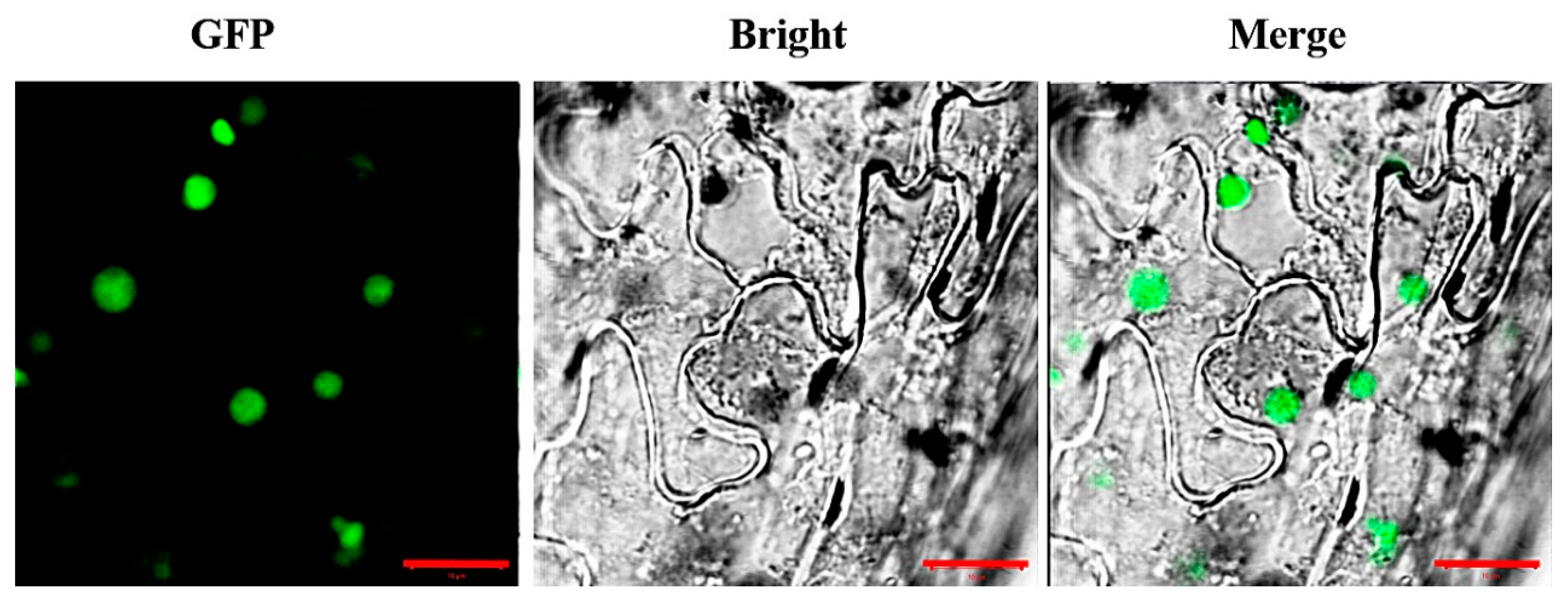
3.4. Subcellular Localization of AaCOI1

To investigate the subcellular localization of AaCOI1, a cassette of AaCOI1::GFP fusion driven by 35S promoter was transformed into N. benthamiana for transient expression analysis. After 48 h post infiltration, the leaf discs were collected for confocal microscopy. The green florescence protein (GFP) signal was predominantly detected in the nuclei (Figure 4). The GFP without AaCOI1 was also introduced into N. benthamiana as the control, and the GFP protein was distributed ubiquitously inside the cells (Figure S1).

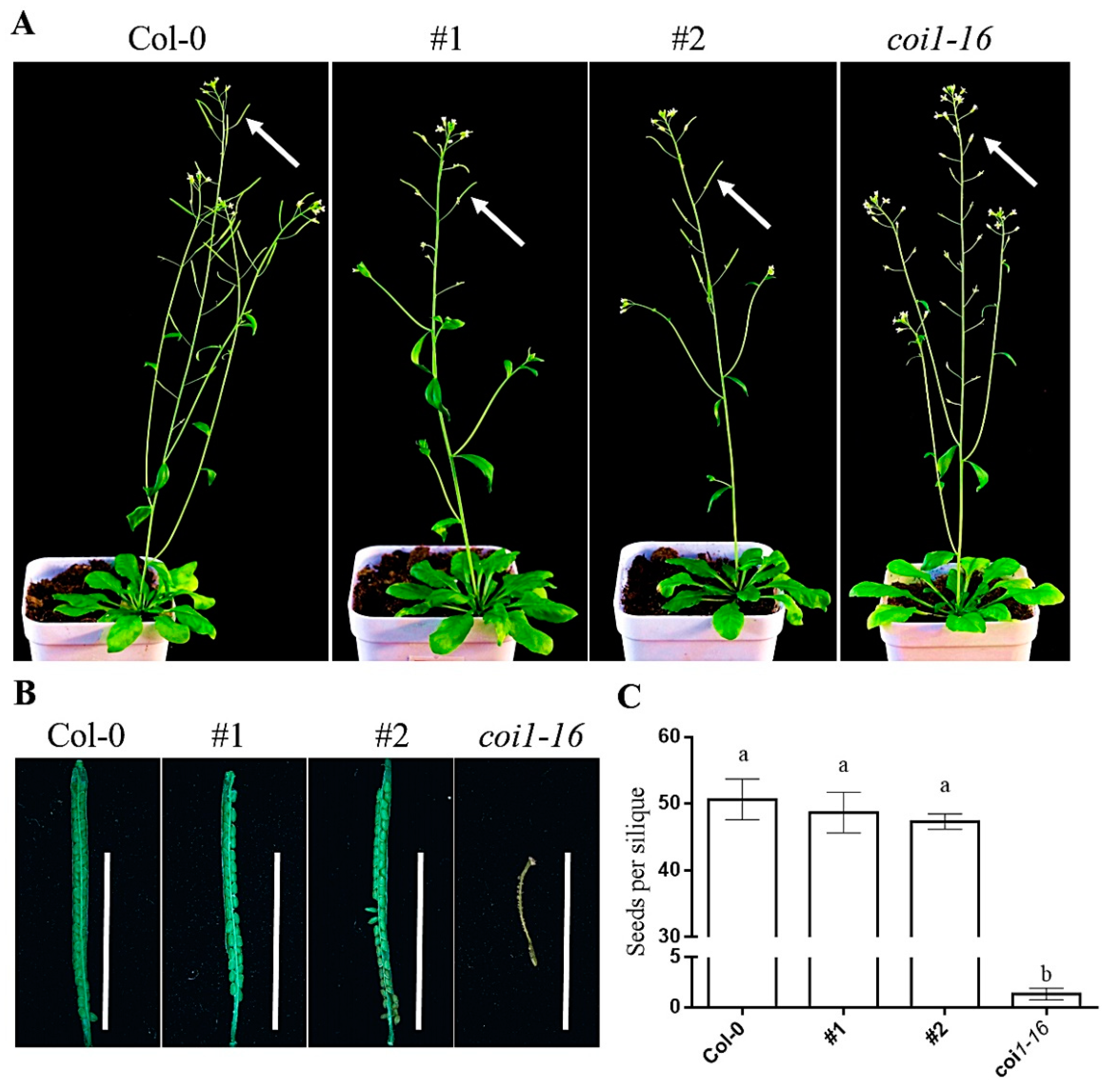
3.5. Ectopic Expression of AaCOI Partially Rescues Developmental Defects of Coi1-16

The ectopic expression of the AaCOI1 coding sequence was driven by the native promoter of Arabidopsis COI1. Two independent lines (#1, #2) ectopically expressing AaCOI1 in the background of coi1-16 were subjected for further analysis (Figure S2). The coi1-16 mutant showed abolished fertility resulting in seedless siliques under 22 °C. In the #1 and #2 transgenic lines, the seed number per silique (Figure 5) was higher than that of coi1-16, which was similar to wild-type plants, indicating that a successful complementation was imparted by ectopic expression of AaCOI1.

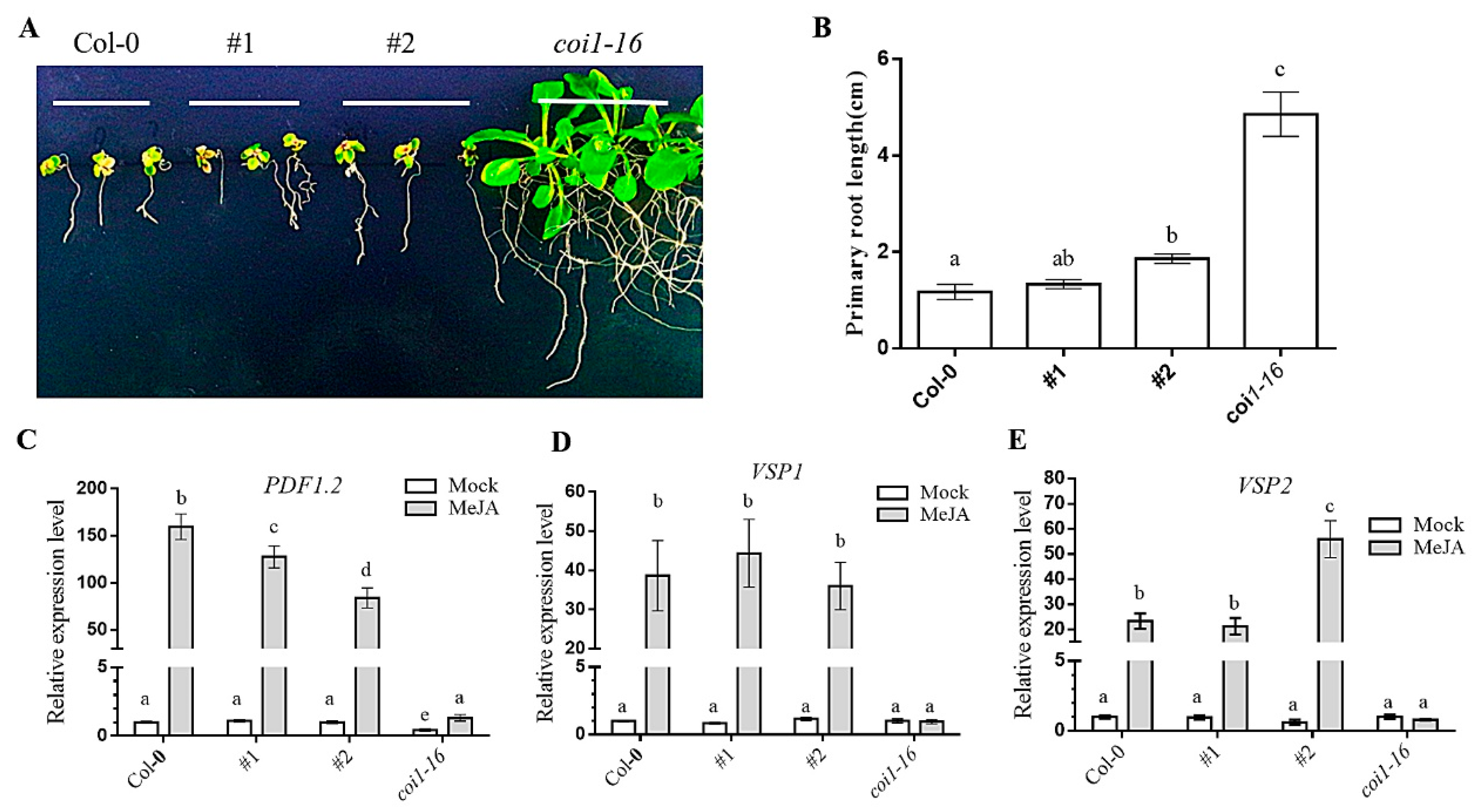
3.6. Ectopic Expression of AaCOI Restores JA-Sensitivity of Coi1-16

The ectopic expression of the AaCOI1 coding sequence was driven by the native promoter of Arabidopsis COI1. Two independent lines (#1, #2) ectopically expressing AaCOI1 in the background of coi1-16 were subjected for further analysis. The coi1-16 mutant showed normal root growth on MS supplemented with 50 μM MeJA (Figure 6A,B) and impaired induction of PDF1.2, VSP1, and VSP2 by MeJA (Figure 6C–E). In contrast, expression of AaCOI1 in #1 and #2 prioritized their JA sensitivity over that of coi1-16, which was indicated by dramatic inhibition of root growth (Figure 6A,B) and significant upregulation of all the three marker genes (Figure 6C–E).

3.7. Ectopic Expression of AaCOI1 Imparts Defense against Herbivory in Coi1-16

The ectopic expression of the AaCOI1 coding sequence was driven by the native promoter of Arabidopsis COI1. Two independent lines (#1, #2) ectopically expressing AaCOI1 in the background of coi1-16 were subjected for further analysis. The coi1-16 mutant showed attenuated resistance to larval feeding compared to the wild-type plants (Figure 7A), reduced inhibitive effects on larval growth (Figure 7B,C). In the #1 and #2 complement lines, the damage caused by herbivory was similar to that of Col-0 but dramatically reduced compared to that of coi1-16.

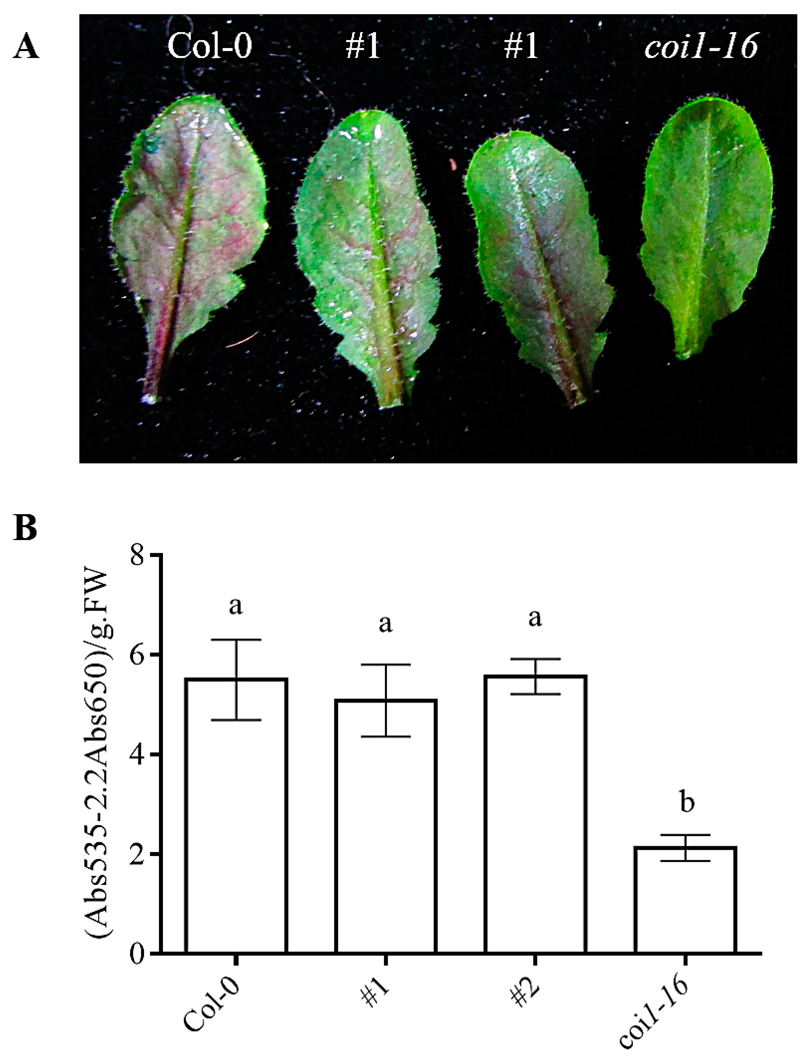
3.8. Ectopic Expression of AaCOI1 Restores the Accumulation of Anthocyanin in Coi1-16

The anthocyanin is an important secondary metabolite for its role as a protective flavonoid in planta and a nutritious diet supplement to human, which also have antioxidant activity and radical scavenging potential, anti-aging effects and antidiabetic potential [46,47,48]. JA and COI1 are important in regulation of secondary metabolites including anthocyanins [4,47,49]. The coil1-16 showed impaired accumulation of anthocyanin upon MeJA treatment. coi1-16 plants expressing AaCOI1 showed almost the same level of MeJA-induced anthocyanin as the wild-type plants (Figure 8A,B).

4. Discussion

Jasmonate-mediated signaling plays multiple roles in plant defense, development, reproduction, and metabolism. Due to its pleiotropic effects, a series of studies have attempted to use jasmonates as a growth regulator to divert the carbon flow to favor the accumulation of the desired metabolites [26,28,29,30,32,33]. Some others have applied genetic engineering to manipulate the JA-governed metabolism [22,23,24,25]. An F-box protein that is encoded by COI1 is a conserved module in the JA signaling pathway. In Arabidopsis thaliana, its role in JA perception and signaling cascade has been extensively studied. Using Arabidopsis thaliana as the model, particularly in the background of the loss-of-function mutant coi1, a plethora of COI1 homologs have been studied in nonmodel plant species, which further supported the conservation of COI1 and its homologs [10,11,12,13,14,15,16].
Artemisinin is currently the most effective drug so is the basis of ACT therapy. Artemisia annua is the main resource of artemisinin, which is not abundant enough for commercial use. The application of jasmonates has great potential to induce the accumulation of artemisinin and tuning of the JA signaling pathway, including the modulation of COI1 activity has also proven potent to regulate plant metabolomics and alter the secondary metabolic profiles [27,29,30,31,32,33,34,50]. In this study, we sought to identify the Artemisia orthologue of AtCOI1, and characterize its functions in JA-responsiveness, defense and regulation of secondary metabolites.
Based on our phylogenetic analysis, two COI1 homologues were identified in O. sativa and Z. mays, respectively, termed OsCOI1a/b and ZmCOI1a/b. By contrast, a single copy of COI1 paralog was identified in Artemisia annua and in another Asteraceae species, Helianthus annuus. Furthermore, the AaCOI1 and HaCOI1 were sequestered in the same clade but clearly separated from those in Arabidopsis thaliana, Z. mays, and O. sativa (Figure 1A). Structural analysis revealed all the COI1 paralogs possessed a F-box, and multiple LRR domains (Figure 1B). In detail, ZmCOI1a and OsCOI1a possess eight LRRs each, while ZmCOI1b and OsCOI1b have seven LRRs. AtCOI1 has six LRRs. In H. annuus, only six LRRs were identified while there were seven predicted LRRs in Artemisia annua. The results strongly suggested the COI1 signaling module had arisen before the separation of monocots and dicots.
In eukaryotes, spatial and temporal transcription of genes is largely dependent on their promoters. Motifs are characteristic cis-elements sequestered on the promoter, which provide binding sites for transcription factors and largely determines the transcription activity of the gene. The 2 kbp upstream of the coding sequence of AaCOI1 was designated as the promoter (Supplementary Data S4) and subjected to motif prediction by the Plantcare [38,39]. Interestingly, plenty of environment-responsive elements were enriched while few of development-related regulatory elements were identified. Those enriched motifs included MeJA-responsive elements, light-responsive elements, and elements conferring drought inducibility (Table S2). The enrichment of JA-responsive elements might be causative of the responsiveness of AaCOI1 to MeJA and wounding. In addition, the oscillation of AaCOI1 mRNA level in the mock group might be conferred by the light-responsive elements as the total illumination dosage increased in the time course. The AaCOI1 mRNA abundance were the highest in the inflorescence and AaCOI1 transcription level was also notably higher in leaves than that in the roots and the stems (Figure 3). Considering the artemisinin content and the expression level of most biosynthesis-related genes are the highest in the flowers and leaves [51], AaCOI1 expression seems to be positively correlated to artemisinin accumulation, which further supports the role of JA signaling in artemisinin biosynthesis. AaCOI1 was upregulated by both MeJA and wounding 6 h post-initial treatment, and by wounding 6 h post-initial treatment. To our surprise, at the time point of 9 h after initial exposure to MeJA, the mRNA level of AaCOI1 was decreased (Figure 3). Based on those results, transcription of AaCOI1 might be partially responsive to stimuli. In addition, the diverse motifs on the promoter gifted it flexibility in response to the environmental fluctuations.
Based on the 3D structure predicted by SWISS-MODEL [37], the AaCOI1 consisted of α-helix, β-sheet, and the uncharacterized random coil (Figure 2). In Arabidopsis and some other species, COI1 is mainly located in the nuclei [12], while the subcellular localization of AaCOI1 remains unknown. The amino acid residue sequence of AaCOI1 contains a predicted NLS (RKIKRLRIE) [52] at the position of 337 amino acid residues from the N-terminus and 7 LRRs [53] (Figure 1B). Transient expression of a fusion cassette of AaCOI1::GFP driven by 35S promoter in the N. benthamiana was carried out to determine the subcellular localization of AaCOI1. The results showed that AaCOI1 was located in the nuclei (Figure 4). The nuclear localization should be reasonable since COI interacts with JAZ proteins, which are transcription repressors in the nuclei.
To further testify the bona fide functions of AaCOI1 in plants, an expression cassette of AaCOI1 driven by the promoter of AtCOI1 was introduced into the Arabidopsis coi1-16 mutant, which is glabrous, male sterile, JA-insensitive, and impaired in plant defense responses [35,36]. In two independent transgenic lines (#1, #2), AaCOI1 complemented the loss-of-function of Arabidopsis COI1 to different extent (Figure 5, Figure 6 and Figure 7). In detail, ectopic expression of AaCOI1 in coi1-16 increased the fertility and seed yield of coi1-16 (Figure 5). The in vitro growth assay showed the inhibition of root growth in coi1-16 by MeJA was dramatically relieved by ectopic expression of AaCOI1 (Figure 6A,B). As JA is a potent elicitor inducing elevation of JA-responsive marker genes (PDF1.2, VSP1, VSP2 etc.) [54,55], whether the coi1-16 mutants expressing AaCOI1 could respond to MeJA treatment on molecular level was investigated. The results showed that introduction of AaCOI1 to coi1-16 restored the JA-induced expression of all the three marker genes (Figure 6C–E). Furthermore, the susceptibility to herbivory of coi1-16 was almost fully complemented by AaCOI1, which was indicated by the leaf damage and larval growth. Interestingly, the capability of complementation of AaCOI in coi1-16 was statistically different. This might be due to the variation of insertion loci resulting different expression level of the introduced AaCOI1. Also, we could not exclude the possibility that AaCOI1 protein level was not as high as that of native AtCOI1 in Arabidopsis plants as the codon preference varies across species [56].
Anthocyanin is a flavonoid that is involved in plant fertility and stress responses [57,58]. The beneficial effects of anthocyanin to human has also been documented [59,60,61,62]. In Arabidopsis and maize (Z. mays), JA-signaling promotes the biosynthesis of anthocyanin dependent of COI1 [47,48,49,63]. To investigate the potential of AaCOI1 in regulation of JA-induced accumulation of anthocyanin, MeJA was applied to Arabidopsis plants in Col-0, #1, #2, or coi1-16 background respectively. The total content of anthocyanin was measured by spectrometry [43]. Interestingly, both #1 and #2 transgenic lines showed no difference in terms of levels of anthocyanin to the wild-type control, indicating the full complementation of coi1 by ectopic expression of AaCOI1. Unlike the results of complementation assays in development, JA sensitivity, and defense response, basal expression level of AaCOI1 might be adequate to exert its function in biosynthesis of secondary metabolites. As has been evidenced, upregulated expression level of abscisic acid receptor led to increased artemisinin content [56]. Considering that the anthocyanin is a universal metabolite and COI1-dependent JA-signaling is a conserved pathway in different plant species, our results offered a novel strategy for future genetic improvement in Artemisia species to favor the secondary metabolites by tailoring the JA signaling pathway.

5. Conclusions

Our study identified AaCOI1, which encodes a F-box and LRR-domain containing protein, as a functional paralog of the jasmonate receptor Arabidopsis COI1. Expression of AaCOI1 was responsive to MeJA and wounding. AaCOI1 was localized in the nuclei. In the background of coi1 null-mutant, ectopic expression of AaCOI1 driven by AtCOI1 native promoter partially restored JA sensitivity in root growth and gene induction, and defense to herbivory, but fully complemented the phenotypes in the fertility and JA-induced anthocyanin accumulation. Our results should be insightful by offering a phytohormone-related target for future genetic improvement to modulate development, defense, and total yield in an important medicinal plant, the Artemisia annua L.

Supplementary Materials

The following are available online at https://0-www-mdpi-com.brum.beds.ac.uk/2073-4425/11/2/221/s1, Figure S1: Subcellular localization of GFP without AaCOI1, Figure S2: Verification of transgenic lines, Table S1: The primers used for constructs and qPCR in the present study, Table S2, Cis-elements identified in the promoter of AaCOI1, Data_S1: Amino acid sequence of AaCOI1, Data S2: Native promoter of AtCOI1, Data S3: AaCOI1 CDS, Data S4: The 2000bp sequence designated as the promoter of AaCOI1.

Author Contributions

M.X. designed the study and composed the manuscript. J.W. and R.L. performed the experiments and processed the data. X.G. assisted in the bioinformatics analysis. J.C. and Y.D. provided insightful suggestions during the submission. All authors have read and agreed to the published version of the manuscript.

Funding

The study was supported by Shandong University Postdoctoral Program (61200089962101) and the Innovation Program for Agronomic Science of Hunan Province (2019ZYC01-1).

Conflicts of Interest

The authors declare no conflicts of interest.

References

  1. Wasternack, C.; Kombrink, E. Jasmonates: Structural requirements for lipid-derived signals active in plant stress responses and development. ACS Chem. Biol. 2010, 5, 63–77. [Google Scholar] [CrossRef]
  2. Fonseca, S.; Chini, A.; Hamberg, M.; Adie, B.; Porzel, A.; Kramell, R.; Miersch, O.; Wasternack, C.; Solano, R. (+)-7-iso-Jasmonoyl-l-isoleucine is the endogenous bioactive jasmonate. Nat. Chem. Biol. 2009, 5, 344–350. [Google Scholar] [CrossRef]
  3. Wasternack, C.; Song, S. Jasmonates: Biosynthesis, metabolism, and signaling by proteins activating and repressing transcription. J. Exp. Bot. 2017, 68, 1303. [Google Scholar] [CrossRef]
  4. Wasternack, C.; Strnad, M. Jasmonates are signals in the biosynthesis of secondary metabolitesPathways, transcription factors and applied aspectsA brief review. New Biotechnol. 2019, 48, 1–11. [Google Scholar] [CrossRef]
  5. Acosta, I.F.; Farmer, E.E. Jasmonates. Arabidopsis Book 2010, 8, e0129. [Google Scholar] [CrossRef]
  6. Feys, B.; Benedetti, C.E.; Penfold, C.N.; Turner, J.G. Arabidopsis mutants selected for resistance to the phytotoxin coronatine are male sterile, insensitive to methyl Jasmonate, and resistant to a bacterial pathogen. Plant Cell 1994, 6, 751–759. [Google Scholar] [CrossRef]
  7. Xie, D.; Feys, B.F.; James, S.; Nieto-Rostro, M.; Turner, J.G. COI1: An Arabidopsis gene required for Jasmonate-regulated defense and fertility. Science 1998, 280, 1091–1094. [Google Scholar] [CrossRef]
  8. Yan, J.; Zhang, C.; Gu, M.; Bai, Z.; Zhang, W.; Qi, T.; Cheng, Z.; Peng, W.; Luo, H.; Nan, F.; et al. The Arabidopsis CORONATINE INSENSITIVE1 protein is a Jasmonate receptor. Plant Cell 2009, 21, 2220–2236. [Google Scholar] [CrossRef]
  9. Sheard, L.B.; Tan, X.; Mao, H.; Withers, J.; Ben-Nissan, G.; Hinds, T.R.; Kobayashi, Y.; Hsu, F.; Sharon, M.; Browse, J.; et al. Jasmonate perception by inositol-phosphate-potentiated COI1-JAZ co-receptor. Nature 2010, 468, 400–405. [Google Scholar] [CrossRef]
  10. Monte, I.; Ishida, S.; Zamarreño, A.M.; Hamberg, M.; Franco-Zorrilla, J.M.; García-Casado, G.; Gouhier-Darimont, C.; Reymond, P.; Takahashi, K.; García-Mina, J.M.; et al. Ligand-Receptor co-evolution shaped the jasmonate pathway in land plants. Nat. Chem. Biol. 2018, 14, 480–488. [Google Scholar] [CrossRef]
  11. Wang, Z.; Dai, L.; Jiang, Z.; Peng, W.; Zhang, L.; Wang, G.; Xie, D. GmCOI1, a soybean F-box protein gene, shows ability to mediate jasmonate-regulated plant defense and fertility in Arabidopsis. Mol. Plant Microbe Interact. MPMI 2005, 18, 1285. [Google Scholar] [CrossRef]
  12. Liao, Y.; Wei, J.; Xu, Y.; Zhang, Z. Cloning, expression and characterization of COI1 gene (AsCOI1) from Aquilaria sinensis (Lour.) Gilg. Acta Pharm. Sin. B 2015, 5, 473–481. [Google Scholar] [CrossRef]
  13. Lee, H.Y.; Seo, J.; Cho, J.H.; Jung, H.; Kim, J.; Lee, J.S.; Rhee, S.; Do Choi, Y. Oryza sativa COI homologues restore jasmonate signal transduction in Arabidopsis coi1-1 mutants. PLoS ONE 2013, 8, e52802. [Google Scholar] [CrossRef]
  14. Lee, S.H.; Sakuraba, Y.; Lee, T.; Kim, K.W.; An, G.; Lee, H.Y.; Paek, N.C. Mutation of Oryza sativa CORONATINE INSENSITIVE 1b (OsCOI1b) delays leaf senescence. J. Integr. Plant Biol. 2015, 57, 562–576. [Google Scholar] [CrossRef]
  15. Schubert, R.; Grunewald, S.; von Sivers, L.; Hause, B. Effects of Jasmonate on ethylene function during the development of tomato stamens. Plants 2019, 8, 277. [Google Scholar] [CrossRef]
  16. Bai, J.; Wang, Y.; Wang, P.; Yuan, S.; Gao, J.; Duan, W.; Wang, N.; Zhang, F.; Zhang, W.; Qin, M.; et al. Genome-Wide identification and analysis of the COI gene family in wheat (Triticum aestivum L.). BMC Genom. 2018, 19, 754. [Google Scholar] [CrossRef]
  17. Tu, Y. The discovery of Artemisinin (qinghaosu) and gifts from Chinese medicine. Nat. Med. 2011, 17, 1217–1220. [Google Scholar] [CrossRef]
  18. Xiang, M.; Chen, Z.; He, L.; Xiong, G.; Lu, J. Transcription profiling of artemisinin-treated diabetic nephropathy rats using high-throughput sequencing. Life Sci. 2019, 219, 353–363. [Google Scholar] [CrossRef]
  19. Kumari, K.; Keshari, S.; Sengupta, D.; Sabat, S.C.; Mishra, S.K. Transcriptome analysis of genes associated with breast cancer cell motility in response to Artemisinin treatment. BMC Cancer 2017, 17, 858. [Google Scholar] [CrossRef]
  20. Wong, Y.K.; Xu, C.; Kalesh, K.A.; He, Y.; Lin, Q.; Wong, W.S.F.; Shen, H.; Wang, J. Artemisinin as an anticancer drug: Recent advances in target profiling and mechanisms of action. Med. Res. Rev. 2017, 37, 1492–1517. [Google Scholar] [CrossRef]
  21. Zhang, B. Artemisinin-Derived dimers as potential anticancer agents: Current developments, action mechanisms, and structure-activity relationships. Arch. Pharm. 2019, 353. [Google Scholar] [CrossRef] [PubMed]
  22. Larson, T.R.; Branigan, C.; Harvey, D.; Penfield, T.; Bowles, D.; Graham, I.A. A survey of artemisinic and dihydroartemisinic acid contents in glasshouse and global field-grown populations of the artemisinin-producing plant Artemisia annua L. Ind. Crops Prod. 2013, 45, 1–6. [Google Scholar] [CrossRef]
  23. Ma, D.M.; Wang, Z.; Wang, L.; Alejos-Gonzales, F.; Sun, M.A.; Xie, D.Y. A Genome-Wide scenario of terpene pathways in self-pollinated Artemisia annua. Mol. Plant 2015, 8, 1580–1598. [Google Scholar] [CrossRef] [PubMed]
  24. Shen, Q.; Zhang, L.; Liao, Z.; Wang, S.; Yan, T.; Shi, P.; Liu, M.; Fu, X.; Pan, Q.; Wang, Y.; et al. The genome of Artemisia annua provides insight into the evolution of Asteraceae family and Artemisinin biosynthesis. Mol. Plant 2018, 11, 776–788. [Google Scholar] [CrossRef] [PubMed]
  25. Xie, D.; Ma, D.; Judd, R.; Jones, A.L. Artemisinin biosynthesis in Artemisia annua and metabolic engineering: Questions, challenges, and perspectives. Phytochem. Rev. 2016, 15, 1093–1114. [Google Scholar] [CrossRef]
  26. Pu, G.; Ma, D.; Chen, J.; Ma, L.; Wang, H.; Li, G.; Ye, H.; Liu, B. Salicylic acid activates Artemisinin biosynthesis in Artemisia annua L. Plant Cell Rep. 2009, 28, 1127–1135. [Google Scholar] [CrossRef] [PubMed]
  27. Maes, L.; Van Nieuwerburgh, F.C.; Zhang, Y.; Reed, D.W.; Pollier, J.; Vande, C.S.; Inze, D.; Covello, P.S.; Deforce, D.L.; Goossens, A. Dissection of the phytohormonal regulation of trichome formation and biosynthesis of the antimalarial compound Artemisinin in Artemisia annua plants. New Phytol. 2011, 189, 176–189. [Google Scholar] [CrossRef]
  28. Banyai, W.; Mii, M.; Supaibulwatana, K. Enhancement of artemisinin content and biomass in Artemisia annua by exogenous GA3 treatment. Plant Growth Regul. 2011, 63, 45–54. [Google Scholar] [CrossRef]
  29. Guo, X.; Yang, X.; Yang, R.; Zeng, Q. Salicylic acid and methyl jasmonate but not Rose Bengal enhance Artemisinin production through invoking burst of endogenous singlet oxygen. Plant Sci. 2010, 178, 390–397. [Google Scholar] [CrossRef]
  30. Dangash, A.; Pandya, N.; Bharillya, A.; Jhala, A.; Jain, D. Impact of exogenous elicitors on Artemisinin production and trichome density in Artemisia annua L. under subtropical conditions. Not. Sci. Biol. 2014, 6. [Google Scholar] [CrossRef]
  31. Ikram, N.K.B.K.; Simonsen, H.T. A Review of Biotechnological Artemisinin Production in Plants. Front. Plant Sci. 2017, 8. [Google Scholar] [CrossRef] [PubMed]
  32. Wang, H.; Ma, C.; Li, Z.; Ma, L.; Wang, H.; Ye, H.; Xu, G.; Liu, B. Effects of exogenous methyl Jasmonate on Artemisinin biosynthesis and secondary metabolites in Artemisia annua L. Ind. Crops Prod. 2010, 31, 214–218. [Google Scholar] [CrossRef]
  33. Xiang, L.; Zhu, S.; Zhao, T.; Zhang, M.; Liu, W.; Chen, M.; Lan, X.; Liao, Z. Enhancement of Artemisinin content and relative expression of genes of artemisinin biosynthesis in Artemisia annua by exogenous MeJA treatment. Plant Growth Regul. 2015, 75, 435–441. [Google Scholar] [CrossRef]
  34. Hao, X.; Zhong, Y.; Fu, X.; Lv, Z.; Shen, Q.; Yan, T.; Shi, P.; Ma, Y.; Chen, M.; Lv, X.; et al. Transcriptome analysis of genes associated with the Artemisinin biosynthesis by Jasmonic acid treatment under the light in Artemisia annua. Front. Plant Sci. 2017, 8. [Google Scholar] [CrossRef] [PubMed]
  35. Adie, B.A.T.; Pérez-Pérez, J.; Pérez-Pérez, M.M.; Godoy, M.; Sánchez-Serrano, J.; Schmelz, E.A.; Solano, R. ABA is an essential signal for plant resistance to pathogens affecting JA biosynthesis and the activation of defenses in Arabidopsis. Plant Cell 2007, 19, 1665–1681. [Google Scholar] [CrossRef] [PubMed]
  36. Ellis, C.; Karafyllidis, I.; Turner, J.G. Constitutive activation of jasmonate signaling in an Arabidopsis mutant correlates with enhanced resistance to Erysiphe cichoracearum, Pseudomonas syringae, and Myzus persicae. Mol. Plant Microbe Interact. MPMI 2002, 15, 1025. [Google Scholar] [CrossRef] [PubMed]
  37. Waterhouse, A.; Bertoni, M.; Bienert, S.; Studer, G.; Tauriello, G.; Gumienny, R.; Heer, F.T.; de Beer, T.A.P.; Rempfer, C.; Bordoli, L.; et al. SWISS-MODEL: Homology modelling of protein structures and complexes. Nucleic Acids Res. 2018, 46, W296–W303. [Google Scholar] [CrossRef]
  38. Lescot, M.; Déhais, P.; Thijs, G.; Marchal, K.; Moreau, Y.; Van de Peer, Y.; Rouzé, P.; Rombauts, S. PlantCARE, a database of plant cis-acting regulatory elements and a portal to tools for in silico analysis of promoter sequences. Nucleic Acids Res. 2002, 30, 325–327. [Google Scholar] [CrossRef]
  39. Rombauts, S.; Dehais, P.; Van Montagu, M.; Rouze, P. PlantCARE, a plant cis-acting regulatory element database. Nucleic Acids Res. 1999, 27, 295–296. [Google Scholar] [CrossRef]
  40. Ishimaru, Y.; Hayashi, K.; Suzuki, T.; Fukaki, H.; Prusinska, J.; Meester, C.; Quareshy, M.; Egoshi, S.; Matsuura, H.; Takahashi, K.; et al. Jasmonic acid inhibits auxin-induced lateral rooting independently of the CORONATINE INSENSITIVE1 receptor. Plant Physiol. 2018, 177, 1704–1716. [Google Scholar] [CrossRef]
  41. Xu, F.; Copeland, C.; Li, X. Protein immunoprecipitation using Nicotiana benthamiana transient expression system. Bio Protoc. 2015, 5. [Google Scholar] [CrossRef]
  42. Xu, F.; Kapos, P.; Cheng, Y.T.; Li, M.; Zhang, Y.; Li, X. NLR-Associating transcription factor bHLH84 and its paralogs function redundantly in plant immunity. PLoS Pathog. 2014, 10, e1004312. [Google Scholar] [CrossRef] [PubMed]
  43. Livak, K.J.; Schmittgen, T.D. Analysis of relative gene expression data using real-time quantitative PCR and the 2-ΔΔCT method. Methods 2001, 25, 402–408. [Google Scholar] [CrossRef] [PubMed]
  44. Li, S.; Wang, W.; Gao, J.; Yin, K.; Wang, R.; Wang, C.; Petersen, M.; Mundy, J.; Qiu, J. MYB75 phosphorylation by MPK4 Is required for light-induced Anthocyanin accumulation in Arabidopsis. Plant Cell 2016, 28, 2866–2883. [Google Scholar] [CrossRef] [PubMed]
  45. Winter, D.; Vinegar, B.; Nahal, H.; Ammar, R.; Wilson, G.V.; Provart, N.J. An “Electronic fluorescent pictograph” browser for exploring and analyzing large-scale biological data sets. PLoS ONE 2007, 2, e718. [Google Scholar] [CrossRef] [PubMed]
  46. Gou, J.; Felippes, F.F.; Liu, C.; Weigel, D.; Wang, J. Negative regulation of Anthocyanin biosynthesis in Arabidopsis by a miR156-targeted SPL transcription factor. Plant Cell 2011, 23, 1512–1522. [Google Scholar] [CrossRef]
  47. Gan, Y.; Li, H.; Xie, Y.; Wu, W.; Li, M.; Wang, X.; Huang, J. THF1 mutations lead to increased basal and wound-induced levels of oxylipins that stimulate anthocyanin biosynthesis via COI1 signaling inArabidopsis. J. Integr. Plant Biol. 2014, 56, 916–927. [Google Scholar] [CrossRef]
  48. Devoto, A.; Ellis, C.; Magusin, A.; Chang, H.; Chilcott, C.; Zhu, T.; Turner, J.G. Expression profiling reveals COI1 to be a key regulator of genesinvolved in wound-and methyl Jasmonate-induced secondary metabolism, defence, and hormone interactions. Plant Mol. Biol. 2005, 58, 497–513. [Google Scholar] [CrossRef]
  49. Ai, Y.; Zhu, Z. Melatonin antagonizes Jasmonate-triggered Anthocyanin biosynthesis in Arabidopsis thaliana. J. Agric. Food Chem. 2018, 66, 5392–5400. [Google Scholar] [CrossRef]
  50. Ma, Y.N.; Xu, D.B.; Li, L.; Zhang, F.; Fu, X.Q.; Shen, Q.; Lyu, X.Y.; Wu, Z.K.; Pan, Q.F.; Shi, P.; et al. Jasmonate promotes Artemisinin biosynthesis by activating the TCP14-ORA complex in Artemisia annua. Sci. Adv. 2018, 4, s9357. [Google Scholar] [CrossRef]
  51. Olofsson, L.; Engström, A.; Lundgren, A.; Brodelius, P.E. Relative expression of genes of terpene metabolism in different tissues of Artemisia annua L. BMC Plant Biol. 2011, 11, 45. [Google Scholar] [CrossRef] [PubMed]
  52. Kosugi, S.; Hasebe, M.; Tomita, M.; Yanagawa, H. Systematic identification of cell cycle-dependent yeast nucleocytoplasmic shuttling proteins by prediction of composite motifs. Proc. Natl. Acad. Sci. USA 2009, 106, 10171–10176. [Google Scholar] [CrossRef] [PubMed]
  53. Bej, A.; Sahoo, B.R.; Swain, B.; Basu, M.; Jayasankar, P.; Samanta, M. LRRsearch: An asynchronous server-based application for the prediction of leucine-rich repeat motifs and an integrative database of NOD-like receptors. Comput. Biol. Med. 2014, 53, 164–170. [Google Scholar] [CrossRef] [PubMed]
  54. Utsugi, S.; Sakamoto, W.; Murata, M.; Motoyoshi, F. Arabidopsis thaliana vegetative storage protein (VSP) genes: Gene organization and tissue-specific expression. Plant Mol. Biol. 1998, 38, 565–576. [Google Scholar] [CrossRef] [PubMed]
  55. Hiraoka, Y.; Kawamata, K.; Haraguchi, T.; Chikashige, Y. Codon usage bias is correlated with gene expression levels in the fission yeast Schizosaccharomyces pombe. Genes Cells 2009, 14, 499–509. [Google Scholar] [CrossRef] [PubMed]
  56. Zhang, F.; Lu, X.; Lv, Z.; Zhang, L.; Zhu, M.; Jiang, W.; Wang, G.; Sun, X.; Tang, K. Overexpression of the Artemisia orthologue of ABA receptor, AaPYL9, enhances ABA sensitivity and improves Artemisinin content in Artemisia annua L. PLoS ONE 2013, 8, e56697. [Google Scholar] [CrossRef]
  57. Winkel-Shirley, B. Biosynthesis of flavonoids and effects of stress. Curr. Opin. Plant Biol. 2002, 5, 218–223. [Google Scholar] [CrossRef]
  58. Gould, K.S. Nature’s swiss army knife: The diverse protective roles of Anthocyanins in leaves. J. Biomed. Biotechnol. 2004, 2004, 314–320. [Google Scholar] [CrossRef]
  59. Matsumoto, H.; Nakamura, Y.; Hirayama, M.; Yoshiki, Y.; Okubo, K. Antioxidant activity of black currant Anthocyanin aglycons and their glycosides measured by chemiluminescence in a neutral pH region and in human plasma. J. Agric. Food Chem. 2002, 50, 5034–5037. [Google Scholar] [CrossRef]
  60. Jing, P.; Bomser, J.A.; Schwartz, S.J.; He, J.; Magnuson, B.A.; Giusti, M.M. Structure-Function relationships of Anthocyanins from various Anthocyanin-rich extracts on the inhibition of colon cancer cell growth. J. Agric. Food Chem. 2008, 56, 9391–9398. [Google Scholar] [CrossRef]
  61. Hwang, J.; Kim, E.; Lee, S.; Kim, Y.; Moon, S.; Jeon, B.; Sung, S.; Kim, E.; Park, P. Antioxidant Activity and protective effect of Anthocyanin oligomers on H2O2-triggered G2/M arrest in retinal cells. J. Agric. Food Chem. 2012, 60, 4282–4288. [Google Scholar] [CrossRef] [PubMed]
  62. Taverniti, V.; Fracassetti, D.; Del Bo, C.; Lanti, C.; Minuzzo, M.; Klimis-Zacas, D.; Riso, P.; Guglielmetti, S. Immunomodulatory effect of a wild blueberry Anthocyanin-rich extract in human Caco-2 intestinal cells. J. Agric. Food Chem. 2014, 62, 8346–8351. [Google Scholar] [CrossRef] [PubMed]
  63. Fu, J.; Liu, L.; Liu, Q.; Shen, Q.; Wang, C.; Yang, P.; Zhu, C.; Wang, Q. ZmMYC2 exhibits diverse functions and enhances JA signaling in transgenic Arabidopsis. Plant Cell Rep. 2019, 39, 1–16. [Google Scholar] [CrossRef] [PubMed]
Figure 1. Phylogenetic tree COI1 paralogs (A) in Artemisia annua, Helianthus annuus, Oryza sativa, Zea mays and Arabidopsis thaliana, and their sequences (B). The relationships were analyzed for deduced full-length amino acid sequences using MEGA7 by using the Maximum Likelihood (ML) method with Partial deletion and Poisson model, and a Bootstrap test of 1000 replicates for internal branch reliability. Bootstrap values were shown near the nodes. Abbreviations: Artemisia annua (Aa), Helianthus annuus (Ha), Oryza sativa (Os), Zea mays (Zm), and Arabidopsis thaliana (At).
Genes 11 00221 g001
Figure 2. The predicted AaCOI1 3D mode (A) and the contribution of amino acid residues to the structure (B). The LRRs and F-box are indicated in the figure. The LRRs are marked by red lines, while the F-box is marked by a yellow line (B).
Genes 11 00221 g002
Figure 3. Transcription analysis of AaCOI in different tissues (A) and in response to wounding and MeJA treatment (B). The error bar represents the mean (±standard error) of three independent biological replicates. Columns marked with different letters (ac) are significantly different from the others (ANOVA followed by Tukey’s multiple comparison test (p < 0.05)); any points of the line chart marked with an asterisk are significantly different (Student’s t-test; ** p < 0.01). Error bars indicate SD.
Genes 11 00221 g003
Figure 4. Subcellular localization of AaCOI1. GFP, GFP fluorescence; Bright, bright-field image; Merge, merged GFP and bright-field image. The scale bar represents 10 μm.
Genes 11 00221 g004
Figure 5. Phenotypes of Col-0, complemented lines #1 and #2, and coi1-16 mutant. Overall appearance of Arabidopsis thaliana plants of different backgrounds (A); close-up of fully developed siliques about 10 days after pollination (B); the fertility was determined by counting the seeds number per silique (C) (N > 10). The error bar represents the mean (±standard error) of 10 independent biological replicates. Columns marked with different letters (ab) are significantly different from the others (ANOVA followed by Tukey’s multiple comparison test (p < 0.05)).
Genes 11 00221 g005
Figure 6. Sensitivity to MeJA of Col-0, complemented lines #1 and #2, and coi1-16 mutant. Growth of Arabidopsis thaliana seedlings on MS supplemented with 50 μM MeJA (A); primary root length (N > 10) of seedlings after growing on MS supplemented with 50 μM MeJA for 10 days (B); the expression of COI1-dependent and JA-responsive genes in Arabidopsis seedlings of different backgrounds ((C), PDF1.2; (D), VSP1; (E), VSP2). The error bar represents the mean (±standard error) of at least three biological replicates. Columns marked with different letters (ac) are significantly different from the others (ANOVA followed by Tukey’s multiple comparison test (p < 0.05)).
Genes 11 00221 g006
Figure 7. The defense response to herbivory in Col-0, #1, #2, and coi1-16 mutant. The overall appearance of Arabidopsis plants after 10 days of feeding by larvae (A); the overall appearance of larvae after feeding on Arabidopsis for 10 days (B); the fresh weight per larva after 10 days of feeding on Arabidopsis plants were measured (C). The error bar represents the mean (±standard error) of at least 20 biological replicates. Columns marked with different letters (ac) are significantly different from the others (ANOVA followed by Tukey’s multiple comparison test (p < 0.05)).
Genes 11 00221 g007
Figure 8. The anthocyanin accumulation in Col-0, #1, #2, and coi1-16 mutant. The overall appearance of Arabidopsis plants after treatment of 50 μM MeJA for 48 h (A); the accumulation content measured from 1 g leaf tissue from MeJA-treated Col-0, #1, #2, coi1-16 (B). The error bar represents the mean (±standard error) of at least three biological replicates. Columns marked with different letters (a,b) are significantly different from the others (ANOVA followed by Tukey’s multiple comparison test (p < 0.05)).
Genes 11 00221 g008

