Case Report

Novel Mutation in CRYBB3 Causing Pediatric Cataract and Microphthalmia

by
Olivia A. Zin
1,
Luiza M. Neves
2,
Fabiana L. Motta
3,
Dafne D. G. Horovitz
2,
Leticia Guida
2,
Leonardo H. F. Gomes
2,
Daniela P. Cunha
2,
Ana Paula S. Rodrigues
1,
Andrea A. Zin
2,4,
Juliana M. F. Sallum
1,3 and
Zilton F. M. Vasconcelos
2,*
1
Department of Ophthalmology, Universidade Federal de São Paulo, Sao Paulo 04039-032, Brazil
2
Instituto Fernandes Figueira-Fundação Oswaldo Cruz, Rio de Janeiro 22250-020, Brazil
3
Instituto de Genética Ocular, São Paulo 04552-050, Brazil
4
Instituto Catarata Infantil, Rio de Janeiro 22250-040, Brazil
*
Author to whom correspondence should be addressed.
Submission received: 25 May 2021 / Revised: 19 June 2021 / Accepted: 28 June 2021 / Published: 13 July 2021

Abstract

:
Up to 25% of pediatric cataract cases are inherited, with half of the known mutant genes belonging to the crystallin family. Within these, crystallin beta B3 (CRYBB3) has the smallest number of reported variants. Clinical ophthalmological and genetic-dysmorphological evaluation were performed in three autosomal dominant family members with pediatric cataract and microphthalmia, as well as one unaffected family member. Peripheral blood was collected from all participating family members and next-generation sequencing was performed. Bioinformatics analysis revealed a novel missense variant c.467G>A/p.Gly156Glu in CRYBB3 in all family members with childhood cataract. This variant is classified as likely pathogenic by ACMG, and no previous descriptions of it were found in ClinVar, HGMD or Cat-Map. The only other mutation previously described in the fifth exon of CRYBB3 is a missense variant that causes a change in amino acid from the same 156th amino acid to arginine and has been associated with pediatric cataract and microphthalmia. To the best of our knowledge, this is the first time the c.467G>A/p.Gly156Glu variant is reported and the second time a mutation in CRYBB3 has been associated with microphthalmia.

1. Introduction

Cataract is defined as lens opacity that results in light scattering, reduced image clarity and visual acuity, as well as impaired contrast sensitivity [1,2]. In children, cataract may be congenital or acquired during childhood and can lead to irreversible visual impairment when it is not time-appropriately treated. Although it is a rare disease, with an overall prevalence ranging from 0.32 to 22.9 per 10,000 children (median 1.03) [2], it represents 21.3% of pediatric blindness worldwide, with higher frequencies in low- and middle-income countries [3]. Cataract in children must be diagnosed and managed early to avoid permanent vision impairment due to amblyopia.
Pediatric cataract can be isolated or associated with anterior chamber developmental anomalies, such as microcornea, microphthalmia and aniridia. They can also be related to multisystem genetic conditions, including chromosome disorders, developmental disorders or metabolic disorders [4]. Inherited congenital cataract comprises between 8.3 and 25% of cases [5], while causal genes have not been identified in one-third of the mapped loci [4]. About 37% of the known mutant genes in cataract families involve mutations in crystallin genes (1). There have been 11 mutations reported within the crystallin beta B3 (CRYBB3) [6]. Supplementary Table S1 describes previously reported mutations in CRYBB3, as well as phenotypes, family origins and inheritance patterns.
We aimed to determine the genetic cause of pediatric cataract related to microphthalmia in three members of a family with an autosomal dominant pattern of inheritance of pediatric cataract and microphthalmia phenotype using next-generation sequencing.

2. Materials and Methods

2.1. Study Design

A case report with clinical and next-generation sequencing of three members of the same autosomal dominant family (father and two children) with a history of pediatric cataract and microphthalmia, as well as one family member who did not present any ocular pathologies.

2.2. Clinical Evaluation

At evaluation, all three patients had already been submitted to cataract surgery, and detailed medical records were available for the two youngest. History of congenital TORCH infections (toxoplasmosis, rubella, cytomegalovirus, herpes simplex, syphilis, varicella zoster, zika), use of corticosteroids and ocular trauma were ruled out.
Ophthalmic exam with slit-lamp biomicroscopy, intraocular pressure (IOP), indirect ophthalmoscopy, ophthalmic ultrasonography and biometry for axial length measurement were performed in all individuals.
Genetic evaluation of family history and origins and pedigree, as well as existence of other systemic conditions, and clinic-morphological evaluation were performed by a clinical geneticist.

2.3. Genomic DNA Preparation

Peripheral blood samples were collected in EDTA tubes from the four family members. Genomic DNA was extracted from peripheral blood leucocytes using PureLink® Genomic DNA Mini Kit Thermofisher (USA), according to the manufacturer’s protocol. DNA concentration in samples were determined by fluorometry using Invitrogen Qubit® 4 Fluorometer. To assess DNA purity, a Spectrophotometer NanoDrop® 2000 was used to evaluate ratio of the absorbance at 260/280 nm (average of 1.90 for all samples) and at 260/230 nm (average of 1.91 for all samples). DNA samples were stored at 4 °C prior to use.

2.4. Library Preparation and Clinical Exome Sequencing

DNA libraries from both parents and children were prepared with 50 ng of DNA using Clinical Exome Solution V2® according to the manufacturer’s instructions (Sophia Genetics, Switzerland). This clinical exome solution covers 4493 genes related to the most common inherited diseases. Sequencing was performed using the NextSeq® 500 system (Illumina, San Diego, CA, USA) using a multiplex system with 16 samples per run with the NextSeq® kit. During library preparation, DNA fragments of 400 bp long on average were evaluated using the Bioanalyzer by Agilent.

2.5. Bioinformatics Analysis

Sequencing data were processed and analyzed using SOPHiA pipelines and DDM® software (Switzerland), and the genetic variant calls were performed against the reference sequence of hg19 from the University of California Santa Cruz (UCSC) Genome Browser. The analysis strategy started with a virtual panel based on Human Phenotype Ontology (HPO) where 658 genes were found related to cataract in general (Supplementary Table S2). Genogram trio analysis was performed on affected and unaffected family members using the trio analysis tools available on SOPHiA DDM®.
During variant interpretation, we considered allele frequency using the Exome Aggregation Consortium database (ExAC), 1000 Genomes Project database, gnomAD and ABraOM, an online archive of Brazilian mutations [7]. Twelve predictors were considered for pathogenicity: BayesDel_addAF, DANN, DEOGEN2, EIGEN, FATHMM-MKL, LIST-S2, M-CAP, MVP, MutationAssessor, MutationTaster, SIFT and PrimateAI. The clinical significance of variants was evaluated with ClinVar, Polymorphism database (dbSNP) and Human Gene Mutation Database (HGMD). Cat-Map, an online chromosome map and reference database for cataract in humans and mice [6], was also searched for previous variant descriptions and clinical associations. Variant naming was based on reference sequence NM_004076.

2.6. Sanger Sequencing
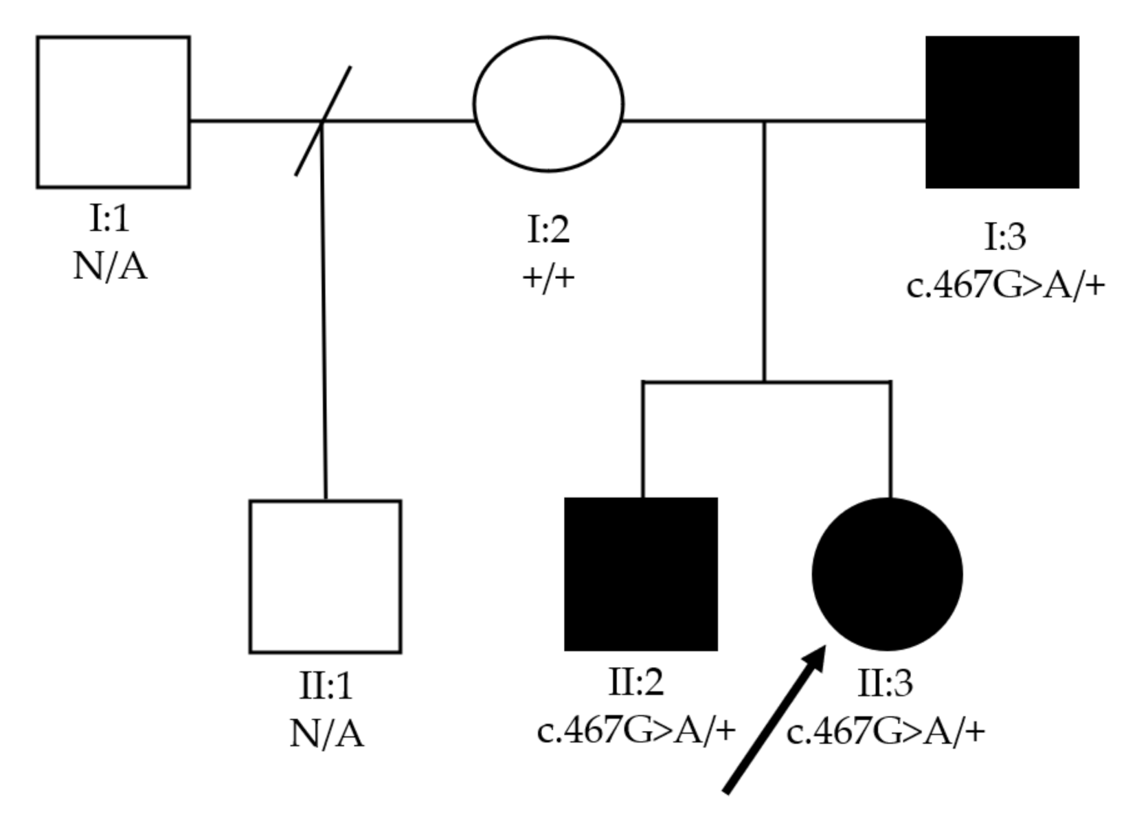
In order to confirm the variant identified by clinical exome analysis, PCR amplification and bi-directional direct Sanger sequencing were performed using the oligonucleotide primers 5′CCTCCTTGACCTCTGTTCTGG3′ and 5′ GGCACTGATTCTGTTTGGAGC3′. Briefly, primers and PCR products were purified using PureLink® (Invitrogen™) and sequenced on an automated sequencer (ABI 3730 Genetic analyzer, Applied Biosystems).

3. Results

Proband was examined at 3 months old and had an unremarkable gestational and peripartum history. An ophthalmological exam revealed nystagmus and bilateral anterior polar cataract, as well as small corneas (7 mm diameter OU) and a reduced axial length (16.06 mm OD, 16.13 mm OS) for her age. Ultrasonography showed no alterations other than lens opacification and a reduced axial length. A diagnosis of bilateral microphthalmia and congenital cataracts was made and lensectomy with anterior vitrectomy was performed on both eyes. Aphakic glaucoma developed four years after surgery on both eyes and was managed with topical antiglaucoma drugs. Figure 1 shows (a) small corneas compatible with the microphthalmia phenotype, as well as an (b) unremarkable fundoscopy.
Family history revealed that the proband’s father was diagnosed with bilateral cataracts at three years old, as well as microphthalmia. The eldest brother also presented with nystagmus and microphthalmia and was diagnosed with cataracts at 18 months old. Two years after lensectomy, the brother presented pain in the left eye. Ophthalmological examination revealed aphakia with a shallow anterior chamber and high intraocular pressure. A diagnosis of aqueous misdirection was made and a surgical approach indicated. Since both the father and eldest son were not initially operated on at our facilities, the records of their cataract phenotype prior to surgery were not available. At examination, the father was microphthalmic, with nystagmus and aphakic bilaterally. Table 1 summarizes the clinical findings of the three family members with cataract.
Next-generation sequencing revealed a novel missense variant c.467G>A/p.Gly156Glu in CRYBB3 in all family members with childhood cataract. No previous descriptions of it were found in ClinVar, HGMD or Cat-Map. Not only is p.Gly156 highly conserved among several species, the variant c.467G>A/p.Gly156Glu in CRYBB3 segregates accordingly in family members (Figure 2). The variant in the same codon c.466G>A (p.Gly156Arg) of CRYBB3, a consolidated gene associated with pediatric cataract, has been described in other ethnic groups, such as Chinese [8] and Turkish families [9]. In addition, c.467G>A/p.Gly156Glu is located in a well-established functional domain (beta/gamma crystallin ‘Greek key’4′) that is also absent from consulted controls. The variant’s pathogenicity is also supported by 11 of the 12 predictors, except PrimateAI. Considering the aforementioned, the variant c.467G>A/p.Gly156Glu in CRYBB3 is classified as likely pathogenic by ACMG criteria [10]. Figure 3 shows confirmation of the variant performed by Sanger sequencing, which was found in all affected family members and not in the healthy parent.

4. Discussion

To the best of our knowledge, this is the second time a mutation in CRYBB3 has been associated with microphthalmia. It is a novel, likely pathogenic variant presenting with an autosomal dominant pattern of inheritance.
Pediatric cataract is a phenotypically and genetically heterogeneous disorder and inherited traits showed interfamily and intrafamily variability [11]. To date, there are 39 cytogenetic loci known to be involved in pediatric cataract. About half of the known mutant genes in families with cataract involve mutations in crystallin genes [6].
Crystallins are the most abundant soluble proteins in the ocular lens (80–90%) and account for its optical transparency and high refractive index [12,13]. They are subdivided into alfa (40%), beta (35%) and gamma (25%) according to the order of their elution on gel exclusion chromatography [1,12,14]. The present study reports a novel mutation which disrupts the beta-crystallin Greek key functional domain, as is the case in most of the congenital cataract’s pathogenic missense mutations in this gene [15].
beta-crystallins can also be subdivided into four acidic (A1, A2, A3, A4) and three basic (B1, B2, B3) isoforms, depending on the isoelectric point and the terminal extension [16]. The least well studied is the crystallin beta B3 (CRYBB3) [17]. There are 9 mutations reported in CRYBB3, 23 in CRYBB1 and 35 in CRYBB2 [6].
Seventy percent of pediatric cataract are isolated [1], and, as expected, most of the CRYBB3 mutations reported in the Cat-Map are also isolated [6]. However, pediatric cataract is also associated with other ocular malformations in 15% of cases, including microphthalmia, aniridia, other anterior chamber developmental anomalies or retinal degenerations [1]. Another report of that same CRYBB3 missense variant c.466G>A (p.Gly156Arg) has included microphthalmia as an additional phenotype of congenital cataract [9].
The phenotype of all these c.466G>A (p.Gly156Arg) mutations was reported as bilateral nuclear cataract [8,9,18]. Conversely, in our present study, the novel c.467G>A (p.Gly156Glu) variant clinically manifested as bilateral anterior polar cataract with microphthalmia.
Interestingly, glaucoma also developed after surgery in the patient with microphthalmia and congenital cataract described by Sekeroglu et al. [9], as was seen in our proband. Glaucoma is a possible event after lensectomy in pediatric patients, its incidence varying between 10 and 58% [19]. According to Chak et al. [20], the age at which surgery is performed is the only factor associated with the development of glaucoma—the earlier surgery is performed, the higher the risk is. An association with microphthalmia and the development of glaucoma was not found. Similar mutations leading to the same complications raise the question as to whether the genotype of patients could be associated with the development of this complication. Are there genes only associated with alterations in the crystalline or could there be other anterior segment malformations associated with and responsible for the development of glaucoma? Further studies are necessary in order to evaluate such a hypothesis.
Genetic diagnosis of developmental eye disorders remains a challenge worldwide due to diverse phenotypes and genetic heterogeneity [18]. However, the evolving knowledge of genetics in medicine enables the phenotypic–genotypic correlation and a better understanding of the disease’s physiopathology. Identification of the causative mutations allows genetic counseling. The benefit regarding de novo mutations is even greater, since the pattern of inheritance and risk for other family members is impossible to predict from the pedigree.

5. Conclusions

Timely pediatric cataract surgical treatment is efficient and does not depend on molecular diagnosis. Nonetheless, the latter is important since it may provide knowledge toward the development of new therapeutic possibilities. Once the causative mechanism of the cataract is identified, the degenerative progressive damage could be delayed and surgical treatment avoided.
Therefore, we believe that our finding can contribute to better understand the role of the CRYBB3 protein in inherited childhood cataract associated with microphthalmia.

Supplementary Materials

The following are available online at https://0-www-mdpi-com.brum.beds.ac.uk/article/10.3390/genes12071069/s1, Table S1: Previously reported mutations in CRYBB3. Table S2: List of 658 genes related to cataract available at Human Phenotype Ontology.

Author Contributions

Conceptualization, O.A.Z., L.M.N., D.D.G.H., L.G., L.H.F.G., A.P.S.R., A.A.Z., J.M.F.S. and Z.F.M.V.; methodology, F.L.M., D.D.G.H., A.A.Z., J.M.F.S. and Z.F.M.V.; validation, O.A.Z., L.M.N., F.L.M., L.G., L.H.F.G., D.P.C. and Z.F.M.V.; formal analysis, O.A.Z., L.M.N., F.L.M., L.G., L.H.F.G., D.P.C. and Z.F.M.V.; investigation, L.G., L.H.F.G., D.P.C. and Z.F.M.V.; resources, A.A.Z. and Z.F.M.V.; data curation, F.L.M., L.G., L.H.F.G., D.P.C. and Z.F.M.V.; writing—original draft preparation, O.A.Z. and L.M.N.; writing—review and editing, O.A.Z., L.M.N., F.L.M., D.D.G.H., L.G., L.H.F.G., D.P.C., A.P.S.R., A.A.Z., J.M.F.S. and Z.F.M.V.; visualization, O.A.Z., L.M.N. and F.L.M. supervision, D.D.G.H., A.P.S.R., A.A.Z., J.M.F.S. and Z.F.M.V.; project administration, A.A.Z., J.M.F.S. and Z.F.M.V.; funding acquisition, D.D.G.H., L.G., L.H.F.G., A.A.Z. and Z.F.M.V. All authors have read and agreed to the published version of the manuscript.

Funding

This research was funded by the Brazilian National Program of Support to the Health Assistance of the Person with Deficiency (PRONAS/PCD), grant number 25000.193451/2019-21, granted in the year 2020.

Institutional Review Board Statement

The study was conducted according to the guidelines of the Declaration of Helsinki and approved by the Ethics Committee of INSTITUTO FERNANDES FIGUEIRA-IFF/FIOCRUZ-RJ/MS (protocol code 21444619.0.0000.5269), 17 October 2019. Approval from the Ethics Committee of UNIVERSIDADE FEDERAL DE SÃO PAULO- UNIFESP was also conceded under the protocol code 4.508.968, 25 January 2021.

Informed Consent Statement

Informed consent was obtained from all subjects involved in the study. Written informed consent was obtained from the patients to publish this paper.

Data Availability Statement

Data is available on the public online database CAT-MAP and from related publications and supplements. This data can be found here: https://cat-map.wustl.edu/.

Acknowledgments

Whole exome sequencing was performed at the platform of New Generation Sequencing of Nucleic Acids BH (RPT01E) of the Technological Networking Platforms Program of FIOCRUZ (PDTIS).

Conflicts of Interest

The authors declare no conflict of interest. The funders had no role in the design of the study; in the collection, analyses, or interpretation of data; in the writing of the manuscript; or in the decision to publish the results.

References

  1. Hejtmancik, J.F. Congenital cataracts and their molecular genetics. Semin. Cell Dev. Biol. 2008, 19, 134–149. [Google Scholar] [CrossRef] [PubMed] [Green Version]
  2. Sheeladevi, S.; Lawrenson, J.; Fielder, A.R.; Suttle, C.M. Global prevalence of childhood cataract: A systematic review. Eye 2016, 30, 1160–1169. [Google Scholar] [CrossRef] [PubMed] [Green Version]
  3. Kong, L.; Fry, M.; Al-Samarraie, M.; Gilbert, C.; Steinkuller, P.G. An update on progress and the changing epidemiology of causes of childhood blindness worldwide. J. Am. Assoc. Pediatr. Ophthalmol. Strabismus 2012, 16, 501–507. [Google Scholar] [CrossRef] [PubMed]
  4. Shiels, A.; Hejtmancik, J.F. Biology of Inherited Cataracts and Opportunities for Treatment. Annu. Rev. Vis. Sci. 2019, 5, 123–149. [Google Scholar] [CrossRef] [PubMed]
  5. Haargaard, B.; Wohlfahrt, J.; Fledelius, H.C.; Rosenberg, T.; Melbye, M. A nationwide Danish study of 1027 cases of congenital/infantile cataracts: Etiological and clinical classifications. Ophthalmology 2004, 111, 2292–2298. [Google Scholar] [CrossRef] [PubMed]
  6. Cat-Map Complete List. Available online: https://cat-map.wustl.edu/ (accessed on 31 March 2021).
  7. ABraOM: Brazilian Genomic Variants. Available online: http://abraom.ib.usp.br/ (accessed on 31 March 2021).
  8. Li, D.; Wang, S.; Ye, H.; Tang, Y.; Qiu, X.; Fan, Q.; Rong, X.; Liu, X.; Chen, Y.; Yang, J.; et al. Distribution of gene mutations in sporadic congenital cataract in a Han Chinese population. Mol. Vis. 2016, 22, 589–598. [Google Scholar] [PubMed]
  9. Sekeroglu, H.T.; Karaosmanoglu, B.; Taskiran, E.Z.; Kiper, P.O.S.; Alikasifoglu, M.; Boduroglu, K.; Coskun, T.; Utine, G.E. Molecular Etiology of Isolated Congenital Cataract Using Next-Generation Sequencing: Single Center Exome Sequencing Data from Turkey. Mol. Syndr. 2020, 11, 302–308. [Google Scholar] [CrossRef] [PubMed]
  10. Richards, S.; Aziz, N.; Bale, S.; Bick, D.; Das, S.; Gastier-Foster, J.; Grody, W.W.; Hegde, M.; Lyon, E.; Spector, E.; et al. Standards and guidelines for the interpretation of sequence variants: A joint consensus recommendation of the American College of Medical Genetics and Genomics and the Association for Molecular Pathology. Genet. Med. 2015, 17, 405–423. [Google Scholar] [CrossRef] [PubMed]
  11. Messina-Baas, O.; Cuevas-Covarrubias, S.A. Inherited Congenital Cataract: A Guide to Suspect the Genetic Etiology in the Cataract Genesis. Mol. Syndr. 2017, 8, 58–78. [Google Scholar] [CrossRef] [PubMed] [Green Version]
  12. Kannabiran, C.; Balasubramanian, D. Molecular genetics of cataract. Indian J. Ophthalmol. 2000, 48, 5–13. [Google Scholar] [PubMed]
  13. Delaye, M.; Tardieu, A. Short-range order of crystallin proteins accounts for eye lens transparency. Nat. Cell Biol. 1983, 302, 415–417. [Google Scholar] [CrossRef] [PubMed]
  14. Zhuang, J.; Cao, Z.; Zhu, Y.; Liu, L.; Tong, Y.; Chen, X.; Wang, Y.; Lu, C.; Ma, X.; Yang, J. Mutation screening of crystallin genes in Chinese families with congenital cataracts. Mol. Vis. 2019, 25, 427–437. [Google Scholar] [PubMed]
  15. Li, J.; Chen, X.; Yan, Y.; Yao, K. Molecular genetics of congenital cataracts. Exp. Eye Res. 2020, 191, 107872. [Google Scholar] [CrossRef] [PubMed]
  16. Santana, A.; Waiswo, M. The genetic and molecular basis of congenital cataract. Arq. Bras. Oftalmol. 2011, 74, 136–142. [Google Scholar] [CrossRef] [PubMed] [Green Version]
  17. Riazuddin, S.A. Mutations in βB3-Crystallin Associated with Autosomal Recessive Cataract in Two Pakistani Families. Investig. Opthalmol. Vis. Sci. 2005, 46, 2100–2106. [Google Scholar] [CrossRef] [PubMed]
  18. Jackson, D.; Malka, S.; Harding, P.; Palma, J.; Dunbar, H.; Moosajee, M. Molecular diagnostic challenges for non-retinal developmental eye disorders in the United Kingdom. Am. J. Med. Genet. Part C Semin. Med. Genet. 2020, 184, 578–589. [Google Scholar] [CrossRef] [PubMed]
  19. Self, J.E.; Taylor, R.; Solebo, A.L.; Biswas, S.; Parulekar, M.; Borman, A.D.; Ashworth, J.; McClenaghan, R.; Abbott, J.; O’Flynn, E.; et al. Cataract management in children: A review of the literature and current practice across five large UK centres. Eye 2020, 34, 2197–2218. [Google Scholar] [CrossRef] [PubMed]
  20. Chak, M.; Rahi, J. Incidence of and Factors Associated with Glaucoma after Surgery for Congenital Cataract: Findings from the British Congenital Cataract Study. Ophthalmology 2008, 115, 1013–1018.e2. [Google Scholar] [CrossRef] [PubMed]
Figure 1. (a) Biomicroscopy photograph of proband’s microphthalmic eye. (b) Retinography showing no fundoscopic alterations in proband.
Genes 12 01069 g001aGenes 12 01069 g001b
Figure 2. Family pedigree of CRYBB3 patients with two generations depicted. Black circles and squares indicate individuals affected by pediatric cataract and microphthalmia. N/A—data not available.
Genes 12 01069 g002
Figure 3. Confirmation of variant by Sanger sequencing of affected and unaffected family members.
Genes 12 01069 g003
Table 1. Clinical features of 3 family members with cataract.
PatientGenderAge at Time of Diagnosis (months)Bilateral Cataract PhenotypeOther Ocular Abnormalities
I:3M36UnknownMicrophthalmia
II:2M18UnknownMicrophthalmia, aqueous misdirection
II:3F3Anterior polarMicrophthalmia, aphakic glaucoma
I:3—father; II:2—son; II:3—daughter; M—male; F—female.
Publisher’s Note: MDPI stays neutral with regard to jurisdictional claims in published maps and institutional affiliations.

