Article

Case Review: Whole-Exome Sequencing Analyses Identify Carriers of a Known Likely Pathogenic Intronic BRCA1 Variant in Ovarian Cancer Cases Clinically Negative for Pathogenic BRCA1 and BRCA2 Variants

by
Wejdan M. Alenezi
1,2,3,
Caitlin T. Fierheller
1,2,
Timothée Revil
1,4,
Corinne Serruya
2,
Anne-Marie Mes-Masson
5,6,
William D. Foulkes
1,2,7,8,9,10,
Diane Provencher
6,11,
Zaki El Haffaf
6,12,
Jiannis Ragoussis
1,4 and
Patricia N. Tonin
1,2,9,*
1
Department of Human Genetics, McGill University, Montreal, QC H3A 0C7, Canada
2
Cancer Research Program, Centre for Translational Biology, The Research Institute of McGill University Health Centre, Montreal, QC H4A 3J1, Canada
3
Department of Medical Laboratory Technology, Taibah University, Medina 42353, Saudi Arabia
4
McGill Genome Centre, McGill University, Montreal, QC H3A 0G1, Canada
5
Département de Médecine, Université de Montréal, Montreal, QC H3T 1J4, Canada
6
Institut du Cancer de Montréal, Centre de Recherche du Centre Hospitalier de l’Université de Montréal, Montreal, QC H2X 0A9, Canada
7
Lady Davis Institute for Medical Research of the Jewish General Hospital, Montreal, QC H3T 1E2, Canada
8
Department of Medical Genetics, McGill University Health Centre, Montreal, QC H3H 1P3, Canada
9
Department of Medicine, McGill University, Montreal, QC H4A 3J1, Canada
10
Gerald Bronfman Department of Oncology, McGill University, Montreal, QC H3A 1G5, Canada
11
Division of Gynecologic Oncology, Université de Montréal, Montreal, QC H4A 3J1, Canada
12
Service de Médecine Génique, Centre Hospitalier de l’Université de Montréal, Montreal, QC H2X 0A9, Canada
*
Author to whom correspondence should be addressed.
Submission received: 8 March 2022 / Revised: 1 April 2022 / Accepted: 14 April 2022 / Published: 15 April 2022
(This article belongs to the Section Molecular Genetics and Genomics)

Abstract

:
Background: Detecting pathogenic intronic variants resulting in aberrant splicing remains a challenge in routine genetic testing. We describe germline whole-exome sequencing (WES) analyses and apply in silico predictive tools of familial ovarian cancer (OC) cases reported clinically negative for pathogenic BRCA1 and BRCA2 variants. Methods: WES data from 27 familial OC cases reported clinically negative for pathogenic BRCA1 and BRCA2 variants and 53 sporadic early-onset OC cases were analyzed for pathogenic variants in BRCA1 or BRCA2. WES data from carriers of pathogenic BRCA1 or BRCA2 variants were analyzed for pathogenic variants in 10 other OC predisposing genes. Loss of heterozygosity analysis was performed on tumor DNA from variant carriers. Results: BRCA1 c.5407-25T>A intronic variant, identified in two affected sisters and one sporadic OC case, is predicted to create a new splice effecting transcription of BRCA1. WES data from BRCA1 c.5407-25T>A carriers showed no evidence of pathogenic variants in other OC predisposing genes. Sequencing the tumor DNA from the variant carrier showed complete loss of the wild-type allele. Conclusions: The findings support BRCA1 c.5407-25T>A as a likely pathogenic variant and highlight the importance of investigating intronic sequences as causal variants in OC families where the involvement of BRCA1 is highly suggestive.

1. Introduction

The major heritable risk factors for ovarian cancer (OC) are pathogenic germline variants in BRCA1 [1] or BRCA2 [2]. Women carrying pathogenic variants in either gene are at significantly increased risk for OC from 17% (95% confidence interval [CI]: 11–25%) to 44% (95%CI: 36–53%) by age 80 depending on the gene involved [3], whereas the lifetime risk for OC in the general population is estimated to be 1.2% by age 80 [4,5]. Depending on the population studied, carriers of pathogenic variants in BRCA1 or BRCA2 have been identified in 40–85% of OC with a family history of breast cancer (BC) and/or OC (i.e., hereditary breast and ovarian cancer (HBOC) syndrome families) and 10–15% of all epithelial OC [6], regardless of the family history of cancer.
Over 20,000 variants in BRCA1 and BRCA2 identified in the context of hereditary BC and/or OC cases have been reported in the literature or in publicly available databases [7,8]. Approximately 15% of all reported variants have been classified as pathogenic or likely pathogenic based on the American College of Medical Genetics and Genomics (ACMG) guidelines [9], and over 90% of these variants were of the nonsense, frameshift or exon-intron splice junction (±1–2 nucleotides from the exon) type, resulting in a purported loss of gene function [7,10]. A pathogenic or likely pathogenic variant classification is mainly under the assumption that such loss-of function (LoF) variants are more likely to result in a premature amino acid termination eliciting nonsense mediated mRNA decay [11,12]. However, other LoF variants located within introns ±3–20 nucleotides from the splice site region that disrupt the normal pattern of mRNA splicing have been described [13,14]. It has been estimated that these splice site variants account for 5% of all pathogenic variants in BRCA1 and BRCA2 [15,16].
With advances in sequencing technology, there have been reports of pathogenic variants located deeper in the intronic regions (beyond ±20 nucleotides). These variants introduce new splice sites, affecting gene function [17,18,19]. Depending on the size and complexity of the intronic sequences, these variants can be identified by targeted whole-gene, or whole-genome or -exome sequencing (WGS or WES) methods [19]. The contribution of these variants in BRCA1 or BRCA2 to hereditary BC and OC is unknown due to the paucity of studies [20,21,22]. In vitro studies demonstrating the biological impact on splicing [15,23] have led to the reclassification of such variants of unknown clinical significance (VUS) to either pathogenic/likely pathogenic or benign/likely benign [24].
Here, we describe sequencing results for BRCA1 and BRCA2 derived from the application of WES and bioinformatic analyses in the re-evaluation of OC cases reported negative for pathogenic variants in these genes by clinical testing. We report the identification of an intronic variant in BRCA1 (NM_007294.4): c.5407-25T>A harbored by sisters affected with OC. We also report the analysis of this variant in WES data from early-onset OC cases not selected for family history of cancer and OC cases from HBOC families, loss of heterozygosity (LOH) analyses of the BRCA1 locus in OC tumor DNA and WES analyses of peripheral blood lymphocyte (PBL) DNA of 10 other OC predisposing genes from variant carriers. We relate our observations to independent findings of all BRCA1 intronic variants that were identified in the context of HBC and HBOC that were curated from ClinVar, a resource that aggregates information about relationships among variation and human health [8], and from a review of the literature.

2. Materials and Methods

2.1. Study Participants

All study participants were selected from biobanks available through adult hereditary cancer clinics in Quebec and/or the Banque de tissus et données of the Réseau de recherche sur le cancer of the Fond de recherche du Québec–Santé (RRCancer biobank) (rrcancer.ca), who had been recruited for research to biobanks in accordance with ethical guidelines of their respective Institutional Research Ethics Boards. All cases and information about their cancer family history, histopathology, tumor grade, disease stage and/or age of diagnoses was anonymized prior to being provided. This project was conducted with approval and in accordance with the guidelines of The McGill University Health Centre Research Ethics Board (MP-37-2019-4783).
The 27 OC cases investigated for the re-evaluation of BRCA1 and BRCA2 included cancers of the ovary or fallopian tubes, or primary peritoneal carcinomas. They are part of 22 OC families defined by having at least two OC cases within first-, second- or third-degree relatives and were selected from the biobanks for this study as they had been previously tested and found negative for pathogenic variants in these genes in medical genetics settings. The characteristics of 16 OC cases from 14 families of French Canadian (FC) ancestry of Quebec have been described previously [25,26,27]; and the remaining 11 OC cases from eight families self-reported having European ancestries. The average age at diagnosis for 25 of 27 OC cases is 55 (median: 57 and range: 25–74) years, as this data was not provided for two cases. These cases comprised serous, high-grade serous carcinoma (HGSC), high-grade endometrioid carcinoma and carcinomas of mixed OC subtypes.
The study participants investigated for the identified BRCA1 variant have been described previously [25,26,27]: 53 sporadic early-onset OC cases, not selected for family history of cancer who were diagnosed before the age of 50 years and tested negative for pathogenic BRCA1 or BRCA2 pathogenic variants; and 24 OC cases from 22 HBOC syndrome families who previously tested positive for pathogenic BRCA1 or BRCA2 variants. All cases self-reported FC ancestry of Quebec and had undergone medical genetic testing in adult hereditary cancer clinics in Quebec, as described elsewhere [27].

2.2. WES Analysis of BRCA1 and BRCA2 Loci

BRCA1 (NM_007294.4) and BRCA2 (NM_000059.4) loci were investigated for pathogenic variants in PBL DNA from 27 OC cases from 22 families. These cases were subjected to WES and a customized bioinformatics pipeline for germline variant calling at the McGill Genome Center, as previously described [27]. NimbleGen SeqCap® EZ Exome Kit v3.0 (Roche, USA) was used to capture 64 mega base pairs of coding, non-coding and flanking intronic regions of up to 100 base pairs, where the average coverage of the coding region was 100×, and that of the flanking intronic region was 60× [28,29]. Then, the annotated variant call format (VCF) files were subjected to additional filtering and prioritizing criteria as follows. We filtered WES data for all rare variants in BRCA1 and BRCA2 with a minor allele frequency (MAF) ≤ 0.005 in the non-cancer general population using Genome Aggregation Database (gnomAD) v2.1.1 (gnomad.broadinstitute.org) [30,31]. This database reports on WES and WGS data from different populations, including MAF of rare pathogenic variants found in less than 1 in 10,000 individuals in the general population [9]. Loci with total coverage of <10 and/or alternative variant frequency of <0.2 or >0.8 were filtered out in order to reduce the rate of false positive variants [32]. Candidate variants were verified manually using the Integrative Genomics Viewer (IGV) v2.8 [33].
The identified candidate variant was verified in the carrier’s PBL DNA using bidirectional Sanger sequencing of amplified PCR products using customized primers (forward: 5′-ACAGTAGGACCTCATGTCTACA-3′; and reverse: 5′-ATGGAAGCCATTGTCCTCTG-3′) at the McGill Genome Center, as previously described [27]. Sequencing chromatograms were then visually inspected for the variant using 4Peaks v1.8 (nucleobytes.com/4peaks/) (The Netherlands Cancer institute, Amsterdam, The Netherlands).

2.3. Databases and In Silico Tools for the Evaluation of BRCA1 and BRCA2 Variants

Variants identified in BRCA1 and BRCA2 were assessed for their conservation and deleteriousness at RNA and protein levels using different in silico tools, which were selected based on their best predictive performance as previously described [19,34,35,36]. Variants were also evaluated for their clinical classification as pathogenic or likely pathogenic in the context of cancer using the ClinVar database [8], ACMG guidelines [9] and BRCA exchange database [7].

2.4. Surveying Carrier Frequencies in Other in-House Sequencing Data of OC Cases

We surveyed our available in-house WES data that had previously been generated from PBL DNA from different study groups to identify additional carriers of the identified BRCA1 variant and verify BRCA1 and BRCA2 status. This group consisted of 53 sporadic early-onset OC cases who were reported to have tested negative in clinical settings for pathogenic variants in BRCA1 and BRCA2 and 24 familial OC cases from 22 HBOC syndrome families who had tested positive for pathogenic variants in BRCA1 and BRCA2. All cases were subjected to the same WES capture kits and bioinformatics pipeline for germline variant calling applied for the 27 cases, as described above.

2.5. Profiling Tumor DNA from BRCA1 c.5407-25T>A Variant Carriers

LOH analysis was performed by Sanger sequencing of OC tumor DNA from BRCA1 c.5407-25T>A variant carriers using customized primers as described above. Extracted DNA from fresh-frozen (FF) or histopathological sections from formalin-fixed paraffin-embedded (FFPE) tumor tissues were provided (RRCancer biobank) for DNA extraction based on the manufacturer’s instructions (Promega, Canada). Sequencing chromatograms were then visually inspected for loss of the wild-type allele, as above.

2.6. Characterization of BRCA1 c.5407-25T>A Carriers for Co-Occurring Pathogenic Variants in Other Known OC Risk Genes

We extracted all variants in the following 10 known OC risk genes [37] from WES data from the three BRCA1 c.5407-25T>A variant carriers and all of the remaining 25 familial OC cases: MLH1 (NM_000249.4), MSH2 (NM_000251.3), MSH6 (NM_000179.3), PMS2 (NM_000535.7), BRIP1 (NM_032043.3), RAD51C (NM_058216.3), RAD51D (NM_001142571.2), STK11 (NM_000455.5), PALB2 (NM_024675.4) and ATM (NM_000051.4) selected based on the National Comprehensive Cancer Network (NCCN) Clinical Practice in Oncology Guidelines 2020 (Version 2.2021)—Genetic/Familial High-Risk Assessment: Breast, Ovarian and Pancreatic [38].
WES data were filtered for rare variants with MAF ≤ 0.005 based on the non-cancer general population using gnomAD v2.1.1 (gnomad.broadinstitute.org) [30,31]. The missense and intronic variants on this list were further prioritized as predicted to be damaging or affect splicing using in silico tools selected based on their best predictive performance [19,34,35,36]. Rare variants from this list were then prioritized for further review based on their clinical classification as pathogenic or likely pathogenic in the context of cancer using the ClinVar database [8] and ACMG guidelines [9].

3. Results

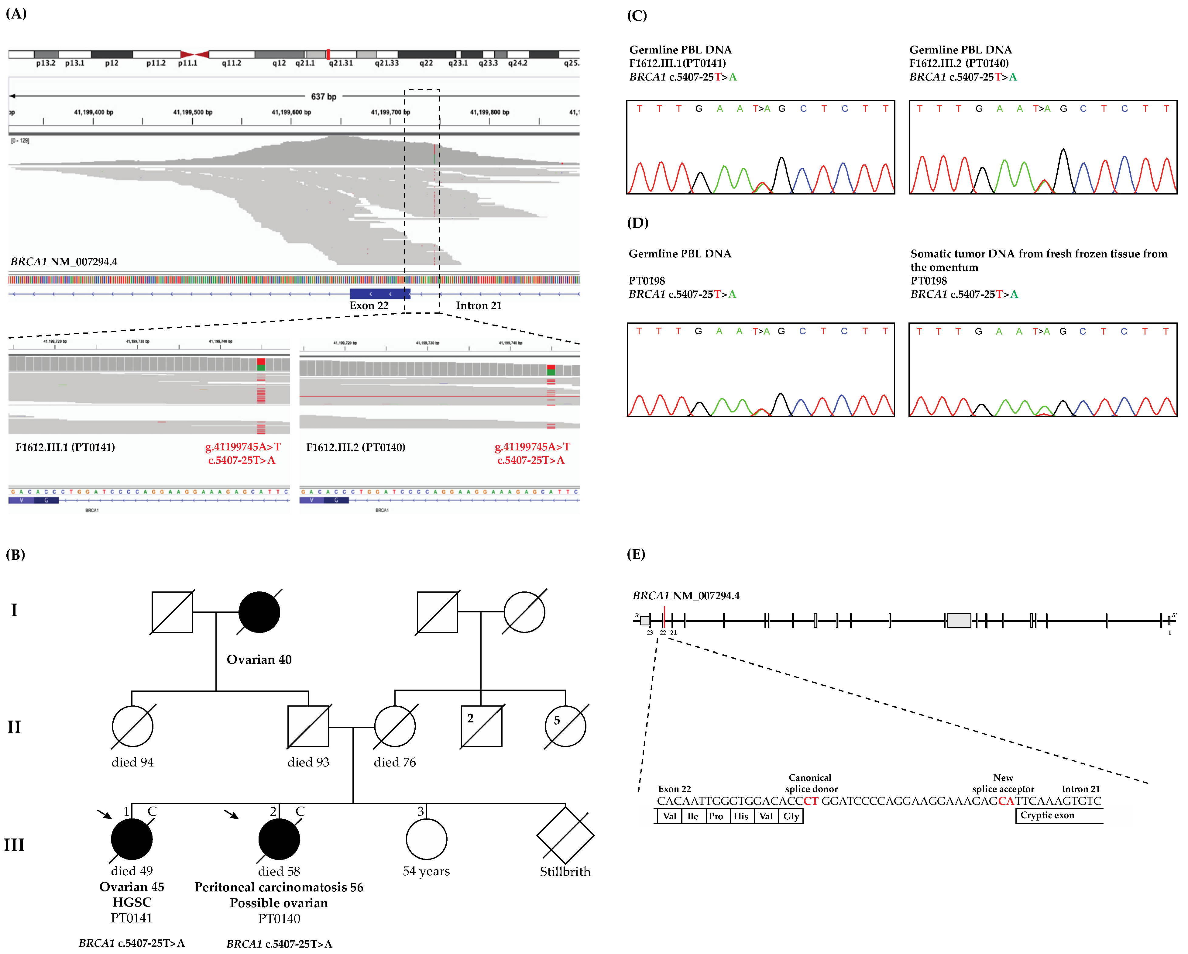
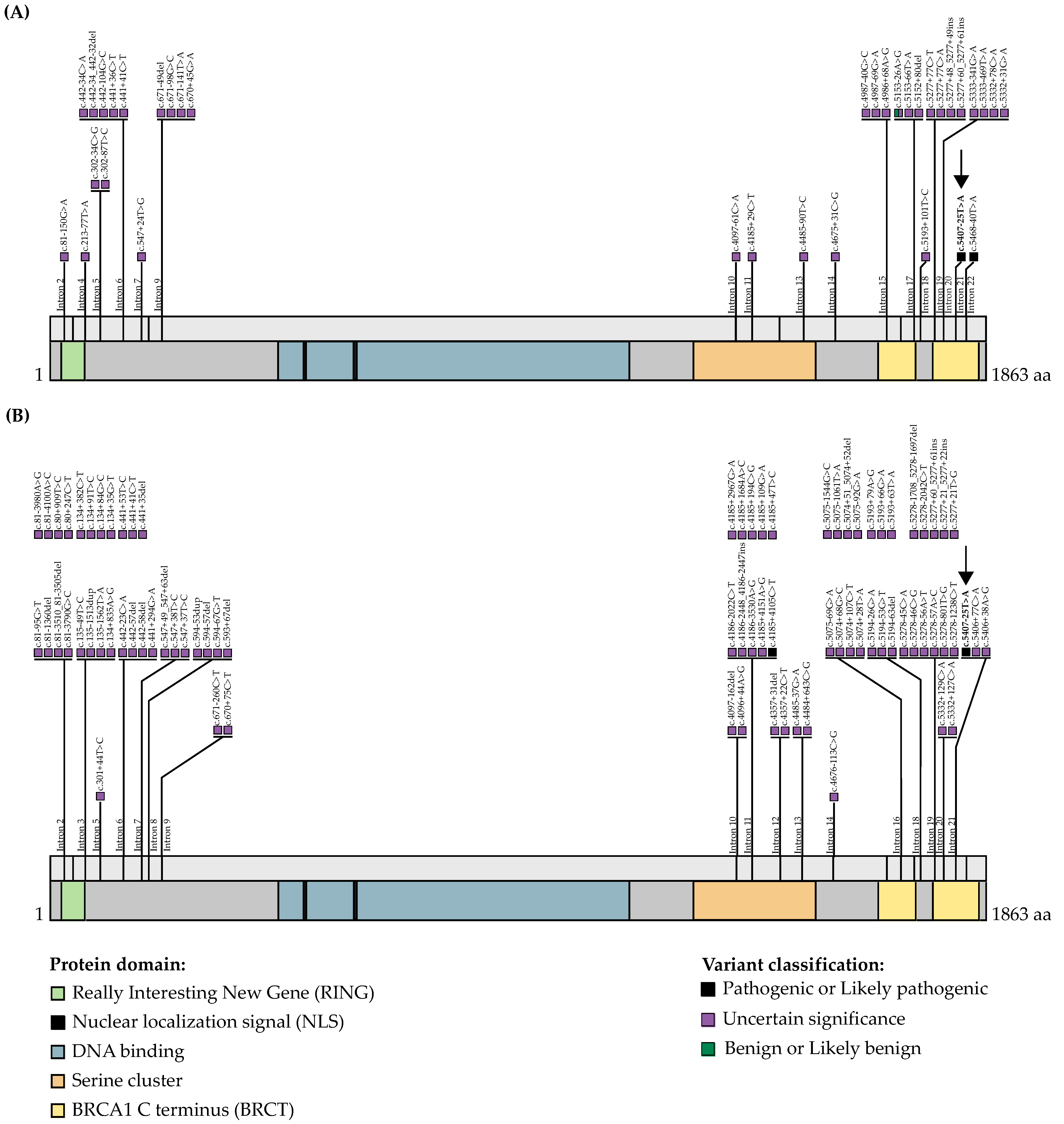
3.1. WES and Bioinformatics Analysis Identified BRCA1 c.5407-25T>A as a Candidate Pathogenic Variant

By applying our bioinformatics pipeline and filtering criteria on WES data from 27 OC cases from 22 families, we identified a total of four BRCA1 and six BRCA2 variants (Table S1). The variants were identified in six cases from five families, meaning some OC cases harbored more than one variant, and as described further below, there was one family of siblings harboring an identical BRCA1 variant.
From the list of seven exonic variants identified by our methods, all but BRCA2 c.4570T>G; p.Phe1524Val were not predicted to be damaging by all seven selected in silico tools (see Table S1). Furthermore, all these coding variants have been classified as benign by ClinVar and/or benign or likely benign by ACMG guidelines in the context of hereditary BC and/or OC and reported as benign in the BRCA exchange database [7]. These observations are consistent with medical genetic reports for cases harboring these variants, as commercial testing should have detected these exonic variants if regions were adequately covered.
The three intronic BRCA1 variants identified by our methods are interesting as they may not have been detectable by commercial testing. The allele frequencies of BRCA1 c.134+1335del and c.4358-722del are unknown, as neither variant was identified in gnomAD. In contrast, in gnomAD BRCA1 c.5407-25T>A is infrequent in the non-cancer general population having a MAF of 7.46 × 10⁻⁶, and a carrier frequency of two out of 134,138 individuals from the non-Finnish European population, a population of ancestral origin closest to the ancestry of our cancer families. As these intronic variants are located beyond ±20 nucleotides from splice sites, none of the prediction scores for affecting splicing were generated by the in silico tools Maximum Entropy Estimates of Splice junction strengths v2.0 (MaxEntScan v2.0) [39], Human Splicing Finder v3.1 (HSF v3.1) [40] and two Database Splicing Consensus Single Nucleotide Variant (dbscSNV) in silico tools: AdaBoost v4.0 (ADA v4.0) and Random Forest v4.0 (RF v4.0) [41] (Table S1). Therefore, we used SpliceAI, a relatively new in silico tool based on a deep learning and pre-mRNA transcript sequencing database, which generates different scores between 0 and 1 that can be interpreted as the probability of the variant affecting splicing by the loss or gain of a splice acceptor or a splice donor site [19]. SpliceAI predicted BRCA1 c.5407-25T>A may result in splice acceptor loss (delta score for acceptor loss = 0.41) (Table S1), suggesting that it might exert a deleterious effect on the transcription of BRCA1. Moreover, the locus of BRCA1 c.5407-25T>A is predicted to be conserved by in silico tools, supporting its biological importance [42]. In contrast, the other two intronic variants were not predicted to affect splicing of BRCA1 by SpliceAI (Table S1). Using IGV, a manual inspection of the sequencing reads for BRCA1 c.134+1335del and c.4358-722del revealed that they are located within repetitive regions deep within introns 3 and 12 of BRCA1, respectively, suggesting that sequencing data could be due to technical artifacts [43]. Indeed, association with risk is questionable as both intronic variants have been classified as benign in ClinVar and by ACMG guidelines, though they had not yet been reviewed in the BRCA exchange database (Table S1).
BRCA1 c.5407-25T>A is located at base pair 25 of intron 21 upstream from the start of exon 22 based on the transcript NM_007294.4 (Figure 1A), or of intron 22 upstream from the start of exon 23 based on the canonical transcript NM_007300.4, a transcript containing exon 4, which was missed due to a historical misannotation of an additional exon 4 in BRCA1 [44]. In this report, we have annotated our variants using the BRCA1 transcript (NM_007294.4), as it is the commonly used in the clinical genetic setting. Interestingly, this variant was identified in two affected sisters with cancer (Figure 1B), and a manual review of their sequencing files using IGV (Figure 1A) shows an average coverage of 60× by our WES capture kit as has been demonstrated by gnomAD v2.1 WES data (gnomad.broadinstitute.org/gene/ENSG00000012048?dataset=gnomad_r2_1 accessed on 5 February 2021). The variant was verified by bidirectional Sanger sequencing of PBL DNA (Figure 1C) from both of our carriers.

3.2. WES Analyses Identified Another OC Case Harboring BRCA1 c.5407-25T>A

To determine whether BRCA1 c.5407-25T>A occurs in other OC cases from our study groups, we reviewed similarly derived WES data sets from familial and sporadic OC cases. This variant was not identified in WES data from 24 OC cases from HBOC families harboring pathogenic BRCA1 or BRCA2 variants. In contrast, a carrier was identified among 53 OC cases who developed HGSC before the age of 50 years. Interestingly, this case (PT0198) had previously been reported as negative for pathogenic variants in BRCA1 or BRCA2 as well as in MLH1, MSH2, MSH6 and PMS2. The sporadic cancer case harboring the BRCA1 variant was diagnosed with HGSC at the age of 45 years, and the variant was verified by bidirectional Sanger sequencing of their PBL DNA (Figure 1D).

3.3. WES Analyses of BRCA1 c.5407-25T>A Carriers Suggest That They Are Unlikely to Harbor Pathogenic Variants in the Other Known OC Risk Genes

To further support the role of BRCA1 c.5407-25T>A in OC risk, we extended our WES and bioinformatic analyses to include an investigation of other known OC risk genes [38] in the familial OC (PT0141 and PT0140, see Figure 1B), and early onset OC (PT0198) cases harboring this BRCA1 allele. A review of WES data for pathogenic variants in MLH1, MSH2, MSH6, PMS2, BRIP1, RAD51C, RAD51D, PALB2, ATM or STK11 identified a carrier of MSH6 (NM_000179.2): c.-18G>T in PT0140 from family F1612. This variant is classified as benign or likely benign by six submissions in ClinVar (Accession number VCV000089159.7) and likely benign by ACMG guidelines. A similar analysis of the WES data from PT0198 did not identify any pathogenic variants in these genes.
A similar analysis of WES data in remaining 25 familial OC cases did not identify any variants classified as pathogenic or likely pathogenic in any of the 10 OC risk genes. Indeed the only rare variant identified, a missense variant in BRIP1 (NM_032043.3): c.2220G>T; p.Gln740His harbored in two sisters (PT0204 and PT0217) from family F1608 was not predicted to be damaging by five out of the seven selected in silico tools. Furthermore, this variant has been classified as likely benign by 6 submissions and VUS by 15 submissions in ClinVar (VCV000133752.33) and VUS by ACMG guidelines in the context of hereditary BC and/or OC.
However, our analyses of the sporadic early-onset OC case (PT0198), which included a thorough investigation of BRCA1 and BRCA2 loci, revealed that they also harbored BRCA2 c.1938C>T; p.Ser646Ser, a variant also found in one of the familial OC carrier cases. As discussed above, this synonymous variant in BRCA2 is classified as benign (Table S1). Thus, our findings suggest that BRCA1 c.5407-25T>A carriers are unlikely to harbor pathogenic, likely pathogenic or VUS in the other known OC predisposing genes based on NCCN guidelines for clinical practice in oncology [38].

3.4. LOH Analysis of the Tumor DNA from BRCA1 c.5407-25T>A Carrier Revealed Loss of the Wild-Type Allele

We performed LOH analysis of tumor DNA from PT0198, the only available OC tumor DNA from BRCA1 c.5407-25T>A carriers. DNA sequencing analyses suggested loss of the wild-type allele had occurred in the development of OC in this case (Figure 1D). This finding is consistent with BRCA1 c.5407-25T>A, playing a role in OC risk, as has been shown with LOH analyses of OC tumor DNA from carriers of BRCA1 pathogenic variants [45].

4. Discussion

Our WES and bioinformatics analyses of 27 familial OC cases who had undergone medical genetic testing and who were reported as negative for pathogenic variants in BRCA1 and BRCA2 identified two sisters harboring BRCA1 c.5407-25T>A. Further investigation of WES data from additional OC cases identified another carrier of this rare BRCA1 variant among the cases who were also previously reported as having tested negative for pathogenic variants in BRCA1 or BRCA2. As our WES data captured some intronic sequencing data, it is possible that this variant was not detected by medical genetic testing efforts due to commercial testing protocols that were applied.
Our application of SpliceAI, a new tool capable of predicting splice sites up to 10 kilobase pairs from the exon-intron junctions [19], predicted that this intronic variant may result in a splice acceptor loss. This suggests that the nucleotide substitution at −25 from exon 22 along with the adjacent nucleotide at −24 created a new splice acceptor site, potentially resulting in the loss of the entire or part of exon 22 of BRCA1 (see Figure 1E). The sensitivity of predicting aberrant splicing effects is estimated to be at least 70% for intronic variants between ±20–50 base pairs from exon–intron junctions [19]. Intronic regions containing sequences of potential exonic characteristics are referred to as pseudoexons or exons where a single substitution or small deletion or insertion may create new splice sites, such that these pseudoexons would be recognized by splicing machinery and result in abnormal patterns of splicing [17,46]. The application of SpliceAI has been used recently in different disorders [47].
BRCA1 c.5407-25T>A was identified in siblings both having OC (one diagnosed with a HGSC of the ovary and the other with primary peritoneal carcinomatosis), cancer phenotypes consistent with harboring a BRCA1 or BRCA2 pathogenic variant [48,49]. Indeed, applying the Manchester Scoring System revealed a probability greater than 10% of either sibling harboring a pathogenic variant in BRCA1 (score = 23) or BRCA2 (score = 15) [50,51,52]. Interestingly, we also found this variant in one of the early-onset OC cases who developed HGSC before the age of 50 years, which is consistent with observations that the average age of diagnosis of HGSC is less than 60 years of age in carriers of pathogenic BRCA1 variants [3]. Moreover, our genetic analysis of OC tumor DNA from this carrier revealed the loss of the wild-type allele and retention of the BRCA1 variant allele. This observation is consistent with BRCA1 c.5407-25T>A, playing a role in OC risk, as has been shown with LOH analyses of OC tumor DNA from those harboring loss-of-function pathogenic variants in BRCA1 [45]. BRCA1 c.5407-25T>A had initially been classified as a VUS in ClinVar based on two submissions of its identification in the context of HBC or HBOC (Table S1) and five independent studies published prior to 2020 that also described its identification in this hereditary cancer context (Table 1). The BRCA1 variant was identified via different detection platforms such as protein truncation test, single-strand conformational polymorphism analysis [53] or multiplex ligation-dependent probe analysis. In 2020, during the course of our investigation, Høberg-Vetti et al. reported the identification of BRCA1 c.5407-25T>A in BC (n = 12) and OC (n = 8) cases, which also included a case of peritoneal carcinomatosis, in 20 cancer families with BRCA1 Manchester scores ranging from 3 to 30 [54]. Indeed, the authors mentioned that they had identified this variant as early as 2006 and had evidence from one case of an effect on BRCA1 transcript [55], though the results were not published [54], highlighting the complexity of interpreting intronic variants. The prevalence of this variant reported in the Norwegian study groups (see Table 1) suggests the possibility of common ancestry for carriers, as has been shown with specific pathogenic variants in defined populations from our study of French Canadians and described in other studies [36]. Høberg-Vetti et al. also provided evidence that BRCA1 c.5407-25T>A creates a new splice site, resulting in the skipping of exon 22, based on a deletion of 61 nucleotides deduced from sequencing the corresponding aberrant size transcript [54]. This could affect protein function as it would result in the partial deletion of the BRCA1 Carboxy-Terminus (BRCT) domain (Figure 2), and thereby affect the binding of several proteins such as BRIP1, RAP80 and CtIP, which mediate the recruitment or stability of BRCA1 [56]. However, this group also demonstrated that the shift in the reading frame, which introduces a premature termination codon after 11 amino acids BRCA1 p.Gly1803GlnfsTer11, likely triggers nonsense-mediated mRNA decay [54]. As RNA is not available from our BRCA1 c.5407-25T>A carriers, we are unable to replicate these findings. Although more research on OC risk associated with BRCA1 c.5407-25T>A is required, collectively, these observations are supportive of the ClinVar classification of likely pathogenic rather than VUS.
The frequency of pathogenic BRCA1 intronic variants is currently unknown but likely underreported due to complex methods used to identify them and assess their biological and clinical impact. While researching independent evidence for the pathogenicity of BRCA1 c.5407-25T>A, we surveyed the ClinVar database (Figure 2A and Table S2) for rare, intronic BRCA1 variants located beyond ±20 nucleotides, rationalizing that this resource would report variants with biologically meaningful associations with cancer risk. A literature review revealed that intronic variants are being identified using a variety of DNA and RNA sequencing technologies, including reverse-transcribed- [60,61], long-range- [62] and multiplex- [63] PCR-based assays, some of which aim to identify variants within intronic regions as large as 10 kilobase pairs [15]. Recently, next-generation sequencing technologies involving RNA [64] or whole genome [18,65] sequencing have been applied. The biological impact of intronic variants can be difficult to discern but usually involve in cellulo assays of genetically engineered cell lines sometimes derived from carriers or minigene constructs [66,67]. Our survey of the ClinVar database revealed that 0.3% (35/11,366) of all reported BRCA1 variants were rare intronic variants that met our criteria (see Figure 2A), where 46% (16/35) were identified between ±20 and ±50 nucleotides and the remaining beyond ±50 nucleotides from exonic-intronic junctions. In total, 2 of the 35 intronic variants were listed as having conflicting interpretation, the variant of our interest c.5407-25T>A (VCV000371817) as VUS or likely pathogenic and c.5153-26A>G (VCV000125786) as VUS or likely benign; and the remaining 33 variants were classified as VUS. Only 3 of 35 BRCA1 intronic variants listed in ClinVar were identified in the gnomAD database, and this included our variant of interest, BRCA1 c.5407-25T>A (Table S2).
From a literature review, we curated a list of 223 original research studies or case reports about rare intronic variants in BRCA1 (Figure 2B and Table S3). This list included 32 reports describing 80 such variants. Of these intronic variants, 21% (17/80) were identified between ±20 and ±50 nucleotides and the remaining beyond ±50 nucleotides from exonic–intronic junctions. Only 2 of 80 variants had been classified as pathogenic c.4185+4105C>T (VCV000632611.2) or likely pathogenic c.5407-25T>A, our variant of interest, and the remaining as VUS. Only 4 of the 80 variants were identified in the ClinVar database, and all had been classified as VUS based on ACMG guidelines [9].
Some studies have argued that the majority of deep intronic variants are unlikely to be associated with cancer risk [68]. Interestingly, the frequency of intronic variants predicted by in silico tools to affect splicing is comparable to those predicted to effect bona fide splice site regions [19,69]. We applied SpliceAI [19] to predict the effect in splicing of BRCA1 to the BRCA1 intronic variants identified in ClinVar and in our literature search. Unlike our findings with BRCA1 c.5407-25T>A, SpliceAI predicted that the majority of curated intronic variants would not affect splicing, though the accuracy of this in silico tool did not reach 95% for all applications (see Table S3). New in silico tools have been developed to predict the splicing impact by these intronic variants using different mathematical models such as CADD-Splice [69] and SQUIRLS [70]. None of these tools have been tested yet on hereditary cancer syndromes. Earlier-developed in silico predictive models include MaxEntScan [39], HSF [40] or both dbscSNV tools [41]: ADA or RF were designed to predict variants within the splice regions. These tools have been tested on different datasets including sequencing data from hereditary BC and OC cases [71]. However, these in silico tools are limited to predicting events that occur within splice regions. Although in cellulo assays would provide supportive evidence for biological impact predicted by bioinformatic tools, the causality of an intronic variant identified in an established highly penetrant cancer predisposing gene such as BRCA1 in conferring risk to cancer remains a challenge.
Clinical testing for pathogenic variants in BRCA1 and BRCA2 has been established in clinical settings as it has been proven to improve cancer risk assessment and management of carriers [37]. OC cases harboring pathogenic variants in these genes are also offered targeted chemotherapies based on poly(ADP-ribose) polymerase (PARP) inhibitors as part of the their standard-of-care treatment regimens, as carriers have shown improvement in overall outcome [72,73]. As sequencing information is gathered from OC patients undergoing different treatment regimens, it will be interesting to investigate the response to PARP inhibitors in carriers of deep intronic variants, particularly those predicted to affect splicing and impact gene function by in silico analyses or by in cellulo assays. Our survey of ClinVar and the literature identified 105 rare intronic BRCA1 variants that are classified as VUS for further confirmation of their pathogenicity. Although our study may have been limited by whole-exome sequencing, our report highlights the importance of the comprehensive sequencing of the entirety of BRCA1 and BRCA2 to capture all possible pathogenic variants in individuals at risk for hereditary OC and BC.

5. Conclusions

Using our WES and bioinformatics analyses, we were able to identify an intronic variant in BRCA1 in one OC family who had tested negative for pathogenic variants in BRCA1 or BRCA2 by commercial testing. We also identified this variant in another OC case diagnosed at an early age and showed loss of the wild-type allele in the tumor DNA using LOH analysis. A splice predictor algorithm suggests that it exerts aberrant splicing affecting gene function. Our findings support BRCA1 c.5407-25T>A as a likely pathogenic variant and highlights the importance of investigating any intronic variants as causal variants in OC families where the involvement of BRCA1 is highly suggestive.

Supplementary Materials

The following supporting information can be downloaded at: https://0-www-mdpi-com.brum.beds.ac.uk/article/10.3390/genes13040697/s1, Table S1: All rare BRCA1 and BRCA2 variants identified in 27 ovarian cancer familial cases by WES analysis; Table S2: All rare intronic BRCA1 variants submitted to ClinVar; and Table S3: All rare intronic BRCA1 variants reported to literature.

Author Contributions

Conceptualization, W.M.A. and P.N.T.; methodology, W.M.A., T.R., C.T.F., J.R. and P.N.T.; investigation and formal analyses, W.M.A., C.T.F., C.S., J.R. and P.N.T.; cases resources: A.-M.M.-M., D.P., W.D.F. and Z.E.H.; writing—original draft preparation, W.M.A. and P.N.T.; writing—reviewing and editing, T.R., C.T.F., C.S., A.-M.M.-M., D.P., Z.E.H., J.R. and P.N.T.; and supervision, P.N.T. All authors have read and agreed to the published version of the manuscript.

Funding

This research was supported in part by the Department of Medicine, McGill University to P.N.T.; Saudi Arabian Cultural Bureau to P.N.T. and W.M.A.; The Canadian Institute of Health Research (CIHR) operating project grant (PJT-156124 to P.N.T. and J.R.); the Fond de la recherche du Québec en santé (FRQS) and Quebec Breast Cancer Foundation network grants to P.N.T.; Compute Canada resource allocation project (wst-164 to J.R.); Ovarian tumor banking was supported by the Banque de tissus et de données of the Réseau de recherche sur le cancer of the FRQS affiliated with the Canadian Tumour Repository Network (CTRNet). W.M.A. was supported by Taibah University Scholarship Award, C.T.F. was supported in part by The Research Institute of the McGill University Health Centre Studentship (RI-MUHC), James O and Maria Meadows Awards and J.R. was supported in part by Genome Canada Genome Technology Platform.

Institutional Review Board Statement

This project was conducted and approved according to the guidelines of The McGill University Health Centre Research Ethic Board (MP-37-2019-4783).

Informed Consent Statement

Informed consent was obtained from all subjects involved in this case report.

Data Availability Statement

Not applicable.

Acknowledgments

The Banque de tissus et données of the Réseau de recherche sur le cancer of the Fond de recherche du Québec–Santé (RRCancer biobank) is affiliated with the Canadian Tissue Repository Network (CTRNet). We acknowledge Liliane Meunier for preparing histopathological blocks, Manon De Ladurantaye and Lise Portelance for providing all clinical data and DNA from the RRCancer biobank; Mary Fujiwara for reviewing the final draft of the manuscript; Nora Wong for providing information for some pedigrees; and Suzanna L. Arcand for organizing samples for WES.

Conflicts of Interest

The authors declare no conflict of interest.

References

  1. Miki, Y.; Swensen, J.; Shattuck-Eidens, D.; Futreal, P.A.; Harshman, K.; Tavtigian, S.; Liu, Q.; Cochran, C.; Bennett, L.M.; Ding, W.; et al. A strong candidate for the breast and ovarian cancer susceptibility gene BRCA1. Science 1994, 266, 66–71. [Google Scholar] [CrossRef] [PubMed]
  2. Wooster, R.; Bignell, G.; Lancaster, J.; Swift, S.; Seal, S.; Mangion, J.; Collins, N.; Gregory, S.; Gumbs, C.; Micklem, G.; et al. Identification of the breast cancer susceptibility gene BRCA2. Nature 1995, 378, 789–792. [Google Scholar] [CrossRef] [PubMed]
  3. Kuchenbaecker, K.B.; Hopper, J.L.; Barnes, D.R.; Phillips, K.A.; Mooij, T.M.; Roos-Blom, M.J.; Jervis, S.; Van Leeuwen, F.E.; Milne, R.L.; Andrieu, N.; et al. Risks of breast, ovarian, and contralateral breast cancer for BRCA1 and BRCA2 mutation carriers. J. Am. Med. Assoc. 2017, 317, 2402–2416. [Google Scholar] [CrossRef] [PubMed]
  4. PDQ Adult Treatment Editorial Board. Ovarian Epithelial, Fallopian Tube, and Primary Peritoneal Cancer Treatment (PDQ®): Patient Version; National Cancer Institute at the National Institutes of Health: Bethesda, MD, USA, 2002; ISBN 0099-2240. [Google Scholar]
  5. Torre, L.A.; Trabert, B.; Desantis, C.E.; Miller, K.D.; Samimi, G.; Runowicz, C.D.; Gaudet, M.M.; Jemal, A.; Siegel, R.L. Ovarian Cancer Statistics, 2018. CA Cancer J. Clin. 2018, 68, 284–296. [Google Scholar] [CrossRef] [PubMed]
  6. Chen, S.; Parmigiani, G. Meta-analysis of BRCA1 and BRCA2 penetrance. J. Clin. Oncol. 2007, 25, 1329–1333. [Google Scholar] [CrossRef] [PubMed]
  7. Cline, M.S.; Liao, R.G.; Parsons, M.T.; Paten, B.; Alquaddoomi, F.; Antoniou, A.; Baxter, S.; Brody, L.; Cook-deegan, R.; Coffin, A.; et al. BRCA Challenge: BRCA Exchange as a global resource for variants in BRCA1 and BRCA2. PLoS Genet. 2018, 14, e1007752. [Google Scholar] [CrossRef]
  8. Landrum, M.J.; Lee, J.M.; Benson, M.; Brown, G.; Chao, C.; Chitipiralla, S.; Gu, B.; Hart, J.; Hoffman, D.; Hoover, J.; et al. ClinVar: Public archive of interpretations of clinically relevant variants. Nucleic Acids Res. 2016, 44, D862–D868. [Google Scholar] [CrossRef]
  9. Richards, S.; Aziz, N.; Bale, S.; Bick, D.; Das, S.; Gastier-Foster, J.; Grody, W.W.; Hegde, M.; Lyon, E.; Spector, E.; et al. Standards and guidelines for the interpretation of sequence variants: A joint consensus recommendation of the American College of Medical Genetics and Genomics and the Association for Molecular Pathology. Genet. Med. 2015, 17, 405–424. [Google Scholar] [CrossRef]
  10. Pavanello, M.; Chan, I.H.; Ariff, A.; Pharoah, P.D.; Gayther, S.A.; Ramus, S.J. Rare germline genetic variants and the risks of epithelial ovarian cancer. Cancers 2020, 12, 3046. [Google Scholar] [CrossRef]
  11. Kurosaki, T.; Maquat, L.E. Nonsense-mediated mRNA decay in humans at a glance. J. Cell Sci. 2016, 129, 461–467. [Google Scholar] [CrossRef]
  12. da Costa, P.J.; Menezes, J.; Romão, L. The role of alternative splicing coupled to nonsense-mediated mRNA decay in human disease. Int. J. Biochem. Cell Biol. 2017, 91, 168–175. [Google Scholar] [CrossRef] [PubMed]
  13. Cooper, T.A.; Wan, L.; Dreyfuss, G. RNA and Disease. Cell 2009, 136, 777–793. [Google Scholar] [CrossRef] [PubMed]
  14. Wieme, G.; Kral, J.; Rosseel, T.; Zemankova, P.; Parton, B.; Vocka, M.; Van Heetvelde, M.; Kleiblova, P.; Blaumeiser, B.; Soukupova, J.; et al. Prevalence of germline pathogenic variants in cancer predisposing genes in czech and belgian pancreatic cancer patients. Cancers 2021, 13, 4430. [Google Scholar] [CrossRef] [PubMed]
  15. Casadei, S.; Gulsuner, S.; Shirts, B.H.; Mandell, J.B.; Kortbawi, H.M.; Norquist, B.S.; Swisher, E.M.; Lee, M.K.; Goldberg, Y.; O’Connor, R.; et al. Characterization of splice-altering mutations in inherited predisposition to cancer. Proc. Natl. Acad. Sci. USA 2019, 116, 26798–26807. [Google Scholar] [CrossRef] [PubMed]
  16. Chen, X.; Truong, T.T.N.; Weaver, J.E.; Bove, B.A.; Cattie, K.; Armstrong, B.A.; Daly, M.B.; Godwin, A.K. Intronic alterations in BRCA1 and BRCA2: Effect on mRNA splicing fidelity and expression. Hum. Mutat. 2006, 27, 427–435. [Google Scholar] [CrossRef] [PubMed]
  17. Sibley, C.R.; Blazquez, L.; Ule, J. Lessons from non-canonical splicing. Nat. Rev. Genet. 2016, 17, 407–421. [Google Scholar] [CrossRef] [PubMed]
  18. Lionel, A.C.; Costain, G.; Monfared, N.; Walker, S.; Reuter, M.S.; Hosseini, S.M.; Thiruvahindrapuram, B.; Merico, D.; Jobling, R.; Nalpathamkalam, T.; et al. Improved diagnostic yield compared with targeted gene sequencing panels suggests a role for whole-genome sequencing as a first-tier genetic test. Genet. Med. 2017, 20, 435–443. [Google Scholar] [CrossRef]
  19. Jaganathan, K.; Panagiotopoulou, S.K.; McRae, J.F.; Darbandi, S.F.; Knowles, D.; Li, Y.I.; Kosmicki, J.A.; Arbelaez, J.; Cui, W.; Schwartz, G.B.; et al. Predicting splicing from primary sequence with deep learning. Cell 2019, 176, 535–548. [Google Scholar] [CrossRef]
  20. Montalban, G.; Bonache, S.; Bach, V.; Gisbert-Beamud, A.; Tenés, A.; Moles-Fernández, A.; López-Fernández, A.; Carrasco, E.; Balmaña, J.; Diez, O.; et al. BRCA1 and BRCA2 whole cDNA analysis in unsolved hereditary breast/ovarian cancer patients. Cancer Genet. 2021, 258, 10–17. [Google Scholar] [CrossRef]
  21. Walsh, T.; Casadei, S.; Munson, K.M.; Eng, M.; Mandell, J.B.; Gulsuner, S.; King, M.C. CRISPR–Cas9/long-read sequencing approach to identify cryptic mutations in BRCA1 and other tumour suppressor genes. J. Med. Genet. 2021, 58, 850–852. [Google Scholar] [CrossRef]
  22. Pirim, D.; Kaya, N.; Yıldırım, E.U.; Sag, Ş.O.; Temel, S.G. Characterization and in silico analyses of the BRCA1/2 variants identified in individuals with personal and/or family history of BRCA-related cancers. Int. J. Biol. Macromol. 2020, 162, 1166–1177. [Google Scholar] [CrossRef] [PubMed]
  23. Montalban, G.; Bonache, S.; Moles-Fernández, A.; Gisbert-Beamud, A.; Tenés, A.; Bach, V.; Carrasco, E.; López-Fernández, A.; Stjepanovic, N.; Balmaña, J.; et al. Screening of BRCA1/2 deep intronic regions by targeted gene sequencing identifies the first germline BRCA1 variant causing pseudoexon activation in a patient with breast/ovarian cancer. J. Med. Genet. 2019, 56, 63–74. [Google Scholar] [CrossRef] [PubMed]
  24. Gelli, E.; Colombo, M.; Pinto, A.M.; De Vecchi, G.; Foglia, C.; Amitrano, S.; Morbidoni, V.; Imperatore, V.; Manoukian, S.; Baldassarri, M.; et al. Usefulness and limitations of comprehensive characterization of mRNA splicing profiles in the definition of the clinical relevance of BRCA1/2 variants of uncertain significance. Cancers 2019, 11, 295. [Google Scholar] [CrossRef] [PubMed]
  25. Rivera, B.; Iorio, M.D.; Frankum, J.; Nadaf, J.; Fahiminiya, S.; Arcand, S.L.; Burk, D.L.; Grapton, D.; Tomiak, E.; Hastings, V.; et al. Functionally null RAD51D missense mutation associates strongly with ovarian carcinoma. Cancer Res. 2017, 77, 4517–4529. [Google Scholar] [CrossRef]
  26. Felicio, P.S.; Grasel, R.S.; Campacci, N.; de Paula, A.E.; Galvão, H.C.R.; Torrezan, G.T.; Sabato, C.S.; Fernandes, G.C.; Souza, C.P.; Michelli, R.D.; et al. Whole-exome sequencing of non-BRCA1/BRCA2 mutation carrier cases at high-risk for hereditary breast/ovarian cancer. Hum. Mutat. 2021, 42, 290–299. [Google Scholar] [CrossRef]
  27. Fierheller, C.T.; Guitton-Sert, L.; Alenezi, W.M.; Revil, T.; Oros, K.K.; Gao, Y.; Bedard, K.; Arcand, S.L.; Serruya, C.; Behl, S.; et al. A functionally impaired missense variant identified in French Canadian families implicates FANCI as a candidate ovarian cancer-predisposing gene. Genome Med. 2021, 13, 186. [Google Scholar] [CrossRef]
  28. Shigemizu, D.; Momozawa, Y.; Abe, T.; Morizono, T.; Boroevich, K.A.; Takata, S.; Ashikawa, K.; Kubo, M.; Tsunoda, T. Performance comparison of four commercial human whole-exome capture platforms. Sci. Rep. 2015, 5, 12742. [Google Scholar] [CrossRef]
  29. Asan; Xu, Y.; Jiang, H.; Tyler-Smith, C.; Xue, Y.; Jiang, T.; Wang, J.; Wu, M.; Liu, X.; Tian, G.; et al. Comprehensive comparison of three commercial human whole-exome capture platforms. Genome Biol. 2011, 12, R95. [Google Scholar] [CrossRef]
  30. Collins, R.L.; Brand, H.; Karczewski, K.J.; Zhao, X.; Alföldi, J.; Khera, A.V.; Franci-Oli, L.C.; Gauthier, L.D.; Wang, H.; Watts, N.A.; et al. gnomAD-SV An open resource of structural variation for medical and population genetics The Genome Aggregation Database (gnomAD) Production Team 7, The gnomAD Consortium. bioRxiv 2019. [Google Scholar] [CrossRef]
  31. Karczewski, K.J.; Francioli, L.C.; Tiao, G.; Cummings, B.B.; Alföldi, J.; Wang, Q.; Collins, R.L.; Laricchia, K.M.; Ganna, A.; Birnbaum, D.P.; et al. Variation across 141,456 human exomes and genomes reveals the spectrum of loss-of-function intolerance across human protein-coding genes. bioRxiv 2019. [Google Scholar] [CrossRef]
  32. Pedersen, B.S.; Brown, J.M.; Dashnow, H.; Wallace, A.D.; Velinder, M.; Tristani-Firouzi, M.; Schiffman, J.D.; Tvrdik, T.; Mao, R.; Best, D.H.; et al. Effective variant filtering and expected candidate variant yield in studies of rare human disease. NPJ Genom. Med. 2021, 6, 60. [Google Scholar] [CrossRef] [PubMed]
  33. Thorvaldsdóttir, H.; Robinson, J.T.; Mesirov, J.P. Integrative Genomics Viewer (IGV): High-performance genomics data visualization and exploration. Brief. Bioinform. 2013, 14, 178–192. [Google Scholar] [CrossRef] [PubMed]
  34. Ghosh, R.; Oak, N.; Plon, S.E. Evaluation of in silico algorithms for use with ACMG/AMP clinical variant interpretation guidelines. Genome Biol. 2017, 18, 225. [Google Scholar] [CrossRef]
  35. Alenezi, W.M.; Fierheller, C.T.; Recio, N.; Tonin, P.N. Literature review of BARD1 as a cancer predisposing gene with a focus on breast and ovarian cancers. Genes 2020, 11, 856. [Google Scholar] [CrossRef] [PubMed]
  36. Fierheller, C.T.; Alenezi, W.M.; Tonin, P.N. The genetic analyses of French Canadians of Quebec facilitate the characterization of new cancer predisposing genes implicated in hereditary breast and/or ovarian cancer syndrome families. Cancers 2021, 13, 3406. [Google Scholar] [CrossRef]
  37. Liu, Y.L.; Breen, K.; Catchings, A.; Ranganathan, M.; Latham, A.; Goldfrank, D.J.; Grisham, R.N.; Long Roche, K.; Frey, M.K.; Chi, D.S.; et al. Risk-reducing bilateral salpingo-oophorectomy for ovarian cancer: A review and clinical guide for hereditary predisposition genes. JCO Oncol. Pract. 2021, 18, 201–209. [Google Scholar] [CrossRef]
  38. Daly, M.B.; Pilarski, R.; Yurgelun, M.B.; Berry, M.P.; Buys, S.S.; Dickson, P.; Domchek, S.M.; Elkhanany, A.; Friedman, S.; Garber, J.E.; et al. Genetic/familial high-risk assessment: Breast, ovarian, and pancreatic, version 1.2020 featured updates to the NCCN guidelines. JNCCN J. Natl. Compr. Cancer Netw. 2020, 18, 380–391. [Google Scholar] [CrossRef]
  39. Shamsani, J.; Kazakoff, S.H.; Armean, I.M.; McLaren, W.; Parsons, M.T.; Thompson, B.A.; O’Mara, T.A.; Hunt, S.E.; Waddell, N.; Spurdle, A.B. A plugin for the Ensembl Variant Effect Predictor that uses MaxEntScan to predict variant spliceogenicity. Bioinformatics 2019, 35, 2315–2317. [Google Scholar] [CrossRef]
  40. Desmet, F.O.; Hamroun, D.; Lalande, M.; Collod-Bëroud, G.; Claustres, M.; Béroud, C. Human Splicing Finder: An online bioinformatics tool to predict splicing signals. Nucleic Acids Res. 2009, 37, e67. [Google Scholar] [CrossRef]
  41. Liu, X.; Wu, C.; Li, C.; Boerwinkle, E. dbNSFP v3.0: A one-stop database of functional predictions and annotations for human nonsynonymous and splice-site SNVs. Hum. Mutat. 2016, 37, 235–241. [Google Scholar] [CrossRef]
  42. Ponting, C.P. Biological function in the twilight zone of sequence conservation. BMC Biol. 2017, 15, 71. [Google Scholar] [CrossRef] [PubMed]
  43. Treangen, T.J.; Salzberg, S.L. Repetitive DNA and next-generation sequencing: Computational challenges and solutions. Nat. Rev. Genet. 2012, 13, 36. [Google Scholar] [CrossRef] [PubMed]
  44. de Garibay, G.R.; Fernandez-Garcia, I.; Mazoyer, S.; de Calais, F.L.; Ameri, P.; Vijayakumar, S.; Martinez-Ruiz, H.; Damiola, F.; Barjhoux, L.; Thomassen, M.; et al. Altered regulation of BRCA1 exon 11 splicing is associated with breast cancer risk in carriers of BRCA1 pathogenic variants. Hum. Mutat. 2021, 42, 1488–1502. [Google Scholar] [CrossRef]
  45. Kanchi, K.L.; Johnson, K.J.; Lu, C.; McLellan, M.D.; Leiserson, M.D.M.; Wendl, M.C.; Zhang, Q.; Koboldt, D.C.; Xie, M.; Kandoth, C.; et al. Integrated analysis of germline and somatic variants in ovarian cancer. Nat. Commun. 2014, 5, 3156. [Google Scholar] [CrossRef] [PubMed]
  46. Sun, H.; Chasin, L.A. Multiple splicing defects in an intronic false exon. Mol. Cell. Biol. 2000, 20, 6414–6425. [Google Scholar] [CrossRef]
  47. Qian, X.; Wang, J.; Wang, M.; Igelman, A.D.; Jones, K.D.; Li, Y.; Wang, K.; Goetz, K.E.; Birch, D.G.; Yang, P.; et al. Identification of deep-intronic splice mutations in a large cohort of patients with inherited retinal diseases. Front. Genet. 2021, 12, 647400. [Google Scholar] [CrossRef]
  48. Nielsen, F.C.; van Overeem Hansen, T.; Sørensen, C.S. Hereditary breast and ovarian cancer: New genes in confined pathways. Nat. Rev. Cancer 2016, 16, 599–612. [Google Scholar] [CrossRef]
  49. Köbel, M.; Kang, E.Y. The evolution of ovarian carcinoma subclassification. Cancers 2022, 14, 416. [Google Scholar] [CrossRef]
  50. Evans, D.G.R.; Eccles, D.M.; Rahman, N.; Young, K.; Bulman, M.; Amir, E.; Shenton, A.; Howell, A.; Lalloo, F. A new scoring system for the chances of identifying a BRCA1/2 mutation outperforms existing models including BRCAPRO. J. Med. Genet. 2004, 41, 474–480. [Google Scholar] [CrossRef]
  51. Evans, D.G.; Lalloo, F.; Wallace, A.; Rahman, N. Update on the Manchester Scoring System for BRCA1 and BRCA2 testing. J. Med. Genet. 2005, 42, e39. [Google Scholar] [CrossRef]
  52. Evans, D.G.; Harkness, E.F.; Plaskocinska, I.; Wallace, A.J.; Clancy, T.; Woodward, E.R.; Howell, T.A.; Tischkowitz, M.; Lalloo, F. Pathology update to the Manchester Scoring System based on testing in over 4000 families. J. Med. Genet. 2017, 54, 674–681. [Google Scholar] [CrossRef] [PubMed]
  53. Hamann, U.; Liu, X.; Bungardt, N.; Ulrich Ulmer, H.; Bastert, G.; Sinn, H.P. Similar contributions of BRCA1 and BRCA2 germline mutations to early-onset breast cancer in Germany. Eur. J. Hum. Genet. 2003, 11, 464–467. [Google Scholar] [CrossRef] [PubMed]
  54. Høberg-Vetti, H.; Ognedal, E.; Buisson, A.; Vamre, T.B.A.; Ariansen, S.; Hoover, J.M.; Eide, G.E.; Houge, G.; Fiskerstrand, T.; Haukanes, B.I.; et al. The intronic BRCA1 c.5407-25T>A variant causing partly skipping of exon 23—A likely pathogenic variant with reduced penetrance? Eur. J. Hum. Genet. 2020, 28, 1078–1086. [Google Scholar] [CrossRef] [PubMed]
  55. Høberg-Vetti, H.; Bjorvatn, C.; Fiane, B.E.; Aas, T.; Woie, K.; Espelid, H.; Rusken, T.; Eikesdal, H.P.; Listøl, W.; Haavind, M.T.; et al. BRCA1/2 testing in newly diagnosed breast and ovarian cancer patients without prior genetic counselling: The DNA-BONus study. Eur. J. Hum. Genet. 2016, 24, 881–888. [Google Scholar] [CrossRef]
  56. Roy, R.; Chun, J.; Powell, S.N. BRCA1 and BRCA2: Important differences with common interests. Nat. Rev. Cancer 2012, 12, 372. [Google Scholar] [CrossRef]
  57. Konstantopoulou, I.; Tsitlaidou, M.; Fostira, F.; Pertesi, M.; Stavropoulou, A.V.; Triantafyllidou, O.; Tsotra, E.; Tsiftsoglou, A.P.; Tsionou, C.; Droufakou, S.; et al. High prevalence of BRCA1 founder mutations in Greek breast/ovarian families. Clin. Genet. 2014, 85, 36–42. [Google Scholar] [CrossRef]
  58. Heramb, C.; Wangensteen, T.; Grindedal, E.M.; Ariansen, S.L.; Lothe, S.; Heimdal, K.R.; Mæhle, L. BRCA1 and BRCA2 mutation spectrum—An update on mutation distribution in a large cancer genetics clinic in Norway. Hered. Cancer Clin. Pract. 2018, 16, 3. [Google Scholar] [CrossRef]
  59. Møller, P.; Dominguez-Valentin, M.; Rødland, E.A.; Hovig, E. Causes for frequent pathogenic BRCA1 variants include low penetrance in fertile ages, recurrent de-novo mutations and genetic drift. Cancers 2019, 11, 132. [Google Scholar] [CrossRef]
  60. Wappenschmidt, B.; Becker, A.A.; Hauke, J.; Weber, U.; Engert, S.; Köhler, J.; Kast, K.; Arnold, N.; Rhiem, K.; Hahnen, E.; et al. Analysis of 30 putative BRCA1 splicing mutations in hereditary breast and ovarian cancer families identifies exonic splice site mutations that escape in silico prediction. PLoS ONE 2012, 7, e50800. [Google Scholar] [CrossRef]
  61. Bonnet, C.; Krieger, S.; Vezain, M.; Rousselin, A.; Tournier, I.; Martins, A.; Berthet, P.; Chevrier, A.; Dugast, C.; Layet, V.; et al. Screening BRCA1 and BRCA2 unclassified variants for splicing mutations using reverse transcription PCR on patient RNA and an ex vivo assay based on a splicing reporter minigene. J. Med. Genet. 2008, 45, 438–446. [Google Scholar] [CrossRef]
  62. Ozcelik, H.; Shi, X.; Chang, M.C.; Tram, E.; Vlasschaert, M.; Nicola, N.D.; Kiselova, A.; Yee, D.; Goldman, A.; Dowar, M.; et al. Long-range PCR and next-generation sequencing of BRCA1 and BRCA2 in breast cancer. J. Mol. Diagn. 2012, 14, 467–475. [Google Scholar] [CrossRef] [PubMed]
  63. Velasco, E.; Infante, M.; Durán, M.; Pérez-Cabornero, L.; Sanz, D.J.; Esteban-Cardeñosa, E.; Miner, C. Heteroduplex analysis by capillary array electrophoresis for rapid mutation detection in large multiexon genes. Nat. Protoc. 2007, 2, 237–246. [Google Scholar] [CrossRef] [PubMed]
  64. Landrith, T.; Li, B.; Cass, A.A.; Conner, B.R.; LaDuca, H.; McKenna, D.B.; Maxwell, K.N.; Domchek, S.; Morman, N.A.; Heinlen, C.; et al. Splicing profile by capture RNA-seq identifies pathogenic germline variants in tumor suppressor genes. NPJ Precis. Oncol. 2020, 4, 4. [Google Scholar] [CrossRef] [PubMed]
  65. Guo, X.; Shi, J.; Cai, Q.; Shu, X.O.; He, J.; Wen, W.; Allen, J.; Pharoah, P.; Dunning, A.; Hunter, D.J.; et al. Use of deep whole-genome sequencing data to identify structure risk variants in breast cancer susceptibility genes. Hum. Mol. Genet. 2018, 27, 853–859. [Google Scholar] [CrossRef]
  66. Fraile-Bethencourt, E.; Valenzuela-Palomo, A.; Díez-Gómez, B.; Acedo, A.; Velasco, E.A. Identification of eight spliceogenic variants in BRCA2 Exon 16 by minigene assays. Front. Genet. 2018, 9, 188. [Google Scholar] [CrossRef]
  67. Acedo, A.; Sanz, D.J.; Durán, M.; Infante, M.; Pérez-Cabornero, L.; Miner, C.; Velasco, E.A. Comprehensive splicing functional analysis of DNA variants of the BRCA2 gene by hybrid minigenes. Breast Cancer Res. 2012, 14, R87. [Google Scholar] [CrossRef]
  68. Parsons, M.T.; Tudini, E.; Li, H.; Hahnen, E.; Wappenschmidt, B.; Feliubadaló, L.; Aalfs, C.M.; Agata, S.; Aittomäki, K.; Alducci, E.; et al. Large scale multifactorial likelihood quantitative analysis of BRCA1 and BRCA2 variants: An ENIGMA resource to support clinical variant classification. Hum. Mutat. 2019, 40, 1557–1578. [Google Scholar] [CrossRef]
  69. Rentzsch, P.; Schubach, M.; Shendure, J.; Kircher, M. CADD-Splice—Improving genome-wide variant effect prediction using deep learning-derived splice scores. Genome Med. 2021, 13, 31. [Google Scholar] [CrossRef]
  70. Danis, D.; Jacobsen, J.O.B.; Carmody, L.C.; Gargano, M.A.; McMurry, J.A.; Hegde, A.; Haendel, M.A.; Valentini, G.; Smedley, D.; Robinson, P.N. Interpretable prioritization of splice variants in diagnostic next-generation sequencing. Am. J. Hum. Genet. 2021, 108, 1564–1577. [Google Scholar] [CrossRef]
  71. Moles-Fernández, A.; Duran-Lozano, L.; Montalban, G.; Bonache, S.; López-Perolio, I.; Menéndez, M.; Santamariña, M.; Behar, R.; Blanco, A.; Carrasco, E.; et al. Computational tools for splicing defect prediction in breast/ovarian cancer genes: How efficient are they at predicting RNA alterations? Front. Genet. 2018, 9, 366. [Google Scholar] [CrossRef]
  72. Kurnit, K.C.; Fleming, G.F.; Lengyel, E. Updates and new options in advanced epithelial ovarian cancer treatment. Obstet. Gynecol. 2021, 137, 108. [Google Scholar] [CrossRef] [PubMed]
  73. Wang, Q.; Peng, H.; Qi, X.; Wu, M.; Zhao, X. Targeted therapies in gynecological cancers: A comprehensive review of clinical evidence. Signal Transduct. Target. Ther. 2020, 5, 137. [Google Scholar] [CrossRef] [PubMed]
Figure 1. Identification of BRCA1 c.5407-25T>A, a likely pathogenic intronic variant. (A) Integrative Genomics Viewer (IGV) v2.8 sequencing data showing coverage of exon 23 of BRCA1 and flanking intronic regions beyond ±20 nucleotides of location of BRCA1 c.5407-25T>A in variant carriers; (B) pedigree of BRCA1 c.5407-25T>A carrier family (F1612) indicating confirmed (“C”) cases of bilateral high-grade serous carcinoma (HGSC) (PT0141) and primary peritoneal carcinomatosis with possible ovarian origin (PT0140) by pathology reports; (C) sequencing chromatogram of peripheral blood lymphocytes (PBL) DNA verifying heterozygous carrier status of both sisters; and (D) sequencing chromatograms from a sporadic early-onset OC case (PT0198) verifying heterozygous BRCA1 variant carrier status in PBL DNA and loss of the wild-type allele in OC tumor DNA; (E) Schema illustrating the location of the intronic BRCA1 variant creating a new splice site as predicted by SpliceAI.
Genes 13 00697 g001
Figure 2. Annotated curated intronic BRCA1 variants. The BRCA1 transcript NM_007294.4 (NCBI Reference Sequence (RefSeq) database (ncbi.nlm.nih.gov/nuccore/NM_007294.4), indicating protein encoded domains was annotated with BRCA1 c.5407-25T>A (indicated with an arrow) and intronic variants classified as pathogenic, likely pathogenic or unknown significance based on ClinVar and/or American College of Medical Genetics and Genomics guidelines were (A) submitted to ClinVar (ncbi.nlm.nih.gov/clinvar/ accessed on 25 January 2022) (see Table S2) or (B) reported in the literature (pubmed.ncbi.nlm.nih.gov accessed on 25 January 2022) (see Table S3). Variants were selected based on: intronic location beyond ±20 nucleotides from splice sites; rarity (minor allele frequency ≤0.005); classification as pathogenic, likely pathogenic or variants of unknown significance (VUS) in BRCA1 in the context of hereditary breast and/or ovarian cancer Search terms for PubMed (as in October 2021) articles included: (“brca1 s”[All Fields] OR “genes, brca1”[MeSH Terms] OR (“genes”[All Fields] AND “brca1”[All Fields]) OR “brca1 genes”[All Fields] OR “brca1”[All Fields]) AND (“intron s”[All Fields] OR “intron”[All Fields] OR “intronically”[All Fields] OR “intronization”[All Fields] OR “introns”[MeSH Terms] OR “introns”[All Fields] OR “intron”[All Fields] OR “intronic”[All Fields]) AND (“variant”[All Fields] OR “variant s”[All Fields] OR “variants”[All Fields]). Intergenic, 3′UTR and 5′UTR variants and large chromosomal rearrangements were excluded.
Genes 13 00697 g002
Table 1. Features of BRCA1 c.5407-25T>A carriers from independent reports.
Year
Reported
Population 1Number of
Carriers per Study Group
Cancer Type
in Carriers
Study Group Investigated 2Reference
2003Germany1/90BreastEarly-onset cases not selected for family history of cancer[53]
2014Greece1/473BreastHBC and HBOC families[57]
2016Norway2/893BreastCases not selected for family history of cancer[55]
2018Norway9/669BreastHBC and HBOC families[58]
2019Norway8/1914Breast or
ovarian
Sporadic cases and families[59]
2020Norway, France,
United States of America
20Breast or
ovarian
Selected HBC and HBOC families[54]
2022French Canadian, Ashkenazi Jewish, Austria, United
Kingdom, Germany, Italy
2/27OvarianFamilies with at least two OC case within first-, second- or third-degree relativesThis report
2022French Canadian1/53OvarianSporadic OC case with early onset of the disease not selected for family history of cancerThis report
1 Geographic origin of population or self-reported as French Canadian from Quebec. 2 Cases investigated include hereditary breast cancer (HBC) and hereditary breast and ovarian cancer (HBOC) families.
Publisher’s Note: MDPI stays neutral with regard to jurisdictional claims in published maps and institutional affiliations.

