Article

Genome-Wide Identification, Characterization and Expression Profiling of ADF Family Genes in Solanum lycopersicum L.

1
Department of Agricultural Industry Economy and Education, Sunchon National University, 413 Jungangno, Suncheon, Jeonnam 540-950, Korea
2
Department of Horticulture, Sunchon National University, 413 Jungangno, Suncheon, Jeonnam 540-950, Korea
3
Department of Horticultural Science, Kyungpook National University, Daegu 702-701, Korea
4
Department of Agricultural Education, Sunchon National University, 413 Jungangno, Suncheon, Jeonnam 540-950, Korea
*
Author to whom correspondence should be addressed.
Submission received: 8 July 2016 / Revised: 12 September 2016 / Accepted: 22 September 2016 / Published: 29 September 2016
(This article belongs to the Section Molecular Genetics and Genomics)

Abstract

:
The actin depolymerizing factor (ADF) proteins have growth, development, defense-related and growth regulatory functions in plants. The present study used genome-wide analysis to investigate ADF family genes in tomato. Eleven tomato ADF genes were identified and differential expression patterns were found in different organs. SlADF6 was preferentially expressed in roots, suggesting its function in root development. SlADF1, SlADF3 and SlADF10 were predominately expressed in the flowers compared to the other organs and specifically in the stamen compared to other flower parts, indicating their potential roles in pollen development. The comparatively higher expression of SlADF3 and SlADF11 at early fruit developmental stages might implicate them in determining final fruit size. SlADF5 and SlADF8 had relatively higher levels of expression five days after the breaker stage of fruit development, suggesting their possible role in fruit ripening. Notably, six genes were induced by cold and heat, seven by drought, five by NaCl, and four each by abscisic acid (ABA), jasmonic acid (JA) and wounding treatments. The differential expression patterns of the SlADF genes under different types of stresses suggested their function in stress tolerance in tomato plants. Our results will be helpful for the functional characterization of ADF genes during organ and fruit development of tomato under different stresses.

1. Introduction

The actin depolymerizing factor (ADF) gene family encodes a group of actin-remodeling proteins that are involved in the reorganization of the actin cytoskeleton. In plant cells, the actin cytoskeleton is involved in various cellular and developmental activities, such as cytoplasmic streaming, cell division, elongation, polar tip growth, and cellular organelle movement [1,2], as well as cell signaling in response to biotic and abiotic stresses [3]. A large number of actin binding proteins, including ADFs, are involved in the reorganization of the actin cytoskeleton. ADF proteins regulate the assembly of globular and filamentous actin to facilitate responses to developmental and environmental stimuli. ADF protein was first isolated from the brains of chick embryos [4] and ADF genes have been identified from a wide range of eukaryotes [5]. The twelve members of the ADF gene family identified in Arabidopsis and rice are grouped into several classes [6,7,8]. The ADFs are ubiquitous, highly conserved, low molecular weight (15–22 kDa) actin-binding proteins [9]. ADFs act as stimulus-responsive modulators in the actin cytoskeleton and are involved in in vitro monomer binding, monomer dissociation inhibition, and actin-filament binding and severing [9]. Several factors, such as pH, phosphatidylinositol 4,5-bisphosphate (PIP2), inorganic phosphate and actin filament-bound nucleotides (ATP or ADP), regulate the activities of ADF in cells [10,11,12]. In Arabidopsis, ADF family genes regulate the organization of filamentous actin and are reported to be involved in organ growth, tissue expansion and flowering [13]. VvADF was found to be involved in the initiation of new root formation from stem cuttings in grape [14]. The over-expression of NtADF1 in tobacco suppressed the NtRac1 mutation, which causes in morphological changes of the pollen tube [15]. ADF proteins are involved in pollen tube growth by controlling cytoskeleton rearrangement [10,16]. Augustine et al. (2008) found strong evidence of the interaction of actin and ADFs in pollen tube growth of plants [11]. ADF family genes also have a vital role in response to various abiotic and biotic stresses. For example, accumulation of TaADF was found in wheat cultivars that were tolerant to freezing temperatures but was absent in sensitive cultivars [1]. Deng et al. (2010) suggested the possible involvement of HbADF in the latex regulation and wound plugging in Hevea brasiliensis [17]. ADFs in Arabidopsis (AtADF2 and AtADF4) and barley are related to resistance against pathogens [18,19,20]. The AtADF4 gene in Arabidopsis participates in mitogen-activated protein kinase signaling and activation of gene-for-gene resistance by regulating the cytoskeletal dynamics and transcription of the R-gene [21].
To date, no studies have investigated the organ-dependent expression profiles of ADF genes in vegetable crops. Tomato (Solanum lycopersicum) is an economically important fruit and vegetable crop world-wide. As the ADF gene family is known to be involved in growth, development and defense in plants, we sought to investigate the function of ADF genes in tomato. In the present study, we used the Sol Genomics databases to identify 11 ADF genes, compare them with other plant ADF genes, and determine their phylogenetic classification. We analyzed the differential expressions of these ADF genes in different organs, including different developmental stages of tomato fruit, as well as in response to different abiotic and phytohormone stresses to investigate their specific functions in tomato.

2. Materials and Methods

2.1. Identification and Analysis of ADF Family Genes

We identified 11 ADF genes in tomato using the InterPro domain accession number IPR002108 (representing a protein family containing the actin-depolymerising factor homology domain, the ADF-H domain) and by BLAST searches of nucleotides in the Sol Genomics Network. The genomic and corresponding protein sequences of the 11 ADF genes were also identified from the Sol Genomics Network (Tomato Genome protein sequences, ITAG 2.40) (http://solgenomics.net/). The primary structure of the ADF proteins was analyzed using the ExPASy-ProtParam tool (http://expasy.org/tools/protparam.html). The SMART web tool (http://smart.embl.de/smart/set_mode.cgi?GENOMIC=1) was used to identify and confirm the presence of the ADF-H domain. The NCBI BLAST search tool (http://0-www-ncbi-nlm-nih-gov.brum.beds.ac.uk/BLAST/) was used to determine the similarity between the tomato ADF proteins and those from other plants. The NCBI protein BLAST tool was used to identify the similarity among the 11 tomato ADF proteins. The Genedoc multiple sequence alignment tool (http://www.nrbsc.org/gfx/genedoc/ebinet.htm) was used to align the protein sequences. The GSDS-2.0 (Gene Structure Display Sever-2.0) web tool (http://gsds.cbi.pku.edu.cn/index.php) was used to determine the exon-intron structure by aligning the CDS and genomic sequences. The ADF protein sequences of Arabidopsis, rice and cotton were collected from the NCBI database and published articles, and their accuracy was confirmed by searching the TAIR (https://www.arabidopsis.org/), RAP-DB (http://rapdb.dna.affrc.go.jp/) and Cottongen (https://www.cottongen.org/) databases, with manual corrections applied as needed. The complete protein sequences of tomato ADF proteins were aligned with other plant proteins using Clustal Omega (http://www.ebi.ac.uk/Tools/msa/clustalo/) and the phylogenetic tree was created using MEGA 6.0 in the Neighbor-Joining (NJ) algorithm method [22]. The significance of nodes in the tree was evaluated by percentage bootstrap analysis with 1000 replicates. Conserved motifs were analyzed using the MEME (Multiple Em for Motif Elicitation, V4.9.0) web tool with the following parameters: optimum motif width ≥6 and ≤200 and maximum motif number 10. The sub-cellular localization of the identified proteins was analyzed using ProtComp 9.0 from the Softberry web tool (http://linux1.softberry.com/berry.phtml). We identified putative cis-regulatory elements of about 5 to 10 bp in around 1500-bp upstream from the start codon (ATG) using the PlantCARE web tool (http://www.dna.affrc.go.jp/PLACE/signalscan.html).

2.2. Analysis of Gene Duplication and Chromosomal Localization

The chromosomal locations of the 11 tomato ADF genes (i.e., start and end positions) were identified using the SGN database (https://solgenomics.net/) and their positions along the 12 chromosomes were mapped using the MapChart software (https://www.wageningenur.nl/en/show/Mapchart-2.30.htm). The NCBI protein BLAST tool was used to find similarity between the 11 tomato ADF genes and the duplication analysis was performed according to Kong et al. (2013); where both the identity and aligned region of the gene sequences covered >80%, the area was defined as a segmental duplication [23].

2.3. Preparation of Plant Materials and Sample Collection

Seeds of tomato cv. Ailsa craig were germinated and the seedlings were grown in potting soil mixture at 25 °C with a 16 h light and 8 h dark cycle in a culture room at the Horticulture Department of Sunchon National University, Korea Republic. For expression analysis of ADF genes, fresh roots, stems and leaves were destructively harvested from seven-week-old plants. The plants were then transferred to a greenhouse to allow their further growth and development for analysis of flowers and fruits. Individual sepals, petals, stamens and ovaries were collected from flowers at the anthesis stage. Fruit samples were collected at six different stages of growth: (i) 1 cm fruit, young fruits around 2 weeks from the date of pollination and about 0.8–1.0 cm in diameter; (ii) immature (IM) fruit, around 20 days from the date of pollination and approximately 2 cm in diameter; (iii) mature green (MG) fruit, 45 days from the date of pollination; (iv) breaker (B), beginning of ripening when the green color changes to light yellow-orange; (v) (B + 5), fruit 5 days after the breaker stage; and (vi) (B + 10), fruit 10 days after the breaker stage. All samples were immediately frozen in liquid nitrogen and stored at −80 °C until RNA isolation.
Four-week-old plants with synchronized growth were selected to study the expression of ADF genes in response to different stress treatments. Seven different treatments, heat, cold, drought, wounding, NaCl, abscisic acid (ABA) and jasmonic acid (JA), were imposed and gene expression was measured at various time points during the treatments (0, 1, 3, 6, 12 and 24 h). To impose the heat and cold treatments, the seedlings were incubated at 40 °C and 4 °C, respectively. For the drought treatment, the plants were gently pulled from the soil, their roots were washed carefully with fresh water, and the plants were placed on a dry paper towel for 24 h. For the wounding treatment, the leaves of the seedlings, including the midrib, were cut with a scalpel. For the NaCl treatment, the roots of the seedlings were submerged in a solution containing 200 mM NaCl for the entire 24 h. For the ABA and JA treatments, seedlings were sprayed with two different concentrations of ABA and JA (100 μM and 50 μM). Samples collected at 0 h were used as a control for all stress treatments. For each treatment three samples were collected from each of three individual plants. All samples were immediately frozen in liquid nitrogen and stored at −80 °C until RNA isolation.

2.4. RNA Extraction and cDNA Synthesis

Qiagen RNeasy mini kits (Qiagen, Valencia, CA, USA) were used for the extraction of RNA form the different organs and stress treated samples. The Qiagen RNase free DNaseSet (Qiagen, Hilden, Germany) was used to remove any genomic DNA contaminants from the RNA. The RNA concentration in each sample was measured by a NanoDrop® 1000 Spectrophotometer (Wilmington, DE, USA). Single-stranded cDNA was synthesized from the RNA with the Superscript® III First-Strand cDNA synthesis kit (Invitrogen, Carlsbad, CA, USA).

2.5. qPCR Expression Analysis

For the qPCR expression analysis, gene-specific primers were designed for all ADF genes using Primer3 software (http://frodo.wi.mit.edu/primer3/input.htm) (Table S1). EF1a (F: TCAGGTAAGGAACTTGAGAAGGAGCCT, R: AGTTCACTTCCCCTTCTTCTGGGCAG) [24] expression was used for normalization. The qPCR was conducted in 10-μL reaction volumes consisting of 1 μL of 50 ng cDNA, 2 μL forward and reverse primers, 5 μL 2× qPCRBIO SyGreen Mix Lo-ROX (PCRBIOSYSTEMS, CA, USA,) and 2 μL double-distilled water. The reaction conditions were: 95 °C for 300 s followed by 40 cycles at 94 °C for 10 s, 58 °C for 10 s and 72 °C for 15 s. The melting temperature was set to 95 °C for 10 s, 65 °C for 60 s and 97 °C for 1 s. The amplification and Cq value of each sample was recorded using the LightCycler96 (Roche, Mannheim, Germany). The relative gene expression was calculated using the 2−∆∆Ct method [25]. To determine significant changes in gene expression levels among the different time points for each treatment, MINITAB statistical software 15 (Minitab Inc., State College, PA, USA) was used to conduct the analysis of variance of the relative expression of each gene following a generalized linear model. Turkey’s pairwise comparison test was conducted for mean separation.

3. Results

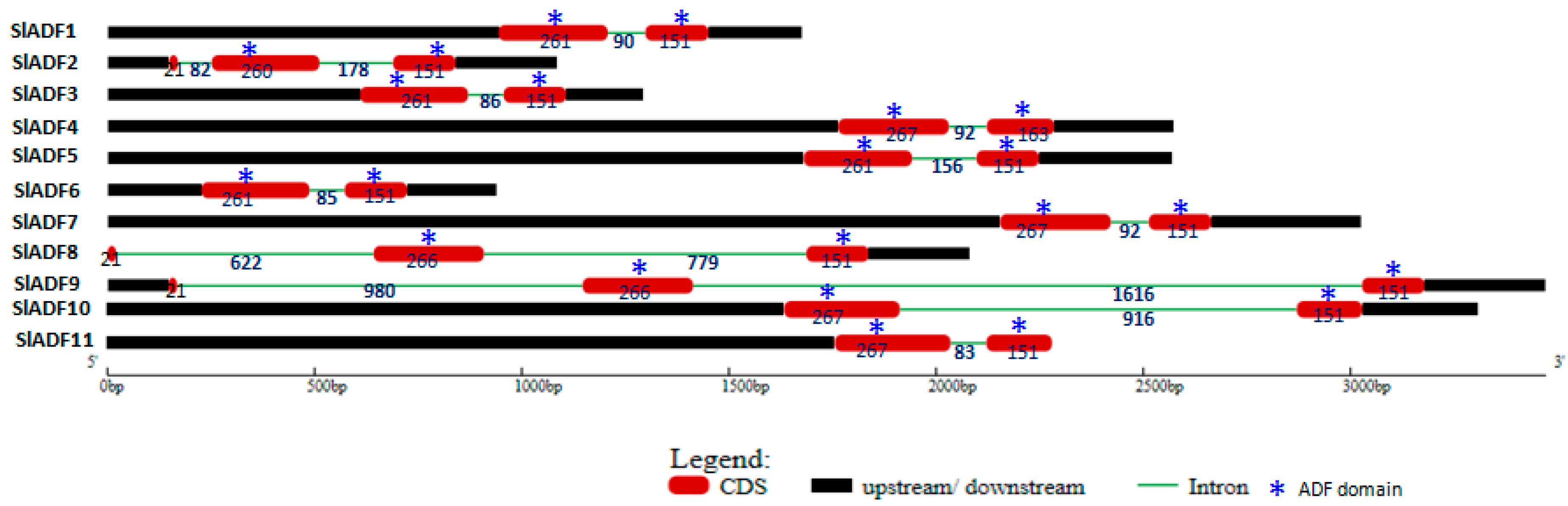
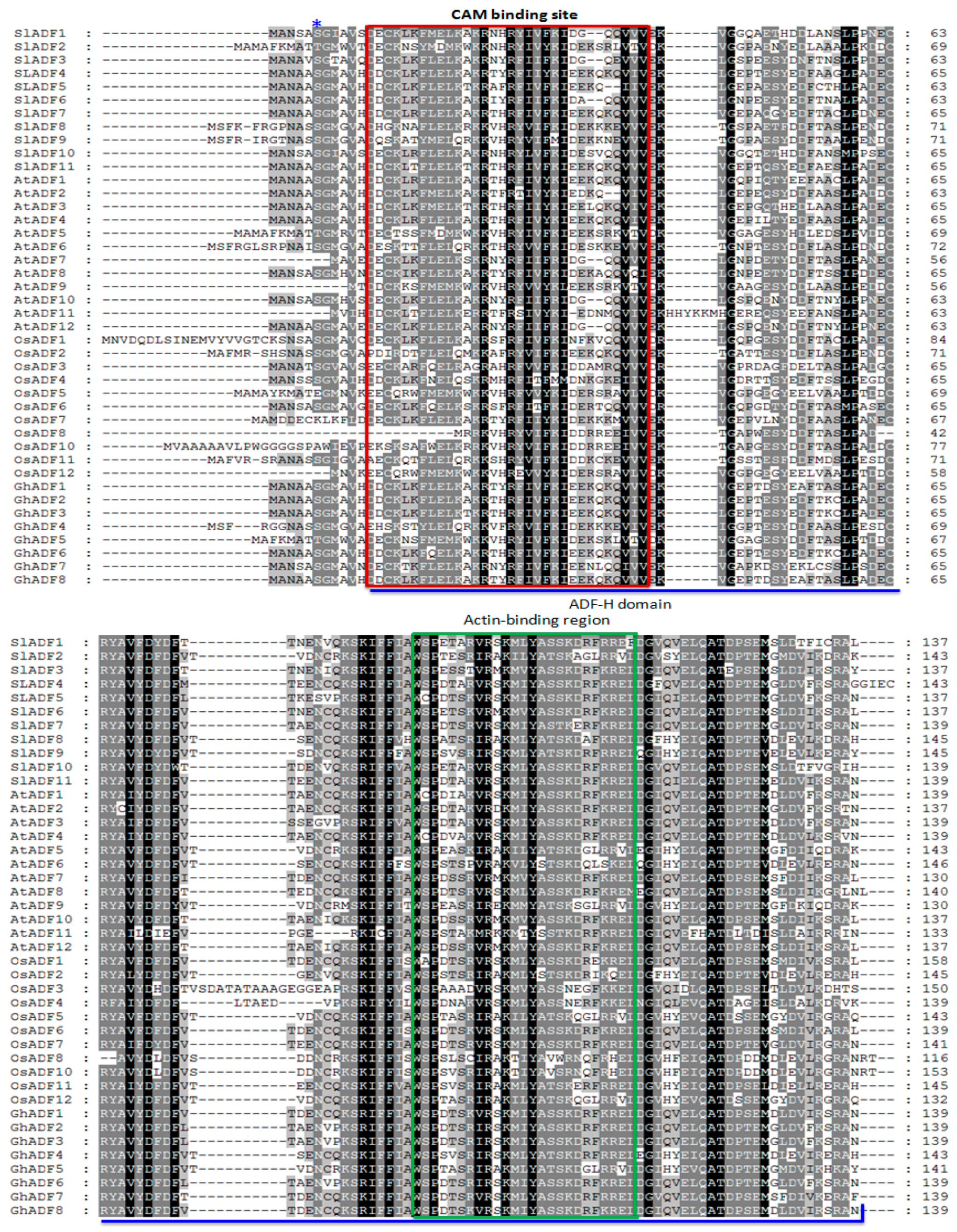
3.1. Sequence Analysis and Genomic Organization of Tomato ADF Genes and Corresponding Proteins

The 11 ADF genes that were identified in this study were designated as S. lycopersicum ADF (SlADF) and the corresponding encoded proteins were named SlADF. The predicted size of the 11 SlADFs ranged from 137–145 amino acids (aa) (Table 1). The ADF-H domain was found in all 11 tomato ADFs, beginning at 12–18 aa and ending at 137–145 aa (Table 1). The predicted isoelectric point of the SlADFs varied from 5.12 to 8.69 indicating some ADF proteins were acidic, while others were basic (Table 1). The grand average of hydropathy (GRAVY) of the SlADFs varied from −0.293 to −0.598, showing that the proteins had hydrophilic characteristics (Table 1). The subcellular localization of the proteins was predicted to be extracellular, and it is possible that all are multi-located proteins found both in the nucleus and in the cytoplasm (Table 1). Analysis of the genomic structure of the ADFs revealed that SlADF2, SlADF8 and SlADF9 contain three exons and two introns at conserved positions, with the first coding sequence was much smaller in size compared to the other two coding sequences (Figure 1). The ADF-H domains were located in the 2nd and 3rd exons of these three genes (Figure 1). The other eight ADF genes contained two exons and one intron and the ADF-H domain were located in both of the exons (Figure 1). The tomato ADFs shared more than 80% sequence similarity with Arabidopsis and other plant ADFs, with a range of 75% to 91% (Table S2). The sequence identity among the 11 tomato ADFs ranged from 51% to 89% (Table S3). SlADF7, SlADF5, SlADF4 and SlADF11 shared more than 80% similarity; SlADF3 shared more than 80% similarity with SlADF6; SlADF4 shared more than 80% similarity with SlADF5; and SlADF1 shared more than 80% similarity with SlADF10, indicating possible gene duplication. Alignment of the predicted tomato ADFs with the ADFs of Arabidopsis and rice revealed that the tomato proteins contained the conserved serine residue that might be the putative phosphorylation site of ADFs and a CAM (calmodulin) binding region at the N-terminus region (Figure 2). The alignment also revealed that the ADF-H domain position and the possible actin-binding region were conserved in all of the ADFs (Figure 2). Motif searches revealed that motifs 2 and 3 were characteristic of the N-terminus and motifs 1 and 4 were characteristic of the C-terminus. The putative phosphorylation site was found in motif 2, the CAM binding region was located in motif 3, and the PIP2/actin binding region was found in motif 1 (Figure S1).

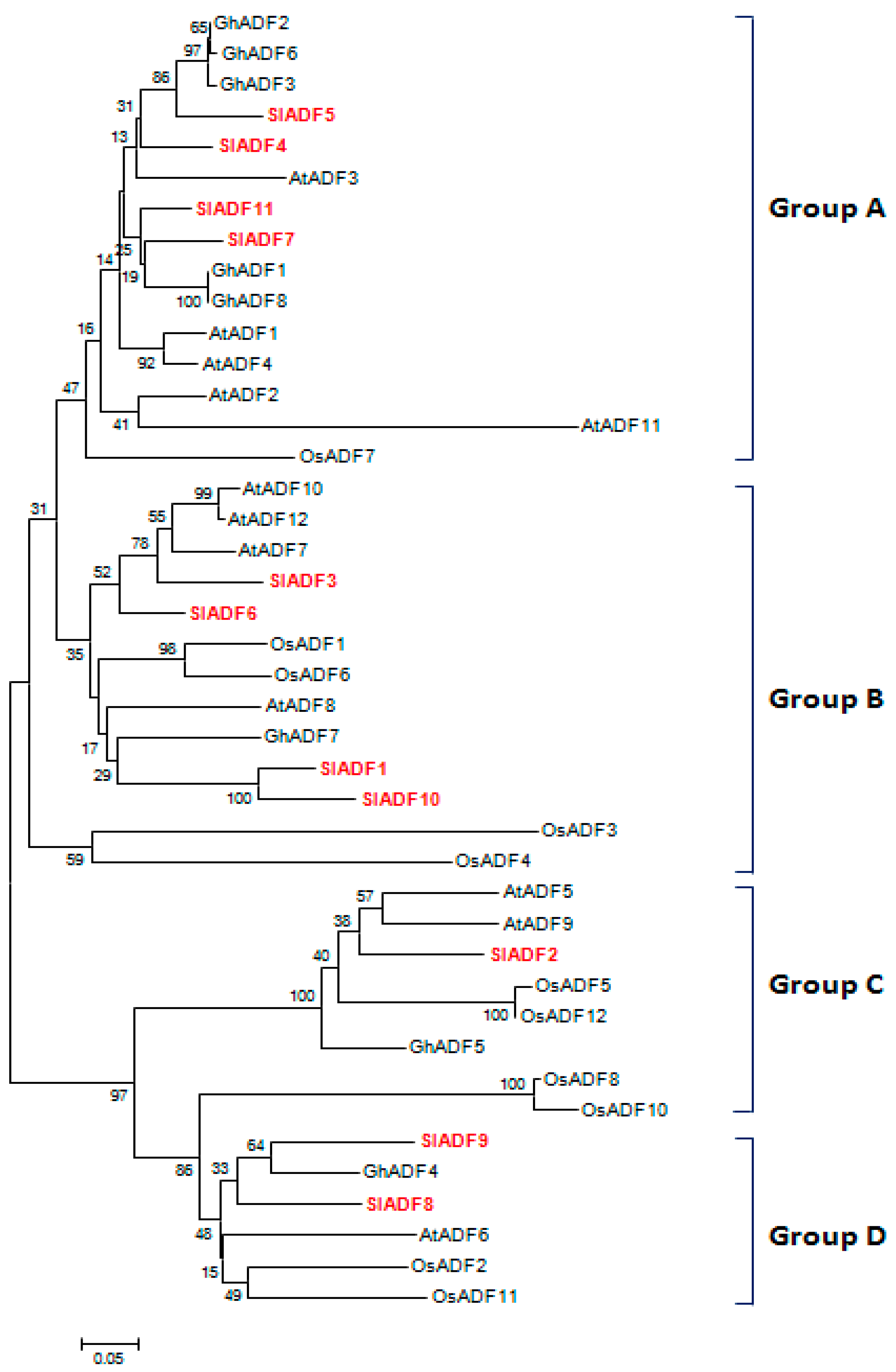
3.2. Phylogenetic Analysis of SlADF Proteins

The phylogenetic tree classified the ADF proteins into four main groups (Figure 3A–D), consistent with published data and expression profiles of ancient ADF proteins [7,8]. SlADF4, SlADF5, SlADF7 and SlADF11 clustered in Group A with Arabidopsis, rice and cotton ADFs; members of group A from these other species are strongly expressed in a wide range of tissues such as root, leaf, and flower (Figure 3) [7]. SlADF1, SlADF3, SlADF6 and SlADF10 belonged to Group B along with Arabidopsis, rice and cotton ADFs that are expressed in root and reproductive tissues (Figure 3) [7]. SlADF2 clustered in Group C, and SlADF8 and SlADF9 clustered in Group D. Group C and D ADFs from other plants are expressed moderately in all tissues (Figure 3) [7].

3.3. Chromosomal Position and Duplication of Tomato ADF Genes

The 11 tomato ADF genes were manually mapped on the 12 tomato chromosomes based on MapChart results (Figure S2). The ADF genes were unevenly distributed, with three genes found on chromosome 9, two on chromosomes 6 and 10, and one on chromosomes 1, 3 and 4. Segmental duplication or tandem duplication during evolution are responsible for generating the different gene families. Among the 11 tomato ADF genes, SlADF1, SlADF3, SlADF4, SlADF5, SlADF6, SlADF7 and SlADF11 were found to be segmentally duplicated as those genes had more than 80% identity and query coverage (Figure S2 and Table S4). The duplicated genes were located on chromosomes 1, 3, 4, 5, 6, 9 and 10. None of the genes had tandem duplication.
Several cis-acting elements related to development, tissue specific expression, seed specific regulation, abiotic and biotic stress response, auxin and ethylene response, and circadian regulation were identified (HD-Zip 1, HD-Zip 2, RY-element, as-2-box, Skn-1_motif, Box-W1, TC-rich repeats, ERE, TGA-element, AuxRR-core, HSE, MBS, CE3, MRE, TCA-element, O2-site and circadian), suggesting the possible involvement of this gene family in development and stress tolerance (Table S5).

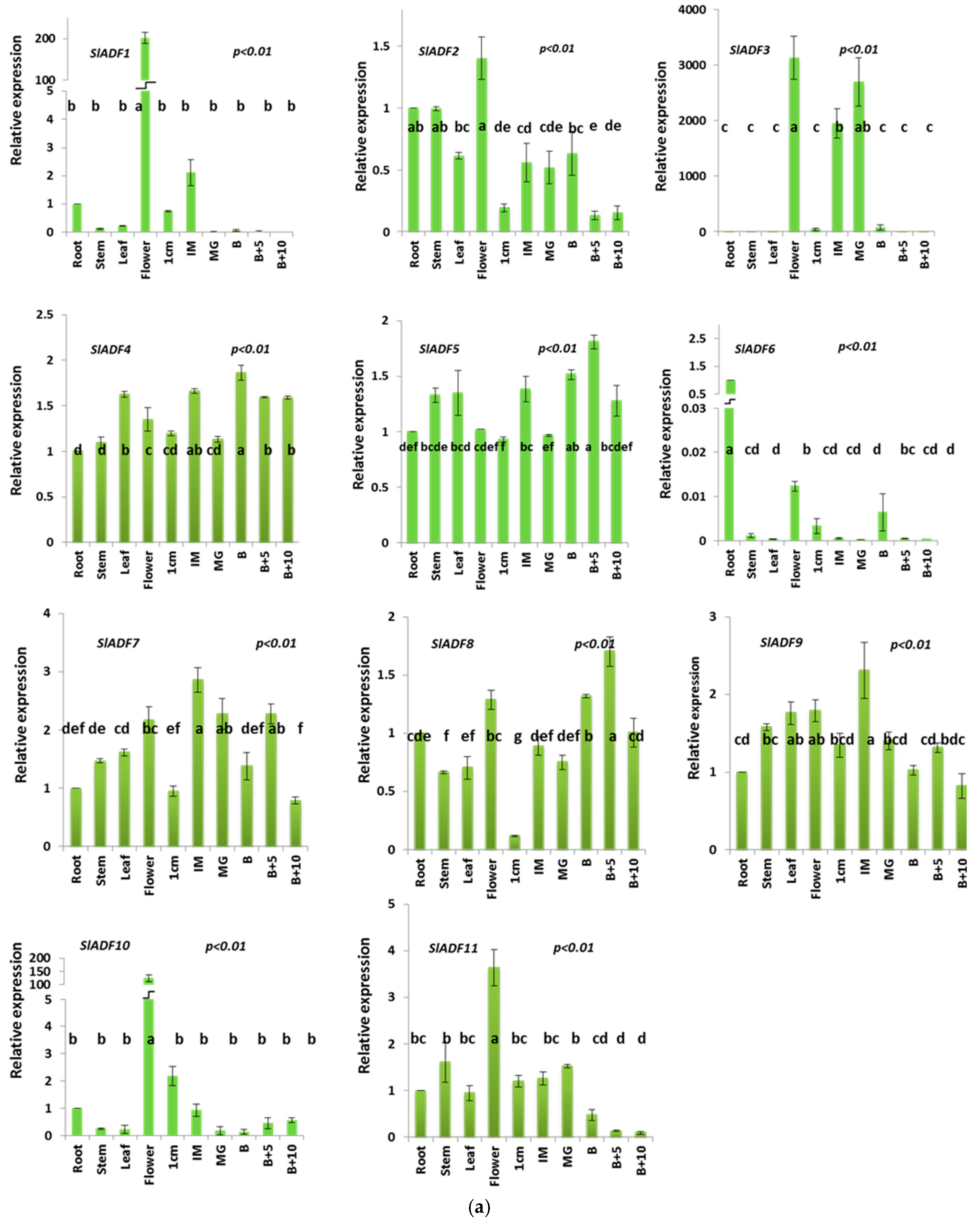
3.4. Expression Analysis of Tomato ADF Genes in Different Organs

The expression patterns of the different tomato ADF genes are shown in Figure 4a. Among the 11 genes, SlADF2, SlADF4, SlADF5, SlADF7, SlADF8, SlADF9 and SlADF11 were expressed at variable levels in all organs examined including the different stages of fruits. SlADF2 and SlADF11 were highly expressed in flowers compared to other organs. The expression of SlADF2 was about three times higher and that of SlADF11 was about four times higher in flowers than fruits at ten days after the breaker stage. SlADF1 and SlADF10 showed flower-specific expression whereas SlADF6 was preferentially expressed in roots. SlADF3 was predominantly expressed in flowers and in fruits at the immature and mature green stage. When the expression profiles at the different fruit developmental stages was compared, we found that SlADF3, SlADF9 and SlADF11 showed significantly higher expression at the early stages, mainly in the immature and mature green stage, compared to 5 and 10 days after breaker stage. By contrast, SlADF5 and SlADF8 strongly expressed at 5 days after the breaker stage, the peak of fruit ripening, compared to other stages of fruit development. Expression of the ADF genes was further investigated in different parts of the flower, sepals, petals, stamen and ovary (Figure 4b). SlADF1, SlADF3 and SlADF10 were predominately expressed in stamen compared to other flower parts. SlADF6, SlADF7, SlADF8 and SlADF9 also had higher expression levels in the stamen compared to other parts of the flower. SlADF2 had about six times higher expression in the petals than in the sepals, stamen and ovary. Expression of SlADF4 and SlADF5 was higher in the petals and stamen compared to the sepals and ovary.

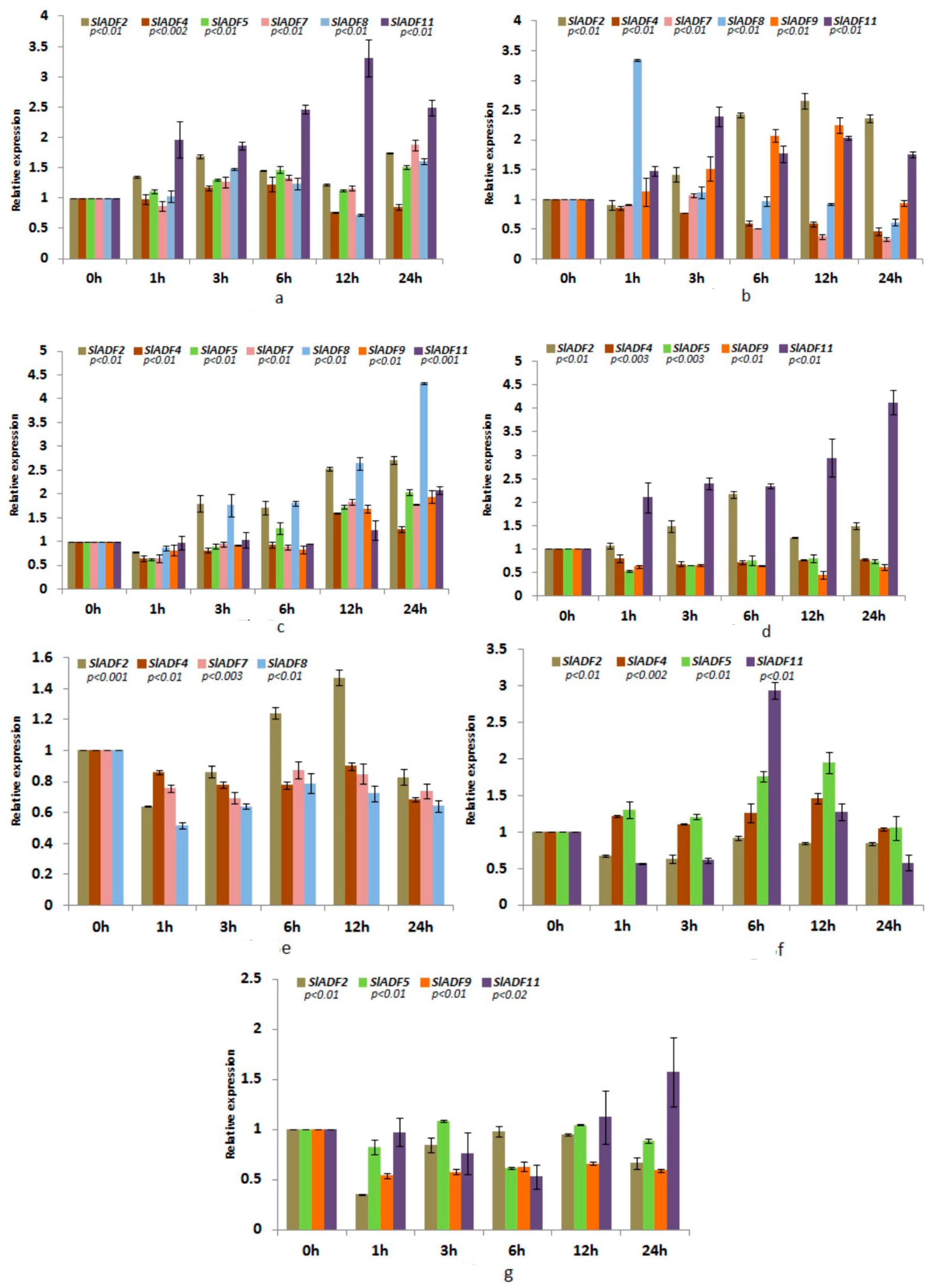
3.5. Expression Analysis of Tomato ADF Genes under Different Abiotic Stresses

Seven of the tomato ADF genes (SlADF2, SlADF4, SlADF5, SlADF7, SlADF8, SlADF9 and SlADF11) were differentially regulated by the abiotic stresses examined in this study (Figure 5a–g).

3.5.1. Cold Stress

The expression patterns of the cold-treated samples are shown in Figure 5a. The expression of SlADF2, SlADF5 and SlADF11 was significantly up-regulated under cold stress compared to the control. SlADF4 showed a slight up-regulation until 6 h of cold treatment but was then down-regulated in the remaining period compared to the control. The expression of SlADF7 remained increased after 1 h and until 24 h of treatment compared to the control. The expression of SlADF8 increased until 6 h of treatment, then decreased at 12 h followed by another increase at 24 h compared to the control.

3.5.2. Heat Stress

The expression patterns of the heat-treated samples are shown in Figure 5b. The expression of SlADF2 and SlADF11 was significantly up-regulated in response to heat treatment compared to the control while SlADF4 was down-regulated. The expression of SlADF7 was reduced compared to the control with the exception of a slight increase at 3 h. Markedly higher expression of SlADF8, around three-fold higher than the control, was found after 1 h of treatment while the expression level remained similar to the control for the remaining treatment period. SlADF9 was gradually up-regulated during treatment until 12 h followed by a decrease at 24 h of treatment.

3.5.3. Drought Stress

The expression patterns of the drought-stressed samples are shown in Figure 5c. The expression of SlADF2 was gradually up-regulated during drought stress treatment. SlADF4, SlADF7, SlADF9 and SlADF11 were slightly down-regulated until 6 h followed by an increase at 12 h and 24 h of treatment compared to the control. The expression of SlADF5 was down-regulated until 3 h and then gradually increased during the remaining treatment period compared to the control. The expression level of SlADF8 significantly increased after 1 h and around four times higher expression was observed after 24 h of treatment compared to the control.

3.5.4. NaCl Stress

The expression patterns of the NaCl-treated samples are shown in Figure 5d. Under NaCl stress, the expression levels of SlADF2 and SlADF11 were significantly up-regulated while SlADF4, SlADF5 and SlADF9 were down-regulated compared to the control.

3.5.5. ABA Treatment

The expression patterns of the ABA-treated samples are shown in Figure 5e. The expression levels of SlADF4, SlADF7 and SlADF8 were down-regulated under ABA treatment compared to the control. The expression of SlADF2 increased only at 6 h and 12 h of treatment compared to the control.

3.5.6. JA Treatment

The expression patterns of the JA-treated samples are shown in Figure 5f. The expression levels of SlADF4 and SlADF5 were slightly up-regulated while SlADF2 was down-regulated under JA treatment compared to the control. SlADF11 was highly expressed, around three-fold higher compared to the control, after 6 h of treatment while its expression was reduced during the remainder of the treatment period.

3.5.7. Wounding

The expression patterns of the wounded samples are shown in Figure 5g. SlADF2 and SlADF9 were slightly down-regulated during wounding stress compared to the control. SlADF5 showed slight up-regulation compared to the control at 3 h and 12 h of treatment but was down-regulated during the other time periods. SlADF11 was gradually down-regulated until 6 h followed by an increase at 12 h and 24 h of treatment compared to the control.

4. Discussion

The ADF protein family is highly conserved both structurally and functionally, even in distantly related species like yeast and mammals and believed to have a similar function in dynamic actin cytoskeleton organization and regulation [26]. The N-terminus serine residue has been found to regulate the activity of ADF proteins both in vertebrates and in maize [27,28,29]. A calcium-dependent calmodulin-like domain protein kinase is involved in the phosphorylation of the serine residue [10]. The phosphorylation of ADF may be linked to the calcium signaling pathway and thereby reorganize the cytoskeleton in response to environmental and developmental signals. Except SlADF2, all other 10 SlADF proteins had a conserved serine residue followed by a glycine residue in their N-terminus region, which might be phosphorylated as in other known plant ADF proteins (Figure 2) [30,31]. The N-terminus serine of SlADF2 was substituted by a threonine residue, which could probably be phosphorylated by a regulatory kinase (Figure 2). The structural similarity of the tomato ADF genes and proteins to those of Arabidopsis and rice suggested that the SlADFs have similar actin binding and severing activity in dynamic actin cytoskeleton reorganization. The phylogenetic classification of 11 tomato ADF proteins clearly confirmed the existence of the four previously identified groups and was consistent with the findings of Feng et al. (2006) [8]. The high homology of the tomato ADFs with those of Arabidopsis and rice in the phylogenetic tree suggests that they evolved from common ancestors and also indicates their functional similarities in flowering plants. Due to considerable variation in the sequences, some of the branch bootstrap values in the phylogenetic tree were found to be insignificant.
Actin binding proteins may changes the actin cytoskeleton rearrangement through different intracellular signaling processes. The ADF/cofilin proteins bind to F-actin by severing the actin filaments and also by dissociating the globular actin (G-actin) from the pointed or minus end of actin and thereby enhancing the filament turnover rate [12,32,33,34,35]. Filamentous actin (F-actin) has been reported to be involved in pollen tube growth, trichome morphogenesis, root hair tip growth and fiber elongation [36,37]. The F-actin cytoskeleton is also involved in flower induction during petal development by transporting signals between cells [38]. In Gossypium hirsutum, the down-regulation of GhADF1 accelerates cotton fiber length and strength as a result of formation of more abundant F-actin filaments [39]. The differential expression of SlADF genes in different tissues implies that functional variation might have arisen during evolution. The expression of different SlADF genes was also consistent with the ancient classification of ADF family genes [7]. The predominant expression of SlADF1, SlADF3 and SlADF10 (Figure 4a,b) in flowers, especially in the stamen suggests that these three genes might have an association with flower and pollen development and other regulatory factors. Members of plant ADFs have been reported to be involved in the flower induction signaling pathway and in pollen tube growth through the rearrangement of the actin cytoskeleton [16,31,36]. AtADF proteins regulate F-actin organization and have important functions in physiological processes including cell and organ expansion in Arabidopsis [13].
Plants absorb water and minerals through their roots for growth and development; accordingly, proper development of roots and root hairs is crucial for adaptation and vigor. Active accumulation of ADF was found in the root tip region to sustain the turnover of the F-actin that is required for the cell elongation [37] and it has been reported that ADFs are associated with root formation and root hair development in plants [14,37]. SlADF6 was predominately expressed in roots (Figure 4a), suggesting that as a regulator of actin dynamics, SlADF6 could reset the cytoskeletal network during root development of tomato and thereby might play a role in root formation and development. The other seven tomato ADF genes were expressed at various levels in different organs, suggesting that they might have various regulatory functions in tomato growth and development. The function of ADF genes in plant growth and development has been confirmed in other species. For instance, reduced expression of AtADF1 stimulated cell expansion and organ growth by promoting actin cable formation, whereas overexpression of AtADF1 resulted in the disappearance of thick actin cables and reduced cell and organ growth [13]. In addition, shorter hypocotyls and decreased root hair formation was observed upon ectopic expression of Gossypium barbadense ADF1 in tobacco [40]. However, it remains to be explored whether SlADF genes regulate growth and development of tomato by organizing actin cytoskeleton structure.
Actin binding proteins (ABPs) control the functional link between the F-actin and the auxin flow [41]. In addition to cell elongation, auxins regulate fruit set, growth and ripening, apical dominance, and leaf senescence, as well as playing an essential role in tropic responses to light and gravity [42]. In tobacco, ADF2 might control the dynamics of cortical actin filaments, which is required for functional auxin-dependent signaling with respect to synchronized cell division [43,44]. Tomato fruit attains its final size through successive cell divisions until 1 cm fruit stage, following by cell expansion until the immature and mature green fruit stage. The cell expansion phases, IM and MG, are the longest phases of fruit development. Depending on the cultivar, about 90% of fruit weight increase occurs during these phases [45]. During the cell expansion phase, auxin produced from the seeds and surrounding tissues accelerates fruit growth [46]. The significantly higher expression of SlADF11 in the early developmental, 1 cm, immature (IM) and mature green (MG) stages, and of SlADF3, SlADF9 and SlADF11 in the IM and MG stages compared to the ripening stages suggested that those SlADF genes might have active roles controlling actin filaments during fruit set and expansion through the auxin signaling pathway. Ripening of tomato fruits is related to the level of ethylene production [47]. Ethylene production starts to increase at the breaker stage and reaches its maximum level at the full ripening stage [47]. SlADF5 and SlADF8 were predominately expressed at five days after breaker stage, the peak of ripening, indicating their relatedness in fruit ripening through the ethylene biosynthesis pathway. Moreover, auxin- and ethylene-responsive cis-elements were also found in most of the SlADF genes, indicating their possible function in the early development and ripening of tomato fruit. Cell expansion and organ growth related functions of ADF genes are also found in Arabidopsis [31]. In maize, ZmADF3 is expressed to different levels in various tissues and developmental stages during kernel development, in terms of both RNA and proteins levels, implicating them as having important roles in kernel development [48]. These possible functions of SlADFs in fruit enlargement and ripening should be the foundation for further functional analysis of this gene family in tomato.
The actin cytoskeleton is reorganized by ADF in response to external and internal stimuli, and thereby changes the morphology of the cell. Accordingly, cytoskeleton rearrangement is used as a marker for various stress signaling pathways such as salt, cold, gravity, osmotic pressure, wounding and pathogen attack [49,50,51,52]. Together, ADF, actin-related protein 2 (ARP2) and actin-binding protein 29 (ABP29) are involved in actin cytoskeleton remodeling under salt stress conditions [53]. The rapid reorganization of the cytoskeleton structure promotes salinity tolerance in cotton roots [54]. The differential assembly of actin filaments under salt stress is crucial for salt stress tolerance in Arabidopsis [54]. The possible association of ADF in dynamic cytoskeleton rearrangement may provide a clue to abiotic stress tolerance mechanisms in plants. Proteomics studies suggested the involvement of OsADF1 and OsADF3 in drought tolerance in an upland rice variety [55,56]. Marked variation in the expression levels of different tomato ADF genes was observed under the different stresses. Based on this differential expression we propose that the SlADF proteins might remodel the cytoskeleton structure in response to different stress signals in cell and this remodeling might be important for the enhancement of stress tolerance in tomato. Notably, abiotic stress-responsive cis-regulatory elements were found in most of the SlADF gene sequences (Supplementary Table S5) supporting the likelihood of abiotic stress tolerance-related functions for the tomato ADF genes. In transgenic Arabidopsis, the ectopic over-expression of OsADF3 conferred mannitol- and drought-stress tolerance by promoting the germination rate, primary root length and survival of seedlings [57]. Further screening and functional analysis of SlADF genes are needed to explore the possible relationship between actin remodeling and the physiological functions of SlADF genes triggered by different stresses.

5. Conclusions

ADFs regulate actin cytoskeletal organization in response to different cellular activities and cell signaling events. In addition to cytoskeleton reorganization, ADFs are involved in organ development in plants. This study represents the first comprehensive study of this important gene family in a vegetable crop. The 11 identified ADF genes were phylogenetically classified into four groups showing consistency with their organ-specific expression patterns. Three genes that were preferentially expressed in the flower and especially the stamen, SlADF1, SlADF3 and SlADF10, are likely associated with flower and pollen development. The unique expression of SlADF6 in roots makes it a good candidate gene for functioning in root development of tomato. We also analyzed the expression of tomato ADFs under different abiotic and phytohormone stresses. The expressions of SlADF2, SlADF4, SlADF5, SlADF6, SlADF7, SlADF8, SlADF9 and SlADF11 were induced by the different stresses, suggesting that these genes may have stress tolerance functions in tomato. These results will be helpful for further functional validation of candidate genes in relation to vegetative growth, reproductive development and stress tolerance of tomato.

Supplementary Materials

The following are available online at www.mdpi.com/2073-4425/7/10/79/s1. Figure S1: Schematic representation of the conserved motif identified in the tomato ADF proteins. Different colors represent different motifs. The motif order in the figure corresponds to their position in the individual protein sequences. The name of each member is shown on the left side of the figure, Figure S2: Positions of the 11 ADF genes along the 12 tomato chromosomes. The black dotted lines represent the duplicated genes in the genome. Chromosome sizes and gene positions were estimated using the scale in megabases (Mb) to the left of the figure, Table S1: Primer sequences used for qPCR analysis of tomato ADF genes, Table S2: Similarity analysis of the tomato ADF gene family, Table S3: Sequence identity among the 11 ADF proteins of tomato, Table S4: Segmentally duplicated genes among the 11 tomato ADF genes, Table S5: Putative cis-elements of more than 5 bp identified in the 11 tomato ADF genes using the PlantCARE database, Additional File 1: Putative protein sequences of tomato, Arabidopsis, rice and cotton used in in silico analysis.

Acknowledgments

This research was supported by the Golden Seed Project (Center for Horticultural Seed Development, No. 213003-04-4-SBV10), Ministry of Agriculture, Food and Rural Affairs (MAFRA), Ministry of Oceans and Fisheries (MOF), Rural Development Administration (RDA) and Korea Forest Service (KFS).

Author Contributions

Mi-Young Chung, Ill-Sup Nou, Chang Kil Kim, Ki-Byung Lim, Min-Bae Kim, Do-Jin Lee and Jong-In Park designed and supervised the work. Mi-Young Chung and Jong-In Park monitored the experimental work. Khadiza Khatun conducted the in silico analysis, grew the plants, isolated RNA and carried out the expression analysis under the guidance of Mi-Young Chung. Arif Hasan Khan Robin analyzed the data and extensively revised the initial manuscript prepared by Khadiza Khatun. All authors contributed to the manuscript.

Conflicts of Interest

The authors declare no conflict of interest exists.

References

  1. Ouellet, F.; Carpentier, E.; Cope, M.J.T.V.; Monroy, A.F.; Sarhan, F. Regulation of a wheat actin-depolymerizing factor during cold acclimation. Plant Physiol. 2001, 125, 360–368. [Google Scholar] [CrossRef] [PubMed]
  2. Hepler, P.K.; Vidali, L.; Cheung, A.Y. Polarized cell growth in higher plants. Annu. Rev. Cell Dev. Biol. 2001, 17, 159–187. [Google Scholar] [CrossRef] [PubMed]
  3. Drobak, B.K.; Franklin-Tong, V.E.; Staiger, C.T. The role of the actin cytoskeleton in plant cell signaling. New Phytol. 2004, 163, 13–30. [Google Scholar] [CrossRef]
  4. Bamburg, J.R.; Harris, H.E.; Weeds, A.G. Partial purification and characterization of an actin depolymerizing factor from brain. FEBS Lett. 1980, 121, 178–182. [Google Scholar] [CrossRef]
  5. Chi, J.; Wang, X.; Zhou, H.; Zhang, G.; Sun, Y.; Li, Z.; Ma, Z. Molecular cloning and characterization of the actin-depolymerizing factor gene in Gossypium barbadense. Genes Genet. Syst. 2008, 83, 383–391. [Google Scholar] [CrossRef] [PubMed]
  6. Maciver, S.K.; Hussey, P.J. The ADF/cofilin family: Actin-remodeling proteins. Genome Biol. 2002. [Google Scholar] [CrossRef]
  7. Ruzicka, D.R.; Kandasamy, M.K.; McKinney, E.C.; Burgos-Rivera, B.; Meagher, R.B. The ancient subclasses of Arabidopsis Actin Depolymerizing Factor genes exhibit novel and differential expression. Plant J. 2007, 52, 460–472. [Google Scholar] [CrossRef] [PubMed]
  8. Feng, Y.; Liu, Q.; Xue, Q. Comparative study of rice and Arabidopsis actin-depolymerizing factors gene families. J. Plant Physiol. 2006, 163, 69–79. [Google Scholar] [CrossRef] [PubMed]
  9. Poukkula, M.; Kremneva, E.; Serlachius, M.; Lappalainen, P. Actin-depolymerizing factor homology domain: A conserved fold performing diverse roles in cytoskeletal dynamics. Cytoskeleton 2011, 68, 471–490. [Google Scholar] [CrossRef] [PubMed]
  10. Allwood, E.G.; Smertenko, A.P.; Hussey, P.J. Phosphorylation of plant actin-depolymerising factor by calmodulin-like domain protein kinase. FEBS Lett. 2001, 499, 97–100. [Google Scholar] [CrossRef]
  11. Augustine, R.C.; Vidali, L.; Kleinman, K.P.; Bezanilla, M. Actin depolymerizing factor is essential for viability in plants, and its phosphoregulation is important for tip growth. Plant J. 2008, 54, 863–875. [Google Scholar] [CrossRef] [PubMed]
  12. Maciver, S.K. How ADF/cofilin depolymerizes actin filaments: Commentary. Curr. Opin. Cell Biol. 1998, 10, 140–144. [Google Scholar] [CrossRef]
  13. Dong, C.H.; Xia, G.X.; Hong, Y.; Ramachandran, S.; Kost, B.; Chua, N.H. ADF proteins are involved in the control of flowering and regulate F-actin organization, cell expansion, and organ growth in Arabidopsis. Plant Cell 2001, 13, 1333–1346. [Google Scholar] [CrossRef] [PubMed]
  14. Thomas, P.; Schiefelbein, J. Cloning and characterization of an actin depolymerizing factor gene from grape (Vitis vinifera L.) expressed during rooting in stem cuttings. Plant Sci. 2002, 162, 283–288. [Google Scholar] [CrossRef]
  15. Chen, C.Y.; Chung, A.Y.; Wu, H. Actin-depolymerizing factor mediates Rac/Rop GTPase-regulated pollen tube growth. Plant Cell 2003, 15, 237–249. [Google Scholar] [CrossRef] [PubMed]
  16. Lopez, I.; Anthony, R.G.; Maciver, S.K.; Jiang, C.J.; Khan, S.; Weeds, A.G.; Hussey, P.J. Pollen specific expression of maize genes encoding actin depolymerizing factor-like proteins. Proc. Natl. Acad. Sci. USA 1996, 93, 7415–7420. [Google Scholar] [CrossRef] [PubMed]
  17. Deng, Z.; Liu, X.; Chen, C.; Tian, W.; Xia, Z.; Li, D. Molecular cloning and characterization of an actin-depolymerizing factor gene in Hevea brasiliensis. Afr. J. Biotechnol. 2010, 9, 7603–7610. [Google Scholar]
  18. Clément, M.; Ketelaar, T.; Rodiuc, N.; Banora, M.Y.; Smertenko, A.; Engler, G.; Abad, P.; Hussey, P.J.; de Almeida Engler, J. Actin-depolymerizing factor2-mediated actin dynamics are essential for root-knot nematode infection of Arabidopsis. Plant Cell 2009, 21, 2963–2979. [Google Scholar] [CrossRef] [PubMed]
  19. Miklis, M.; Consonni, C.; Bhat, R.A.; Lipka, V.; Schulze-Lefert, P.; Panstruga, R. Barley MLO modulates actin-dependent and actin-independent antifungal defense pathways at the cell periphery. Plant Physiol. 2007, 144, 1132–1143. [Google Scholar] [CrossRef] [PubMed]
  20. Tian, M.; Chaudhry, F.; Ruzicka, D.R.; Meagher, R.B.; Staiger, C.J.; Day, B. Arabidopsis actin-depolymerizing factor AtADF4 mediates defense signal transduction triggered by the Pseudomonas syringae effector AvrPphB. Plant Physiol. 2009, 150, 815–824. [Google Scholar] [CrossRef] [PubMed]
  21. Porter, K.; Shimono, M.; Tian, M.; Day, B. Arabidopsis actin-depolymerizing factor-4 links pathogen perception, defense activation and transcription to cytoskeletal dynamics. PLoS Pathog. 2012, 8, e1003006. [Google Scholar] [CrossRef] [PubMed]
  22. Tamura, K.; Stecher, G.; Peterson, D.; Filipski, A.; Kumar, S. MEGA6: Molecular evolutionary genetics analysis version 6.0. Mol. Biol. Evol. 2013, 30, 2725–2729. [Google Scholar] [CrossRef] [PubMed]
  23. Kong, X.; Lv, W.; Jiang, S.; Zhang, D.; Cai, G.; Pan, J.; Li, D. Genome-wide identification and expression analysis of calcium-dependent protein kinase in maize. BMC Genom. 2013. [Google Scholar] [CrossRef] [PubMed]
  24. Aoki, K.; Yano, K.; Suzuki, A.; Kawamura, S.; Sakurai, N.; Suda, K.; Ooga, K. Large-scale analysis of full length cDNAs from the tomato (Solanum lycopersicum) cultivar Micro-Tom, a reference system for the Solanaceae genomics. BMC Genom. 2010, 11, 210. [Google Scholar] [CrossRef] [PubMed]
  25. Schmittgen, T.D.; Livak, K.J. Analyzing real-time PCR data by the comparative CT method. Nat. Protoc. 2008, 3, 1101–1108. [Google Scholar] [CrossRef] [PubMed]
  26. Aizawa, H.; Fukui, Y.; Yahara, I. Live dynamics of Dictyostelium cofilin suggests a role in remodeling actin lattice work into bundles. J. Cell Sci. 1997, 110, 2333–2344. [Google Scholar] [PubMed]
  27. Agnew, B.J.; Minamide, L.S.; Bamburg, J.R. Reactivation of phosphorylated actin depolymerizing factor and identification of the regulatory site. J. Biol. Chem. 1995, 270, 17582–17587. [Google Scholar] [CrossRef] [PubMed]
  28. Moriyama, K.; Iida, K.; Yahara, I. Phosphorylation of Ser-3 of cofilin regulates its essential function on actin. Genes Cells 1996, 1, 73–86. [Google Scholar] [CrossRef] [PubMed]
  29. Smertenko, A.P.; Jiang, C.J.; Simmons, N.J.; Weeds, A.G.; Davies, D.R.; Hussey, P.J. Ser6 in the maize actindepolymerizing factor, ZmADF3, is phosphorylated by calcium stimulated protein kinase and is essential for the control of functional activity. Plant J. 1998, 14, 187–193. [Google Scholar] [CrossRef] [PubMed]
  30. Zhang, C.; Guo, L.; Wang, X.; Zhang, H.; Shi, H.; Xu, W.; Li, X. Molecular characterization of four ADF genes differentially expressed in cotton. J. Genet. Genom. 2007, 34, 347–354. [Google Scholar] [CrossRef]
  31. Dong, C.H.; Kost, B.; Xia, G.; Chua, N.H. Molecular identification and characterization of the Arabidopsis AtADF1, AtADF5 and AtADF6 genes. Plant Mol. Biol. 2001, 45, 517–527. [Google Scholar] [CrossRef] [PubMed]
  32. Carlier, M.F.; Laurent, V.; Santolini, J.; Melki, R.; Didry, D.; Xia, G.X.; Hong, Y.; Chua, N.H.; Pantaloni, D. Actin depolymerizing factor (ADF/cofilin) enhances the rate of filament turnover: Implication in actin-based motility. J. Cell Biol. 1997, 136, 1307–1322. [Google Scholar] [CrossRef] [PubMed]
  33. Bamburg, J.R. Proteins of the ADF/cofilin family: Essential regulators of actin dynamics. Annu. Rev. Cell Dev. Biol. 1999, 15, 185–230. [Google Scholar] [CrossRef] [PubMed]
  34. Chen, H.; Bernstein, B.W.; Bamburg, J.R. Regulating actin-filament dynamics in vivo. Trends Biochem. Sci. 2000, 25, 19–23. [Google Scholar] [CrossRef]
  35. Yeoh, S.; Pope, B.; Mannherz, H.G.; Weeds, A. Determining the differences in actin binding by human ADF and cofilin. J. Mol. Biol. 2002, 315, 911–925. [Google Scholar] [CrossRef] [PubMed]
  36. Allwood, E.G.; Anthony, R.G.; Smertenko, A.P.; Reichelt, S.; Drobak, B.K.; Doonan, J.H.; Weeds, A.G.; Hussey, P.J. Regulation of the pollen-specific actin-depolymerizing factor LlADF1. Plant Cell 2002, 14, 2915–2927. [Google Scholar] [PubMed]
  37. Jiang, C.J.; Weeds, A.G; Hussey, P.J. The maize actin-depolymerizing factor, ZmADF3, redistributes to the growing tip of elongating root hairs and can be induced to translocate into the nucleus with actin. Plant J. 1997, 12, 1035–1043. [Google Scholar] [CrossRef] [PubMed]
  38. McLean, B.G.; Hempel, F.D.; Zambryski, P.C. Plant intercellular communication via plasmodesmata. Plant Cell 1997, 9, 1043–1054. [Google Scholar] [CrossRef] [PubMed]
  39. Wang, H.Y.; Wang, J.; Gao, P.; Jiao, G.L.; Zhao, P.M.; Li, Y.; Wang, G.L.; Xia, G.X. Down-regulation of GhADF1 gene expression affects cotton fibre properties. Plant Biotechnol. J. 2009, 7, 13–23. [Google Scholar] [CrossRef] [PubMed]
  40. Chi, J.; Han, Y.; Wang, X.; Wu, L.; Zhang, G.; Ma, Z. Overexpression of the Gossypium barbadense actin-depolymerizing factor 1 gene mediates biological changes in transgenic tobacco. Plant Mol. Biol. Rep. 2013, 31, 833–839. [Google Scholar] [CrossRef]
  41. Durst, S.; Nick, P.; Maisch, J. Nicotiana tabacum actin-depolymerizing factor 2 is involved in actin-driven, auxin-dependent patterning. J. Plant Physiol. 2013, 170, 1057–1066. [Google Scholar] [CrossRef] [PubMed]
  42. Davies, P.J. The plant hormones: Their nature, occurrence, and functions. In Plant Hormones; Springer: Dordrecht, The Netherlands, 2010; pp. 1–15. [Google Scholar]
  43. Campanoni, P.; Blasius, B.; Nick, P. Auxin transport synchronizes the pattern of cell division in a tobacco cell line. Plant Physiol. 2003, 133, 1251–1260. [Google Scholar] [CrossRef] [PubMed]
  44. Maisch, J.; Nick, P. Actin is involved in auxin-dependent patterning. Plant Physiol. 2007, 143, 1695–1704. [Google Scholar] [CrossRef] [PubMed]
  45. Lemaire-Chamley, M.; Petit, J.; Garcia, V.; Just, D.; Baldet, P.; Germain, V.; Rothan, C. Changes in transcriptional profiles are associated with early fruit tissue specialization in tomato. Plant Physiol. 2005, 139, 750–769. [Google Scholar] [CrossRef] [PubMed]
  46. Gillaspy, G.; Ben-David, H.; Gruissem, W. Fruits: A developmental perspective. Plant Cell 1993, 5, 1439–1451. [Google Scholar] [CrossRef] [PubMed]
  47. Giovannoni, J.J. Genetic regulation of fruit development and ripening. Plant Cell 2004, 16, 170–180. [Google Scholar] [CrossRef] [PubMed]
  48. Qiao, D.; Dong, Y.; Zhang, L.; Zhou, Q.; Hu, C.; Ren, Y.; Li, Y. Ectopic expression of the maize ZmADF3 gene in Arabidopsis revealing its functions in kernel development. PCTOC 2016, 126, 239–253. [Google Scholar] [CrossRef]
  49. Abdrakhamanova, A.; Wang, Q.Y.; Khokhlova, L.; Nick, P. Is microtubule disassembly a trigger for cold acclimation? Plant Cell Physiol. 2003, 44, 676–686. [Google Scholar] [CrossRef] [PubMed]
  50. Nick, P. Microtubules as sensors for abiotic stimuli. In Plant Microtubules; Springer: Berlin, Germany, 2008; pp. 175–203. [Google Scholar]
  51. Engler, J.A.; Rodiuc, N.; Smertenko, A.; Abad, P. Plant actin cytoskeleton remodeling by plant parasitic nematodes. Plant Signal. Behav. 2010, 5, 213–217. [Google Scholar] [CrossRef]
  52. Wang, C.; Zhang, L.; Huang, R.D. Cytoskelton and plant salt stress tolerance. Plant Signal. Behav. 2011, 6, 29–31. [Google Scholar] [CrossRef] [PubMed]
  53. Li, W.; Zhao, F.A.; Fang, W.; Xie, D.; Hou, J.; Yang, X.; Lv, S. Identification of early salt stress responsive proteins in seedling roots of upland cotton (Gossypium hirsutum L.) employing iTRAQ-based proteomic technique. Front. Plant Sci. 2015. [Google Scholar] [CrossRef] [PubMed]
  54. Wang, C.; Zhang, L.; Yuan, M.; Ge, Y.; Liu, Y.; Fan, J.; Ruan, Y.; Cui, Z.; Tong, S.; Zhang, S. The microfilament cytoskeleton plays a vital role in salt and osmotic stress tolerance in Arabidopsis. Plant Biol. 2010, 12, 70–78. [Google Scholar] [CrossRef] [PubMed]
  55. Yang, L.; Zheng, B.; Mao, C.; Yi, K.; Liu, F.; Wu, Y.; Tao, Q.; Wu, P. cDNA-AFLP analysis of inducible gene expression in rice seminal root tips under a water deficit. Gene 2003, 314, 141–148. [Google Scholar] [CrossRef]
  56. Chen, C.W.; Yang, Y.W.; Lur, H.S.; Tsai, Y.G.; Chang, M.C. A novel function of abscisic acid in regulation of rice (Oryza sativa L.) roots growth and development. Plant Cell Physiol. 2006, 47, 1–13. [Google Scholar] [CrossRef] [PubMed]
  57. Huang, Y.C.; Huang, W.L.; Hong, C.Y.; Lur, H.S.; Chang, M.C. Comprehensive analysis of differentially expressed rice actin depolymerizing factor gene family and heterologous overexpression of OsADF3 confers Arabidopsis Thaliana drought tolerance. Rice 2012. [Google Scholar] [CrossRef] [PubMed]
Figure 1. Schematic representation of the exon-intron distribution in the tomato actin depolymerizing factor (ADF) gene family. The red boxes represent the exons and the green lines represent the introns. Asterisks indicate the ADF-H domain position in the exon. The numbers represent the length of exons and introns in bp.
Genes 07 00079 g001
Figure 2. Alignment of all deduced tomato ADF polypeptides with those of Arabidopsis, rice and cotton. Asterisks indicate the putative serine phosphorylation site. The red box indicates the putative CAM binding site and the green box indicates the amino acid residues essential for actin binding. The dark black background highlights the identical amino acids and the light background highlights the amino acids with >50% identity. The blue underline represents the ADF-H domain position.
Genes 07 00079 g002
Figure 3. Phylogenetic tree of tomato actin depolymerizing factor (ADF) proteins with those of Arabidopsis, rice and cotton. The 11 tomato ADF proteins are shown in red. The deduced full-length polypeptide sequences were used to create the tree. The tree was constructed by the Neighbor-joining method in MEGA 6.0 software following the Poisson-model. The bootstrap values were calculated as a percentage of 1000 replicates. Bootstrap values were shown next to the branches. The scale represents the units of the number of amino acid substitutions per site. Protein sequences of Arabidopsis, rice and cotton were taken from published literature, the TAIR, RAP-DB, Cottongen and the NCBI database.
Genes 07 00079 g003
Figure 4. Tissue specificity of tomato ADF gene expression: (a) Expression levels of the 11 ADF genes via quantitative PCR in different organs; root, stem, leaf, whole flower and fruit at six developmental stages (1 cm: 1 centimeter sized fruit; IM: immature fruit; MG: mature green fruit; B: breaker; B + 5: five days after breaker; B + 10: 10 days after breaker). The standard error of the means of three independent replicates is represented by the error bars. p values indicate statistically significant variations of expression. Different lowercase letters (a, b, c, etc.) indicate statistically significant difference. (b) Expression levels of the 11 ADF genes via quantitative PCR in the different floral organs; sepal, petal, stamen and ovary. The standard error of the means of three independent replicates is represented by the error bars. p values indicate the statistically significant variations of expression. Mean values at different sampling points are represented by different letters.
Genes 07 00079 g004aGenes 07 00079 g004b
Figure 5. Stress-related ADF gene expression in tomato. Expression levels of the 11 ADF genes via quantitative PCR under different stresses: (a) cold; (b) heat; (c) drought; (d) NaCl; (e) abscisic acid (ABA); (f) Jasmonic acid (JA); and (g) wounding. The standard error of the means of three independent replicates is represented by the error bars. P-values indicate the statistically significant variations of expression. Mean values at different sampling points are separated by different letters.
Genes 07 00079 g005
Table 1. In silico analysis of the actin depolymerizing factor (ADF) genes collected from the Solanum lycopersicum database.
Gene NameLocus NameORF (bp)Chromosomal LocationProteinSubcellular LocalizationNo. of Introns
Length (aa)ADF Domain Start–End (aa)MW (KDa)pIGRAVY
SlADF1Solyc01g094400414113712–13715.636.13−0.437Extracellular1
SlADF2Solyc01g111380432114316–14316.448.69−0.397Extracellular2
SlADF3Solyc03g025750414313712–13715.805.12−0.442Extracellular1
SlADF4Solyc04g011370432414312–13916.495.33−0.469Extracellular1
SlADF5Solyc06g005360414613712–13715.846.15−0.350Extracellular1
SlADF6Solyc06g035980414613712–13715.795.50−0.293Extracellular1
SlADF7Solyc09g010440420913912–13916.006.73−0.465Extracellular1
SlADF8Solyc09g072590450914518–14516.697.77−0.594Extracellular2
SlADF9Solyc09g0901104569 14518–14516.846.74−0.598Extracellular2
SlADF10Solyc10g0175504201013912–13915.985.63−0.524Extracellular1
SlADF11Solyc10g0846604201013912–13916.115.29−0.523Extracellular1
Abbreviations: ORF, open reading frame; bp, base pair; aa, amino acid; MW, molecular weight; KDa, kilo Dalton; pI, iso-electric point; GRAVY, grand average of hydropathy.

