Article

M1 Polarized Tumor-Associated Macrophages (TAMs) as Promising Prognostic Signature in Stage I–II Gastric Adenocarcinomas

1
Department of Human Pathology in Adult and Developmental Age “Gaetano Barresi”, University of Messina, 98125 Messina, Italy
2
Translational Molecular Medicine and Surgery, Department of Biomedical and Dental Sciences and Morphofunctional Imaging, University of Messina, 98125 Messina, Italy
3
Integrated Cancer Registry of Oriental Sicily, 95123 Catania, Italy
*
Author to whom correspondence should be addressed.
Gastrointest. Disord. 2021, 3(4), 207-217; https://0-doi-org.brum.beds.ac.uk/10.3390/gidisord3040020
Submission received: 15 October 2021 / Revised: 26 October 2021 / Accepted: 27 October 2021 / Published: 30 October 2021

Abstract

:
Tumor-associated macrophages (TAMs) may be noticed in gastric carcinomas (GC), but their clinicopathological significance has not been yet explored. From a histological review of 400 cases of tubular/papillary adenocarcinomas, 24 cases of stage I–II gastric adenocarcinomas with intraglandular and stromal TAMs were identified. Their clinicopathological features were compared with 72 pT-matched as well as stage-matched control cases of adenocarcinomas without TAMs. TAMs present in GC cases were present either in glands or in neoplastic stroma, showing an immunoreactivity for CD68 and CD80; sometimes, they were organized in mature granulomas with occasional giant cells. Therefore, the stained TAMs were reminiscent of a specific polarized macrophage M1 phenotype; however, in any case of our cohort, no M2 phenotype macrophages were documented by CD 163 and CD 204 immunostainings. Statistically, no significant differences in age, gender, tumor location, size, and lymphovascular and perineural invasion between the case group with TAMs and pT- as well as stage-matched controls were reported; furthermore, the case group showed lower frequency of lymph node metastasis (p = 0.02). In addition, a significantly different clinical course and overall survival rate were also observed in gastric adenocarcinomas with M1 TAMs (p = 0.02) in comparison to controls. These results suggest that tumor-associated M1 macrophages are related to a quite indolent growth and a better prognosis of patients with this peculiar variant of gastric adenocarcinomas.

1. Introduction

Tumor progression has been traditionally considered to be an evolutionary process with genetic/epigenetic alterations targeting only cancer cells [1]. However, it has become increasingly evident that the development and clinical behavior of a given tumor depend on tumor microenvironment (TME), which is constituted by blood vessels, immune cells, and extracellular matrix [2]. Macrophages are cellular protagonists of TME in many tumors [2,3,4]; specifically, tumor-associated macrophages (TAMs) have been demonstrated to provide a favorable microenvironment for tumor expansion and survival stimulating angiogenesis [2,3,4,5]. Environmental situations such as tumor hypoxia may activate this latter event; consequently, TAMs increase in hypoxic regions of neoplasm with the consequent acquisition of an invasive phenotype [4,5,6]. In detail, M1/M2 functional polarization of macrophages has been identified [5,6]; very simply, M1 macrophages kill infectious microorganisms or tumor cells, whereas M2 macrophages are associated with tumor progression and invasion, favoring TME [4,5,6]. However, recent evidence suggests that macrophages exhibit differences in their activation states and the identification of their phenotypes should be based on specific activation stimulus rather than a generic definition of M1/M2 macrophages, the first defined as “classically activated” (pro-inflammatory) and the second as “alternatively activated” (anti-inflammatory) [4,5,6,7,8,9]. Although TAMs infiltration alone has not been considered a prognostic significant parameter, it has been reported that high M1 macrophage infiltration is correlated with a better prognostic situation in colon-rectal cancer (CRC) patients in a stage-dependent manner [4,10]. By contrast, an increase in the proportion of M2/M1 type TAMs has been found to be directly related to the presence of liver metastases in CRC patients [4].
In gastric cancer (GC), depth of invasion, nodal status, and clinical stage have been positively associated with TAMs infiltration [11,12,13]. In addition, the role of stromal micro-hemorrhages in GC has been not well clarified, although some studies have analyzed the distribution pattern of TAMs [14,15,16,17]. Moreover, total TAMs as well the number of infiltrating M2 have been suggested as negative prognostic factors for GC patients, while M1 macrophage infiltration has been related to a better favorable prognosis [18]. To resolve these inconsistencies as well as to clarify the clinicopathological significance of TAMs, we have compared clinicopathological features of stage I–II gastric adenocarcinomas with pT- and stage-matched gastric controls.

2. Results

2.1. Incidence and Clinical Manifestations of Gastric Cancers with TAMs

From January 2005 to December 2020, 400 patients with tubular/papillary/mucinous gastric cancers underwent gastrectomy at the University Hospital of Messina (Italy). Of these, 24 patients (6%) histologically showed TAMs in neoplastic tissue. The age of patients ranged from 60 to 83 years (median: 72 years). The follow-up of patients ranged from 15 to 60 months (mean follow-up 57.2 months). Fifteen patients were men and nine were women. Tumors were in the fundus (n = 1), corpus (n= 10) or antrum (n = 13) of the stomach. Tumor sizes ranged from 30 to 70 mm (median: 52 mm). According to invasion gastric wall (pT), 2 cases were pT1, 14 pT2, and 8 pT3; stage I was encountered in 11 cases, while stage II was recorded in 13 cases. Only three patients died for the disease, and they were all in stage II; the other 21 were still alive with no evidence of disease (Table 1).
The age of 72 patients used as control group ranged from 61 to 84 years (median: 72.5 years). The follow-up ranged from 18 to 64 months (mean follow-up 59 months). Forty-three patients were men and 29 were women. Tumors were in the fundus (n = 2), corpus (n= 29) or antrum (n = 41) of the stomach. Tumor sizes ranged from 32 to 74 mm (median: 55 mm). According to invasion gastric wall (pT), 6 cases were pT1, 42 pT2, and 24 pT3; stage I was encountered in 33 cases, while stage II was recorded in 39 cases. Twenty-six patients died for the disease either in stage I or II; the other 46 were still alive with no evidence of disease (Table 1).

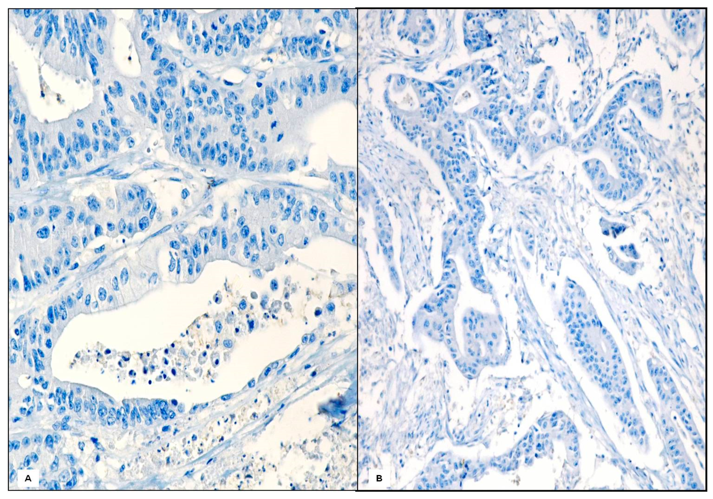
2.2. Histological Findings

At light microscopy, the tumors were tubular or papillary adenocarcinomas composed of medium to large glands lined by columnar cells with an eosinophilic or clear cytoplasm. Maximal depth of invasion was into the submucosa in 2 cases, muscularis propria in 14 cases and subserosa in 8 cases. Lymphovascular invasion was present in 8 cases, perineural invasion in 2 cases, whereas lymph node metastases were found in 7 cases. In these tumors, some neoplastic glands contained only foamy macrophages (Figure 1A,B) and other ones showed foamy macrophages in intimate contact with erythrocytes (Figure 1C). Sometimes, mainly when the glandular profile was interrupted, a mature granuloma with multinucleated giant cells was appreciable (Figure 1D).
By immunohistochemistry, the intraglandular TAMs exhibited an evident cytoplasmic immunoreactivity for CD68 (Figure 2A) and CD80 (Figure 2B) antisera. In any case of our cohort, the lack of CD 204 cells was documented by the negative immunostaining (Figure 2C). An equivalent immunohistochemical pattern was also encountered when TAMs were in tumor stroma along the front edge of the invasion (Figure 2D,E).
Obviously, no evidence of TAMs was documented in control GC cases and therefore, no immunoexpression of CD68 was recorded (Figure 3).

2.3. Comparison of Clinicopathologic Parameters in Case and Control Groups

Table 1 displays clinicopathological features in the case group compared with those in the pT- as well as stage-matched controls. The case group presented a lower frequency of lymph node metastases (p = 0.022) and better clinical course (p = 0.023). The case and control groups did not differ significantly with respect to age, gender, Hp status, tumor location, tumor size, gastric wall invasion (pT) lympho-vascular and/or perineural invasion.
By univariate analysis of cancer-specific mortality by Mantel–Cox log-rank test, stage (p = 0.000) and M1 macrophage presence (p = 0.035) emerged as prognostic parameters in the whole casuistry of gastric adenocarcinomas (Table 1).
In addition, by multivariate analysis, stage (p = 0.001) as well as M1 macrophages presence (p = 0.037) maintained their role as independent prognostic variables (Table 2).
Finally, survival analysis with Kaplan–Meier and log-rank tests showed the 5-year survival rate of the case group with TAMs was higher than pT- as well as stage-matched control group (Figure 4).

3. Discussion

TAMs are one of the most prominent immune components present in neoplastic conditions and their various unfavorable functions, such as contribution in metastatic mechanism, immune suppression and chemotherapy resistance, have been hypothesized [15]. However, the role of TAMs in GC is contradictory; in fact, TAMs were shown to be related to a stromal-associated gene link and poor patient outcome, but they have also been related to a high level of tumor cell apoptosis and good prognosis [19,20]. These conflicting data may be associated with their heterogeneity nature inside individual cancers [21,22,23,24,25,26,27,28,29]. Moreover, the description of TAMs in GC has been complicated due to their adaptive changes to environmental stimuli, the lack of limited markers between populations and the differences between human and animal models [25,26,27]. Hence, although numerous studies on macrophage heterogeneity, their significance in human tumors remains unclear [21,22,23,24,25,26,27,28,29].
It is well known that the main biological characteristic of macrophages is to express different functional strategies in response to different micro-environmental stimuli, usually evident in all pathological conditions, such as infections and cancer [5,7]. Typically, chronic injuries can strongly regulate the immune responses, being able to activate highly polarized type I or type II immunity [9,10]. Crucial to the development of type I or type II polarization is the specificity of the host-pathogen interaction [7,8,9,10]. Although intracellular pathogens induce a type I polarized inflammation, with numerous neutrophils, macrophage infiltrate, typical in granulomas, some agents trigger strong type II inflammation, characterized by extensive eosinophilia, mastocytosis and tissue remodeling [7,8,9,10]. However, granulomatous stromal reaction is rarely observed in GC, and it has been reported as epithelioid granuloma (sarcoid-like) [30,31,32].
In our GC study group with TAMs, foamy macrophages have been encountered either inside glandular lumen or in neoplastic stroma; moreover, sometimes TAMs were in intimate contact with erythrocytes. Occasionally, mainly when the glandular profile was interrupted, multinucleated giant cell granulomas were noted. By immunohistochemistry, the polarization of TAMs may be attributed to M1 phenotype, since an evident cytoplasmic immunoreactivity for CD68 and CD80 antisera was constantly encountered. By contrast, the absence of CD 163 and 204 immunostaining allows us to exclude M2 polarization in our GC cases. The same immunoreactive pattern was encountered in TAMs located in tumor stroma as well as in those present along the front edge of the invasion.
Literature provides positive or negative evidence on the prognostic significance of TAMs in cancer patients [18,33,34]. In some studies, CD204-positive tumor-associated M2 macrophages have been reported as a significant risk factor for adenocarcinoma development in gastric adenoma [35]; moreover, a high density of CD163+ TAMs has been found to be positively associated with a worse prognosis in GC [34,35]. Huang et al. [34] showed that increased density of CD163+ (CD206-) TAMs with concurrent high CD68 expression is associated with improved patient survival by univariate, but not by multivariate analysis. In contrast with the above-mentioned results, a meta-analysis demonstrated that a high density of TAMs is associated with better prognosis in patients with colorectal cancer [4]. However, these studies show a remarkable degree of heterogeneity in terms of ethnicity (Asian/Caucasian), stage (I, I–III, I–IV), macrophage identification (CD68, CD163 and/or CD206 stain/immunohistochemistry; antibody clones) and scoring [33,34,35,36,37]. This heterogeneity may account for prognostic differences observed between studies regarding TAMs (reviewed by Galdiero et al.) [38]. Moreover, TAMs have been identified by immunohistochemical techniques (CD68, CD80, CD163, CD 204 and/or CD206), but not related to their erythrophagocytic activity [39]. Recent investigations underline the relevance of intra-tumor bleeding, since massive hemorrhage has been a common and serious complication in cancer patients, where it is associated with severe prognosis [40]. However, the role of micro-hemorrhages in TME is still controversial; in fact, Yin et al. [41] showed pro-tumoral functions of extra-vascular erythrocytes and hemoglobin able to determine tumor cell proliferation, inflammation, angiogenesis, and macrophage recruitment. On the other hand, Costa da Silva et al. [42] showed a co-localization of TAMs in hemorrhagic areas at the invasive front and in central areas of non-small cell lung cancer. In this study, patients with lung adenocarcinoma accumulating iron in the TME showed higher numbers of M1-like pro-inflammatory TAMs and a survival advantage compared to iron-negative patients [42]. In a subsequent study [43], they demonstrated that iron accumulation in TAMs does not influence survival in patients with lung squamous cell carcinoma. According to these results, we showed that stage I-II GC with infiltration of M1-like macrophages were associated with lower frequency of lymph node metastases and better clinical course with a significant longer survival compared to pT- as well as stage-matched GC controls.

4. Materials and Methods

4.1. Case Selection

The paraffin-embedded pathologic specimens from 656 patients with gastric cancer, diagnosed between 2005–2020, were obtained from the archives of Department of Human Pathology, University Hospital, Messina Italy. This project was conducted in agreement with Good Clinical Practice guidelines and the Declaration of Helsinki (1975, revised in 2013); its retrospective nature did not need any informed consent, even if written informed consent had been obtained from each patient before surgical procedures. The clinical information has been retrieved from the patient’s medical records and pathology reports.
All these resection samples have a uniform fixation, dissection, and processing protocol. The pathology reports for these cases were analyzed by a computer search for cases which included papillary, tubular, mucinous, and poorly cohesive (including signet ring cell) descriptors in the final diagnosis, according to WHO classification [44]. In this way, 400 gastric adenocarcinomas with papillary, tubular, or mucinous features were selected. Twenty-four cases (6%) of gastric adenocarcinomas, EBV negative and without mismatch repair (MMR) defect, but with a peculiar diffuse presence of macrophages were selected. These patients formed the case cohort of the study (Table 3) and neither received chemotherapy nor radiation therapy before surgery. Tumors were restaged according to the 2017 American Joint Committee on Cancer (AJCC)/International Union Against Cancer (UICC) TNM classification system (8th edition) [45] and they were found all in stage I and II.

4.2. Case-Control Study and Matching

The study design was a retrospective case-control study, and the case-control ratio was 1:3. Control subjects included cases of tubular/papillary/mucinous adenocarcinomas, where neoplastic glands did not contain macrophages and/or erythrocytes. Thus, 72 pT- as well as stage-matched controls were considered; this control cohort did not received any pre-surgical treatment, similarly to that occurred to the GC macrophage-rich group. During the matching process, the investigators were blinded to the clinical outcomes of the tumors. Information about tumor size, depth of invasion, lymphovascular invasion (LVI) (defined as tumor cells within endothelium lined space, confirmed by CD34 and D2-40 immunostaining), perineural invasion (morphologically and S-100 immunohistochemically defined when tumor cells infiltrated into perineurium or neural fascicles), nodal and distant metastases, tumor stage were obtained from the review of all Hematoxylin and Eosin (H&E) slides and pathology reports of the case group and pT- as well as stage-matched controls. Helicobacter pylori (Hp) status (presence or absence), was determined by Giemsa staining. All pathological findings have been confirmed by four (RAC, AI, GF and GT) well experienced gastrointestinal pathologists.

4.3. Immunohistochemistry

Four micrometer thick consecutive sections were cut from the paraffin blocks (selected for the more representative neoplastic picture in which the tumor component consisted > of 75%) of the 96 gastric adenocarcinomas have been deparaffinized, then washed in descending alcohol scale; after rinsing in water, antigen heat-mediated retrieval procedure has been performed in a microwave oven at 750 W with three rinses (3 min each) in a citrate buffer solution (pH 6.0). Successively, sections have been treated by 3% hydrogen peroxide for 10 min, washed again in deionized water for three times and incubated with normal sheep serum to prevent unspecific adherence of serum proteins for 30 min at room temperature. Subsequently, sections have been washed with deionized water and submitted to the immunohistochemical procedure against prediluted ready to use antibodies: CD68 (clone KP-1), CD 80 (clone 16-10A1), CD163 (clone MRQ-26), CD 204 (clone MA5-29733), podoplanin (clone D2-40), S-100 (clone 4C4.9) and CD34 (clone QBEnd/10) on a Ventana BenchMark Ultra (Roche Diagnostics, Rotkreuz, Switzerland). Antigen retrieval was performed in a high pH Ultra cell conditioning solution (CC1, Roche Diagnostics) for 52 min followed by incubation with CD68, CD 80, CD163, D2-40, S-100, CD34 (Roche Diagnostics) and CD 204 (ThermoFisher, Waltham, MA, USA) at 36 °C for 32 min. UltraView Universal DAB detection kit (Roche Diagnostics) was used in accordance with the manufacturer’s instructions. Slides were then removed from the Autostainer, counterstained with Mayer’s Hematoxylin, mounted with Permount and coverslipped.

4.4. Follow-Up

Patients with gastric cancer were followed-up to 5 years or until the time of their death. Vital status, date of death and the cause of death for all patients were obtained from the Integrated Cancer Registry of Eastern Sicily on 30 December 2020.

4.5. Statistical Analysis

Statistical evaluation was performed using the SPSS version 13.0 software package (SPSS, Inc., Chicago, IL, USA). Fisher’s exact or Chi-square test were used to compare categorical variables among tubular/papillary adenocarcinomas with or without TAMs. Cancer-specific survival analysis was performed by the Kaplan–Meier method, and for comparison of the survival curves, the Mantel–Cox log-rank test was used. A multivariate analysis (Cox regression model) was used to determine the independent effects of variables on overall survival. A value less than 0.05 was considered statistically significant.

5. Conclusions

Finally, even if GC is one of the most aggressive tumors and metastases occur in most patients, stage I–II GC may present a better survival. In these latter cases, according to our data, the presence of M1 macrophages may represents a new potential morphological feature of prognostic value; this suggestion is based on our univariate and multivariate analyses, in which TAM occurrence represents an independent variable together with the stage of disease. Therefore, M1 macrophages may be an intriguing and attractive morphological finding, symbol of a prognostic positive outcome in GC patients, although further studies are required to avoid the potential limitation or bias due to the number of recruited GC patients. This point should be furtherly investigated taking also into consideration any demographic characteristics of patient’s population (White, Non-Hispanic, African and Asian), and the influence of different environmental factors able to modify the rate of incidence of GC. Finally, the development of new tailored therapeutical approaches may be addressed to macrophage-targeting strategies, which may encompass several antibodies as well as tyrosine kinase inhibitors, currently under investigation in clinical trials.

Author Contributions

R.A.C. and G.T. designed the project and performed the experimental research. C.P. and E.I. collected casuistry. R.A.C., G.N., G.F. and L.R. analyzed data. G.G. performed statistical analysis. R.A.C., G.T. and A.I. wrote the paper. All authors have read and agreed to the published version of the manuscript.

Funding

This research received no external funding.

Institutional Review Board Statement

Ethical review and approval were waived for this study, due to the retrospective nature of the research.

Informed Consent Statement

Informed consent was obtained from all subjects involved in the study.

Data Availability Statement

The data that support the findings of this study are available from the corresponding author upon reasonable request.

Acknowledgments

The authors would like to thank Sandra De Dominici for her skilled technical assistance in reviewing the English style and grammar of the manuscript.

Conflicts of Interest

All authors declare that there are no conflict of interest.

References

  1. Podlaha, O.; Riester, M.; De, S.; Michor, F. Evolution of the cancer genome. Trends Genet. 2012, 28, 155–163. [Google Scholar] [CrossRef] [Green Version]
  2. Dvorak, H.F.; Weaver, V.M.; Tlsty, T.D.; Bergers, G. Tumor microenvironment and progression. J. Surg. Oncol. 2011, 103, 468–474. [Google Scholar] [CrossRef] [Green Version]
  3. Chanmee, T.; Ontong, P.; Konno, K.; Itano, N.; Chanmee, T. Tumor-Associated Macrophages as Major Players in the Tumor Microenvironment. Cancers 2014, 6, 1670–1690. [Google Scholar] [CrossRef] [Green Version]
  4. Wang, H.; Tian, T.; Zhang, J. Tumor-Associated Macrophages (TAMs) in Colorectal Cancer (CRC): From Mechanism to Therapy and Prognosis. Int. J. Mol. Sci. 2021, 22, 8470. [Google Scholar] [CrossRef]
  5. Rihawi, K.; Ricci, A.; Rizzo, A.; Brocchi, S.; Marasco, G.; Pastore, L.; Llimpe, F.; Golfieri, R.; Renzulli, M. Tumor-Associated Macrophages and Inflammatory Microenvironment in Gastric Cancer: Novel Translational Implications. Int. J. Mol. Sci. 2021, 22, 3805. [Google Scholar] [CrossRef] [PubMed]
  6. Mills, C.D. Anatomy of a Discovery: M1 and M2 Macrophages. Front. Immunol. 2015, 6, 212. [Google Scholar] [CrossRef] [PubMed]
  7. Mantovani, A.; Marchesi, F.; Malesci, A.; Laghi, L.; Allavena, P. Tumour-associated macrophages as treatment targets in oncology. Nat. Rev. Clin. Oncol. 2017, 14, 399–416. [Google Scholar] [CrossRef]
  8. Condeelis, J.; Pollard, J.W. Macrophages: Obligate Partners for Tumor Cell Migration, Invasion, and Metastasis. Cell 2006, 124, 263–266. [Google Scholar] [CrossRef] [PubMed] [Green Version]
  9. Yang, M.; McKay, D.; Pollard, J.W.; Lewis, C.E. Diverse Functions of Macrophages in Different Tumor Microenvironments. Cancer Res. 2018, 78, 5492–5503. [Google Scholar] [CrossRef] [Green Version]
  10. Edin, S.; Wikberg, M.L.; Dahlin, A.; Rutegård, J.; Öberg, Å.; Oldenborg, P.-A.; Palmqvist, R. The Distribution of Macrophages with a M1 or M2 Phenotype in Relation to Prognosis and the Molecular Characteristics of Colorectal Cancer. PLoS ONE 2012, 7, e47045. [Google Scholar] [CrossRef] [Green Version]
  11. Laforga, J.B.M. Foamy macrophages in pylorocardiac gastric carcinoma: A source of confusion with signet ring cell carcinoma. Histopathology 2003, 43, 98–100. [Google Scholar] [CrossRef]
  12. Go, Y.; Tanaka, H.; Tokumoto, M.; Sakurai, K.; Toyokawa, T.; Kubo, N.; Muguruma, K.; Maeda, K.; Ohira, M.; Hirakawa, K. Tumor-Associated Macrophages Extend Along Lymphatic Flow in the Pre-metastatic Lymph Nodes of Human Gastric Cancer. Ann. Surg. Oncol. 2015, 23, 230–235. [Google Scholar] [CrossRef]
  13. Zhu, Q.; Wu, X.; Tang, M.; Wu, L. Observation of tumor-associated macrophages expression in gastric cancer and its clinical pathological relationship. Medicine 2020, 99, e19839. [Google Scholar] [CrossRef] [PubMed]
  14. Park, J.Y.; Sung, J.Y.; Lee, J.; Park, Y.K.; Kim, Y.W.; Kim, G.Y.; Won, K.Y.; Lim, S.J. Polarized CD163+ tumor-associated macrophages are associated with increased angio-genesis and CXCL12 expression in gastric cancer. Clin. Res. Hepatol. Gastroenterol. 2016, 40, 357–365. [Google Scholar] [CrossRef]
  15. Yamaguchi, T.; Fushida, S.; Yamamoto, Y.; Tsukada, T.; Kinoshita, J.; Oyama, K.; Miyashita, T.; Tajima, H.; Ninomiya, I.; Munesue, S.; et al. Tumor-associated macrophages of the M2 phenotype contribute to pro-gression in gastric cancer with peritoneal dissemination. Gastric Cancer 2016, 19, 1052–1065. [Google Scholar] [CrossRef] [PubMed]
  16. Xu, J.; Yu, Y.; He, X.; Niu, N.; Li, X.; Zhang, R.; Hu, J.; Ma, J.; Yu, X.; Sun, Y.; et al. Tumor-associated macrophages induce invasion and poor prognosis in human gastric cancer in a cyclooxygenase-2/MMP9-dependent manner. Am. J. Transl. Res. 2019, 11, 6040–6054. [Google Scholar]
  17. Liu, L.; Ye, Y.; Zhu, X. MMP-9 secreted by tumor associated macrophages promoted gastric cancer metastasis through a PI3K/AKT/Snail pathway. Biomed. Pharmacother. 2019, 117, 109096. [Google Scholar] [CrossRef] [PubMed]
  18. Gambardella, V.; Castillo, J.; Tarazona, N.; Gimeno-Valiente, F.; Martínez-Ciarpaglini, C.; Cabeza-Segura, M.; Roselló, S.; Roda, D.; Huerta, M.; Cervantes, A.; et al. The role of tumor-associated macrophages in gastric cancer development and their potential as a therapeutic target. Cancer Treat. Rev. 2020, 86, 102015. [Google Scholar] [CrossRef] [PubMed] [Green Version]
  19. Huang, Y.K.; Wang, M.; Sun, Y.; Di Costanzo, N.; Mitchell, C.; Achuthan, A.; Hamilton, J.A.; Busuttil, R.A.; Boussioutas, A. Macrophage spatial heterogeneity in gastric cancer defined by multiplex immuno-histochemistry. Nat. Commun. 2019, 10, 3928. [Google Scholar] [CrossRef] [PubMed] [Green Version]
  20. Chayanon, N.; Heather, H.; Suzie, H.P. Progress in tumor-associated macrophage (TAM)-targeted therapeutics. Adv. Drug Deliv. Rev. 2017, 114, 206–221. [Google Scholar]
  21. Luo, Q.; Zheng, N.; Jiang, L.; Wang, T.; Zhang, P.; Liu, Y.; Zheng, P.; Wang, W.; Xie, G.; Chen, L.; et al. Lipid accumulation in macrophages confers protumorigenic polarization and immunity in gastric cancer. Cancer Sci. 2020, 111, 4000–4011. [Google Scholar] [CrossRef] [PubMed]
  22. Nie, K.; Zheng, Z.; Wen, Y.; Pan, J.; Liu, Y.; Jiang, X.; Yan, Y.; Jiang, K.; Liu, P.; Xu, S.; et al. A novel ceRNA axis involves in regulating immune infiltrates and macrophage polarization in gastric cancer. Int. Immunopharmacol. 2020, 87, 106845. [Google Scholar] [CrossRef] [PubMed]
  23. Zhou, W.; Zhang, Y.; He, F.; Lv, S.; Zhang, X.; Fei, C. Abundance of CD163-Positive Tumor-Associated Macrophages in the Early Gastric Cancer Predicts the Recurrence after Curative Resection. Dig. Dis. 2020, 38, 458–465. [Google Scholar] [CrossRef]
  24. Oya, Y.; Hayakawa, Y.; Koike, K. Tumor microenvironment in gastric cancers. Cancer Sci. 2020, 111, 2696–2707. [Google Scholar] [CrossRef] [PubMed]
  25. Li, W.; Zhang, X.; Wu, F.; Zhou, Y.; Bao, Z.; Li, H.; Zheng, P.; Zhao, S. Gastric cancer-derived mesenchymal stromal cells trigger M2 macrophage polarization that promotes metastasis and EMT in gastric cancer. Cell Death Dis. 2019, 10, 918. [Google Scholar] [CrossRef] [Green Version]
  26. Lu, J.; Xu, Y.; Wu, Y.; Huang, X.-Y.; Xie, J.-W.; Wang, J.-B.; Lin, J.-X.; Li, P.; Zheng, C.-H.; Huang, A.-M.; et al. Tumor-infiltrating CD8+ T cells combined with tumor-associated CD68+ macrophages predict postoperative prognosis and adjuvant chemotherapy benefit in resected gastric cancer. BMC Cancer 2019, 19, 920. [Google Scholar] [CrossRef]
  27. Wei, C.; Yang, C.; Wang, S.; Shi, D.; Zhang, C.; Lin, X.; Liu, Q.; Dou, R.; Xiong, B. Crosstalk between cancer cells and tumor associated macrophages is required for mesenchymal circulating tumor cell-mediated colorectal cancer metastasis. Mol. Cancer 2019, 18, 64. [Google Scholar] [CrossRef]
  28. Umakoshi, M.; Takahashi, S.; Itoh, G.; Kuriyama, S.; Sasaki, Y.; Yanagihara, K.; Yashiro, M.; Maeda, D.; Goto, A.; Tanaka, M. Macrophage-mediated transfer of cancer-derived components to stromal cells contributes to establishment of a pro-tumor microenvironment. Oncogene 2018, 38, 2162–2176. [Google Scholar] [CrossRef]
  29. Räihä, M.R.; Puolakkainen, P.A. Tumor-associated macrophages (TAMs) as biomarkers for gastric cancer: A review. Chronic Dis. Transl. Med. 2018, 4, 156–163. [Google Scholar] [CrossRef]
  30. Shigematsu, H.; Kurita, A.; Omura, Y.; Kubo, Y.; Takashima, S.; Mandai, K. Gastric cancer with sarcoid reactions in the regional lymph nodes, the stomach wall, and the splenic parenchyma: Report of a case. Surg. Today 1999, 29, 549–552. [Google Scholar] [CrossRef]
  31. Nakamura, M.; Mizuta, E.; Morioka, H.; Nakamura, M.; Isiglo, K. Multiple early gastric cancer associated with sarcoid-like reaction in the regional lymph nodes. J Gastroenterol 2001, 36, 710–717. [Google Scholar] [CrossRef] [PubMed]
  32. Kojima, M.; Nakamura, S.; Fujisaki, M.; Hirahata, S.; Hasegawa, H.; Maeda, D.; Suito, T.; Motoori, T.; Joshita, T.; Suzuki, K.; et al. Sarcoid-like reaction in the regional lymph nodes and spleen in gastric carci-noma: A clinicopathologic study of five cases. Gen. Diagn. Pathol. 1997, 142, 347–352. [Google Scholar]
  33. Zhang, W.-J.; Zhou, Z.-H.; Guo, M.; Yang, L.-Q.; Xu, Y.-Y.; Pang, T.-H.; Gao, S.-T.; Xu, X.-Y.; Sun, Q.; Feng, M.; et al. High Infiltration of Polarized CD163+ Tumor-Associated Macrophages Correlates with Aberrant Expressions of CSCs Markers, and Predicts Prognosis in Patients with Recurrent Gastric Cancer. J. Cancer 2017, 8, 363–370. [Google Scholar] [CrossRef] [Green Version]
  34. Huang, X.; Pan, Y.; Ma, J.; Kang, Z.; Xu, X.; Zhu, Y.; Chen, J.; Zhang, W.; Chang, W.; Zhu, J. Prognostic significance of the infiltration of CD163+macrophages combined with CD66b+neutrophils in gastric cancer. Cancer Med. 2018, 7, 1731–1741. [Google Scholar] [CrossRef] [PubMed] [Green Version]
  35. Taniyama, D.; Taniyama, K.; Kuraoka, K.; Zaitsu, J.; Saito, A.; Nakatsuka, H.; Sakamoto, N.; Sentani, K.; Oue, N.; Yasui, W. Long-term follow-up study of gastric adenoma; tumor-associated macrophages are associated to carcinoma development in gastric adenoma. Gastric Cancer 2017, 20, 929–939. [Google Scholar] [CrossRef] [PubMed] [Green Version]
  36. Zhang, Q.W.; Liu, L.; Gong, C.Y.; Shi, H.S.; Zeng, Y.H.; Wang, X.Z.; Zhao, Y.W.; Wei, Y.Q. Prognostic significance of tumor-associated macrophages in solid tumor: A me-ta-analysis of the literature. PLoS ONE 2012, 7, e50946. [Google Scholar] [CrossRef] [PubMed] [Green Version]
  37. Zhou, K.; Cheng, T.; Zhan, J.; Peng, X.; Zhang, Y.; Wen, J.; Chen, X.; Ying, M. Targeting tumor-associated macrophages in the tumor microenvironment. Oncol. Lett. 2020, 20, 1. [Google Scholar] [CrossRef] [PubMed]
  38. Galdiero, M.R.; Marone, G.; Mantovani, A. Cancer Inflammation and Cytokines. Cold Spring Harb. Perspect. Biol. 2018, 10, a028662. [Google Scholar] [CrossRef] [Green Version]
  39. Jayasingam, S.D.; Citartan, M.; Thang, T.H.; Zin, A.A.M.; Ang, K.C.; Ch’Ng, E.S. Evaluating the Polarization of Tumor-Associated Macrophages into M1 and M2 Phenotypes in Human Cancer Tissue: Technicalities and Challenges in Routine Clinical Practice. Front. Oncol. 2020, 9, 1512. [Google Scholar] [CrossRef] [PubMed] [Green Version]
  40. Avvisati, G.; Tirindelli, M.C.; Annibali, O. Thrombocytopenia and hemorrhagic risk in cancer patients. Crit. Rev. Oncol. 2003, 48, S13–S16. [Google Scholar] [CrossRef] [PubMed]
  41. Yin, T.; He, S.; Liu, X.; Jiang, W.; Ye, T.; Lin, Z.; Sang, Y.; Su, C.; Wan, Y.; Shen, G.; et al. Extravascular Red Blood Cells and Hemoglobin Promote Tumor Growth and Therapeutic Resistance as Endogenous Danger Signals. J. Immunol. 2014, 194, 429–437. [Google Scholar] [CrossRef] [PubMed] [Green Version]
  42. Da Silva, M.C.; Breckwoldt, M.O.; Vinchi, F.; Correia, M.P.; Stojanovic, A.; Thielmann, C.M.; Meister, M.; Muley, T.; Warth, A.; Platten, M.; et al. Iron Induces Anti-tumor Activity in Tumor-Associated Macrophages. Front. Immunol. 2017, 8, 1479. [Google Scholar] [CrossRef] [PubMed] [Green Version]
  43. Thielmann, C.M.; Costa da Silva, M.; Muley, T.; Meister, M.; Herpel, E.; Muckenthaler, M.U. Iron accumulation in tu-mor-associated macrophages marks an improved overall survival in patients with lung adenocarcinoma. Sci. Rep. 2019, 9, 11326. [Google Scholar] [CrossRef] [PubMed] [Green Version]
  44. Lauwers, G.Y.; Carneiro, F.; Graham, D.Y.; Curado, M.P.; Franceschi, S.; Montgomery, E.; Tatematsu, M.; Hattori, T. Gastric carcinoma. In WHO Classification of Tumours of the Digestive System; Bosman, F.T., Carneiro, F., Hruban, R.H., Theise, N.D., Eds.; WHO Press: Geneva, Switzerland, 2010; pp. 48–58. [Google Scholar]
  45. Sobin, L.H.; Gospodarowicz, M.K.; Christian Wittekind, C. International Union Against Cancer (UICC): TNM Classification of Malignant Tumours, 8th ed.; Wiley-Blackwell: Hoboken, NJ, USA, 2017. [Google Scholar]
Figure 1. Neoplastic glands diffusely containing foamy macrophages (double headed black arrow) (A) Hematoxylin and Eosin, ×200; (B) H&E, ×320). In other neoplastic glands both erythrocytes and macrophages were present ((C) Hematoxylin and Eosin, ×200). Mature granuloma with giant cells mixed to foamy macrophages and erythrocytes were sometimes encountered ((D) Hematoxylin and Eosin, ×120).
Gastrointestdisord 03 00020 g001
Figure 2. A large amount of CD68+ TAMs was observed either in neoplastic glands or in tumor stroma ((A) Mayer’s Hematoxylin counterstain, ×120); at higher magnification, TAMs showed a strong membrane expression for CD80 ((B) Mayer’s Hematoxylin counterstain, ×200). No immunoreactivity for CD204 was evident in TAMs present in GC ((C) Mayer’s Hematoxylin counterstain, ×120). Diffuse cytoplasmic immunoreactivity for CD 68 ((D) Mayer’s Hematoxylin counterstain, ×200) and CD 80 ((E) Mayer’s Hematoxylin counterstain, ×200) was revealed also in extraglandular TAMs.
Gastrointestdisord 03 00020 g002
Figure 3. No immunoreactivity for CD163 was noted in intraglandular macrophages ((A) Mayer’s Hematoxylin counterstain, ×300); in neoplastic control case, any macrophage was seen neither CD68 immunostained ((B) Mayer’s Hematoxylin counterstain, ×200).
Gastrointestdisord 03 00020 g003
Figure 4. Kaplan–Meier survival curves in 96 gastric carcinoma cases.
Gastrointestdisord 03 00020 g004
Table 1. Prognostic parameters examined in 96 gastric carcinoma cases: a univariate analysis of cancer-specific mortality by Mantel–Cox log-rank test (df degrees of freedom; TAMs tumor-associated macrophages).
VariablesX2dfp Value
Lymphovascular invasion1.88610.170
Perineural invasion1.78810.181
Stage13.83110.000
TAMs presence4.44510.035
Table 2. Multivariate survival analysis by Cox regression model in 96 gastric carcinoma cases (β regression coefficient, SE standard error, Exp(β) ratio of risk; TAMs tumor-associated macrophages).
VariablesβSEExp (β)p Value
Stage1.6710.4935.3180.001
TAMs presence−1.2700.6100.2810.037
Table 3. Comparison of clinicopathological characteristic of the case cohort and controls.
VariablesCase (n = 24)Controls (n= 72)p Value
Age (years), median7272.50.830
Sex
Male1543
Female9290.503
Location
Fundus12
Corpus1029
Antrum13410.930
Size (mm) median52550.200
Invasion
T126
T21442
T38241.000
Lymphovascular invasion
Absent1633
Present8390.062
Perineural invasion
Absent2268
Present240.469
Nodal metastases
Absent1732
Present7400.022
Stage
I1133
II13390.592
Helicobacter pylori status
Presence1854
Absence6180.615
Clinical course
Alive2146
Death from gastric cancer3260.023
Publisher’s Note: MDPI stays neutral with regard to jurisdictional claims in published maps and institutional affiliations.

