Review

A Historical Perspective of Cardiac Implantable Electronic Device Infection: How a Menace Can Drive Technological and Clinical Improvement

Cardiology Unit, Cardio-Thoracic-Vascular Building, S.Orsola-Malpighi Policlinic, University of Bologna, 40126 Bologna, Italy
*
Author to whom correspondence should be addressed.
These authors equally contributed to this work.
Submission received: 1 February 2021 / Revised: 15 March 2021 / Accepted: 17 March 2021 / Published: 30 March 2021

Abstract

:
In recent decades there has been a relevant increase in the implantation rate of cardiac implantable electronic devices (CIEDs), albeit with relevant geographical inhomogeneities. Despite the positive impact on clinical outcomes, the possibility of major complications is not negligible, particularly with respect to CIED infections. CIED infections significantly affect morbidity and mortality, especially in instances of delayed diagnosis and appropriate treatment. In the present review, we will start to depict the factors underlying the development of CIED infection as well as the difficulties related to its diagnosis and treatment. We will explain the reasons underlying the need to focus on prophylaxis rather than treatment, in view of the poor outcomes despite improvements in lead extraction procedures. This will lead to the consideration of management of this complication in a hub-spoke manner, and to our analysis of the several technological and procedural improvements developed to minimize this complication. These include prolongation of CIED longevity, the development of leadless devices, and integrated prophylactic approaches. We will conclude with a discussion regarding new devices and strategies under development. This complete excursus will provide the reader with a new perspective on how a major complication can drive technological improvements.

1. The Issue of Cardiac Implantable Electronic Device Infection: Implantation Rates and Socio-Economic Burden

We are experiencing a dramatic increase in the implantation rates of cardiac implantable electronic devices (CIEDs), which include permanent pacemakers (PMs), implantable cardioverter defibrillators (ICDs), and devices for cardiac resynchronization therapy (CRT) [1,2]. Considering an estimate from 61 countries across Europe, Asia, the Pacific region, and Africa, over 1,000,000 PM and 300,000 ICD implantations have been performed. Greenspon et al. reported a rise in the CIED implantation rate in the United States of 96% between 1993 and 2008, which was mainly due to an increase in ICD implantations [3]. However, there are several geographical inhomogeneities among different countries, as evidenced by a systematic review [1] that consider the European scenario. The range for PM implantations per center varied between less than 50 and more than 1000 per million inhabitants. Similarly, ICD implantation rates ranged between less than 10 to more than 500 per million inhabitants. Moreover, the authors reported significant intra-country variability (ranging between a factor of 2 for PMs and over 20 for ICDs). Notably, there were several factors accounting for this variability, ranging from the types of health care systems and economics to demographic patterns and cultural influences on demographic outcomes [1,4]. However, in the literature there are few systematic analyses aimed at providing a complete picture of the relationship between the CIED implantation rate and these variables. It is also possible that national, regional, provincial, and hospital variables have been inconsistently reported by the available studies, providing only a patchwork of pacing and ICD practice [1].
Therapy based on CIED implantation constitutes a very peculiar model in medicine, differing from other treatments in several aspects. Costs for pharmacological therapy are spread over the time of patient follow-up, whereas for CIED and surgical/interventional therapy the costs are apparent upfront. However, pharmacological treatments do not have a predefined termination date, whereas CIEDs inevitably experience battery depletion. Moreover, surgical or interventional treatments do not require device tailoring, and when the intervention requires the implantation of a device, this cannot (realistically) be upgraded but at most repaired (e.g., the coronary angioplasty of a previous coronary bypass or transcatheter prosthetic valve implantation). In addition, unlike all other treatments, CIED replacement, which carries a low risk of serious complications, is mandatory to ensure continued therapy delivery when the battery is drained. These factors demonstrate the increased complexity associated with long-term CIED therapy, as well as the risk of developing CIED-related complications. Several registries were designed to explore the incidence and predisposing factors associated with these complications [5]. Notably, there is a huge variability in terms of incidence of major complications, ranging from 0.5% to 36.5% [5] depending on the type of implanted CIED (PM vs. ICD vs. CRT), type of procedures (first implant, upgrade, or device replacement), temporal window, and classification of the various events. Two recent papers highlight the importance of these factors. Ezzat et al. provided a systematic review [6] of ICD complications, comparing figures provided by randomized control trials and registries. The pooled complication rate from the trials was 9.1% (including lead displacement (3.1%), infections (1.5%), pneumothorax (1.1%), and hematoma (1.2%)). On the contrary, data from the largest ICD registry™ (United States National Cardiovascular Data Registry (NCDR®)) reported a statistically significant three-fold lower total major complication rate of 3.08% (including lead displacement (1.02%), hematoma (0.86%), and pneumothorax (0.44%)). These findings call for the standardization of the international classification of CIED-related complications as well as methods to reduce/eliminate underreporting. The second paper from Palmisano et al. [7] reported the results of a prospective, multicenter (six high-volume centers), Italian registry specifically designed to collect data on complications within three years after de novo CIED implantation. A complication was defined as any CIED-related adverse event requiring surgical revision. They compared patients with complications to those without, and used unadjusted rates. The authors observed 283 complications in 2811 enrolled patients. In particular, early complications (occurring within 30 days after implantation) were associated with an increase in cardiovascular and all-cause mortality (1.7-fold and 2.8-fold increase, respectively). The association between complications and all-cause mortality was already known; however, the authors highlighted that the impact of early complications on cardiovascular and all-cause mortality could extend up to 96 months. Moreover, they underlined the relevance of CIED infection as the most important complication in relation to cardiovascular mortality, mainly associated with heart failure and sudden death due to non-reimplantation after CIED extraction. However, another more plausible explanation is the development of multi-organ-failure secondary to unsolved systemic infections in these patients, especially considering the absence of a correlation between re-implantation and post-extraction survival [8]. It is interesting to note that CIED infections can occur both early on and several years after CIED procedures, probably due to different factors, especially in patients with more complex devices (Figure 1) [9,10].
The relevance of CIED infections has increased in recent years, particularly in view of the growing awareness of the associated financial burden. Treatment of CIED infections requires prolonged hospitalization, high-cost antimicrobial therapy, the need for device extraction, and frequently the need for device reimplantation. An analysis from the Medicare database revealed that admission for CIED infection was associated with significant increased length of hospital stay and adjusted in-hospital and long-term mortality. The standardized adjusted incremental and total admission costs with infection were $14,360 to $16,498 and $28,676 to $53,349, according to the device type. The largest incremental cost caused by CIED infection was intensive care, accounting for more than 40% of the difference [11]. Elevated costs have also been provided by European countries: in the United Kingdom it was shown that the cost of infection ranged from £5139 ($7013.75) for PPM to £24,318 ($33,198.28) for CRT-D [12]; similarly, in Germany an analysis was performed based on health insurance claims that the CIED infection prevalence was 3.4% overall (2.9% for de novo procedures, 4.4% for replacement procedures), causing a 3-year incremental expenditure of €31,493 ($37,856.47) for de-novo implanted patients and €33,777 ($40,601.97) for replacement patients [13].

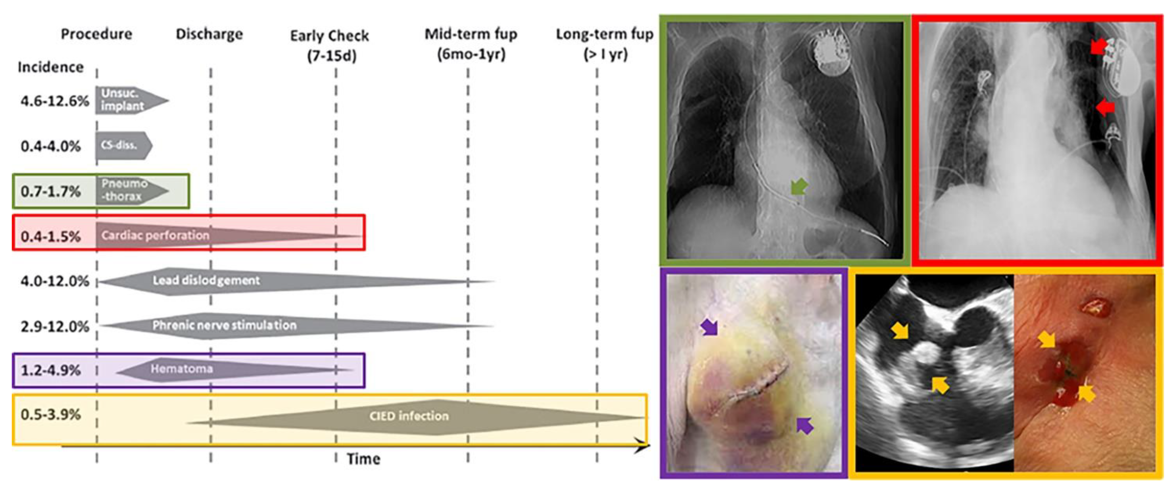
2. Risk Factors for CIED Infections

The relevance of CIED infections has progressively increased from the beginning of the 2000s, as pointed out by an alarming report [2] that showed a rise in these complications that was much higher than the increase in device implantations during the same period. The authors underlined that in the period between 1996 and 2003, which was characterized by no significant changes in the demographic profile of patients receiving CIED implantation, hospitalizations for CIED infections increased 3.1-fold (2.8-fold for PMs and 6-fold for ICDs). Notably, the only factor that changed during the same period was the type of implanted CIED, with an increase of ICDs vs. PMs (from 14% to 27%, p < 0.001) (Figure 2) [14]. A possible explanation for this phenomenon can be provided by the disproportion between survival of ICD and PM recipients vs. implanted devices. In fact, as later underlined [15], the three to five-fold longer survival of PM generators (in that period) dramatically increased the rate of multiple re-interventions in ICD recipients (vs. PM-implanted patients). In fact, looking at Figure 2 it is impressive to see that the steep increase in CIED infections occurred just four years after the MADIT trial, which led to adoption of ICDs for the primary prevention of sudden cardiac death. This time window was exactly the same as the longevity of ICD batteries at that particular moment. Moreover, the slope preceded the publication of the MADIT II trial by two years.
Similar evidence, coupled with data underlying the association of CIED infections with re-do procedures [5], encouraged the development of devices with extended longevity, aimed not only at reducing the costs related to device replacement but also at reducing the occurrence of CIED infections. A relevant issue related to device infections is the number of implanted leads; in fact, more leads can drive a higher incidence of infections, especially in more complex CIED systems like CRT-D. Two factors hindered the benefit of extending CIED longevity: comorbidities and lead failures. One possible explanation for the disproportionate rise in CIED infections is the increased prevalence of comorbidities. We are currently experiencing an imbalance between fertility rates and life expectancy, leading to an increase in median age of the general population. In particular, patients aged ≥65 years represent 8.5% of the worldwide population, and this proportion is estimated to increase up to 17% in the upcoming years. The aging of the general population correlates with the development of chronic diseases like kidney disease, diabetes mellitus, and chronic obstructive pulmonary disease. As reported by Greenspon et al., the incidence of the four major comorbidities (heart failure, diabetes, renal disease, and respiratory disease) in patients with CIED infections remained relatively constant from 1993 through 2004, when a noticeable increase was seen, with a similar trend observed in infection rate during the same period [3].
The second factor promoting the increase in CIED infections, which became evident after the improvement in CIED longevity, was the malfunction (or potential malfunction) of CIED leads. Since 2007, two major recalled ICD leads (the Sprint Fidelis (Medtronic Corp, Dublin, Ireland) and Riata (St. Jude Medical Inc, Saint Paul, MN, USA)) have revealed this problem [16], leading to different responses from clinicians. These ranged from those that were more conservative to extract-all approaches [17]. These issues promoted the development and spread of two great advancements in current CIED technology: remote CIED monitoring (to reveal early signs of lead malfunction before clinical manifestations) and leadless technology. However, the leadless revolution was not only driven by recalled leads, but also by the presence of several reports regarding the suboptimal performance of ICD leads [18]. These reports highlighted a huge gap between the longevity of CIED leads purported by manufacturers and real service life. This could be due to the fact that survival of CIED leads can also be affected by several other factors beyond manufacturing: the implantation approach (in particular, cephalic vs. subclavian access), patient behavior, modifications of the lead–heart interface, lead dislodgment, and the necessity for CIED upgrade. However, cephalic vein access is more secure for lead integrity in the long term; nowadays, direct puncture of the subclavian vein is still the preferred approach for most implanting centers. Despite case-specific reasons for suboptimal lead performance, in the last decade intravascular leads were signaled as the Achilles’ heel of conventional CIED [17].

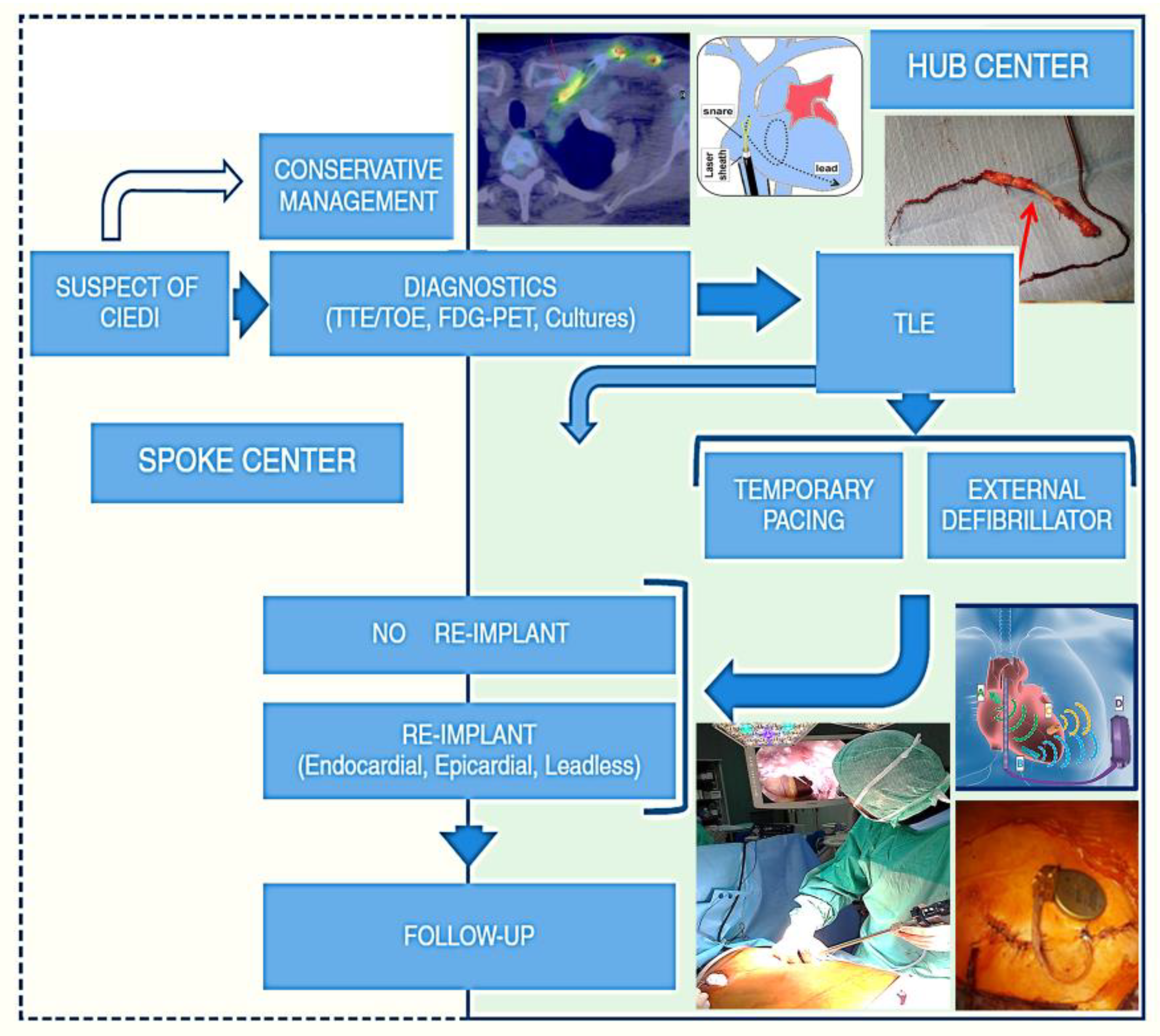
3. The Complexity of the Treatment of CIED Infection

Early after the development of the first PM, it became necessary to develop an approach for extracting pacing leads in case of CIED infections, since the simple removal of the generator or other conservative approaches were associated with failure to resolve the infection [15]. The first approach that was adopted was a simple and direct traction of the lead after disconnection from the generator. This approach was frequently associated with lead fracture, especially with the development of thinner leads to limit vascular encumbrance, and of systems to limit lead displacement. In a short time, several tools were developed for mechanical lead extraction, including polypropylene sheaths and snares, increasing extraction rates to above 80%. More recently, with the development of internal jugular approach by the school of Pisa [19] and the availability of powered sheaths (either mechanical-rotational or laser), a dramatic increase in the number of lead extraction procedures has occurred.
While in the early lead extraction era this was considered the highest-risk procedure in the field of CIED, current figures provided by international registries confirm the safety and efficacy of this procedure. The results of the European Lead Extraction ConTRolled Registry (ELECTRa) [20], a large multicenter prospective registry of consecutive patient candidates for transvenous lead extraction, showed that the efficacy of current approach is very high, with clinical and complete radiological success in more than 95% of cases. The primary endpoint was a safety endpoint, showing that procedure-related major complications (including death) occurred in 1.7% patients, with an incidence of 0.5% procedure-related deaths. Notably, clinical outcomes were heavily affected by center volumes, with a doubled rate of overall major complications and intra-hospital death in low-volume centers. However, it has to be noted that despite few procedure-related complications (mortality 0.5%, 95% CI 0.3–0.8%), the in-hospital overall mortality was not negligible (1.4%, 95% CI 1.1–1.9%%), with systemic CIED infections being the strongest independent risk factor (OR 4.93, 95% CI 2.72–8.93; p < 0.001). In a recently published German registry on laser lead extraction the authors reported similar procedure-related outcomes, with a complete procedural success rate in 97.8% of the patients, and an incidence of major procedure-related complications of 2.05% [21]; the procedure-related mortality was 0.55%. Notably, the in-hospital mortality was 3.55%, with a significantly higher mortality rate in patients with systemic infection compared with patients with local infection or non-infectious indications for lead extraction (11.08 vs. 0.55%, p < 0.0001). Taken together, these data highlight the importance of CIED infections (especially when associated with systemic involvement) as a major promoter of adverse events despite effective and safe lead extraction. Data on long-term survival after lead extraction for CIED infections are limited, but the available information suggest that reimplantation is not a major determinant of survival; on the contrary, renal failure, systemic infection, and the presence of vegetations, especially involving cardiac valves or in case of post-extraction remnants (i.e., ghosts) [8], are associated with poorer survival. In regard to these findings, it is interesting to review the results of a multicenter study we organized to explore if the predictors of development of CIED infections were at the same time effective in predicting post-extraction survival in the long term. We followed 169 patients for an average follow-up of 20 ± 12 months after transvenous lead extraction for CIED infection (48.5% systemic) with complete radiological success in 96.4%, and for each patient we calculated the Shariff score at the last CIED procedure (i.e., before developing CIED infection). This score, elaborated by Shariff et al. in a previous paper [22], presents good accuracy in predicting the risk of developing CIED infection before any procedure. In our population a score ≥3 was present in 60.3% of the patients, and proved to be an extremely good predictor of mortality after lead extraction at three years (HR 10.833, 95% CI 2.544–46.129; p = 0.001), together with presence of vegetations (HR 3.324, 95% CI 1.530–7.221; p = 0.002). These data, beyond confirming the poor survival of patients with CIED infections despite effective lead extraction, confirmed the hypothesis that the struggle against this complication must focus on primary prevention. Two trials addressed this issue by improving antibiotic prophylaxis: the Prevention of Arrhythmia Device Infection Trial (PADIT) [23], and the Worldwide Randomized Antibiotic EnveloPe Infection PrevenTion Trial (WRAP-IT) [24]. The first was based on a prolonged antibiotic prophylaxis scheme adopting treatment with multiple conventional antibiotics, while the second assessed the efficacy of a new device, an antibacterial polypropylene mesh sleeve (TYRX™ Absorbable Antibacterial Envelope, Medtronic, Minneapolis, MN, USA) that releases minocycline and rifampicin into the generator pocket within approximately seven days. Both intervention arms performed better than the control groups, but only the WRAP-IT trial reached significance in terms of reduction of CIED infections, albeit with a number needed to treat (NNT) score of 200. Importantly, both studies were characterized by an incidence of CIED infections in the control arm that was significantly lower than expected, similar to more recent observational data [25], evidencing that, despite the usefulness of prolonged antibiotic prophylaxis to reduce CIED infections, it should be remembered that preventive strategies should consider all the various aspects of CIED therapy, from patient selection to implanting procedure and follow-up [14,26]. In this regard, the management of CIED infections should be considered a complex issue, from diagnosis to the decisions for device extraction and re-implantation. It requires a multidisciplinary and tailored approach, with relevant economic implications [13]. Each patient should be managed with an “hub-and-spoke” organization, with referral centers that have at their disposal a team of expert operators from the beginning of the diagnostic process [27] (Figure 3). Despite some available recommendations supporting antibiotic therapy and device extraction, there are still issues regarding the need for re-implantation, optimal timing, technique, and device selection. Furthermore, the diagnostic process requires experienced clinicians, especially in case of possible systemic infection without overt CIED pocket involvement. In this case, it is relevant to perform appropriate culturing and integrate imaging data. Trans-esophageal echocardiography (TEE) is mandatory, but there is a relevant number of patients presenting with lead masses not related to CIED infection; in fact, incidental masses attached to CIED leads were reported in up to 22% of the patients [28,29,30,31]. In a large retrospective study considering about 2000 TEE scans to identify patients with visible leads, 15/125 TTE scans with explorable CIED leads presented a mass, and 9/15 patients with a lead mass presented with clinical suspicion of CIED infection; 6 patients with incidental mass were only treated with medical therapy without sequelae [30]. Recently, the availability of positron emission tomography (PET) with fluorodeoxyglucose marked by fluorine-18 (18F-FDG) has dramatically improved the diagnostic accuracy for these cases; in fact, this method provides functional data on organs and tissue [32,33]. PET scans reveal the intensity of glucose uptake among body’s tissues, giving information about the presence of increased metabolic activity in a particular body region; it usually correlates with neoplastic, inflammatory, or regenerative processes. Moreover, in our experience 18F-FDG PET can reclassify one quarter of patients in terms of Duke score [32], and help predict long-term survival with higher accuracy; in particular, we found that patients with lead infection without CIED pocket involvement (the so-called “cold closed pocket”) have significantly poorer long-term survival, with mortality of about 60% at 1 year (Figure 4).

4. Current Prevention of CIED Infections and Future Perspectives

As previously discussed, the battle against CIED infections has driven the development of several improvements both in terms of new technologies and clinical management. Current preventive strategies should be based on tailoring CIED procedures to patient requirements, and the utilization of all strategies to reduce surgical site infections [34,35]. This was highlighted by Ahsan et al., who reported the results of a retrospective study analyzing the impact of a specifically designed infection-control protocol (that includes antibiotic prophylaxis chosen by risk stratification, improved glycemic control, specific skin preparation, electrocautery setting, and closure techniques) on the incidence of CIED infections in a tertiary referral center [36]. They found a 54% reduction in CIED infections (from 1.3 to 0.6%, p < 0.03) associated with consequent relevant cost savings (about £70,000 per year), while patient costs varied between £85 and £115 according to infection risk and drug intolerance. Notably, despite not being fully confirmed, the available data suggest that from a fifth to one quarter of CIED infections are not the result of CIED pocket manipulation, but are seeded by bacteria from distant sites, as occurs with infections of native cardiac valves [26,28,32]. This concept clearly emphasizes the need for the development of leadless technologies to avoid or at least limit intravascular hardware associated with CIED therapy.
In the field of cardiac pacing, Medtronic Micra systems, which allow cardiac pacing without the necessity for transvenous leads, are now available [37]. Their newest device makes it possible to achieve (with some limitations) atrioventricular synchrony with a leadless system [38,39] (Figure 5). Although there is considerable enthusiasm about leadless systems, some issues have to be mentioned about these devices; in fact, there are some reports of infections of leadless devices [40]. Furthermore, long-term results are lacking.
The availability of subcutaneous defibrillators should limit the complications related to high voltage transvenous lead systems in a specific subgroup of patients [41,42]. However, a recent recall of EMBLEM S-ICD Subcutaneous Electrodes (Boston Scientific) took place because of the increased risk of fractures at a specific point (distal to the proximal sense ring). The most recent technology innovation relates to the availability of an investigational extravascular system (EV ICD Medtronic, Minneapolis, MN, USA), implanted behind the sternum, that can treat fatal ventricular arrhythmias [43,44].

Funding

This research received no external funding.

Conflicts of Interest

The authors declare no conflict of interest.

References

  1. Valzania, C.; Torbica, A.; Tarricone, R.; Leyva, F.; Boriani, G. Implant rates of cardiac implantable electrical devices in Europe: A systematic literature review. Health Policy 2016, 120, 1–15. [Google Scholar] [CrossRef]
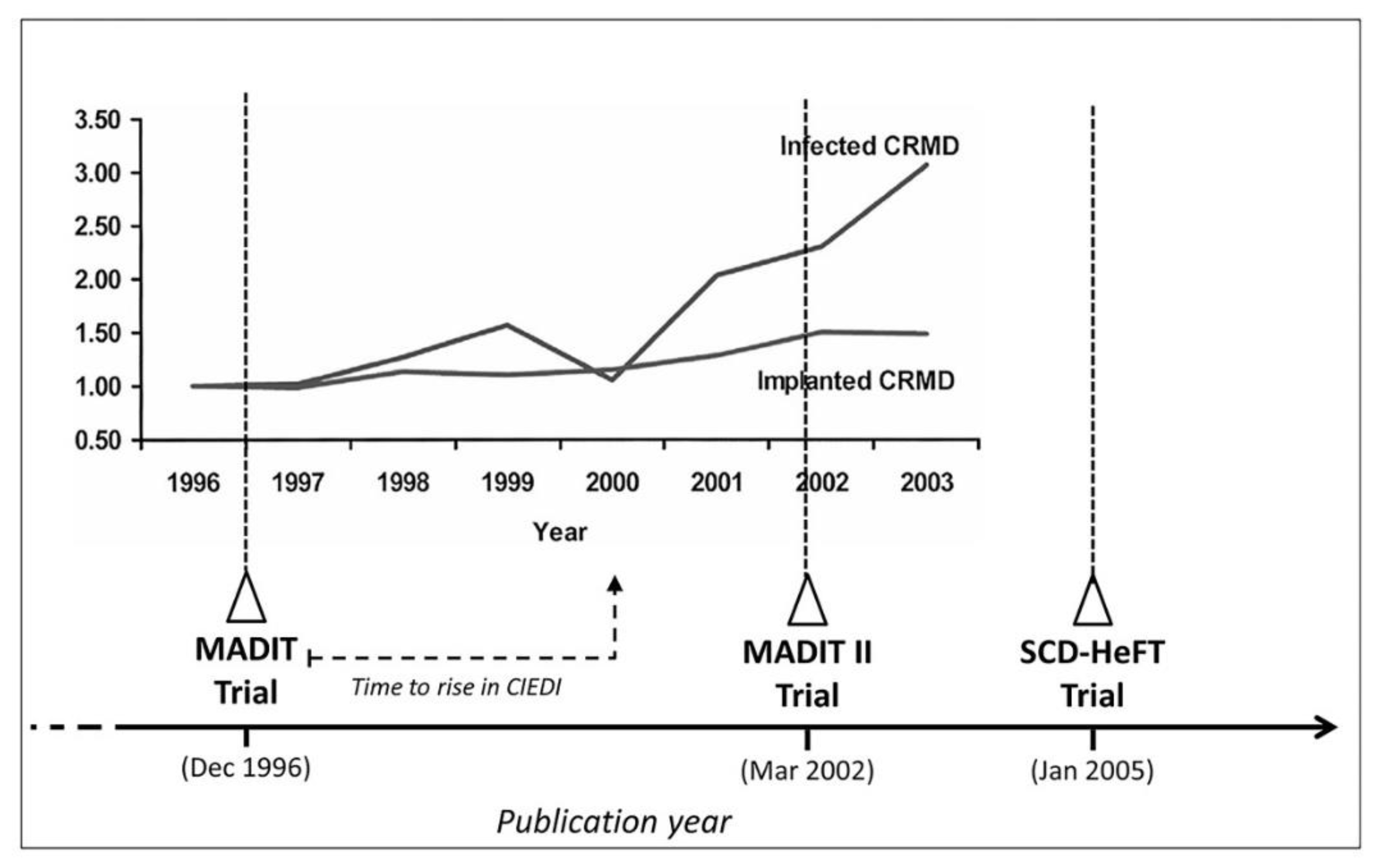
  2. Voigt, A.; Shalaby, A.; Saba, S. Rising rates of cardiac rhythm management device infections in the United States: 1996 through 2003. J. Am. Coll. Cardiol. 2006, 48, 590–591. [Google Scholar] [CrossRef] [Green Version]
  3. Greenspon, A.J.; Patel, J.D.; Lau, E.; Ochoa, J.A.; Frisch, D.R.; Ho, R.T.; Pavri, B.B.; Kurtz, S.M. 16-year trends in the infection burden for pacemakers and implantable cardioverter-defibrillators in the United States 1993 to 2008. J. Am. Coll. Cardiol. 2011, 58, 1001–1006. [Google Scholar] [CrossRef] [Green Version]
  4. Bachrach, C.A. Culture and demography: From reluctant bedfellows to committed partners. Demography 2014, 51, 3–25. [Google Scholar] [CrossRef] [Green Version]
  5. Diemberger, I.; Parisi, Q.; De Filippo, P.; Narducci, M.L.; Zanon, F.; Potenza, D.R.; Ciaramitaro, G.; Malacrida, M.; Boriani, G.; Biffi, M. Detect Long-term Complications After ICD Replacement (DECODE): Rationale and Study Design of a Multicenter Italian Registry. Clin. Cardiol. 2015, 38, 577–584. [Google Scholar] [CrossRef] [Green Version]
  6. Ezzat, V.A.; Lee, V.; Ahsan, S.; Chow, A.W.; Segal, O.; Rowland, E.; Lowe, M.D.; Lambiase, P.D. A systematic review of ICD complications in randomised controlled trials versus registries: Is our ‘real-world’ data an underestimation? Open Heart 2015, 2, e000198. [Google Scholar] [CrossRef]
  7. Palmisano, P.; Guerra, F.; Dell′Era, G.; Ammendola, E.; Ziacchi, M.; Laffi, M.; Troiano, F.; Prenna, E.; Russo, V.; Angeletti, A.; et al. Impact on All-Cause and Cardiovascular Mortality of Cardiac Implantable Electronic Device Complications: Results from the POINTED Registry. JACC Clin. Electrophysiol. 2020, 6, 382–392. [Google Scholar] [CrossRef] [PubMed]
  8. Diemberger, I.; Biffi, M.; Lorenzetti, S.; Martignani, C.; Raffaelli, E.; Ziacchi, M.; Rapezzi, C.; Pacini, D.; Boriani, G. Predictors of long-term survival free from relapses after extraction of infected CIED. EP Eur. 2018, 20, 1018–1027. [Google Scholar] [CrossRef] [PubMed]
  9. Diemberger, I.; Lorenzetti, S.; Vitolo, M.; Boriani, G. Infective endocarditis in patients with cardiac implantable electronic devices: Impact of comorbidities on outcome. Eur. J. Intern. Med. 2019, 66, e9–e10. [Google Scholar] [CrossRef] [PubMed]
  10. Tarakji, K.G.; Mittal, S.; Kennergren, C.; Corey, R.; Poole, J.E.; Schloss, E.; Gallastegui, J.; Pickett, R.A.; Evonich, R.; Philippon, F.; et al. Antibacterial Envelope to Prevent Cardiac Implantable Device Infection. N. Engl. J. Med. 2019, 380, 1895–1905. [Google Scholar] [CrossRef] [PubMed]
  11. Sohail, M.R.; Henrikson, C.A.; Braid-Forbes, M.J.; Forbes, K.F.; Lerner, D.J. Mortality and cost associated with cardiovascular implantable electronic device infections. Arch. Intern. Med. 2011, 171, 1821–1828. [Google Scholar] [CrossRef] [PubMed] [Green Version]
  12. Ahmed, F.Z.; Fullwood, C.; Zaman, M.; Qamruddin, A.; Cunnington, C.; Mamas, M.A.; Sandoe, J.; Motwani, M.; Zaidi, A. Cardiac implantable electronic device (CIED) infections are expensive and associated with prolonged hospitalisation: UK Retrospective Observational Study. PLoS ONE 2019, 14, e0206611. [Google Scholar] [CrossRef]
  13. Blomstrom-Lundqvist, C.; Traykov, V.; Erba, P.A.; Burri, H.; Nielsen, J.C.; Bongiorni, M.G.; Poole, J.; Boriani, G.; Costa, R.; Deharo, J.C.; et al. European Heart Rhythm Association (EHRA) international consensus document on how to prevent, diagnose, and treat cardiac implantable electronic device infections-endorsed by the Heart Rhythm Society (HRS), the Asia Pacific Heart Rhythm Society (APHRS), the Latin American Heart Rhythm Society (LAHRS), International Society for Cardiovascular Infectious Diseases (ISCVID), and the European Society of Clinical Microbiology and Infectious Diseases (ESCMID) in collaboration with the European Association for Cardio-Thoracic Surgery (EACTS). Eur. Heart J. 2020, 41, 2012–2032. [Google Scholar] [CrossRef] [PubMed] [Green Version]
  14. Diemberger, I.; Boriani, G.; Deharo, J.C. Prevention of Device Infection: Procedural Aspects, Drugs, and Preventive Tools. In Infections of Cardiac Implantable Devices; Springer: Cham, Switzerland, 2020; pp. 177–208. [Google Scholar] [CrossRef]
  15. Diemberger, I.; Biffi, M.; Martignani, C.; Boriani, G. From lead management to implanted patient management: Indications to lead extraction in pacemaker and cardioverter-defibrillator systems. Expert Rev. Med. Devices 2011, 8, 235–255. [Google Scholar] [CrossRef] [PubMed]
  16. Brunner, M.P.; Cronin, E.M.; Jacob, J.; Duarte, V.E.; Tarakji, K.G.; Martin, D.O.; Callahan, T.; Borek, P.P.; Cantillon, D.J.; Niebauer, M.J.; et al. Transvenous extraction of implantable cardioverter-defibrillator leads under advisory—A comparison of Riata, Sprint Fidelis, and non-recalled implantable cardioverter-defibrillator leads. Heart Rhythm 2013, 10, 1444–1450. [Google Scholar] [CrossRef] [PubMed]
  17. Diemberger, I.; Mazzotti, A.; Giulia, M.B.; Cristian, M.; Matteo, M.; Letizia, Z.M.; Reggiani, B.; Battistini, P.; Boriani, G. From lead management to implanted patient management: Systematic review and meta-analysis of the last 15 years of experience in lead extraction. Expert Rev. Med. Devices 2013, 10, 551–573. [Google Scholar] [CrossRef] [PubMed]
  18. Maisel, W.H.; Kramer, D.B. Implantable cardioverter-defibrillator lead performance. Circulation 2008, 117, 2721–2723. [Google Scholar] [CrossRef] [Green Version]
  19. Bongiorni, M.G.; Segreti, L.; Di Cori, A.; Zucchelli, G.; Paperini, L.; Viani, S.; Soldati, E. Overcoming the current issues surrounding device leads: Reducing the complications during extraction. Expert Rev. Med. Devices 2017, 14, 469–480. [Google Scholar] [CrossRef]
  20. Bongiorni, M.G.; Kennergren, C.; Butter, C.; Deharo, J.C.; Kutarski, A.; Rinaldi, C.A.; Romano, S.L.; Maggioni, A.P.; Andarala, M.; Auricchio, A.; et al. The European Lead Extraction ConTRolled (ELECTRa) study: A European Heart Rhythm Association (EHRA) Registry of Transvenous Lead Extraction Outcomes. Eur. Heart J. 2017, 38, 2995–3005. [Google Scholar] [CrossRef]
  21. Pecha, S.; Burger, H.; Möller, V.; Madej, T.; Osswald, B.; Maali, A.; De Simone, R.; Monsefi, N.; Ziaukas, V.; Erler, S.; et al. The German Laser Lead Extraction Registry: GALLERY. Thorac. Cardiovasc. Surg. 2019, 67, S1–S100. [Google Scholar] [CrossRef]
  22. Shariff, N.; Eby, E.; Adelstein, E.; Jain, S.; Shalaby, A.; Saba, S.; Wang, N.C.; Schwartzman, D. Health and Economic Outcomes Associated with Use of an Antimicrobial Envelope as a Standard of Care for Cardiac Implantable Electronic Device Implantation. J. Cardiovasc. Electrophysiol. 2015, 26, 783–789. [Google Scholar] [CrossRef] [PubMed]
  23. Krahn, A.D.; Longtin, Y.; Philippon, F.; Birnie, D.H.; Manlucu, J.; Angaran, P.; Rinne, C.; Coutu, B.; Low, R.A.; Essebag, V.; et al. Prevention of Arrhythmia Device Infection Trial: The PADIT Trial. J. Am. Coll. Cardiol. 2018, 72, 3098–3109. [Google Scholar] [CrossRef] [PubMed]
  24. Tarakji, K.G.; Mittal, S.; Kennergren, C.; Corey, R.; Poole, J.; Stromberg, K.; Lexcen, D.R.; Wilkoff, B.L. Worldwide Randomized Antibiotic EnveloPe Infection PrevenTion Trial (WRAP-IT). Am. Heart J. 2016, 180, 12–21. [Google Scholar] [CrossRef]
  25. Biffi, M.; Ammendola, E.; Menardi, E.; Parisi, Q.; Narducci, M.L.; De Filippo, P.; Manzo, M.; Stabile, G.; Potenza, D.R.; Zanon, F.; et al. Real-life outcome of implantable cardioverter-defibrillator and cardiac resynchronization defibrillator replacement/upgrade in a contemporary population: Observations from the multicentre DECODE registry. EP Eur. 2019, 21, 1527–1536. [Google Scholar] [CrossRef] [PubMed]
  26. Bartoletti, M.; Viale, P. Microbiological Background: Biofilm, Culturing, and Antibiotics. In Infections of Cardiac Implantable Devices; Springer: Cham, Switzerland, 2020; pp. 17–32. [Google Scholar] [CrossRef]
  27. Boriani, G.; Elsner, C.; Diemberger, I. The struggle against infections of cardiac implantable electrical devices: The burden of costs requires new personalized solutions. EP Eur. 2018, 20, 1877–1879. [Google Scholar] [CrossRef] [PubMed]
  28. Diemberger, I.; Lorenzetti, S.; Bonfiglioli, R. Building Up the Diagnosis of Cardiac Device Infections: The Role of Imaging. In Infections of Cardiac Implantable Devices; Springer: Cham, Switzerland, 2020; pp. 65–94. [Google Scholar] [CrossRef]
  29. Downey, B.C.; Juselius, W.E.; Pandian, N.G.; Estes, N.A., 3rd; Link, M.S. Incidence and significance of pacemaker and implantable cardioverter-defibrillator lead masses discovered during transesophageal echocardiography. Pacing Clin. Electrophysiol. PACE 2011, 34, 679–683. [Google Scholar] [CrossRef]
  30. Lo, R.; D′Anca, M.; Cohen, T.; Kerwin, T. Incidence and prognosis of pacemaker lead-associated masses: A study of 1,569 transesophageal echocardiograms. J. Invasive Cardiol. 2006, 18, 599–601. [Google Scholar]
  31. Victor, F.; De Place, C.; Camus, C.; Le Breton, H.; Leclercq, C.; Pavin, D.; Mabo, P.; Daubert, C. Pacemaker lead infection: Echocardiographic features, management, and outcome. Heart 1999, 81, 82–87. [Google Scholar] [CrossRef] [Green Version]
  32. Diemberger, I.; Bonfiglioli, R.; Martignani, C.; Graziosi, M.; Biffi, M.; Lorenzetti, S.; Ziacchi, M.; Nanni, C.; Fanti, S.; Boriani, G. Contribution of PET imaging to mortality risk stratification in candidates to lead extraction for pacemaker or defibrillator infection: A prospective single center study. Eur. J. Nucl. Med. Mol. Imaging 2019, 46, 194–205. [Google Scholar] [CrossRef]
  33. Graziosi, M.; Nanni, C.; Lorenzini, M.; Diemberger, I.; Bonfiglioli, R.; Pasquale, F.; Ziacchi, M.; Biffi, M.; Martignani, C.; Bartoletti, M.; et al. Role of (1)(8)F-FDG PET/CT in the diagnosis of infective endocarditis in patients with an implanted cardiac device: A prospective study. Eur. J. Nucl. Med. Mol. Imaging 2014, 41, 1617–1623. [Google Scholar] [CrossRef]
  34. Biffi, M.; Angeletti, A.; Ziacchi, M. Prevention of Infection: Indications, Device Programming, Patient Follow-Up. In Infections of Cardiac Implantable Devices; Springer: Cham, Switzerland, 2020; pp. 209–229. [Google Scholar] [CrossRef]
  35. De Maria, E.; Diemberger, I.; Vassallo, P.L.; Pastore, M.; Giannotti, F.; Ronconi, C.; Romandini, A.; Biffi, M.; Martignani, C.; Ziacchi, M.; et al. Prevention of infections in cardiovascular implantable electronic devices beyond the antibiotic agent. J. Cardiovasc. Med. 2014, 15, 554–564. [Google Scholar] [CrossRef]
  36. Ahsan, S.Y.; Saberwal, B.; Lambiase, P.D.; Koo, C.Y.; Lee, S.; Gopalamurugan, A.B.; Rogers, D.P.; Lowe, M.D.; Chow, A.W. A simple infection-control protocol to reduce serious cardiac device infections. EP Eur. 2014, 16, 1482–1489. [Google Scholar] [CrossRef] [PubMed]
  37. Beurskens, N.E.G.; Breeman, K.T.N.; Dasselaar, K.J.; Meijer, A.C.; Quast, A.B.E.; Tjong, F.V.Y.; Knops, R.E. Leadless cardiac pacing systems: Current status and future prospects. Expert Rev. Med. Devices 2019, 16, 923–930. [Google Scholar] [CrossRef] [PubMed] [Green Version]
  38. Chinitz, L.; Ritter, P.; Khelae, S.K.; Iacopino, S.; Garweg, C.; Grazia-Bongiorni, M.; Neuzil, P.; Johansen, J.B.; Mont, L.; Gonzalez, E.; et al. Accelerometer-based atrioventricular synchronous pacing with a ventricular leadless pacemaker: Results from the Micra atrioventricular feasibility studies. Heart Rhythm 2018, 15, 1363–1371. [Google Scholar] [CrossRef] [PubMed]
  39. Steinwender, C.; Khelae, S.K.; Garweg, C.; Chan, J.Y.S.; Ritter, P.; Johansen, J.B.; Sagi, V.; Epstein, L.M.; Piccini, J.P.; Pascual, M.; et al. Atrioventricular Synchronous Pacing Using a Leadless Ventricular Pacemaker: Results From the MARVEL 2 Study. JACC Clin. Electrophysiol. 2020, 6, 94–106. [Google Scholar] [CrossRef]
  40. Ellison, K.; Hesselson, A.; Ayoub, K.; Leung, S.; Gurley, J. Retrieval of an infected leadless pacemaker. Heart Rhythm Case Rep. 2020, 6, 863–866. [Google Scholar] [CrossRef]
  41. Viani, S.; Migliore, F.; Tola, G.; Pisano, E.C.L.; Russo, A.D.; Luzzi, G.; Sartori, P.; Piro, A.; Rordorf, R.; Forleo, G.B.; et al. Use and outcomes of subcutaneous implantable cardioverter-defibrillator (ICD) after transvenous ICD extraction: An analysis of current clinical practice and a comparison with transvenous ICD reimplantation. Heart Rhythm 2019, 16, 564–571. [Google Scholar] [CrossRef]
  42. Ziacchi, M.; Corzani, A.; Diemberger, I.; Martignani, C.; Marziali, A.; Mazzotti, A.; Massaro, G.; Rapezzi, C.; Biffi, M.; Boriani, G. Electrocardiographic Eligibility for Subcutaneous Implantable Cardioverter Defibrillator: Evaluation during Bicycle Exercise. Heart Lung Circ. 2016, 25, 476–483. [Google Scholar] [CrossRef] [PubMed] [Green Version]
  43. Boersma, L.V.A.; Merkely, B.; Neuzil, P.; Crozier, I.G.; Akula, D.N.; Timmers, L.; Kalarus, Z.; Sherfesee, L.; DeGroot, P.J.; Thompson, A.E.; et al. Therapy From a Novel Substernal Lead: The ASD2 Study. JACC Clin. Electrophysiol. 2019, 5, 186–196. [Google Scholar] [CrossRef]
  44. Chan, J.Y.S.; Lelakowski, J.; Murgatroyd, F.D.; Boersma, L.V.; Cao, J.; Nikolski, V.; Wouters, G.; Hall, M.C.S. Novel Extravascular Defibrillation Configuration with a Coil in the Substernal Space: The ASD Clinical Study. JACC Clin. Electrophysiol. 2017, 3, 905–910. [Google Scholar] [CrossRef]
Figure 1. Incidence and common periods of greater vulnerability to complications of cardiac resynchronization implants or upgrades. Modified from “Cardiac resynchronization therapy in the real world: need to upgrade outcome research” by Boriani, G., Diemberger, I., 2018, European Journal of Heart Failure, 20, 1469–1471, Copyright [2018] by European Society of Cardiology. CIED: cardiac implantable electronic device; CS-diss: coronary sinus dissection; d: days; fup: follow-up; mo: months, unsuc: unsuccessful; yr: years.
Hearts 02 00016 g001
Figure 2. Relationship between the publication date of three leading randomized trials on implantable defibrillators for the primary prevention of sudden cardiac death and the disproportionate increase in the number of CIED infections with respect to implantation rate. Modified from “Infections of Cardiac Implantable Devices” by Diemberger, I.; Boriani, G., 2020, Infections of Cardiac Implantable Devices, VI, 229, Copyright [2020] by the Springer International Publishing. CMRD: cardiac rhythm management devices.
Hearts 02 00016 g002
Figure 3. Proposed hub-spoke approach for the management of CIED infection. Modified from ref 27 (From “The Struggle against Infections of Cardiac Implantable Electrical Devices: The Burden of Costs Requires New Personalized Solutions.” by Boriani, G.; Elsner, C.; Diemberger I. 2018, Europace, 20, 1877–1879, Copyright [2018] by the Oxford University Press).
Hearts 02 00016 g003
Figure 4. Survival curves (Kaplan–Meier) considering all-cause mortality according to the presence/absence of a “cold closed pocket”, defined as the absence of pocket skin lesion or metabolic activity revealed by 18F-FDG PET/CT scan. Legend: A, C, D = cases of CIED infection with overt pocket involvement. B = CIED infection with hidden pocket involvement, as revealed by 18F-FDG PET/CT. Reproduced from “Contribution of PET imaging to mortality risk stratification in candidates to lead extraction for pacemaker or defibrillator infection: a prospective single center study” Diemberger, I. et al., 2019, European Journal of Nuclear Medicine and Molecular Imaging, 46, 194–205, Copyright [2019] by the Springer Nature.
Hearts 02 00016 g004
Figure 5. Heart activity monitoring with leadless devices. (1) A magnet placed in the right atrium, through which movements are revealed during the cardiac cycle using an external subcutaneous Hall effect sensor (HES) (2). The distance between magnet and HES decreases with atrial diastole (A), with a consequent increase in the magnetic field that reaches a maximum when the distance is minimal ((B), atrial systole). Once atrial activity is revealed, the external device (4) can drive a ventricular leadless pacemaker (PM) (3). To avoid the necessity for external subcutaneous devices, an HES could be probably inserted directly in the leadless PM (5). Modified from “Infections of Cardiac Implantable Devices” by Diemberger, I.; Boriani, G., 2020, Infections of Cardiac Implantable Devices, VI, 229, Copyright [2020] by the Springer International Publishing.
Hearts 02 00016 g005
Publisher’s Note: MDPI stays neutral with regard to jurisdictional claims in published maps and institutional affiliations.

