Article

Plant Production and Leaf Anatomy of Mertensia maritima (L.) Gray: Comparison of In Vitro Culture Methods to Improve Acclimatization

1
CREA—Centro di Ricerca Orticoltura e Florovivaismo, Corso Inglesi 508, 18038 Sanremo, Italy
2
Dipartimento di Scienze della Terra, dell’Ambiente e della Vita, Università di Genova, Corso Europa 26, 16132 Genova, Italy
3
Dipartimento di Scienze Agrarie, Alimentari e Agro-alimentari, Università di Pisa, Via del Borghetto 80, 56124 Pisa, Italy
4
Centro Interdipartimentale di Ricerca “Nutraceutica e Alimentazione per la Salute” (NUTRAFOOD), Università di Pisa, Via del Borghetto 80, 56124 Pisa, Italy
*
Author to whom correspondence should be addressed.
Submission received: 14 April 2021 / Revised: 6 May 2021 / Accepted: 8 May 2021 / Published: 13 May 2021
(This article belongs to the Collection Application of Tissue Culture to Horticulture)

Abstract

:
Mertensia maritima is a commercially interesting herb with edible leaves and flowers, characterized by oyster flavor and taste. Plant propagation and traditional cultivation are challenging for this species. Therefore, the main purpose of the present study was to establish successful protocols aimed at ensuring oyster plant shoot propagation, rooting and in vivo acclimatization. Both micropropagation and rooting were tested, comparing the traditional in vitro solid substrate in jar vs. the liquid culture in a temporary immersion system (TIS) bioreactor (Plantform™). A Murashige and Skoog (MS) medium added with 4-μM thidiazuron (TDZ) and 1-μM α-naphthaleneacetic acid (NAA) was employed for micropropagation, while a half-strength MS medium supplemented with 4-μM indole−3-butyric acid (IBA) was used for rooting. Different acclimatization conditions in the greenhouse or in growth chamber were tested. Morphometric and microscopical analyses were performed on the oyster plant leaves at the propagation, rooting and acclimatization stages both in a jar and in a TIS. Micropropagation in a TIS allowed to obtain large shoots, while a great number of shoots was observed in the jar. M. maritima shoots rooted in TIS produced more developed roots, leaves with more developed waxy glands and well-formed stomata; moreover, the plants coming from the TIS showed the best acclimatization performances.

1. Introduction

Mertensia maritima (L.) Gray (Boraginaceae family) is a perennial herb, which has a circumboreal distribution, and it is found in sea coastal habitats like shingle beaches, growing around the high-tide mark and, rarely, on sand [1]. The plant is an upright rosette consisting of glaucous blue-green leaves with wax glands on the upper surface and can reach 20 cm in height. Between June and September, the plant produces branched cymose inflorescences with leaf-like bracts. The flowers are 4–6 mm long with an initial pink color that gradually changes to blue or pink/blue hues because of the flower’s development process [1]. The interest in this herb is due to the surprising marine oyster-like aroma and taste of its edible leaves and flowers, as reported by Delort and collaborators [2], who analyzed, in 2012, the volatile composition of M. maritima leaves. Therefore, M. maritima is also called a vegetarian oyster or oyster plant, and their leaves are used to prepare culinary recipes. Some information about the volatile organic compounds (VOCs) and the nutraceutical contents of oyster leaf extracts were investigated [2], as well as their digestive and anticancer properties [3].
Oyster plants are included in the “Red List” in North America and in the “Pink List” in Britain, because they are threatened with extinction, since their habitat is subject to Atlantic storms and, hence, strongly influenced by the ongoing climate changes. The oyster plant is difficult to cultivate in gardens, because it is almost impossible to replicate the original habitat of this species, due to the limitations caused by temperature, and the poor germination aptitude owing to seed dormancy [1,4]. In order to overcome all these constraints, the in vitro micropropagation technique is the best solution for the germplasm conservation and extensive propagation of oyster plants.
Park and collaborators [5] established an efficient protocol for the large-scale propagation of oyster plants, evaluating also their carotenoid, fatty acid and tocopherol contents in the leaves of in vitro regenerated shoots. The phytochemical profile and biological activities of the extracts obtained from calli, shoots and in vitro seedlings were also studied in a more recent paper [6]. A successful acclimatization of well-rooted plantlets has been reported [5], previously grown on agarized substrate enriched with 4-μM indole–3-butyric acid (IBA), even if no information about the applied method, the results of the acclimatization and the number of obtained plants were provided.
Our preliminary acclimatization tests for M. maritima plants gave unsatisfactory results; therefore, the experiments to improve plant development and their acclimatization rate were carried out. To this purpose, in this study, different micropropagation methods were compared, including liquid culture in bioreactors in temporary immersion systems (TIS), a method already applied to the micropropagation of different species [7,8,9,10,11]. Regarding M. maritima, only one micropropagation protocol using agar-solidified medium has been reported by two different papers [5,6], and no studies have been conducted to verify the potential of the liquid medium for in vitro sprout multiplication and rooting.
The aim of the study was to establish an efficient and reproducible method for in vitro M. maritima shoot propagation and to improve their in vivo acclimation. Furthermore, the morphoanatomical features of leaves and roots during acclimatization are investigated to establish if the morphometrical, histochemical and anatomical parameters of the leaves can affect the acclimatization performance.

2. Materials and Methods

2.1. Plant Material and In Vitro Establishment

The plants of M. maritima were provided by the Chambre d’Agriculture des Alpes-Maritimes (CREAM, Nice, France) and were grown at the Research Centre for Vegetable and Ornamental Crops (CREA, Sanremo, Imperia, Italy; GPS: 43.816887, 7.758900). Microcuttings (1 cm) were deprived of the leaves, washed under running tap water for 10 min, superficially sterilized with 1.5% NaClO for 20 min and then rinsed twice in sterile deionized water for 10 min. After sterilization, the microcuttings were placed in jars containing a culture medium composed by MS salts and vitamins [12], 3% sucrose, 1.33-μM 6-benzyl-aminopurine (BAP) and 0.75% agar. The pH was adjusted to 5.80 ± 0.01 before autoclaving at 121 °C for 20 min. The microcuttings were sighted in the culture chamber at 23 ± 1 °C with a 16/8-h light/dark cycle and 209 ± 5-μmol m−2 s−1 light irradiances at jar level (white fluorescent Philips Master TL-D 36W/840 lamp). After two weeks, only the aseptic microcuttings were used to induce shoot development.

2.2. Multiple Shoot Induction

The microcuttings were transferred to jars with solid substrates for multiple shoot inductions consisting of MS salts and vitamins [12], 3% sucrose, 4-μM thidiazuron (TDZ), 1-μM α-naphthalene acetic acid (NAA) and 0.75% agar (pH 5.8) [5]. The microcuttings were placed in the same environmental conditions mentioned above. After five weeks, the induced shoots were separated and subcultured into a fresh medium. This operation was repeated every five weeks, until obtaining enough shoots to perform further experiments.

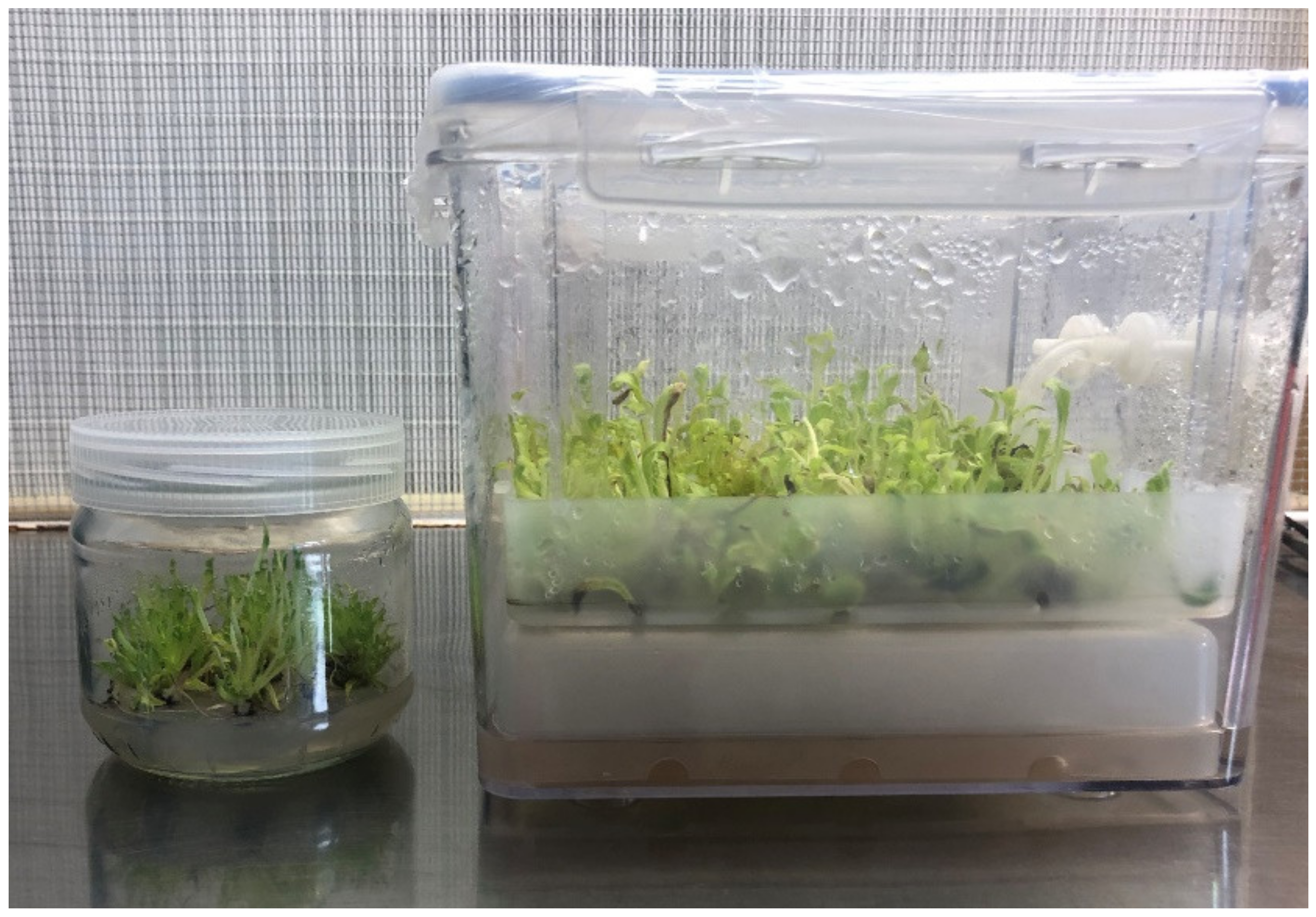
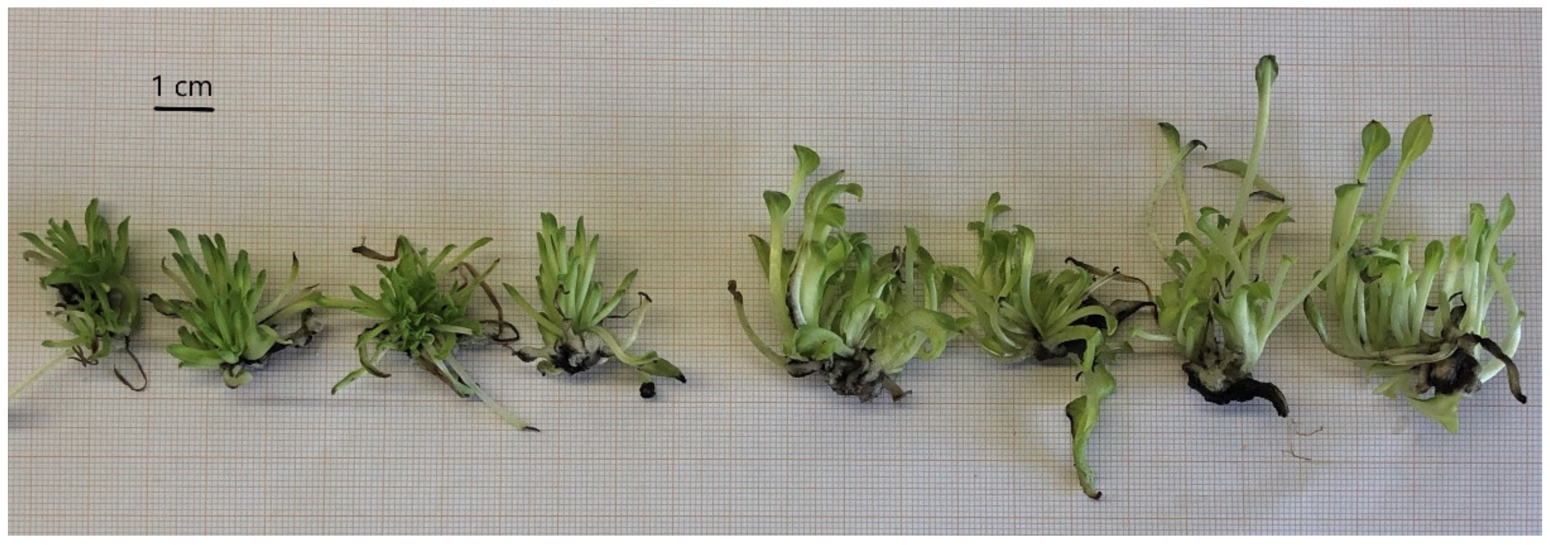
2.3. Comparison between Solid and TIS Culture Systems for Shoot Micropropagation

Three trials were carried out to compare two different culture systems: the solid medium (in jar) and TIS culture systems. Homogenous explants were used to evaluate the shoot proliferation, and they were 1.0–1.5 cm long with 3 to 4 leaves and nodes. The explants were transplanted into jars (85-mm diameter; 85-mm height) filled with 60 mL of multiple shoot induction agarized substrate (solid medium—see above) or in the polypropylene basket of a Plantform™ bioreactor (180 × 150 × 160-mm length × width × height) (www.plantform.se, Sweden) filled on the bottom with 240-mL multiple shoot induction not-agarized substrate (liquid medium) (Figure 1). The Plantform parts were sterilized in an autoclave for 20 min at 120 °C.
In order to compare the same number of explants, in each trial, four jars containing ten shoots each and one Plantform bioreactor with forty shoots were prepared. In the jars, the shoots were always in contact with the solid substrate, while, for the bioreactor, a flooding time of 3 min every 3 h was set up; to maintain sterility, the injected air was filtered by 0.20-μm pore filters. Both jars and Plantform systems were placed in the growth chamber at the same distance from the light source and at the same conditions previously mentioned. After 4 weeks of culture, the shoot tips produced shoot clusters. The fresh weight of the shoot clusters, the shoot number per cluster and the leaf number per cluster were recorded. Ten clusters per each treatment were dried at 60 °C for one week, and then, the shoot dry weight was evaluated. The third apical leaves of twenty clusters per treatment were collected and measured in total length, length and width of the leaf blade. The third leaf from the tip was chosen, because it was fully developed and was certainly generated during the cluster growth.

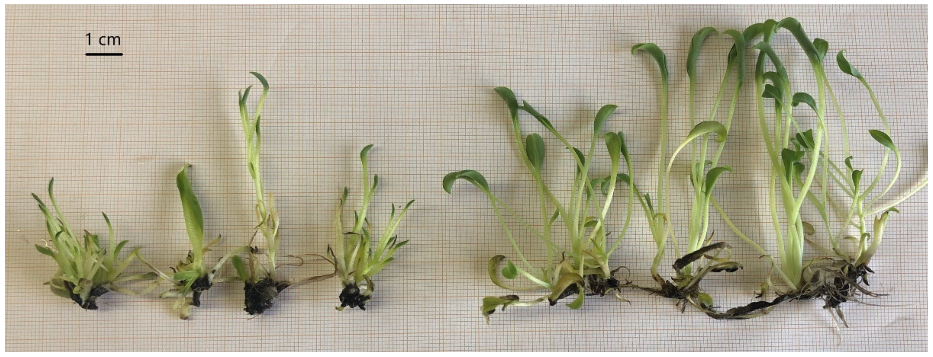
2.4. Comparison between Solid and TIS Culture Systems for Rooting Induction

Similar trials were performed to compare the two culture systems (solid in jar vs. liquid in Plantform) for rooting induction. The trials were repeated in triplicate using shoot tips cultured in jars with a solid multiple shoot induction substrate. The shoot rooting substrate was composed by vitamins MS, half-strength MS salts [12], 3% sucrose and 4-μM indole–3-butyric acid (IBA) (pH 5.8) [5]. Agar (0.75% w/v) was added only in the solid substrate (jars). Four jars, each containing ten shoot tips, were set up for each Plantform bioreactor containing forty shoot tips. The culture conditions applied for the shoot rooting were the same described for the shoot micropropagation, also including the immersion cycles of the Plantform bioreactor. After four weeks of culture, the shoot fresh weight, the root number per explants and the leaf number per explants were recorded. As for the explants used for shoot induction, the third apical leaves of twenty explants per treatment were collected and measured in total length, length and width of the leaf blade. Finally, the rooted M. maritima plants of each treatment were used for acclimatization tests and microscopic analysis.

2.5. Acclimatization Tests

Forty rooted shoots of M. maritima from jars and forty rooted shoots from the Plantform bioreactor were transplanted into plastic pots (14-cm diameter) filled with 250 mL of peat:pumice:perlite 1:1:1 as substrate and covered with a perforated plastic cup (250 mL in volume). The pots were kept in a greenhouse or in a growth chamber with a 16/8-h light/dark photoperiod, 22 °C thermoperiod, 200 ± 5-μmol m−2 s−1 light irradiances at pot level (white fluorescent Philips Master TL-D 36W/840 lamp) and watered two times per week with 50-mL tap water. Four treatments were considered: plants from jars and cultured in the greenhouse (JG), plants from Plantform and cultured in the greenhouse (PG), plants from jars and cultured in a growth chamber (JC) and plants from Plantform and cultured in a growth chamber (PC). A total of 20 plant per treatment were prepared. After two weeks of culture, the plastic cups were removed, and after another four weeks, the survival rate of the acclimatized plants was recorded. The trial was repeated in triplicate.

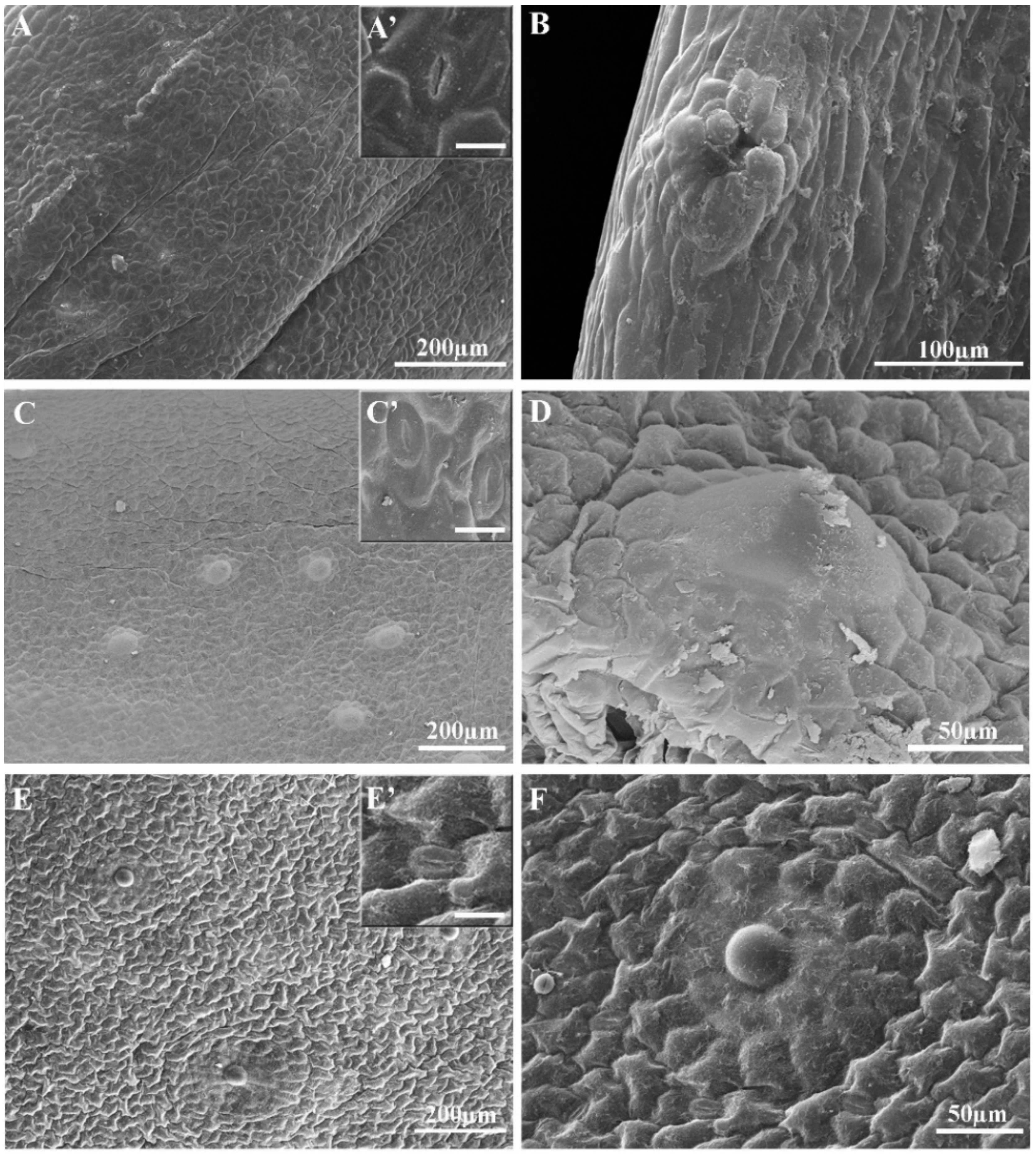
2.6. Leaf Morphological and Anatomical Examination

We collected in vitro developed leaves from plants of M. maritima previously rooted in a jar and Plantform. Leaves obtained from treated plants were compared with mature leaves from in vivo plants. To clear the tissues, three leaves for treatment were placed on a microscope slide with two drops of Visikol solution (2B Scientific Limited, Upper Heyford, UK). A coverslip was then applied, and the slides were put on a hot plate until air bubbles moved out of the edges of slides. Microscopic examination and image analysis were obtained with a DM 4000B optical microscope (Leica Microsystems, Wetzlar, Germany) equipped with an AxioCam ERc 5s (Carl Zeiss Microimaging Goettingen, Germany) [13].
For the Scanning Electron Microscope (SEM) analyses, small leaf pieces were fixed in Finefix working solution (Milestone S.r.l., Bergamo, Italy) with 70% ethanol, incubated overnight at 4 °C, dehydrated by an ethanol series (70–100%) at 60-min intervals and critically dried using a K850CPD 2M processor (Strumenti S.r.l., Roma, Italy). Dried samples were mounted on aluminum stubs and coated with a 10-nm layer of gold. Finally, specimens were analyzed with a VEGA3-Tescan-type LMU microscope (Tescan Orsay Holding, a.s., Brno, Czech Republic) at 20-kV voltage [14].
The micromorphological features of the leaves were investigated using light and scanning electron microscopy (LM and SEM, respectively). For LM, the epidermal peels or sections of leaves were immediately examined by transmitted light or epifluorescence with a Leica DM 2000 microscope. To detect the autofluorescence of the phenolic compounds, untreated small portions were directly observed under a UV filter (340–380 nm). Fluorol Yellow 088 was used for the lipids [15], and the observations were carried out under the UV filter. In addition, the following histochemical reactions were carried out: Toluidine Blue O (TBO) for polyphenols and tannins detection, Sudan III for total lipids and Ruthenium Red for non-cellulosic polysaccharides [16].
Other leaves, collected at the same stages, were fixed overnight in paraformaldehyde solution (4% w/v) and phosphate buffer 0.1 M, pH 7.0, washed with the same phosphate buffer, dehydrated in the ethanol series and embedded in paraffin (Paraplast, Merck, Germany). Then, the paraffin blocks were sectioned with a rotative microtome (Leica RM2265), and the cross-sections (10-μm thick) were deparaffinized and stained with TBO [17,18]. Microscope slides were observed with a Leica DM 4000B microscope, and images acquisition was performed with an AxioCam ERc 5s (Carl Zeiss Microimaging, Goettingen, Germany). Anatomical structures were observed, and the thickness of the palisade tissue was measured.

2.7. Statistical Analysis

Data were statistically analyzed by one-way ANOVA, followed by Fisher’s probable least-squares difference test with a cut-off significance at p ≤ 0.05. All the data were normally distributed as verified by the Shapiro–Wilk and Levene’s tests. The shoot multiplication and root induction trials were repeated in triplicate. In each trial, forty explants per treatment were analyzed. The data about the survival rate of the acclimatized plants were reported as the average of three trials performed.

3. Results

3.1. Shoot Micropropagation

The shoots micropropagated in the temporary immersion system (TIS) appeared larger than the micropropagated shoots with the classic jar system with a solid substrate (Figure 2). Nevertheless, after four weeks of culture, the shoot clusters from TIS did not only show a significantly higher biomass (both fresh and dry), but longer leaves with longer and wider leaf blades compared to shoots grown in a jar (Table 1). Moreover, the plants in the TIS showed the lowest number of shoots per cluster, and some hyperhydricity symptoms (17 ± 2%) were observed, especially at the base of the clusters. For these last two reasons, and because the explants produced in the jar had more homogeneous dimensions, the micropropagation in the jar system was chosen to produce M. maritima shoots to be used for the subsequent experiments of rooting induction.

3.2. Rooting Induction

All the explants transferred in the rooting substrate both in the jar and in the Plantform bioreactor developed roots. As shown in Table 2 and Figure 3, the plants cultured in the Plantform bioreactor showed a higher biomass and more numerous and longer roots compared to those grown in jars. No difference was recorded in the number of leaves, while the length and width of the leaf blades were greater in the plants from Plantform.

3.3. Plant Acclimatization

The best results in M. maritima acclimatization were obtained in the growth chamber, showing a statistically significant increase in the survival rate (%) compared to the survival rate of those transferred in the greenhouse (Table 3). Moreover, the survival rate of the plants coming from Plantform and cultured in the growth chamber (PC in Table 3) was much higher than that of the plants coming from the jars and acclimatized under the same cultivation conditions (JC). After acclimatization in the climatic chamber, the plants were transferred outdoors, and their survival rate was 100%.

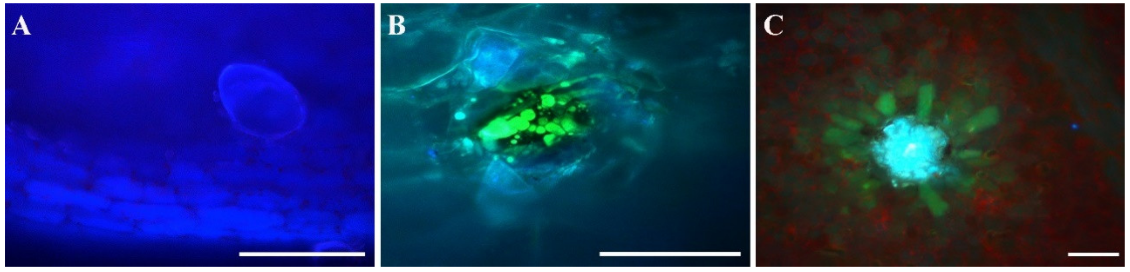
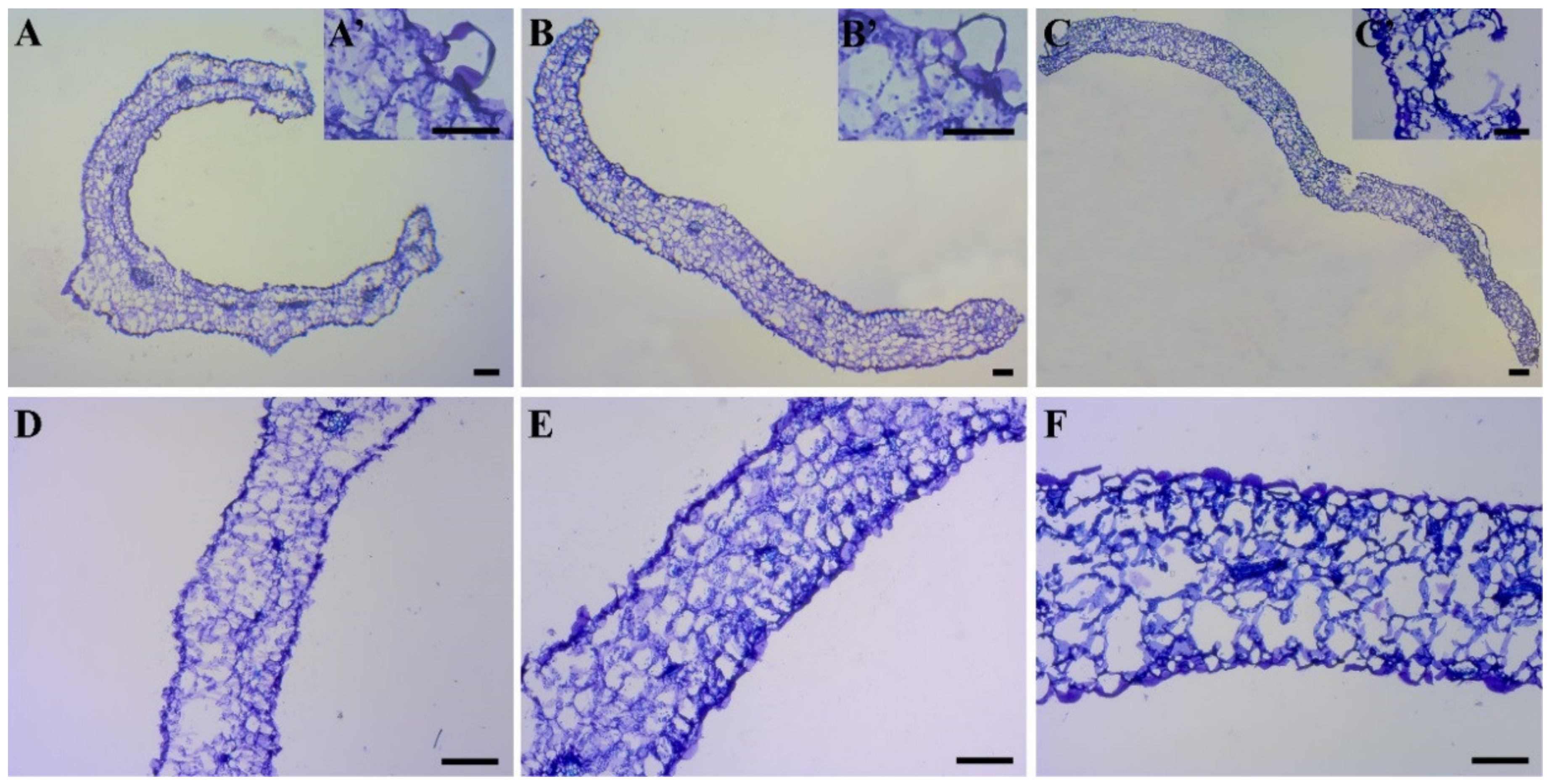
3.4. Leaf Anatomical Analysis

The leaf of M. maritima is characterized by dorsiventral and amphistomatous leaves, with an anomocytic stomatal apparatus. The adaxial surface shows a greater number of stomata than the abaxial one. The epidermis is monolayered, and both adaxial and abaxial surfaces are covered with waxes. Epidermal cells are round to polygonal-shaped, mildly swollen, with straight or slightly sinuous anticlinal walls in the adaxial epidermal surface of the leaf and strongly sinuous in the abaxial one. The leaves show peculiar glandular trichomes, composed by a glandular head surrounded by complexes of elongated epidermal cells, which radially converge, sinking at the gland base. Both converging cells and the central gland resemble a “rosette” structure (see below).
Leaves sampled from all the rooting treatments (R-Jar, R-TIS and in vivo plants) were examined by clarification with Visikoll solution in order to observe the development of the vascular system of the leaves (Figure 4A–C). By light microscopy, not fully developed rosettes were visible along the leaf margins in all the treatments (Figure 4D–F). Mature and fully formed rosettes were clearly visible only on the surface of the leaves harvested from plants grown in in vivo conditions. In this case, veins located in the mesophyll were always visible, in correspondence with the rosette structures present on the surface (Figure 4C).
The SEM images highlighted the further differences between the treatments. The leaf blades from the jars showed open stomata (Figure 5A’) and a few rosette primordia (Figure 5A). These were mainly concentrated in the central portion and, generally, were not fully developed and sometimes deformed or lacking the central glandular head (Figure 5B). On the contrary, the leaves from Plantform displayed closed stomata (Figure 5B) and a higher number of immature rosettes (Figure 5C) with non-turgid radial cells but well-developed and swollen glandular heads (Figure 5D). Finally, the leaves collected from the plants grown in pots (in vivo) showed closed stomata (Figure 5E) and many mature rosettes distributed throughout the adaxial side (Figure 5E,F). These glandular structures showed prominent heads and turgid radial cells, which sink at the gland base, crossing the mesophyll (not shown). Radial cells of the rosettes present along the leaf margins appeared more flattened, with a button-like shape (Figure 4F).
The histochemical analyses showed differences among the leaves coming from the three different treatments. Under UV excitation, the leaves from the jars showed low signals attributable to the presence of chlorophylls; neither secondary metabolite was detected, except for a weak blue color, indicating the presence of polyphenols, emitted by the button-like glands scattered at the leaf margin (Table 4 and Figure 6).
In some cases, these glands appeared birefringent under polarized light. In the leaves from Plantform, by polarized light, the presence of crystals was confirmed also in the radial cells of the rosettes. In general, a higher number of crystals was detected in the leaf mesophyll of the plants coming from this treatment in respect to the previous ones. In some cases, whole gland structures were stained in blue/green with TBO and were positive to Fluorol Yellow. The cells of the rosette appeared a light-blue color when observed under UV light (Figure 6B). The center of the gland showed yellow/greenish drops, indicating a flavin/flavonoid/terpenoid content. In the leaf margin, a high presence of button-like glands, light-blue under UV light, was recorded. In leaves from in vivo plants, the rosettes were birefringent, revealing crystals within the glandular head when observed in a transversal section (not shown). In addition, in the transversal section, the glands appeared structured on several layers, occupying the entire mesophyll up to protruding on the abaxial surface. TBO, Ruthenium Red and Sudan III staining showed the presence of polyphenolic, polysaccharidic and lipidic substances, respectively. The center of the gland, in some cases covered by a cuticle, appeared light blue under UV excitation, confirming the presence of phenolic substances; in some cases, the rosette cells were yellow/greenish, indicating a flavin/flavonoid/terpenoid content (Figure 6C). Fluorol yellow showed the presence of terpenoids and lipidic substances within the glandular head and in the surrounding cells.
The sections of leaves coming from plants rooted in the solid substrate had thin, poorly organized palisade tissue 266 ± 27 μm thick (Figure 7A), spongy tissue with few intracellular spaces, a few poorly structured glands (Figure 7B) and a waxy layer almost completely lost during the deparaffinization of the leaf samples. The leaves harvested from the plants rooted in Plantform (liquid medium) maintained a waxy layer at the leaf margins and showed a wider (389 ± 1-μm thick) and more organized palisade layer than those grown in a jar (solid medium), more expanded spongy tissue with larger intracellular spaces and more organized rosettes. The in vivo leaves were well-organized, with a compact palisade layer 563 ± 57 μm thick, large spongy tissue rich in spaces, a thick, waxy superficial layer and very organized large and deep rosettes that sink into the palisade, touching the vascular tissue. Crystals of different shapes were present in the rosette cells (not shown).

4. Discussion

The great interest in edible flowers of Mertensia maritima, together with their poor germination and extinction hazard, led germplasm conservation as an essential goal. Micropropagation is the pivotal technique to overcome these problems, although the rooting and acclimatization phases represented a critical bottleneck [11]. Recently, in our laboratory, many rooted plants were produced with the micropropagation method [5], but, during the acclimatization phase, their survival rate was extremely unsatisfactory (data not shown). Therefore, this work tried to improve the acclimatization protocol by testing both different rooting conditions (both jar and Plantform) and different acclimatization conditions (both greenhouse and a climatic room). For this purpose, two in vitro culture methods (solid and TIS) were compared to establish the best shoot multiplication and protocols.
The shoot multiplication is significantly greater in the culture in a solid substrate than in the liquid one, while the leaf parameters were higher in the liquid culture. The shoot number per cluster observed in the solid culture was comparable to that registered to Park et al. [5], also with a similar visual appearance of the in vitro plants. The liquid culture in the temporary immersion system (TIS) did not increase the biomass of the micropropagated shoots (both fresh and dry) but allowed to obtain longer leaves with longer and wider leaf blades in comparison with the common jar system with an agarized solid substrate. However, this latter culture system decreased the number of shoots per cluster and, hence, the multiplication rate. Generally, the multiplication rate increases in a liquid culture. Liquid cultures in bioreactors in temporary immersion systems (TIS) are largely used to increase the quantity and quality of micropropagated plants in different ornamental plant species (such as Anthurium, Hydrangea, Lilium and Zantedeschia), fruit trees and herbs [7,8,9,10]. This increment is observed in other genera belonging to the Boraginaceae family [19]; in other herbaceous species such as foxglove, echinacea [20] and carnation [21] and in species such as caper [22] and goji [10]. The literature reports that plant multiplication in a bioreactor with a liquid medium is better compared to a static, solid culture, because the tissue area in contact with the medium is greater, and therefore, this factor increases the absorption of both the nutrients and plant growth regulators. Moreover, the periodic agitation of the medium and air exchange allows to avoid the problems of asphyxia often present in a solid culture [11]. We can assume that the culture condition in Plantform has improved the development of large leaves of M. maritima at the expense of the number of shoots.
The concentrations of 4-μM IBA and ½ MS salts were chosen for the in vitro rooting, because they were already used successfully to induce the formation of adventitious roots in M. maritima, as shown in previous studies [5,6]. In our study, the rooting occurred both in the solid and liquid substrates, but in the TIS culture, the roots were more abundant and longer. These results are in line with those reported for other species. In Arnebia euchroma (Boraginaceae), the rooting began 8/10 days earlier in the liquid culture than that observed in the agarized medium, and moreover, the roots were more numerous and more developed [19]. Similar results were found in the Eucalyptus clones [23] and Siraitia grosvenorii [24]. Probably, the aeration system connected to the TIS culture improved the root formation [11]. Indeed, in pistachios, the rooting in the TIS was lower than that observed in the solid medium [25]. In nature, M. maritima lives on very ventilated gravel often rearranged by storms, which can leave the roots exposed [1]; therefore, its roots are well-adapted to an open habitat. Probably, rooting in TIS is more similar to the conditions of the root growth in nature, and for this reason, this root development is better compared to the jar system.
For the same reason, a very porous and easily reproducible substrate was chosen for the acclimatization. No information is available in the literature on the topic, since no authors have reported data on M. maritima in vivo acclimatization. Park and collaborators [5] affirmed that 56-day-old rooted Mertensia plants were acclimatized in a greenhouse, but no indication of their survival rate was provided. In our study, the survival rate was greater when M. maritima was acclimatized in a climatic room at 22 °C. The constant condition of the climatic room was the same in which the plants were grown during the multiplication and rooting phases; probably, this environmental condition avoids further stress for the plants in the subsequent acclimatization phase. Moreover, plant survival rate was greater in the plants rooted coming from the TIS system compared to those coming from jars and acclimated in a climatic room. This difference may depend on the root development. Although of different sizes, the roots from both the jar and Plantform were functional, because the vascular system of the roots was continuous with that of the stem (data not shown), resulting in an efficient water transfer between the roots and shoots.
The anatomical and morphological differences found on the leaves from the jar and TIS cultures can affect the survival rates of transplanted plants. In different plant species, the high mortality rate during the acclimatization process is linked to stomatal dysfunction and/or poor cuticle development [26]. In our case, the leaves from the jars always have open stomata, while the leaves from Plantform have closed stomata similar to those observed in in vivo leaves and are, apparently, active and functioning. Similar results were also observed in Anthurium andreanum [27]; the plants placed to rooting in the TIS showed a high percentage of closed stomata and a low stomatal index compared to those from the solid medium system. The authors also demonstrated a greater accumulation of total chlorophylls, and they hypothesized that the improvement of the stomatal function, the low rate of transpiration and the high content of photosynthetic pigments found in the rooting plants from the TIS system led to an increase in the photosynthetic rate.
Morphological data about the stomata and leaf surface allowed us to corroborate the available information reported in the literature for M. maritima and M. sibirica [3,28]. In addition, we provided new information on the presence, distribution and micro-morphology of the glandular structures in plants grown in different in vitro and in vivo conditions. The micromorphological characterization of the production sites of secondary metabolites was obtained by means of SEM and LM. As already reported by Burkovskaya and Khrolenko [3], who provided the first morphological description of M. maritima leaf blades and glandular structures, plants grown in vivo show complex “waxy glands” in the shapes of rosettes. These glands have prominent heads and turgid radial cells; as reported for other Boraginaceae [29,30], they are structured on several layers crossing the mesophyll, and they are distributed throughout the adaxial side of the leaves. LM and SEM analyses allowed us to observe that plants grown in jars present undeveloped stomata and leaf blades and immature or not well-developed wax glands. On the other hand, leaves from the TIS culture display rosettes with more defined and turgid radial cells, together with a more swollen central head. These characters suggest that the glands from this stage can undertake a secretory activity, an aspect indeed confirmed by the histochemical analyses. Phytochemical data on M. maritima have been already provided by several authors [5,6,31], but in these cases, the production sites were not identified and characterized. During these surveys, the metabolite contents were evaluated for different plant growth conditions. The detected metabolites belonged to carotenoids, phenolic acids, terpenoids and fatty acids, with many of the recognized compounds well-known for their bioactivities. It has been observed that the phytochemical contents of plant materials vary both qualitatively and quantitatively, depending on the tissues examined [31] or on the different treatments [6]. Song et al. [6] found a major number of different secondary metabolites in the shoots and seedling extracts than in the callus extract. However, Fedoreyev et al. [31] detected a higher concentration of rosmarinic acid, rabdosiin and an ureide, allantoin, in the callus culture with respect to the root or leaf tissues. Our histochemical tests revealed a complex mixture of phenolic, flavonoid and terpenoid/lipid compounds both in the leaves of plants grown in Plantform and in vivo. However, a clear increase in the presence of the metabolites was observed in the latter case. In both situations, secondary compounds were secreted and accumulated mainly in the central region of the gland. In addition, they were also observed in the surrounding radial cells, both in those of the innermost layers that plunged into the mesophyll (not shown) and those of the epidermal layer (Figure 6B,C). In the gland center of some rosettes, metabolites were accumulated in a space delimited by a relatively thick cuticle. This characteristic has been already recorded for other members of Boraginaceae, for example, in Cordia verbenacea [32]. Terpenes, terpenoids and general lipid compounds were localized with Fluorol Yellow 088 and Sudan III dyes. Among the fatty acids, α-linolenic acid and stearidonic acid were detected by Park and collaborators [5], while Song et al. [6] also recorded undecanedioic, dodecanedioic, tetradecanedioic and hexadecanedioic acids. Autofluorescence and TBO staining confirmed the presence in the glands of other flavonoid or phenolic compounds. Their production and accumulation in M. maritima have also been previously recorded by Song et al. [6]. By means of polarized light, crystals were detected on the leaf surface, especially in the cells of the rosettes. Furthermore, the LM analysis of the leaf samples treated with Visikoll (Figure 4) highlighted that glandular structures develop in correspondence of the terminal veins. The accumulation of mineralized materials in the basal cells of the rosettes has been already reported for other Boraginaceae [30]. Dassanayake and Larkin [33] reviewed the evolutionary and structural diversity of the salt glands, considering more than 50 species belonging to 14 different families. They stated that glands from related clades can be grouped into four structural classes, sharing convergently evolved features specialized for salt compartmentalization and excretion. The shape of multicellular multilayered glands, as well as the presence of an evident cuticle in M. maritima, are similar to the type II glands described by the above-mentioned authors for Asterids, a group that phylogenetically includes the Boraginaceae family [34]. Therefore, considering their shape, the presence of crystals in their cells and the fact that they are distributed in correspondence to the vascular vessels, we can assume that the glands of M. maritima are involved in the excretion of salts coming from the growth substrate. A more structured vascular system, like that observed in the case of the plants grown in Plantform, would favor the development of leaf structures and glands, which can therefore prepare the plant for the natural environmental conditions, contributing to a successful acclimatization. In addition, and in support of this, it should be considered the fact that glandular structures at the mature stage (like the in vivo situation) are also responsible for wax excretion. Indeed, plants grown in jars and Plantform completely lack or show, only in some cases, waxes in the glands or on the leaf surface (Figure 6A,B and Figure 7A,B). Conversely, in the leaves sampled from the in vivo plants, a higher presence of waxes was detected (Figure 6C and Figure 7C). The mature leaves from the TIS system are larger and thicker than those from the solid system; they have a more differentiated mesophyll with a more developed and thicker palisade layer, as well as more formed and complex epithelial structures. We can then confirm that, at their full development stage, the glands are responsible for wax production, giving protection against exsiccating agents such as the wind, intense sunlight and salt water [3]. In fact, the leaf, in addition to carrying out photosynthetic activity, also allows water storage [3].

5. Conclusions

The propagation protocol that allows to obtain the greatest number of Mertensia maritima plants provides the multiplication of a solid substrate (in a jar) and rooting in a TIS. In vivo transplantation under controlled conditions (growth chamber) increases the survival of plants deriving from rooting in a TIS. We hypothesized that the environment created in the TIS system prepares plants for acclimatization stress more than that in a jar (solid substrate). The complex data suggests that wax glands are involved in acclimatization, as well as both in regulatory and excretory functions, enhancing the ability of this species to adapt to high-salinity environmental conditions. This protocol is appropriate both for the commercial production of M. maritima plants and in reintroduction programs in nature.

Author Contributions

Conceptualization, A.C. (Andrea Copetta) and B.R.; methodology, A.C. (Andrea Copetta), M.B., A.C. (Arianna Cassetti) and I.M.; software, A.C. (Andrea Copetta) and M.B.; validation, A.C. (Andrea Copetta), M.B., L.C. and B.R.; formal analysis, A.C. (Andrea Copetta), M.B. and A.C. (Arianna Cassetti); investigation, A.C. (Andrea Copetta) and M.B.; resources, A.C. (Andrea Copetta) and B.R.; data curation, A.C. (Andrea Copetta), M.B. and C.M.; writing—original draft preparation, A.C. (Andrea Copetta) and M.B.; writing—review and editing, A.C. (Andrea Copetta), M.B., A.C. (Arianna Cassetti), C.M., L.C., L.P. and B.R.; visualization, A.C. (Andrea Copetta), M.B., A.C. (Arianna Cassetti) and I.M.; supervision, B.R., L.C. and L.P.; project administration, B.R. and funding acquisition, B.R. All authors have read and agreed to the published version of the manuscript.

Funding

This research was funded by the INTERREG-ALCOTRA UE 2014–2020 “ANTEA” Project—Attività innovative per lo sviluppo della filiera transfrontaliera del fiore edule (n. 1139), grant number: CUP C12F17000080003.

Institutional Review Board Statement

Not applicable.

Informed Consent Statement

Not applicable.

Data Availability Statement

Data are contained within the article.

Acknowledgments

The authors thank Laura Negretti and Federica Betuzzi for their precious support during the microscopic analyses, Manuela Pamato for her support with the in vitro culture and Sergio Ariano and Alberto Lanteri for the in vivo cultivation of the plant materials.

Conflicts of Interest

The authors declare no conflict of interest.

References

  1. Alton, S.; FitzGerald, R. Mertensia maritima Boraginaceae. Curtis’s Bot. Mag. 2009, 26, 96–110. [Google Scholar] [CrossRef]
  2. Delort, E.; Jaquier, A.; Chapuis, C.; Rubin, M.; Starkenmann, C.J. Volatile composition of oyster leaf (Mertensia maritima (L.) Gray). J. Agric. Food Chem. 2012, 60, 11681–11690. [Google Scholar] [CrossRef]
  3. Burkovskaya, E.V.; Khrolenko, Y.A. Ecological features of the photosynthetic complex structure of Mertensia maritima (Boraginaceae) from different geographic locations. Bull. KrasGAU. Biol. Sci. 2015, 8, 53–58. (In Russian) [Google Scholar]
  4. Skarpaas, O.; Stabbetorp, E. Diaspore ecology of Mertensia maritima: Effects of physical treatments and their relative timing on dispersal and germination. Oikos 2003, 95, 374–382. [Google Scholar] [CrossRef]
  5. Park, H.Y.; Kim, D.H.; Saini, R.K.; Gopal, J.; Keum, Y.S.; Sivanesan, I. Micropropagation and quantification od bioactive compounds in Mertensia maritima (L.) Gray. Int. J. Mol. Sci. 2019, 20, 2141. [Google Scholar] [CrossRef] [PubMed] [Green Version]
  6. Song, K.; Sivanesan, I.; Ak, G.; Zengin, G.; Cziàky, Z.; Jeko, J.; Rengasamy, K.R.R.; Lee, O.N.; Kim, D.H. Screening of biological activities of calli, shoots, and seedlings of Mertensia maritima (L.) Gray. Plants 2020, 9, 155. [Google Scholar] [CrossRef] [PubMed]
  7. Ruffoni, B.; Savona, M. The temporary immersion system (T.I.S.) for the improvement of micropropagation of ornamental plants. Acta Hortic. 2003, 683, 445–454. [Google Scholar] [CrossRef] [Green Version]
  8. Barberini, S.; Savona, M.; Ruffoni, B. Temporary immersion culture of Lilium bulbiferum. Acta Hortic. 2010, 900, 377–383. [Google Scholar] [CrossRef]
  9. Savona, M.; Sacco, E.; Ruffoni, B. Improving micropropagation performances in Hydrangea spp.: Temporary immersion shoot culture and induction of morphogenetic events. Acta Hortic. 2011, 961, 457–464. [Google Scholar] [CrossRef]
  10. Ruta, C.; De Mastro, G.; Ancona, S.; Tagarelli, A.; De Cillis, F.; Benelli, C.; Lambardi, M. Large-scale plant production of Licium barbarum L. by liquid culture in temporary immersion system and possible application to the synthesis of bioactive substance. Plants 2020, 9, 844. [Google Scholar] [CrossRef] [PubMed]
  11. Etienne, H.; Berthouly, M. Temporary immersion systems in plant micropropagation. Plant Cell Tissue Organ Cult. 2002, 69, 215–231. [Google Scholar] [CrossRef]
  12. Murashige, T.; Skoog, F. A revised medium for rapid growth and bioassays with tobacco tissue cultures. Physiol. Plant. 1962, 15, 473–497. [Google Scholar] [CrossRef]
  13. Koroch, A.R.; Villani, T.S.; Pyne, R.M.; Simon, J.E. Rapid staining method to detect and identify downy mildew (Peronospora belbahrii) in basil. Appl. Plant Sci. 2013, 1, 1300032. [Google Scholar] [CrossRef] [PubMed]
  14. Chieco, C.; Rotondi, A.; Morrone, L.; Rapparini, F.; Baraldi, R. An ethanol-based fixation method for anatomical and micro-morphological characterization of leaves of various tree species. Biotech. Histochem. 2013, 88, 109–119. [Google Scholar] [CrossRef] [PubMed]
  15. Brundrett, M.C.; Kendrick, B.; Peterson, C.A. Efficient lipid staining in plant material with Sudan Red/B or Fluoral Yellow 088 in polyethylene glycol-glicerol. Biotech. Histochem. 1991, 66, 111–116. [Google Scholar] [CrossRef]
  16. Tateo, F.; Bononi, M.; Mariotti, M.G.; Cornara, L.; Serrato-Valenti, G. Trichomes on vegetative and reproductive organs of Stevia rebaudiana (Asteraceae). Structure and secretory products. Plant Biosyst. 2001, 135, 25–37. [Google Scholar] [CrossRef]
  17. Ruzin, S.E. Plant Microtechnique and Microscopy; Oxford University Press: New York, NY, USA, 1999; ISBN 0-19-508956-1. [Google Scholar]
  18. Bassolino, L.; Giacomelli, E.; Giovanelli, S.; Pistelli, L.; Cassetti, A.; Damonte, G.; Bisio, A.; Ruffoni, B. Tissue culture and aromatic profile in Salvia dolomitica Codd. Plant Cell Tiss. Organ. Cult. 2015, 121, 83–95. [Google Scholar] [CrossRef]
  19. Malik, S.; Sharma, M.; Ahuja, P.S. An efficient and economic method for in vitro propagation of Arnebia euchroma using liquid culture system. Am. J. Biotech. Med. Res. 2016, 1, 19–25. [Google Scholar] [CrossRef] [Green Version]
  20. Welander, M.; Persson, J.; Asp, H.; Zhu, L.H. Evaluation of a new vessel system based on temporary immersion system for micropropagation. Sci. Hortic. 2014, 179, 227–232. [Google Scholar] [CrossRef]
  21. Ahmadian, M.; Babaei, A.; Shokri, S.; Hessami, S. Micropropagation of carnation (Dianthus caryophyllus L.) in liquid medium by temporary immersion bioreactor in comparison with solid culture. J. Genet. Eng. Biotechnol. 2017, 15, 309–315. [Google Scholar] [CrossRef] [PubMed]
  22. Gianguzzi, V.; Inglese, P.; Barone, E.; Sottile, F. In vitro regeneration of Capparis spinosa L. by using a temporary immersion system. Plants 2019, 8, 177. [Google Scholar] [CrossRef] [Green Version]
  23. Mc Alister, B.; Finnie, J.; Watt, M.P.; Blakeway, F. Use of the temporary immersion bioreactor system (RITA®) for production of commercial Eucalyptus clonesin mondi Forest (SA). Plant Cell Tiss. Organ. Cult. 2005, 81, 347–358. [Google Scholar] [CrossRef]
  24. Yan, H.; Liang, C.; Li, Y. Improved growth and quality of Siraitia grosvenorii plantlets using a temporary immersion system. Plant Cell Tiss. Organ. Cult. 2010, 103, 131–135. [Google Scholar] [CrossRef]
  25. Akdemir, H.; Suzerer, V.; Onay, A.; Tilkat, E.; Ersali, Y.; Ciftci, Y.O. Micropropagation of the pistachio and its rootstocks by temporary immersion system. Plant Cell Tiss. Organ. Cult. 2014, 117, 65–76. [Google Scholar] [CrossRef]
  26. Kumar, K.; Rao, I.U. Morphophysiological problems in acclimatization of micropropagated plants in ex vitro conditions—A reviews. J. Ornam. Hortic. Plants 2012, 2, 271–283. [Google Scholar]
  27. Martinez-Estrada, E.; Islas-Luna, B.; Pérez-Sato, A.; Bello-Bello, J.J. Temporary immersion improves in vitro multiplication and acclimatization of Anthurium andreanum Lind. Sci. Hortic. 2019, 249, 185–191. [Google Scholar] [CrossRef]
  28. Belaeva, T.N.; Butenkova, A.N. Leaf blade anatomy of the rare Siberian flora species Mertensia sibirica (L.) G. Don fil. (Boraginaceae). Ukr. J. Ecol. 2020, 10, 186–191. [Google Scholar] [CrossRef]
  29. Selvi, F.; Bigazzi, M. Leaf surface and anatomy in Boraginaceae tribe Boragineae with respect to ecology and taxonomy. Flora 2001, 196, 269–285. [Google Scholar] [CrossRef]
  30. Barykina, R.P.; Alyonkin, V. Pubescence of vegetative organs and trichome micromorphology in some Boraginaceae at different ontogenetic stages. Wulfenia 2016, 23, 1–29. [Google Scholar]
  31. Fedoreyev, S.A.; Inyushkina, Y.V.; Bulgakov, V.P.; Veselova, M.V.; Tchernoded, G.K.; Gerasimenko, A.V.; Zhuravlev, Y.N. Production of allantoin, rabdosiin and rosmarinic acid in callus cultures of the seacoastal plant Mertensia maritima (Boraginaceae). Plant Cell Tiss. Organ Cult. 2012, 110, 183–188. [Google Scholar] [CrossRef]
  32. Ventrella, M.C.; Marinho, C. Morphology and histochemistry of glandular trichomes of Cordia verbenacea DC. (Boraginaceae) leaves. Braz. J. Bot. 2008, 31, 457–467. [Google Scholar] [CrossRef] [Green Version]
  33. Dassanayake, M.; Larkin, J.C. Making plants break a sweat: The structure, function, and evolution of plant salt glands. Front. Plant Sci. 2017, 8, 406. [Google Scholar] [CrossRef] [Green Version]
  34. Simpson, M.G. Diversity and classification of flowering plants: Eudicots. In Plant Systematics, 2nd ed.; Academic Press: Cambridge, MA, USA, 2010; pp. 275–448. [Google Scholar]
Figure 1. M. maritima in vitro culture in jars (left) and Plantform™ (right).
Horticulturae 07 00111 g001
Figure 2. M. maritima shoot clusters from the jars (left) and Plantform (right) after 4 weeks of culture.
Horticulturae 07 00111 g002
Figure 3. M. maritima rooted plants from the jars (left) and Plantform (right) after 4 weeks of culture.
Horticulturae 07 00111 g003
Figure 4. Leaves sampled of M. maritima plants from the jar (A,D), Plantform (B,E) and in vivo plants (C,F) after clarification with the Visikoll solution. (AC) Leaf epidermis and veins and (DF) waxy glands of the leaf margin. In (AE), bars = 100 μm; in (F), bar = 50 μm.
Horticulturae 07 00111 g004
Figure 5. Scanning micrographs of the epidermal tissue from the abaxial leaf surface of M. maritima leaves from the jar (A,A′,B); Plantform (C,C′,D) and in vivo plants (E,E′,F). (A,C,E) Leaf epidermis; (A′,C′,E′) stomata and (B,D,F) wax gland. In (A,C,E), bars = 200 μm; in (B), bar = 100 μm and in (A′,C′,D,E′,F) bars = 50 μm.
Horticulturae 07 00111 g005
Figure 6. Autofluorescence analysis of epidermal tissue and wax glands from the abaxial leaf surface of M. maritima plants from a jar (A), Plantform (B) and in vivo plants (C). In (A,C) bars = 100 μm; in (B), bar = 50 μm.
Horticulturae 07 00111 g006
Figure 7. Sections of the leaf samples of M. maritima plants from a jar (A,A′,D); Plantform (B,B′,E) and in vivo plants (C,C′,F). (AC) Complete section; (A′C′) wax gland section and (DF) magnification of the leaf transversal section. In (C), bars = 200 μm; in (A,B,DF,C′), bars = 100 μm and in (A′,B′), bars = 50 μm.
Horticulturae 07 00111 g007
Table 1. Shoot micropropagation: comparison between solid (jar) and temporary immersion system (TIS—Plantform) culture systems.
ParametersJarTIS
Shoot cluster fresh weight (g)0.909 ± 0.121 a1.040 ± 0.193 a
Shoot cluster dry weight (g)0.033 ± 0.003 a0.036 ± 0.013 a
Shoot number per cluster9 ± 1 a6 ± 1 b
Leaf number25 ± 2 a15 ± 2 a
Leaf length (mm)13 ± 1 b42 ± 2 a
Length of leaf blade (mm)5 ± 1 b15 ± 1 a
Width of leaf blade (mm)2 ± 1 b7 ± 1 a
Values are the means ± standard errors (N = 40). Different letters indicate statistically significant differences (p < 0.05) comparing the treatments (across the rows of the table).
Table 2. Root induction: comparison between the solid (jar) and temporary immersion system (TIS—Plantform) culture systems.
ParametersJarTIS
Shoot fresh weight (g)0.392 ± 0.033 a0.925 ± 0.102 b
Root number4 ± 1 a14 ± 1 b
Root length (mm)2 ± 1 a9 ± 1 b
Leaf number11 ± 1 a12 ± 1 a
Leaf length (mm)20 ± 1 a37 ± 1 b
Length of leaf blade (mm)6 ± 1 a12 ± 1 b
Width of leaf blade (mm)3 ± 1 a5 ± 1 b
Values are the means ± standard errors (N = 40). Different letters indicate statistically significant differences (p < 0.05) comparing the treatments (across the rows of the table).
Table 3. The survival rate of M. maritima plants from the jar (J) or Plantform (P) and acclimatized in a greenhouse (G) or in a growth chamber (C).
Treatments 1Survival Rate (%)
JG3.3 ± 3.3 a
PG5.0 ± 2.9 a
JC11.7 ± 1.7 b
PC56.7 ± 4.4 c 2
1 Treatments: JG—plants from jars and cultured in a greenhouse, PG—plants from Plantform and cultured in a greenhouse, JC—plants from jars and cultured in a growth chamber and PC—plants from Plantform and cultured in a growth chamber. 2 Values are the means ± standard errors. Different letters indicate statistically significant differences (p < 0.05) comparing the treatments (across the column of the table).
Table 4. Histochemical analyses performed on leaf samples from the different treatments.
TreatmentsTestTarget CompoundsResponse
JarToluidine Blue OPolyphenols and tannins-
Fluorol yellowTerpenoids and lipids-
Ruthenium RedNon cellulosic polysaccharides-
Sudan IIITotal lipids-
AutofluorescenceVarious+
PlantformToluidine Blue OPolyphenols and tannins+/−
Fluorol YellowTerpenoids and lipids+/−
Ruthenium RedNon cellulosic polysaccharides-
Sudan IIITotal lipids-
AutofluorescenceVarious+
In vivo plantToluidine Blue OPolyphenols and tannins+
Fluorol YellowTerpenoids and lipids+
Ruthenium RedNon cellulosic polysaccharides+
Sudan IIITotal lipids+
AutofluorescenceVarious+
Publisher’s Note: MDPI stays neutral with regard to jurisdictional claims in published maps and institutional affiliations.

