Article

Downregulation of the Expression of GLUT1 Plays a Role in Apoptosis Induced by Sodium Butyrate in HT-29 Cell Line

1
Department of Gastroenterology, Renmin Hospital of Wuhan University, Wuhan 430060, People’s Republic of China
2
Department of General Surgery, Renmin Hospital of Wuhan University Wuhan 430060, People’s Republic of China
3
Zhangzhou Medical and Nursing Vocational School, Zhangzhou, Fujian 363000, People’s Republic of China
*
Author to whom correspondence should be addressed.
Int. J. Mol. Sci. 2006, 7(2), 59-70; https://0-doi-org.brum.beds.ac.uk/10.3390/i7020059
Submission received: 11 November 2005 / Accepted: 24 January 2006 / Published: 28 February 2006

Abstract

:
The regulation of glucose and sodium butyrate transporters(glucose transporter1-5 and Monocarboxylate transporter 1) and their relationship with cell apoptosis induced by sodium butyrate in colonic caner cell line HT-29 were studied. Cell apoptosis was detected by flow cytometric assay. The expression of MCT1 and GLUT1-5 mRNA were detected by RT-PCR and the uptake of glucose was detected using 2-deoxy-[3H]glucose. The expression of bax and bcl-x/l were detected by westernblot assay. We found that sodium butyrate induced apoptosis in HT-29 cell line. The expression of GLUT1 mRNA, bcl-x/l, as well the uptake of glucose was inhibited by sodium butyrate. The expression of MCT1 and GLUT2, GLUT3, GLUT5 was not regulated by sodium butyrate. However, the concentration of glucose had positive correlation with the expression of bcl-x/l protein and negative correlation with the apoptosis induced by sodium butyrate. All the results suggested thatdownregulation of the expression of GLUT1 was associated with the apoptosis induced by sodium butyrate inHT-29 cell line.

1. Introduction

Epidemiologic studies indicate that high fiber diet have a protective role in the colon cancer [1].The four-carbon short-chain fatty acid (SCFA)butyrate is a metabolic by-product of dietary fiber, which is shown to have anti-poliferative, pro-differentiating and pro-apoptotic properties in cancer cell cultures and to inhibit the growth of colon cancer in animal models [1,2,3,4]. In contrast, butyrate can promote the growth and proliferation of normal colonic mucosa [5,6].
Short-chain fatty acids(SCFA), which occur in millimolar amounts, are rapidly absorbed in the colon, providing important energy source for the colorectal epithelium [7]. Out of three SCFA, butyrate is considered to be the main energy source and it accounts for ~70% of total energy utilization in normal colonocytes [8]. But butyrate may not be the main source of energy in colonic cancer cells. In colorectal cancer cell line Caco-2, butyrate was not significantly metabolized within 10 min [9]. Previous studies showed high glucose uptake and utilization in malignant tissues [10,11]. Colon cancer cells may utilize 30 to 40 times more glucose for energy than normal cells .
Glucose transport is the rate-limiting step in glucose metabolism and is mainly controlled through a family of glucose transporter proteins (GLUTs). The distributions of GLUT isoforms in solid tumors are different. In gastric cancer, all GLUT isoforms have been found, while only GLUT1 and GLUT3 are found in head and neck tumors and in non-small cell lung cancer. In breast cancer, GLUT1, GLUT3 and GLUT4 have been detected, while GLUT1-3, GLUT5 have been detected in colon cancer tissue [12,13,14,15,16]. Further studies reveal that GLUT1 is the most important isoform in human colonic cancer cells [17,18]. Y. Nagachi et al have found that GLUT1 can be expressed in all the colonic tumor samples, while none of the samples of normal epithelium tissues can be used for the expression of GLUT1 [17]. This facilative expression of GLUT1 is compatible with high metabolic rates in tissues. And it may be an advantageous for tumors to acquire GLUT1 during malignant transformation in order to cope with the increased need of energy [18].
MCT1 is a member of family of monocarboxylate transporters(MCTs) which mediate the transport of monocarboxylates across the plasma membrane of a variety of cell types. It is also involved in cellular transportation of butyrate [19,20]. Meanwhile, MCT1 has been found to be subject to upregulation by sodium butyrate in human normal colonic epithelial cell line AA/C1. This upregulatin involves both transcriptional and post-transcriptional mechanisms. The increase of expression and activity of MCT1 may serve as a mechanism to maximize intracellular availability of sodium butyrate. A more rapid accumulation of intracellular sodium butyrate may promote its own effect [21].
Butyrate is the main energy source for normal colonic epithelial cells, but its regulation on itself and glucose uptake of colon cancer cell is not clear. The purpose of present study is to examine the regulation of the expression of MCT1 and GLUT1-5 in human colonic cell line HT-29 treated with sodium butyrate, and to explore the relationship between the regulation and apoptosis induced by sodium butyrate.

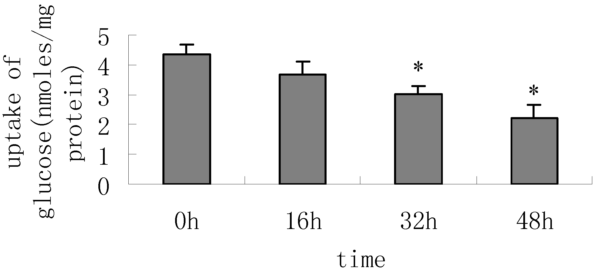
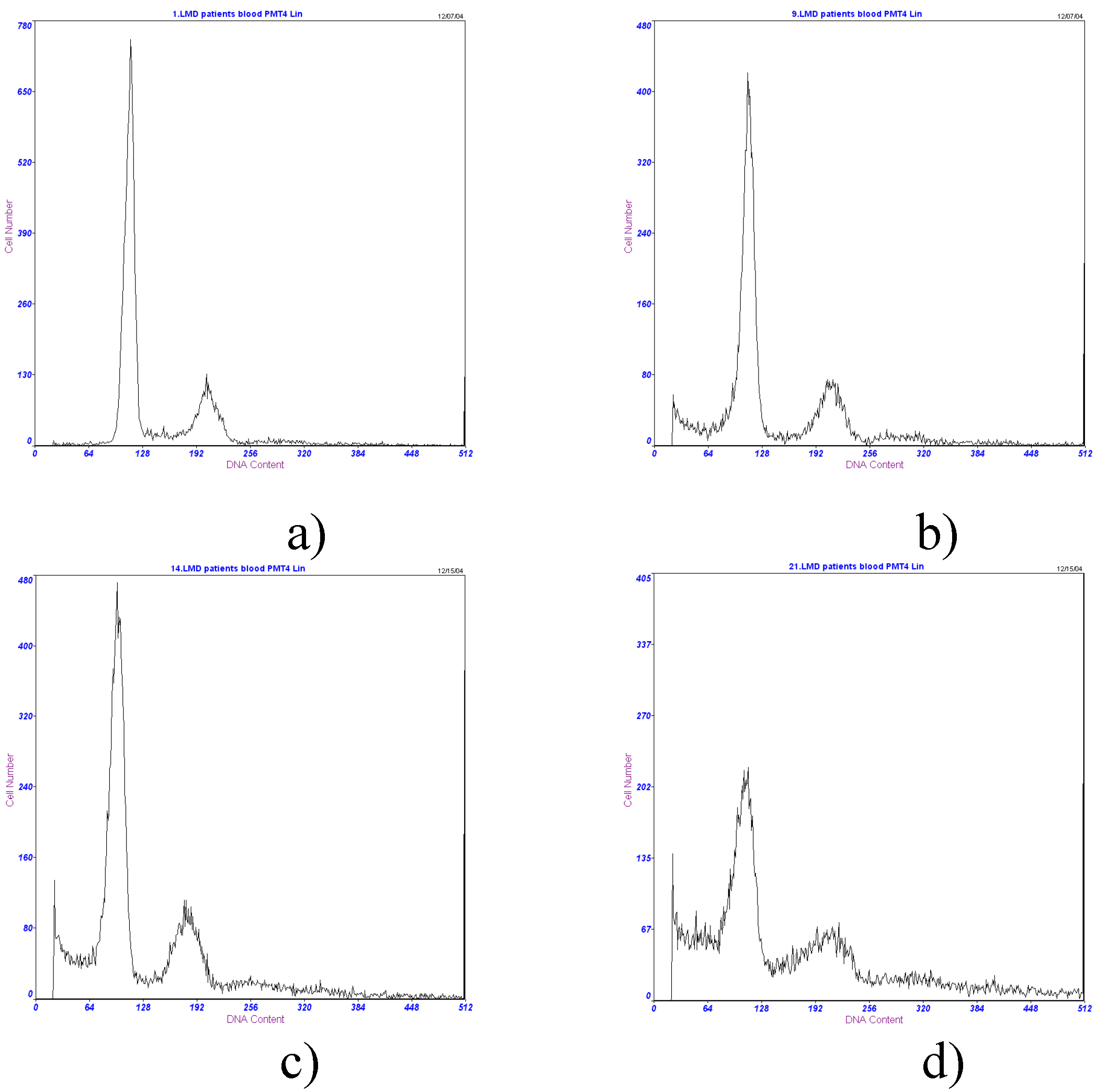
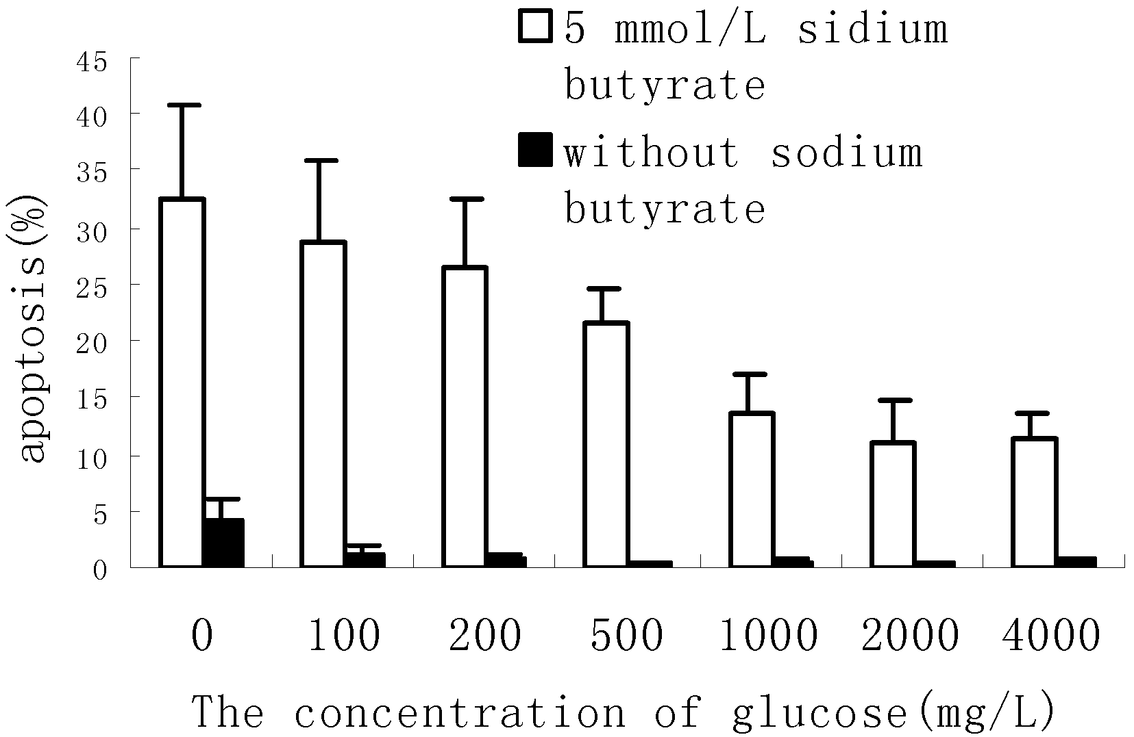
Result and discussion

By using RT-PCR, the effect of sodium butyrate on MCT1 and GLUT1-5 mRNA was investigated. When HT-29 cell line was treated with 5mmol/L sodium butyrate, a decrease of GLUT1 mRNA was detected (p*<0.05). Sodium butyrate inhibited the expression of GLUT1 in a time ­dependent manner. After treated with sodium butyrate for 48h, the GLUT1 mRNA/β-actin mRNA ratio decreased 2.4±0.4 folds compared with the control without treatment. But no significant changes of GLUT2, GLUT3, GLUT5 mRNA were observed(p>0.05) and GLUT4 mRNA can not be detected in HT-29 cell line. Glucose transport assay showed that when cells were treated with sodium butyrate more than 16h, the uptake of 2-deoxy-[3H]glucose decreased (p*<0.05). All these results showed that the regulation of GLUT1 may be responsible for the decrease of the uptake of glucose. MCT1 mRNA also had no significant change, suggesting that sodium butyrate may not regulate the uptake of colon cancer cell line on itself (figure 1, figure 2).
Figure 1. With the treatment of 5 mmol/L sodium butyrate in complete medium on HT-29 cell line for 16,32,48h, the expression of GLUT1 mRNA decreased obviously comparing with the cells without treatment (p*<0.05), but there was no significant change of GLUT2, GLUT3, GLUT5, MCT1mRNA, and GLUT4 was not detected in HT-29 cell line.
Ijms 07 00059 g001
Figure 2. 1×10⁶ cells were seeded in 12 wells multiplates, and cultured for 48 h until they reached confluence. Then the cells were treated with 5mmol/L sodium butyrate for 0 (without treating with sodium butyrate), 16, 32, 48h before glucose transport measurements. After washing, cells were incubated for 5 minutes in the present of 2-deoxy-D[3H]-glucose, the glucose transported was detected as described in the “materials and methods”. With the treatment of sodium butyrate more than 16h, the transport of glucose decreased obviously compared to that without adding sodium butyrate (p *<0.05).
Ijms 07 00059 g002
Sodium butyrate can induce apoptosis in some colon cancer cell lines by regulating bax or bcl-x/l. In our study, sodium butyrate can downregulate the expression of bcl-x/l after adding sodium butyrate for 48 h, however, bax expression had no significant change after treatment with sodium butyrate (figure 3). When HT-29 cells was treated with 5mmol/L sodium butyrate, with the concentration of glucose increased from 0mg/L to 2000mg/L, the expression of Bcl-x/l increased obviously (1.6±0.5 folds), but the expression of bax had no significant change (figure 3) 5 mmol/L
sodium butyrate + + + -
glucose (mg/L) 0 500 2000 2000
Figure 3. When HT-29 cell line was treated with 5 mmol/L sodium butyrate (containing 2000mg/Lglucose ) for 48 h, the expression of Bax didn’t change obviously, but the expression of Bcl-x/l decreased obviously compared with that without treating with sodium butyrate (containing 2000mg/Lglucose ). When HT-29 cell line was treated with 5 mmol/L sodium butyrate for 48 h, with the concentration of glucose increased from 0mg/L to 500,2000mg/L,the expression of Bcl-x/l protein increased obviously, but the expression of bax protein didn’t change.
Ijms 07 00059 g003
After treatment with sodium butyrate more than 24 h, the apoptosis in HT-29 cells can be observed and sodium butyrate induced apoptosis in a time-depended manner.(data not show). When treated with 5mmol/L sodium butyrate, with the concentration of glucose increasing from 0mg/L to 2000mg/L, the apoptosis decreased obviously (p<0.05). When the concentration of glucose was 0mg/L, the apoptosis rate was 32.7±8.1%, and when the concentration of glucose reached to 2000mg/L, the apoptosis decreased to 11.0±3.7%. When the concentration of glucose reached to 4000mg/L from 2000mg/L, the apoptosis did not decrease continuously in HT-29 cell line. But it was interest that in the absence of sodium butyrate, low concentration of glucose (0,100,200mg/L) can induce only a few cells apoptosis in HT-29. When the concentration of glucose was 0mg/L, the apoptosis rate was 4.1±1.8%. But when the concentration of glucose was more than 200mg/L, there is no significant apoptosis (figure 4, figure 5) .
Figure 4. a) In the condition of 2000mg/L glucose for 48h, without sodium butyrate, the apoptosis was very low.(0.4±0.2%). b) In the condition of 0mg/L glucose for 48h, without sodium butyrate, apoptosis was observed in a few cells (4.1±1.8%). c) In the condition of 2000mg/L,treated with 5mmol/Lsodium butyrate for 48 h,apoptosis of many cancer cells was observed (11.0±3.7%). d) In the condition of 100mg/L glucose,treated with 5mmol/Lsodium butyrate for 48 h,a great deal of apoptosis in HT-29 colonic cancer cell were found (28.7±7.2%)(FCM).
Ijms 07 00059 g004
Figure 5. Treated with 5mmol/L sodium butyrate for 48 h,with the increase of concentration of glucose from 0mg/L to 2000mg/L, the apoptosis of HT-29 colonic cancer cell decreased obviously (p<0.05). Without sodium butyrate, when the concentration of glucose was less than 200mg/L, apoptosis in a few cells was observed, but when the concentration of glucose was more than 200mg/L, the apoptosis was very low.
Ijms 07 00059 g005
Butyrate and other short chain fatty acids (SCFA) are generated in the intestine by the bacterial metabolism of dietary fiber. SCFA, especially butyrate, benefit the normal coloncytes which utilize it as their primary energy source. Butyrate has been shown to inhibit proliferation and induce differentiation and apoptosis in various tumor cell line including colorectal cancer cell.
Healthy colonocytes derive ~70% of their energy supply from SCFA, but malignant tissues show increased glucose uptake and utilization. Glucose is an important energy source for colonic cancer cell. Glucose transport is controlled through GLUTs with GLUT1 being the most important isoform in human colon cancer, while the lower expression and lower affinity of other GLUT comparing to GLUT1 has slight influence on the uptake and intracellular of glucose.
GLUT1 has a high affinity for glucose. The expression of GLUT1 is important for tumor cell to cope with the increased need of energy. Several studies have shown that a significant number of malignant tumors including colorectal carcinomas expressed GLUT1 which is not detected in nomal colonic epithelium [25,26], whereas GLUT2, GLUT3, GLUT5 can be detected in both normal and malignant colonic epithelium. The present results showed that GLUT2, GLUT3, GLUT5 was not obviously regulated by sodium butyrate and GLUT4 was not detected, while only GLUT1 decreased and accompanied with the decrease of the uptake of glucose in HT-29 colon cancer cell. This showed the downregulation of GLUT1 must be responsible for the decrease of the uptake of glucose.
The downregulation of GLUT1 induced by sodium butyrate with the decrease of uptake of glucose was accompanied with increased apoptosis of cells. In the present study, sodium butyrate induced apoptosis and downregulated GLUT1 expression by a time-dependent manner. Downregulation of GLUT1 mRNA was observed after treated with sodium butyrate for 16h, while apoptosis was observed after treated with sodium butyrate for 24h. Some other researches have showed that the uptake and the intracellular concentration of glucose which are controlled by GLUT1 are associated with apoptosis. Higer levels of GLUT1 in tumor tissue reflect an increased uptake of glucose and glycolylic metabolism. Overexpression of GLUT1 with the high uptake of glucose have been shown to inhibit cytochrome C release and downstream caspase activation and delay the onset of apoptosis [27,28]. The decrease of GLUT1 may down regulate the uptake and the intracellular concentration of glucose and both nonxidative and oxidative glucose meatabolism. Low uptake of glucose and intracellular concentration of glucose may induce ATP depletion and stimulation of mitochondrial death pathway cascade, and may induce oxidative stress and trigger of bax-associated events including the JNK/MAPK signal pathway [29,30,31].Those all suggest that GLUT1 may play an role in apoptosis in HT-29 cell line.
When the concentration of glucose increased obviously, the uptake of glucose and the intracellular concentration of glucose also increased, and the downregulation of uptake and intracellular concentration of glucose controlled by decreased GLUT1 can be inverted by the increased glucose concentration in medium. If the expression of GLUT1 was associated with apoptosis, the increased glucose in medium may help decrease the apoptosis in HT-29 cell line. In fact we found that the increased concentration of glucose can obviously reduce the apoptosis induced by sodium butyrate. These results also suggested that the downregulation of GLUT1 with the decreased uptake and intracellular concentration of glucose may play a role in the course of apoptosis induced by sodium butyrate.
Bax and bcl-x/l are closely related to apoptosis. In our study, Sodium butyrate downregulated the expression of Bcl-x/l and didn’t change the expression of Bax. Many other researches showed the similar results that sodium butyrate can induce apoptosis by regulating the expression of Bcl-x/l in some colon cancer cell lines [32,33]. The change of concentration of glucose can not regulate the expression of bax, but can influence the expression of Bcl-x/l. This suggests that GLUT1 regulated the change of the uptake of glucose and the intracellular concentration of glucose may influence apoptosis by Bcl-x/l.
But it was hard to explain that low concentration of glucose in the absence of sodium butyrate can induce apoptosis only in a few cells. And when the concentration of glucose was more than 200mg/L, there was no significant apoptosis. The possible mechanism is that downregulation of uptake of glucose and intracellular glucose concentration and glucose metabolism may not directly induce apoptosis in HT-29 cell line, but only increase the sensitivity of cells to apoptosis induced by sodium butyrate. Some researches showed that inhibition of glucose metabolism may sensitize tumor cells to death receptor-triggered apoptosis [34]. In the present study, the change of glucose concentration did not regulate the expression of bax which was one of the most important apoptosis promoters, but only regulated the expression of bcl-x/l. Bcl-x/l may play a more important antiapoptotic role than bcl-2 in some colon tumour cells. In the presence of antiapoptotic bcl-x/l, cytochrome-c release is dramatically inhibited. But the reduce of bcl-x/l is not sufficient to allow translocation of cytochrome-c, with subsequent formation of an apoptosome. Downregulation of antiapoptotic bcl-xL is insuffcient to induce Caco-2 cell apoptosis, but it may decrease the threshold to undergo apoptosis [35]. So, the decrease of the uptake and metabolism of glucose caused by the down regulation of GLUT1 may sensitize tumor cells to apoptosis induced by sodium butyrate.
MCT1 is involved in cellular transportation of sodium butyrate . When HT-29 cells was treated with sodium butyrate, there was no significant influence on MCT1 mRNA. Many of the cellular effects of butyrate are concentration-dependent, and time-dependent. The ability of sodium butyrate to exert its effects may depend upon its intracellular concentration. MCT1 was upregulated by sodium butyrate in AA/C1 cell line. This may serve as a mechanism to maximize intracellular availability of butyrate. We repeated the experiment many times, and didn’t find that sodium butyrate can obviously regulate the expression of MCT1 in HT-29 cell line, so it may not influence the uptake and the intracellular concentration of itself in HT-29 cell line by regulating its receptors MCT1.

Conclusion

Sodium butyrate and glucose are important energy source for cells. As the main energy supply for nomal coloncytes, sodium butyrate can downregulate the expression of GLUT1 which may sensitize the cancer cells to apoptosis by regulating the expression of bcl-x/l in HT-29 colon cell line, suggesting that the uptake and metabolism of energy source may be associated with the effect of sodium butyrate, and GLUT1 could be a target for colon cancer therapy.

Experimental

Cells and Cell Culture

HT-29 human colonic cancer cells were grown as a monolayer in RPMI-1640 medium (Sigma) supplemented with 10% fetal calf serum (GIBCO), cultured in T-75 cm2 culture flasks, maintained at 37°C in 5% CO2 humidified atmosphere. At the beginning of the experiment, mycoplasma-free cells in the exponential growth phase were removed from the flask with 0.25% trypsin and 0.02% EDTA solution and seeded in T-75 cm2 flask in RPMI-1640 medium with 10% fetal calf serum. The cells were allowed to adhere for 24h. The seeding medium was removed and then replaced with experimental medium.

RNA Preparation

Cells were kept for 0,16,32,48h in the complete medium supplemeted with 5mmol/L sodium butyrate (Sigma). After harvesting of the cells, total RNA was extracted by means of Trizol (Invitrogen) according to the manufactures instruction. The concentration of RNA was measured by absorbance 260 and 280nm. Total RNA was suspended in DEPC-treated water and stored at –80°C.

Reverse Transcriptase-polymerase Chain Reaction

Sigle-stranded cDNA was prepared using 2ug of total RNA, 200U MMLV Reverse transciptase (Promega), 5ul MMLV buffer (Promega), 1.25ul 10mmol/LdNTP (Promega), 1ug oligod(dT)15 (Promega), 25U Rnasin (Biostar) in a 25ul solution and incubated for 90 minutes at 42°C. An amplification of the resulting cDNA sequence was carried out using polymerase chain reaction (PCR). 1ul cDNA was combined with 1pmol oligonucleotide primers specific for human GLUT1-5, MCT1,β-actin(table 1), and 0.5U Taq DNA Polymerase (Biostar), 2.5ul Tap buffer (Biostar), 0.5ul 10mmol/LdNTP in a 25ul solution. The conditions for the reaction were the following: 5 minutes at 95°C (predenaturation), followed by 28 cycles of 1min at 95°C (denaturation), 45 seconds at 60°C (annealing), and 1minute at 72°C (extension) and 7 minutes at 72°C (final extension). Then 5ul samples of amplified products were resolved by electrophoresis in 2% agarose gel, stained with ethidium bromide. The level of each PCR product was semiquantitatively evaluated using a digital camera and an image analysis system (Viberlourmat, France), and normalized to β-actin. Each experiments was done in triplicate.

Western Blot

Equal amounts of protein were loaded and run on a 12% denaturing polyacrylamide gel as described previously[22]. Separated proteins were transferred to a nitrocellulose membrane by electroblotter. The membrane was placed into blocking buffer for one hour at room temperature. Blocking buffer was decanted and the membrane was incubated with the primary antibody (Bax antibody:Santa Cruz Biotechnologies.Bcl-x/l antibody:Wuhan Boster Biological technology LTD. )on a shaker at 4°C overnight. After being washed, the membrane was incubated with a peroxidase conjugated secondary antibody, which was diluted in 5% non-fat milk in wash buffer (one hour; room temperature; gentle shaking). After washed, the membrane was exposed to sensitive film several minutes after incubating in western blotting luminol reagent(Cell Signal Corp). The bands were quantified by densitometry.
Table 1. The primers of GLUT1, MCT1 and β-actin.
Primers (sense and anti-sense)Size of PCR product (bp)
GLUT15’ CGGGCCAAGAGTGTGCTAAA 3’ 283bp
5’ TGACGATACCGGAGCCAATG 3’
GLUT25’CGTCTCCTTTGACATTTCCTTC3’221bp
5’GGTGGAGAAAACAGCCTAGAGAT3’
GLUT35’CCAACTTCCTAGTCGGATTG3’250bp
5’AGGAGGCACGACTTAGACAT3’
GLUT45’CCCCCTCAGCAGCGAGTGA3’319bp
5’GCACCGCCAGCACATTGTTG3’
GLUT55’GCAACAGGATCAGAGCATGA3’316bp
5’TCGCAGGCACGATAGAAAAT3’
MCT15’ CACCACCAGCGAAGTGTC 3’158bp
5’ AGAAAGAAGCTGCAATCAAG 3’
β-actin 5’ CGAGCGGGAAATCGTGCGTGACATTAAGGAGA 3’479bp
5’ CGTCATACTCCTGCTTGCTGATCCACATCTGC 3’

The Uptake of Glucose Assay

The uptake of glucose assay was similar as described before[23].The 1×10⁶ Cells were seeded in 12 well culture plates.Cells were further cultured for 48h until they reach confluency. Then adding sodium butyrate 5mmol/L for 0(without treating with sodium butyrate),16,32,48h.Then the cells were rinsed three times with phosphate buffered saline (PH=7.4) and incubated for designated time in Krebs-henseleit Hepes buffer buffer with 0.5μM(2mCi) 2-deoxy-[3H]glucose. The isotopes remaining in the media were washed three times with phosphate buffered saline after the designated incubation times. The cells were solubilized in 1.2 ml of 2% SDS and 0.8 ml lysates were taken to measure the amount of transported glucose in liquid scintillation counter. Each experiment were done in triplicate and protein concentration was measured by Bradgord method [24].

Apoptosis Assay

After HT-29 cells were harvested, they were fixed in 70% ethanol overnight at 4°C, washed by PBS, then mixed with PI staining fluid for 20 min at 4°C, and filtered. At last, the samples were examed by FCM. The apoptosis assay was similar as described before [36].

Acknowledgments

This work was supported by the Key Technologies R&D Program of Hubei Province under Grant No. 2004AA304B08.

References and Notes

  1. Jacobs, L.R. Fiber and colon cancer. Gastroenterol Clin. North. Am. 1988, 17, 747–60. [Google Scholar]
  2. Hernandez, A.; Thomas, R.; Smith, F.; Sandberg, J.; Kim, S.; Chung, DH.; Evers, B.M. Butyrate sensitizes human colon cancer cells to TRAIL-mediated apoptosis. Surgery 2001, 130, 265–72. [Google Scholar] [CrossRef]
  3. Wang, Q.; Wang, X.; Hernandez, A.; Kim, S.; Evers, B.M. Inhibition of the phosphatidylinositol 3-kinase pathway contributes to HT29 andCaco-2 intestinal cell differentiation. Gastroenterology 2001, 120, 1381–1392. [Google Scholar]
  4. Otaka, M.; Singhal, A.; Hakomori, S. Antibody-mediated targeting of differentiation inducers to tumor cells:inhibition of colonic cancer cell growth in vitro and in vivo. Biochem.Biophys. Res. Commun. 1989, 158, 202–208. [Google Scholar] [CrossRef]
  5. Vidyasagar, S.; Ramakrishna, B.S. Effects of butyrate on active sodium and chloride transport in rat and rabbitdistal colon. J. Physiol. 2002, 539, 163–173. [Google Scholar] [CrossRef]
  6. Scheppach, W.; Bartram, P.; Richter, A.; Richter, F.; Liepold, H.; Dusel, G.; Hofstetter, G.; Ruthlein, J.; Kasper, H. Effect of short-chain fatty acids on the human colonic mucosa in vitro. JPEN. J. Parenter. Entera. l. Nutr. 1992, 16, 43–48. [Google Scholar] [CrossRef]
  7. Butzner, J.D.; Meddings, J.B.; Dalal, V. Inhibition of short-chain fatty acid absorption and Na+ absorption during acute colitis in the rabbit. Gastroenterology 1994, 106, 1190–1198. [Google Scholar]
  8. Duncan, S.H.; Louis, P.; Flint, H.J. Lactate-utilizing bacteria, isolated from human feces, that produce butyrate as a major fermentation product. Appl. Environ. Microbiol. 2004, 70, 5810–5817. [Google Scholar] [CrossRef]
  9. Stein, J.; Zores, M.; Schroder, O. Short-chain fatty acid (SCFA) uptake into Caco-2 cells by a pH-dependent and carrier mediated transport mechanism. Eur. J. Nutr. 2000, 39, 121–125. [Google Scholar] [CrossRef]
  10. Younes, M.; Lechago, L.V.; Somoano, J.R.; Mosharaf, M.; Lechago, J. Wide expression of the human erythrocyte glucose transporter Glut1 in human cancers. Cancer Res. 1996, 56, 1164–1167. [Google Scholar]
  11. Bozzetti, F.; Gavazzi, C.; Mariani, L.; Crippa, F. Glucose-based total parenteral nutrition does not stimulate glucose uptake by humans tumours. Clin. Nutr. 2004, 23, 417–421. [Google Scholar] [CrossRef]
  12. Noguchi, Y.; Marat, D.; Saito, A.; Yoshikawa, T.; Doi, C.; Fukuzawa, K.; Tsuburaya, A.; Satoh, S.; Ito, T. Expression of facilitative glucose transporters in gastric tumors. Hepatogastroenterology. 1999, 46, 2683–2689. [Google Scholar]
  13. Mellanen, P.; Minn, H.; Grenman, R.; Harkonen, P. Expression of glucose transporters in head-and-neck tumors. Int. J. Cancer. 1994, 6, 622–629. [Google Scholar]
  14. Younes, M.; Brown, R.W.; Stephenson, M.; Gondo, M.; Cagle, P.T. Overexpression of Glut1 and Glut3 in stage I nonsmall cell lung carcinoma is associated with poor survival. Cancer 1997, 80, 1046–1051. [Google Scholar] [CrossRef]
  15. Brown, R.S.; Wahl, R.L. Overexpression of Glut-1 glucose transporter in human breast cancer. Cancer 1993, 72, 2979–2985. [Google Scholar] [CrossRef]
  16. Rivenzon-Segal, D.; Rushkin, E.; Polak-Charcon, S.; Degani, H. Glucose transporters and transport kinetics in retinoic acid-differentiated T47D human breast cancer cells. Am. J. Physiol. Endocrinol. Metab. 2000, 279, 508–519. [Google Scholar]
  17. Kang, S.S.; Chun, Y.K.; Hur, M.H.; Lee, H.K.; Kim, Y.J.; Hong, S.R.; Lee, J.H.; Lee, S.G.; Park, Y.K. Clinical significance of glucose transporter 1 (GLUT1) expression in human breast carcinoma . Jpn. J. Cancer Res. 2002, 93, 1123–1128. [Google Scholar] [CrossRef]
  18. Lambert, D.W.; Wood, I.S.; Ellis, A.; Shirazi-Beechey, S.P. Molecular changes in the expression of human colonic nutrient transporters during the transition from normality to malignancy. Br. J. Cancer 2002, 86, 1262–1269. [Google Scholar] [CrossRef]
  19. Hadjiagapiou, C.; Schmidt, L.; Dudeja, P.K.; Layden, T.J. Mechanism(s) of butyrate transport in Caco-2 cells: role of monocarboxylatetransporter 1. Am. J. Physiol. Gastrointest. Liver Physiol. 2000, 279, 775–780. [Google Scholar]
  20. Ritzhaupt, A.; Wood, I.S.; Ellis, A.; Hosie, K.B.; Shirazi-Beechey, S.P. Identification and characterization of a monocarboxylate transporter (MCT1) inpig and human colon: its potential to transport L-lactate as well as butyrate. J. Physiol. 1998, 513, 719–732. [Google Scholar] [CrossRef]
  21. Cuff, M.A.; Lambert, D.W.; Shirazi-Beechey, S.P. Substrate-induced regulation of the human colonic monocarboxylate transporter,MCT1. J. Physiol. 2002, 539, 361–371. [Google Scholar] [CrossRef]
  22. Pellizzaro, C.; Coradini, D.; Daidone, M.G. Modulation of angiogenesis-related proteins synthesis by sodium butyrate in colon cancer cell line HT29. Carcinogenesis 2002, 23, 735–740. [Google Scholar] [CrossRef]
  23. Choi, J.W.; Yoon, D.J.; Lee, H.W.; Han, D.P.; Ahn, Y.H. Antisense GLUT1 RNA suppresses the transforming phenotypes of NIH 3T3 cells transformed by N-Ras. Yonsei Med. J. 1995, 36, 480–486. [Google Scholar]
  24. Bradford, M.M. A rapid and sensitive method for the quantitation of microgram quantities of protein utilizing the principle of protein-dye binding. Anal. Biochem. 1976, 72, 248–254. [Google Scholar] [CrossRef]
  25. Noguchi, Y.; Okamoto, T.; Marat, D.; Yoshikawa, T.; Saitoh, A.; Doi, C.; Fukuzawa, K.; Tsuburaya, A.; Satoh, S.; Ito, T. Expression of facilitative glucose transporter 1 mRNA in colon cancer was not regulated by k-ras. Cancer Lett. 2000, 154, 137–142. [Google Scholar] [CrossRef]
  26. Younes, M.; Lechago, L.V. Lechago, Overexpression of the human erythrocyte glucose transporter occurs as a late event in human colorectal carcinogenesis and is associated with an increased incidence of lymph node metastases. J. Clin.Cancer Res. 1996, 2, 1151–1154. [Google Scholar]
  27. Loberg, R.D.; Vesely, E.; Brosius, F.C. Enhanced glycogen synthase kinase-3beta activity mediates hypoxia-induced apoptosis of vascular smooth muscle cells and is prevented by glucose transport and metabolism. J. Biol. Chem. 2002, 277, 41667–41673. [Google Scholar] [CrossRef]
  28. Vander Heiden, M.G.; Plas, D.R.; Rathmell, J.C.; Fox, C.J.; Harris, M.H.; Thompson, C.B. Growth factors can influence cell growth and survival through effects on glucose metabolism. Mol. Cell. Biol. 2001, 21, 5899–5912. [Google Scholar] [CrossRef]
  29. Moley, K.H.; Mueckler, M.M. Glucose transport and apoptosis. Apoptosis 2000, 5, 99–105. [Google Scholar] [CrossRef]
  30. Takata, K.; Hirano, H. Mechanism of glucose transport across the human and rat placental barrier. Microsc. Res. Tech. 1997, 38, 145–152. [Google Scholar] [CrossRef]
  31. Sato, Y.; Ito, T.; Udaka, N.; Kanisawa, M.; Noguchi, Y.; Cushman, S.W.; Satoh, S. Immunohistochemical localization of facilitated-diffusion glucose transportersin rat pancreatic islets. Tissue Cell 1996, 28, 637–643. [Google Scholar] [CrossRef]
  32. Kovarikova, M.; Hofmanova, J.; Soucek, K.; Kozubik, A. The effects of TNF-alpha and inhibitors of arachidonic acid metabolism on human colon HT-29 cells depend on differentiation status. Differentiation 2004, 72, 23–31. [Google Scholar] [CrossRef]
  33. Ruemmele, F.M.; Schwartz, S.; Seidman, E.G.; Dionne, S.; Levy, E.; Lentze, M. J. Butyrate induced Caco-2 cell apoptosis is mediated via the mitochondrial pathway. Gut. 2003, 52, 94–100. [Google Scholar] [CrossRef]
  34. Munoz-Pinedo, C.; Ruiz-Ruiz, C.; Ruiz de Almodovar, C.; Palacios, C.; Lopez-Rivas, A. Inhibition of glucose metabolism sensitizes tumor cells to death receptor-triggered apoptosis through enhancement of death-inducing signalingcomplex formation and apical procaspase-8 processing. J. Biol. Chem. 2003, 278, 12759–12768. [Google Scholar] [CrossRef]
  35. Wacheck, V.; Selzer, E.; Gunsberg, P.; Lucas, T.; Meyer, H.; Thallinger, C.; Monia, B.P.; Jansen, B. Bcl-x(L) antisense oligonucleotidesradiosensitise colon cancer cells. Br. J. Cancer 2003, 89, 1352–1357. [Google Scholar] [CrossRef] [Green Version]
  36. Zheng, S.Y.; Li, D.C.; Zhang, Z.D.; Zhao, J.; Ge, J.F. Adenovirus-mediated FasL gene transfer into human gastric carcinoma. World. J. Gastroenterol. 2005, 11, 3446–3450. [Google Scholar]

