Article

Analysis of Protein–Protein Interactions in MCF-7 and MDA-MB-231 Cell Lines Using Phthalic Acid Chemical

1
Department of Biotechnology, College of Life Science, Kaohsiung Medical University, Kaohsiung 80708, Taiwan
2
Department of Public Health, College of Health Science, Kaohsiung Medical University, Kaohsiung 80708, Taiwan
3
Graduate Institute of Medicine, College of Medicine, Kaohsiung Medical University, Kaohsiung 80708, Taiwan
4
Center for Resources, Research and Development, Kaohsiung Medical University, Kaohsiung 80708, Taiwan
5
Center of Excellence for Environmental Medicine, Kaohsiung Medical University, Kaohsiung 80708, Taiwan
*
Authors to whom correspondence should be addressed.
Int. J. Mol. Sci. 2014, 15(11), 20770-20788; https://0-doi-org.brum.beds.ac.uk/10.3390/ijms151120770
Submission received: 12 September 2014 / Revised: 16 October 2014 / Accepted: 27 October 2014 / Published: 13 November 2014
(This article belongs to the Special Issue Proteins and Protein-Ligand Interactions)

Abstract

:
Phthalates are a class of plasticizers that have been characterized as endocrine disrupters, and are associated with genital diseases, cardiotoxicity, hepatotoxicity, and nephrotoxicity in the GeneOntology gene/protein database. In this study, we synthesized phthalic acid chemical probes and demonstrated differing protein–protein interactions between MCF-7 cells and MDA-MB-231 breast cancer cell lines. Phthalic acid chemical probes were synthesized using silicon dioxide particle carriers, which were modified using the silanized linker 3-aminopropyl triethoxyslane (APTES). Incubation with cell lysates from breast cancer cell lines revealed interactions between phthalic acid and cellular proteins in MCF-7 and MDA-MB-231 cells. Subsequent proteomics analyses indicated 22 phthalic acid-binding proteins in both cell types, including heat shock cognate 71-kDa protein, ATP synthase subunit beta, and heat shock protein HSP 90-beta. In addition, 21 MCF-7-specific and 32 MDA-MB-231 specific phthalic acid-binding proteins were identified, including related proteasome proteins, heat shock 70-kDa protein, and NADPH dehydrogenase and ribosomal correlated proteins, ras-related proteins, and members of the heat shock protein family, respectively.

1. Introduction

Phthalates such as di-(2-ethylhexyl) phthalate (DEHP), butyl benzyl phthalate (BBP), di-isononyl phthalate (DINP), di-isodecyl phthalate (DIDP), di-ethyl phthalate (DEP), di-isobutyl phthalate (DIBP), and di-n-butyl phthalate (DBP) are globally used to increase the plasticity of plastic products, and are easily detected in bags, infants’ toys, bottles, flooring, and cosmetics. Moreover, chronic kidney disease (CKD) patients undergoing hemodialysis are exposed to DEHP and polyvinyl chloride (PVC) from dialysis tubes and bag filters [1,2]. The phthalates DEHP, BBP, and DBP and their metabolites are classified as endocrine disruptors, and have been shown to adversely affect sexual development in rats [3,4,5] and male rabbits exposed to DBP during adolescence [6]. Moreover, an increased concentration of phthalates’ metabolites reportedly induced asthma and allergic diseases, and they were easily detected in urine [7,8]. Accordingly, simulations of the Comparative Toxicogenomics Database (CTD) associate phthalates with cardiotoxicity, hepatotoxicity, and endocrine and genital diseases. In particular, the relationship between DEHP and mono-2-ethylhexyl phthalate MEHP and nephrotoxicity has been shown in GeneOntology pathways using network analyses with Ingenuity Pathways Analysis (IPA, QIAGEN’s Ingenuity Systems, Redwood, CA, USA), Pathway Studio (Ariadne, Inc., Rockville, MD, USA), and MetaCore Analytical Suite (GeneGO, Inc., St. Joseph, MI, USA) software [9]. The U.S. Centers for Disease Control and Prevention (CDC) announced that >97% of Americans had detectable phthalate metabolites in urine samples, including monobutyl phthalate (MBP), monobenzyl phthalate (MBzP), and monoethyl phthalate (MEP). In addition, urine samples from >75% of Americans contain DEHP and dimethyl phthalate (DMP) metabolites, such as MEHP and monomethyl phthalate (MMP) [10,11], and urine samples from >92% of Americans contain bisphenol A (BPA) [11,12].
Proteomic techniques using tandem mass spectrometry (MS) and bioinformatics have been developed rapidly over the past two decades. Whereas proteomics studies previously relied on gel electrophoresis [13], gel-free shotgun proteomics techniques are now widely used to screen target proteins [14]. However, tandem MS remains critical to the evaluation and verification of biomarkers after identification with genomics and quantitative proteomic comparison of normal and abnormal specimens [15,16]. Multidimensional gel electrophoresis or multidimensional liquid chromatography (MDLC) techniques can limit numbers of candidate biomarkers [17,18,19,20]. However, numbers of identified nonspecific biomarker candidates often hamper evaluation and verification. As alternatives, target protein screening can be achieved using activity-based chemical probes that detect proteomic profiles according to carbon electrophiles [21] or activity-based proteomics that generate serine hydrolase enzymes [22]. Similar methods involve the design of probes using click chemistry to connect proteins and carriers [21,23], the use of chemiluminescent bioprobes [24], and Au nanoparticles linked with synthetic DNA to detect estrogen receptors [25].
In previous studies, phthalic acid was observed as a secondary metabolite from phthalate derivation, which is observed in dialysis patients [2]. In addition, the phthalic acid metabolites of DEHP and MEHP were described [26]. In the present study, we used phthalic acid as a phthalate precursor to synthesize esterified phthalic acid chemical probes and detect protein–protein interactions. Previously, we developed chemical probes that generate phthalic acid or nicotinic acid using 3-aminopropyl triethoxyslane (APTES) linkers on silicon dioxide particles [27,28]. In our previous study, BBP promoted progression of a breast cancer cell line by inducing lymphoid enhancer factor 1 [29]. For the present study, we used chemical probes to characterize phthalic acid-binding proteins in MCF-7 and MDA-MB-231 cells. Subsequently, quantitative proteomics analyses identified 22 binding proteins that were common to both cell types, including heat shock cognate 71-kDa protein, ATP synthase subunit beta, and heat shock protein HSP 90-beta. Finally, ATP synthase subunit beta, heat shock protein HSP 90-beta, and heat shock cognate 71-kDa protein-linked proteasome protein were identified as exclusive MCF-7 proteins, and connected ribosomal correlated proteins were identified as specific to MDA-MB-231 cells.

2. Results and Discussion

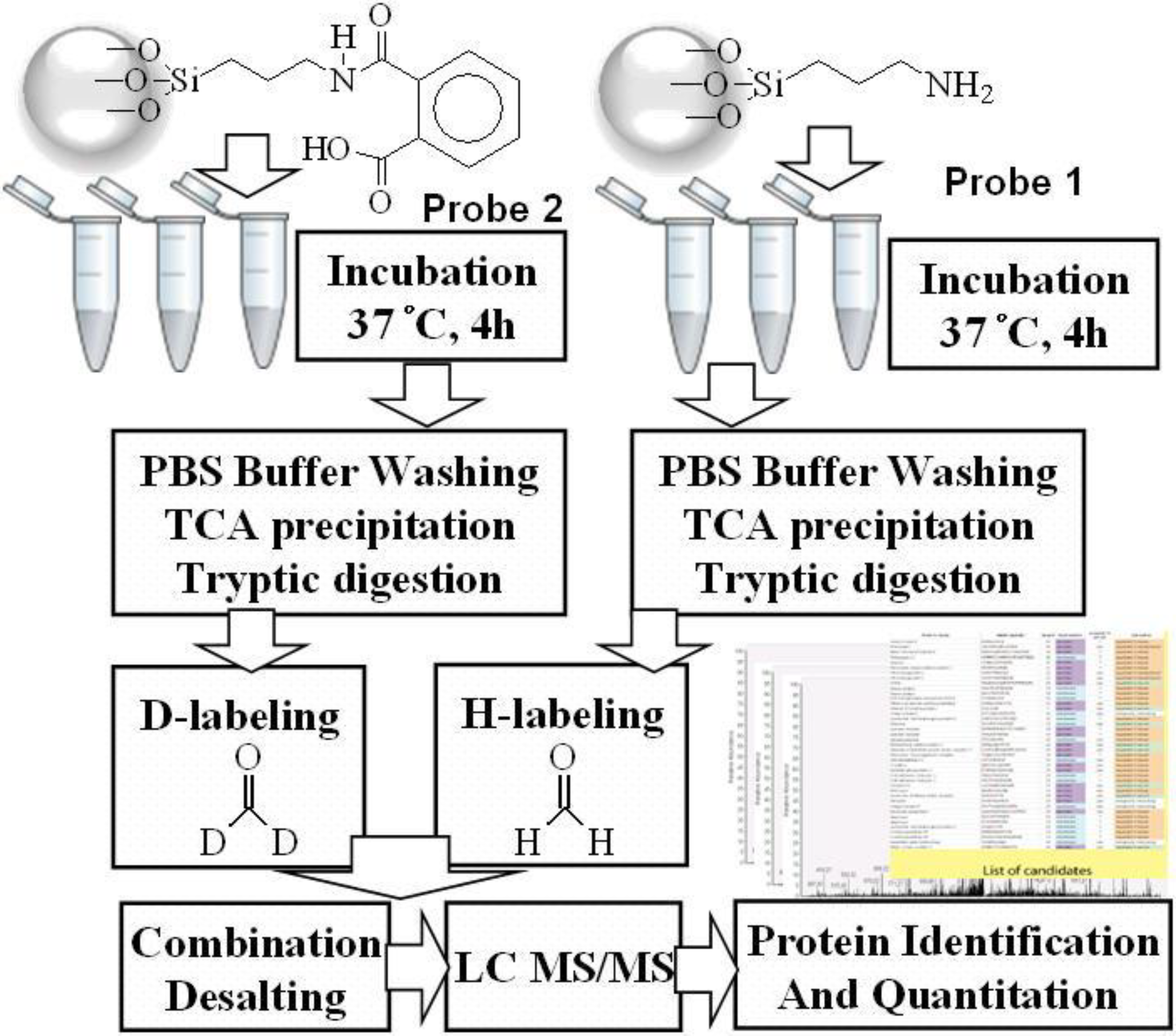
2.1. Phthalic Acid Chemical Probe Synthesis and Characterization

Phthalic acid chemical probes were synthesized and characterized as shown in previous studies [27,28]. SiO2 surfaces were modified by APTES using a silanized modification technique [30]. After reaction of carboxylic groups with 1-Ethyl-3-(3-dimethylaminopropyl) carbodiimide (EDC)/N-hydroxysuccinimide (NHS), phthalic acid was generated for 12 h and synthetic probes were characterized using infrared spectroscopy (IR) [31].

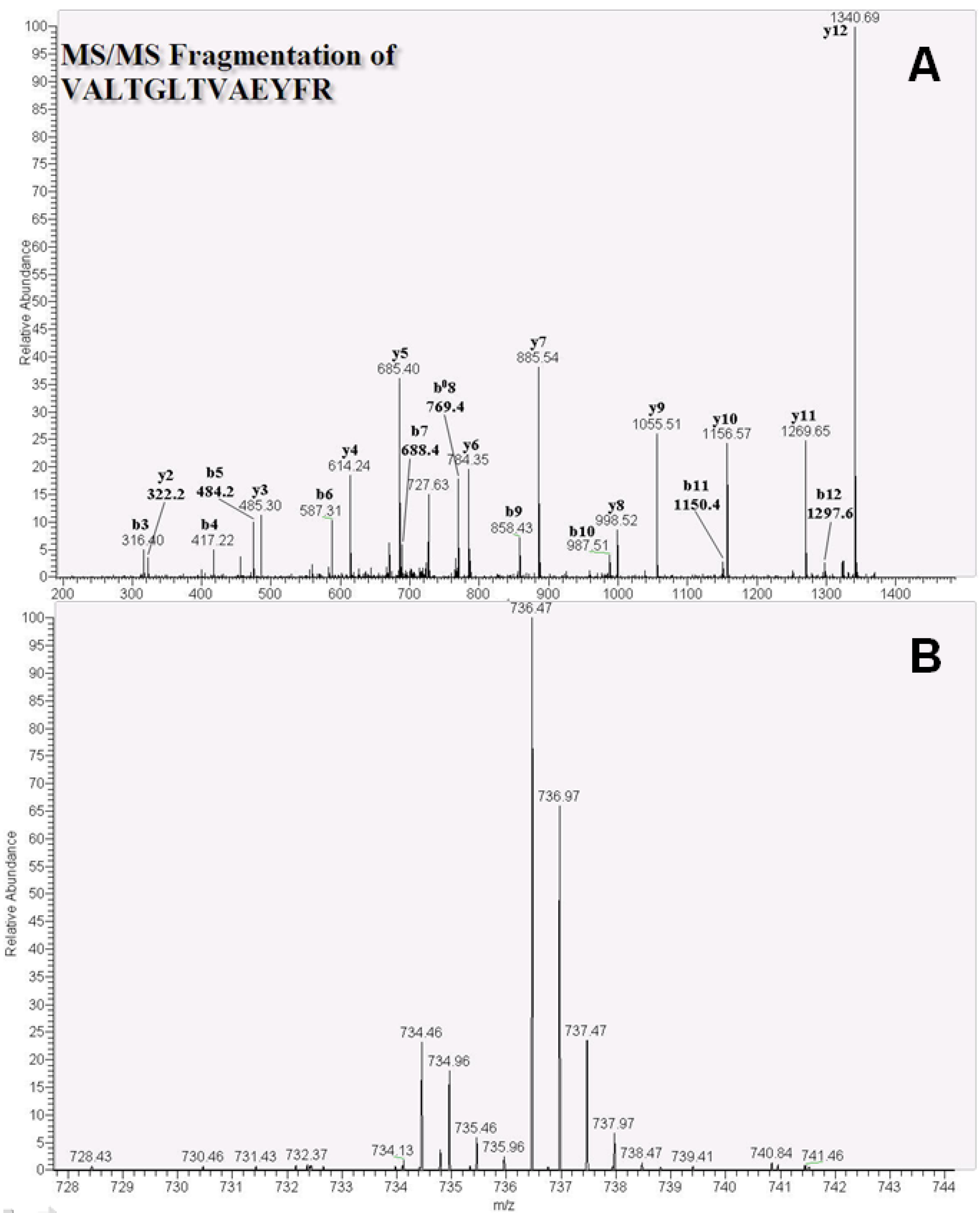
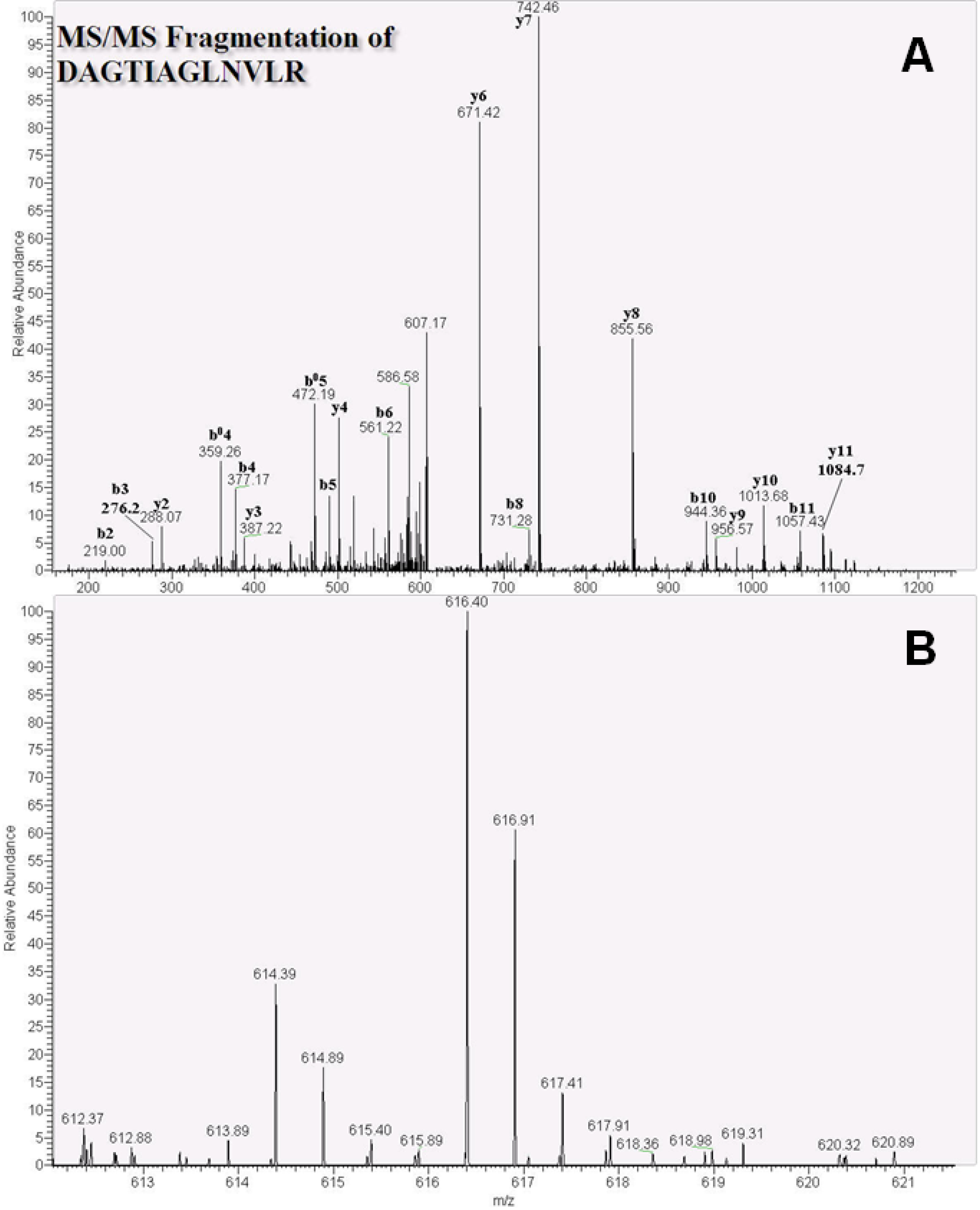
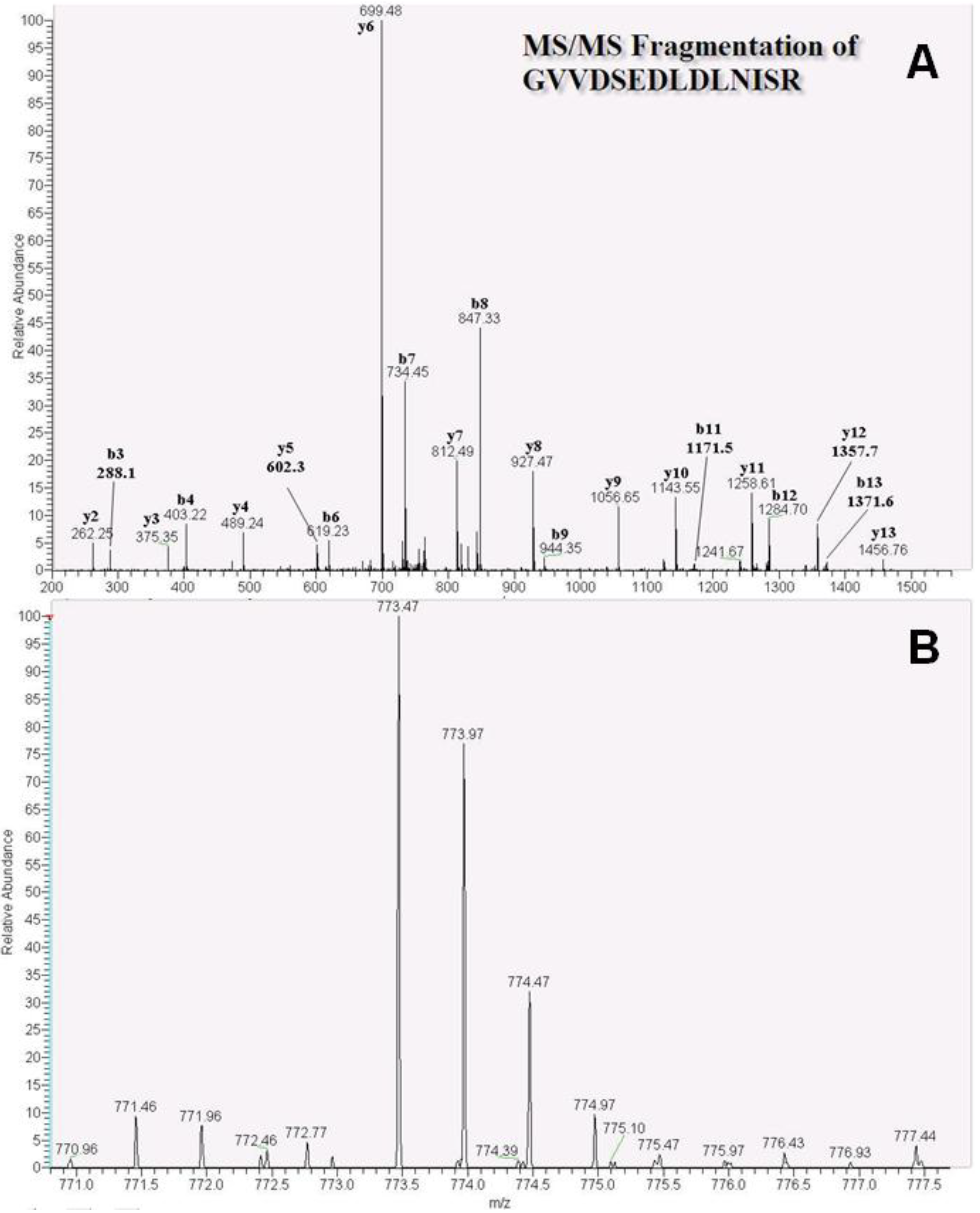
2.2. Identification and Quantitation of Phthalic Acid-Binding Proteins Using Proteomics

Chemical probes were individually incubated with MCF-7 and MDA-MB-231 cell lysates, and phthalic acid-bound proteins were identified using LC-MS/MS (Figure 1). After reduction by dl-dithiothreitol (DTT) and alkylation by iodoacetamide (IAM), related proteins were extracted and eluted using 1% sodium dodecyl sulfate (SDS). SDS was then removed by trichloroacetic acid (TCA) precipitation and proteins were subjected to tryptic digestion. Tryptic peptides bound to probes 1 and 2 were labeled with formaldehyde-H2 and formaldehyde-D2, respectively. Labeled samples were acidized using 10% trifluoroacetic acid (TFA) and were then desalted using a C18 desalting cartridge. Subsequently, eluted samples containing peptide mixtures were examined using LC-MS/MS, and raw data were generated using Raw2MSM (version 1.10_2007.06.14) [32] for protein characterization and Mascot Distiller (version 2.4.2.0 (64 bits)) for protein quantitation.
Figure 1. Flowchart of cell treatments and quantitation of proteins in MCF-7 and MDA-MB-231 cell using a phthalic acid chemical probe.
Ijms 15 20770 g001

2.3. Phthalic Acid-Binding Proteins in MCF-7 and MDA-MB-231 Cell Lines

Protein quantitation using Mascot Distiller and manual statistics showed that ATP synthase subunit beta, the heat shock protein family, and elongation factor 1-alpha 1 bound phthalic acid chemical probes, and related proteins were found in MCF-7 and MDA-MB-231 cell lines. Furthermore, phthalic acid chemical probes were bound to numerous proteasome-related and energy-correlated proteins such as NAD(P)H dehydrogenase, UDP-glucose 6-dehydrogenase, and fatty acid synthase in MCF-7 cells. Among the correlated proteins (Table 1), the phthalic acid probes detected ribosomal proteins such as 60S acidic ribosomal proteins and 40S ribosomal proteins, ras-related proteins, and heat shock proteins including 60-kDa heat shock protein and heat shock 70-kDa protein 6.
Table 1. Phthalic acid chemical probe related proteins with significant D/H labeling ratios in MCF-7 and MDA-MB-231 breast cancer cell lines.
Accession aGene Name bProtein IdentificationMDA-MB-231 H/L(I) cMDA-MB-231 H/L(II) cMDA-MB-231 H/L(III) cAverageMCF-7 H/L(I) cMCF-7 H/L(II) cMCF-7 H/L(III) cAverage
Phthalic acid-binding proteins identified in MCF-7 and MDA-MB-231
HSP7C_HUMANHSPA8Heat shock cognate 71-kDa protein5.04.03.44.2n.a.0.48.74.6
ATPB_HUMANATP5BATP synthase subunit beta, mitochondrial5.93.53.04.121.42.25.09.5
HS90B_HUMANHSP90AB1Heat shock protein HSP 90-beta8.5108.69.117n.a.1315.0
HS90A_HUMANHSP90AA1Heat shock protein HSP 90-alpha12.615.68.012.121.42.31111.7
NPM_HUMANNPM1Nucleophosmin4.56.64.15.1222.21011.4
G3P_HUMANGAPDHGlyceraldehyde-3-phosphate dehydrogenase4.63.32.23.4111.38.16.8
LDHA_HUMANLDHAl-lactate dehydrogenase A chain7.69.16.97.9161.47109.2
ENOA_HUMANENO1Alpha-enolase4.43.25.04.213.82.99.28.6
KPYM_HUMANPKM2Pyruvate kinase isozymes M1/M25.47.35.86.29.13.13.25.1
ADT2_HUMANSLC25A5ADP/ATP translocase 27.15.28.16.847.13.112.020.7
ARF4_HUMANARF4ADP-ribosylation factor 47.06.814.19.330.3n.a.n.a.30.3
TBA1B_HUMANTUBA1BTubulin alpha-1B chain8.08.17.47.815.33.515.111.3
TBB2C_HUMANTUBB2CTubulin beta-2C chain16.05.47.99.814.21.1n.a.7.7
TBB5_HUMANTUBBTubulin beta chain12.15.18.28.514.11.00.95.3
RS2_HUMANRPS240S ribosomal protein S23.24.15.34.22.81.1n.a.2.0
PHB2_HUMANPHB2Prohibitin-23.42.95.03.822.91.713.012.5
CATD_HUMANCTSDCathepsin D3.32.52.52.88.22.06.55.6
K2C8_HUMANKRT8Keratin, type II cytoskeletal 83.23.32.02.86.92.63.74.4
K1C18_HUMANKRT18Keratin, type I cytoskeletal 183.52.43.13.018.5n.a.15.617.1
ACTB_HUMANACTBActin, cytoplasmic 19.35.56.57.113.94.28.68.9
EF1A1_HUMANEEF1A1Elongation factor 1-alpha 15.24.74.94.919.13.0n.a.11.1
TERA_HUMANVCPTransitional endoplasmic reticulum ATPase0.74.33.72.950.3n.a.n.a.50.3
Phthalic acid-binding proteins identified in MCF-7
PSA4_HUMANPSMA4Proteasome subunit alpha type-4----6.40.85.14.1
PSB6_HUMANPSMB6Proteasome subunit beta type-6----6.30.93.53.6
PSB5_HUMANPSMB5Proteasome subunit beta type-5----6.00.73.03.2
PSA6_HUMANPSMA6Proteasome subunit alpha type-6----5.90.84.23.6
PSA7_HUMANPSMA7Proteasome subunit alpha type-7----5.80.84.73.8
PSA2_HUMANPSMA2Proteasome subunit alpha type-2----5.31.04.63.6
PSA1_HUMANPSMA1Proteasome subunit alpha type-1----5.30.94.63.6
PSB7_HUMANPSMB7Proteasome subunit beta type-7----4.50.84.33.2
PSB2_HUMANPSMB2Proteasome subunit beta type-2----8.0n.a.3.55.8
PSA3_HUMANPSMA3Proteasome subunit alpha type-3----12.40.7n.a.6.6
PSB1_HUMANPSMB1Proteasome subunit beta type-1----26.20.84.510.5
HSP71_HUMANHSPA1AHeat shock 70-kDa protein 1A/1B----16.7n.a.n.a.16.7
HS71L_HUMANHSPA1LHeat shock 70-kDa protein 1-like----n.a.2.17.54.8
NQO1_HUMANNQO1NAD(P)H dehydrogenase [quinone] 1----33.42.9n.a.18.2
UGDH_HUMANUGDHUDP-glucose 6-dehydrogenase----22.51.4n.a.12.0
ADT3_HUMANSLC25A6ADP/ATP translocase 3----19.0n.a.n.a.19.0
FAS_HUMANFASNFatty acid synthase----16.62.79.69.6
H2B1A_HUMANHIST1H2BAHistone H2B type 1-A----16.92.310.49.9
K1C19_HUMANKRT19Keratin, type I cytoskeletal 19----2.20.73.92.3
EF1A2_HUMANEEF1A2Elongation factor 1-alpha 2----n.a.3.010.16.6
VDAC1_HUMANVDAC1Voltage-dependent anion-selective channel protein 1----6.61.76.14.8
Phthalic acid-binding proteins identified in MDA-MB-231
RLA0_HUMANRPLP060S acidic ribosomal protein P010.4n.a.3.36.9----
RL7A_HUMANRPL7A60S ribosomal protein L7a2.9n.a.n.a.2.9----
RL6_HUMANRPL660S ribosomal protein L66.54.62.74.6----
RL23A_HUMANRPL23A60S ribosomal protein L23a2.52.95.13.5----
RL5_HUMANRPL560S ribosomal protein L55.818.68.911.1----
RL11_HUMANRPL1160S ribosomal protein L113.9n.a.n.a.3.9----
RL31_HUMANRPL3160S ribosomal protein L311.92.33.32.5----
RS7_HUMANRPS740S ribosomal protein S72.42.45.83.5----
RS13_HUMANRPS1340S ribosomal protein S132.12.03.92.7----
RS23_HUMANRPS2340S ribosomal protein S233.23.94.73.9----
PSMD2_HUMANPSMD226S proteasome nonATPase regulatory subunit 25.6n.a.33.719.7----
CH60_HUMANHSPD160 kDa heat shock protein, mitochondrial6.46.54.85.9----
HSP76_HUMANHSPA6Heat shock 70-kDa protein 63.9n.a.n.a.3.9----
RB11A_HUMANRAB11ARas-related protein Rab-11A3.94.87.65.4----
RAB10_HUMANRAB10Ras-related protein Rab-10n.a.4.37.66.0----
PTRF_HUMANPTRFPolymerase I and transcript release factor3.43.62.33.1----
VIME_HUMANVIMVimentin3.1n.a.1.72.4----
ACTN1_HUMANACTN1Alpha-actinin-13.5n.a.n.a.3.5----
ACTN4_HUMANACTN4Alpha-actinin-4n.a.7.87.97.9----
ATPA_HUMANATP5A1ATP synthase subunit alpha, mitochondrial5.34.75.65.2----
LDHB_HUMANLDHBL-lactate dehydrogenase B chain5.912.713.410.7----
PSA5_HUMANPSMA5Proteasome subunit alpha type-52.01.60.71.4----
YBOX1_HUMANYBX1Nuclease-sensitive element-binding protein 11.32.71.31.8----
EF1G_HUMANEF1GElongation factor 1-gamma6.80.713.87.1----
RAN_HUMANRANGTP-binding nuclear protein Ran5.86.65.86.1----
H12_HUMANHIST1H1CHistone H1.22.63.492.632.9----
SMD3_HUMANSNRPD3Small nuclear ribonucleoprotein Sm D365.3n.a.67.966.6----
NP1L1_HUMANNAP1L1Nucleosome assembly protein 1-like 18.49.65.98.0----
IF4A1_HUMANEIF4A1Eukaryotic initiation factor 4A-I7.85.66.36.6----
ARF1_HUMANARF1ADP-ribosylation factor 17.4n.a.n.a.7.4----
SET_HUMANSETProtein SET6.98.63.76.4----
ENPL_HUMANHSP90B1Endoplasmin6.97.78.37.6----
a Accessions are presented according to Mascot software; b Gene names are listed according to the STRING software; c Ratio values are listed according to analyses using Mascot Distiller software.

2.4. Identification of Related Proteins Using Phthalic Acid Chemical Probes

Proteins were identified after conversion of raw MS data using Raw2MSM software with the Mascot search engine. ATP synthase subunit beta, heat shock protein HSP 90-beta, and heat shock cognate 71-kDa protein were characterized according to MS/MS patterns, and the peptide VALTGLTVAEYFR of the ATP synthase subunit beta protein showed b- and y-ion patterns (Figure 2A). Mean ATP synthase subunit beta protein quantities were 4.1- and 9.5-fold in MCF-7 and MDA-MB-231 cells (n = 3), respectively. Subsequent isotope labeling of VALTGLTVAEYFR using formaldehyde-D2 and formaldehyde-H2 (Figure 2B) gave an m/z of 734.46 with a charge 2+ and an m/z of 736.47, respectively.
Figure 2. Nano-LC tandem MS spectra of the ATP synthase subunit beta protein peptide VALTGLTVAEYFR in MDA-MB-231 cells. (A) Product ion scan spectra of the identified peptide VALTGLTVAEYFR (m/z 736.47, 2+ charge); (B) The identified light (L, formaldehyde-H2) and heavy (H, formaldehyde-D2) isotope-labeled peptides (m/z 734.46 and 736.47, respectively).
Ijms 15 20770 g002
The heat shock cognate 71-kDa protein was also characterized using tandem MS, and the representative peptide DAGTIAGLNVLR was identified with a charge of 2+. MS/MS spectra (Figure 3A) and quantitative data showed 4.2- and 4.6-fold increases in this peptide in MCF-7 and MDA-MB-231 cells (n = 3), respectively with m/z values of 616.40 (D-labeled peptide) and 614.39 (H-labeled peptide; Figure 3B). Finally, the related heat shock protein HSP 90-beta (Figure 4A) and the peptide GVVDSEDLDLNISR were identified with a charge of 2+, along with a D-labeled peptide m/z of 773.47 and a H-labeled peptide m/z of 771.46 (Figure 4B). Finally, quantitative ratios showed 9.1- and 15.0-fold increases in MDA-MB-231 and MCF-7 cell lines, respectively.
Figure 3. Nano-LC tandem MS analytic spectra of the heat shock cognate 71-kDa protein peptide DAGTIAGLNVLR in MDA-MB-231 cells. (A) Patterns of b- and y-ions were used to arrange the DAGTIAGLNVLR (m/z 616.40, 2+ charge) amino acid sequence; (B) Formaldehyde-H2 and formaldehyde-D2 isotope-labeled peptides with m/z 616.40 and 614.39, respectively.
Ijms 15 20770 g003
Figure 4. MS and MS/MS spectra of the heat shock protein HSP 90-beta peptide GVVDSEDLDLNISR. (A) MS/MS fragmentation of GVVDSEDLDLNISR (m/z 773.47, 2+ charge), showing b- and y-ion patterns; (B) Formaldehyde-D2 labeled peptide (m/z 773.47) and formaldehyde-H2 labeled peptide (m/z 771.46) were co-eluted using HPLC and had differing intensities for protein quantitation.
Ijms 15 20770 g004

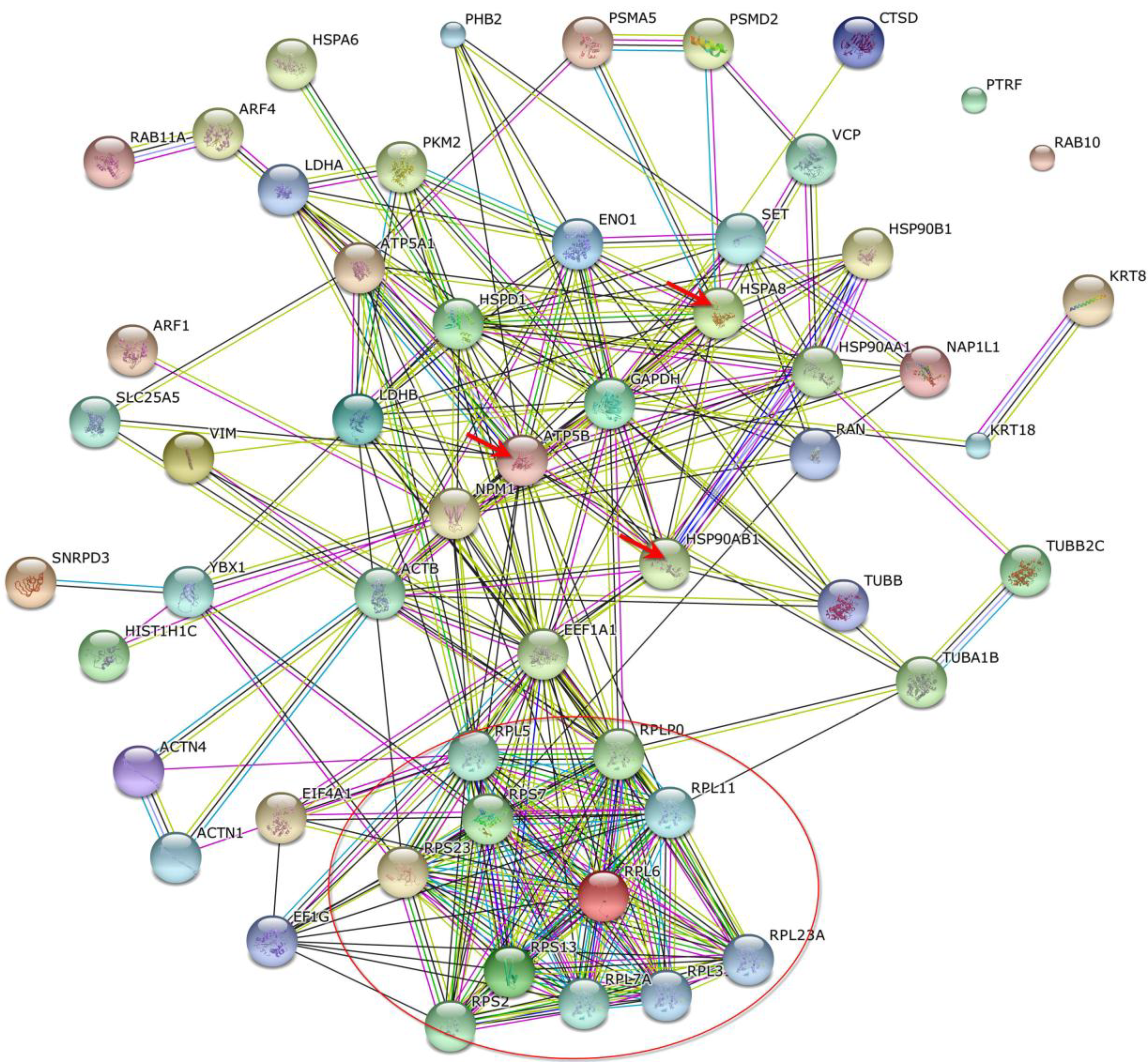
2.5. Relationships between Protein–Protein Interactions in MCF-7 and MDA-MB-231 Cells

Proteins that bind phthalic acid chemical probes were identified and relationships between these were characterized by organized protein–protein interactions using STRING software. Phthalic probes demonstrated arrangements of proteasome subunit proteins (red circle), and interactions with ATP synthase subunit beta (ATP5B) and heat shock 70-kDa protein 1A/1B (HSPA8) in MCF-7 cells (Figure 5). Furthermore, HSPA8 proteins interacted with heat shock protein HSP 90-beta (HSP90AB1), heat shock protein HSP 90-alpha (HSP90AA1), and nucleophosmin protein (NPM1). In MDA-MB-231 cells, protein–protein interactions (Figure 6) were found between HSPA8 and HSP90AB1, NPM1, ATP5B, glyceraldehyde-3-phosphate dehydrogenase (GAPDH), and elongation factor 1-alpha 1 (EEF1A1). Moreover, among related proteins, NPM1, GAPDH, HSP90 AB1, and ATP5B interacted with EEF1A1, which was correlated with a group of ribosomal proteins (red circle).
Figure 5. STRING protein–protein interactions between phthalic acid-binding proteins in MCF-7 cells. Red circles containing proteasome subunit proteins and the three red arrows indicate heat shock cognate 71-kDa protein, heat shock protein HSP 90-beta, and ATP synthase subunit beta.
Ijms 15 20770 g005
Figure 6. Schematic of the relationship between phthalic acid-binding proteins in MDA-MB-231 cells. High fold ratios were observed in triplicate experiments; the red circle includes numerous ribosomal proteins and the three red arrows indicate heat shock cognate 71-kDa protein, heat shock protein HSP 90-beta, and ATP synthase subunit beta.
Ijms 15 20770 g006

3. Experimental Section

3.1. Materials and Chemicals

dl-dithiothreitol (DTT), trifluoroacetic acid (TFA), 1-Ethyl-3-(3-dimethylaminopropyl) carbodiimide (EDC), sodium acetate, and sodium cyanoborohydride (NaBH3CN) were purchased from Sigma-Aldrich (St. Louis, MO, USA). Acetonitrile (MeCN), ammonium hydrogen carbonate (NH4HCO3), sodium hydroxide, hydrochloric acid, sodium dodecyl sulfate (SDS), acetic acid, ethanol, and urea were purchased from J.T.Baker (Phillipsburg, NJ, USA). Formaldehyde solution (36.5%–38% in H2O), phenylmethane sulfonyl fluoride (PMSF), leupeptin, sodium orthovanadate (Na3VO4), sodium chloride (NaCl), formic acid (FA), potassium chloride (KCl), potassium dihydrogen phosphate (KH2PO4), sodium dihydrogen phosphate (NaH2PO4), and iodoacetamide (IAM) were purchased from Sigma (St. Louis, MO, USA). Formaldehyde-D2 (20% solution in D2O) was obtained from Isotec Corp. (Miamisburg, OH, USA). Trypsin was purchased from Promega (Madison, WI, USA). Phthalic acid, APTES, potassium bromide (FTIR grade), and N-hydroxysuccinimide (NHS) were purchased from Alfa Aesar (Heysham, UK). Deionized water was obtained with a resistance of 18.2 MΩ using a Millipore water system (Millipore, Bedford, MA, USA).

3.2. Synthesis and Characterization of Phthalic Acid Chemical Probes

The chemical probes were synthesized and characterized according to previous studies [27,28]. Briefly, 200 mg of silicon dioxide (SiO2, 400 mesh, approximately 40 μm; Acros Organics, Geel, Belgium) was activated using 0.5 M HCl and 0.5 M NaOH, then washed and dried with distilled water and ethanol to remove and evaporate HCl and NaOH. Surface silanization of SiO2 was performed by reacting with APTES (5% in ethanol), and the SiO2 was washed two times with 1 mL of ethanol and was baked overnight in an oven at 50 °C. Subsequently, 13 mg of EDC and 5 mg of NHS were added in 1 mL of deionized water to react with 10 mg of phthalic acid. After activation by EDC/NHS, the phthalic acid was conjugated to SiO2 via the amino groups of APTES. Functional groups of APTES-modified SiO2, phthalic acid SiO2, phthalic acid, and SiO2 were then identified using infrared spectroscopy (IR). Particles were ground to flat wafers with KBr (FTIR grade) under pressure, and were characterized using IR spectroscopy (Perkin-Elmer Spectrum RX1 spectrometer, Canton, MA, USA).

3.3. Culture of MCF-7 and MDA-MB-231 Breast Cancer Cells

MCF-7 and MDA-MB-231 cells were cultured in Dulbecco’s modified Eagle’s medium (DMEM, Sigma-Aldrich) supplemented with 1% penicillin (GibcoBRL, Grand Island, NY, USA) and 5% fetal bovine serum (FBS). Cells were cultured to 80% confluence in 100-mm dishes, at 37 °C in a 5% CO2 incubator. Cells were then lysed in a modified RIPA buffer containing 150 mM NaCl, 50 mM Tris-HCl, 1% NP-40, 0.1% SDS, 0.5 mM PMSF, 2 μg/mL leupeptin, and 1 mM Na3VO4 at pH 7.5. Protein concentrations of MCF-7 and MDA-MB-231 cell lysates were determined using the Bradford assay (Thermo, Rockford, IL, USA).

3.4. Chemical Probe Conditions for MCF-7 and MDA-MB-231 Cell Lysates

The individual and triplicate MCF-7 and MDA-MB-231 cell lysates containing 100 μg of protein were incubated with APTES-modified (10 mg; probe 1) and phthalic acid-modified (10 mg; probe 2) chemical probes diluted to 400 μL in a phosphate-buffer saline (PBS) containing 2.7 mM KCl, 137 mM NaCl, 8 mM NaH2PH4, and 1.4 mM KH2PO4, at 37 °C for 4 h. After centrifugation, supernatants were removed and chemical probes were washed with 200 μL of PBS buffer and incubated at 37 °C for 4 h, three times.

3.5. Tryptic Digestion and Quantitative Dimethyl Labeling

After centrifugation, supernatants were removed and protein-bound chemical probes were eluted in 0.1% SDS. Before tryptic digestion, SDS was removed by TCA precipitation and the extracted proteins were reduced by DTT, alkylated by IAM, and digested using 0.2 μg trypsin. After 4 h, an additional 0.2 μg of trypsin was added and samples were incubated at 37 °C for a further 18 h. Bound proteins on chemical probes were quantitated using dimethyl labeling, and tryptic peptide solutions were dried using a centrifuge vacuum [33]. Subsequently, lyophilized samples were redissolved in 180 μL of 100 mM sodium acetate at pH 5.5. Tryptic peptides bound to probe 1 were modified using 10 μL of formaldehyde-H2, and those bound to probe 2 were reacted with 10 μL of formaldehyde-D2. After 5 min of vortexing, labeled samples were reduced using 10 μL of 600 mM sodium cyanoborohydride for 1 h. Labeled solutions were then combined and desalted by adjusting the pH to 2–3 using 10% TFA/H2O. The desalting kit comprised a C18 reverse-phase chromatography column packed with C18 powder in an in-house column cartridge and holder. Eluted solutions were vacuum dried for nano-LC-MS/MS.

3.6. Nano-LC-Tandem MS Analysis, Protein Identification and Quantitation

The combined labeled and lyophilized fractions were redissolved in 10 μL of 0.1% FA in H2O and analyzed using a Thermo LTQ Orbitrap XL system (Thermo Fisher Scientific, San Jose, CA, USA). Separate analyses were performed using a Waters ACQUITY nanoflow system (nanoUPLC, Waters Corp., Manchester, UK). In these experiments, 3-μL aliquots of redissolved samples were injected into a C18 capillary pretrapped column (20 mm × 180 μm), and protein separation was performed using a reverse-phase Waters BEH C18 column (i.d. 75 μm × 150 mm, 1.7 μm particle size). UPLC flow rates were set at 5 μL/min (loading pump) and 300 nL/min (gradient pump); mobile phases were prepared in bottles A and B, which contained 0.1% FA in water and 0.1% FA in 100% MeCN, respectively. The linear gradient comprised 2% (B) for 2 min, 2%–40% (B) for 40 min, 40%–98% (B) for 8 min, 98% (B) for 2 min, 98% to approximately 2% (B) over 1 min, and then 2% (B) for 7 min. Separated peptides were nebulized using a voltage of 1.8 kV in the positive ion mode and detected by tandem MS using a scan mode of m/z 400–1600 Da with 30,000 resolutions in the Orbitrap chamber. Separated peptides were predominantly detected in the MS mode, and five with high-intensity signals were selected and transferred into a collision-induced dissociation (CID) chamber with nitrogen collision gas and 35 eV collision energy for MS/MS fragmentation. The second scan mode for the MS/MS analyzer was set at m/z 100–1600 Da, with exclusion of similar m/z ions, evasion of interferences, and a repeat duration of 30 s in data-dependent mode. The integrated UPLC loading and analytic pump was controlled by MassLynx 4.1 and Global ProteinLynx softwares, and MS data were managed and acquired using Xcalibur software (version 2.0.7, Thermo-Finnigan, Inc., San Jose, CA, USA). Raw MS data were converted to the Mascot generic data format by using Raw2MSM (version 1.10_2007.06.14) [32], and protein quantities were converted using Mascot Distiller software (version 2.4.2.0; 64 bits, Matrix Science Ltd., London, UK) with Orbitrap_res_MS2 (default parameter setting) for peak list transformation and Homo sapiens (human) taxonomy in the Swiss-Prot database for the Mascot search engine, coupled with the Universal Protein Resource (UniPort) 2013 database (accessed on 21 September 2011) [34]. The following parameter settings for the Mascot search program [35] were installed: allow up to zero missed cleavages for tryptic digestion, dimethylation [MD] for quantitation, carbamidomethyl cysteine set for fixed modifications, mass tolerance of 10 ppm with precursor ions, and 0.8 Da for fragment ions. Peptides that had charges of 1+, 2+, or 3+ and Mascot ion scores of >20 (p < 0.05, individual peptides) were selected. Subsequently, quantitative ratios of D-label (Probe 2)/H-label (Probe 1) were generated and listed for each protein.

3.7. Establishment of STRING Protein–Protein Interaction Networks

Interactions between proteins and phthalic acid were predicted using the STRING database (version 9.1), and relationships between phthalate-related proteins and associated proteins were demonstrated.

4. Conclusions

In this study, phthalic acid chemical probes were used to demonstrate relationships between phthalic acid-sensitive proteins in MCF-7 and MDA-MB-231 cells. In these experiments, phthalate-like (phthalic acid) structures interacted with a group of proteasome subunit proteins in MCF-7 cells, and with a group of ribosomal proteins in MDA-MB-231 cells. Moreover, these proteins were connected by heat shock cognate 71-kDa protein, ATP synthase subunit beta, and heat shock protein HSP 90-beta. Finally, protein networks in MCF-7 and MDA-MB-231 cells were established using STRING protein–protein interaction software. Future studies may identify phthalate receptors using chemical probes.

Acknowledgments

This work was supported by grants from the National Science Council (NSC 100-2113-M-037-012-MY2, NSC 102-2113-M-037-013-MY2 and NSC 102-2632-B-037-001-MY3), Taipei, Taiwan, and Kaohsiung Medical University “Aim for the Top Universities Grant, grant No. KMU-TP103A15 and grant No. KMU-TP103A16”. We also thank the Center for Resources, Research and Development of Kaohsiung Medical University for support with proteomic techniques.

Author Contributions

Tsu-Nai Wang and Shih-Shin Liang carried out sample pretreatments, protein identification, and bioinformatics data processing. Eing-Mei Tsai and Shih-Shin Liang designed the experiment and wrote the manuscript. All authors approved and read the final manuscript.

Conflicts of Interest

The authors declare no conflict of interest.

References

  1. Nassberger, L.; Arbin, A.; Ostelius, J. Exposure of patients to phthalates from polyvinyl chloride tubes and bags during dialysis. Nephron 1987, 45, 286–290. [Google Scholar] [PubMed]
  2. Mettang, T.; Alscher, D.M.; Pauli-Magnus, C.; Dunst, R.; Kuhlmann, U.; Rettenmeier, A.W. Phthalic acid is the main metabolite of the plasticizer di(2-ethylhexyl) phthalate in peritoneal dialysis patients. Adv. Perit. Dial. 1999, 15, 229–233. [Google Scholar] [PubMed]
  3. Ema, M.; Miyawaki, E. Effects on development of the reproductive system in male offspring of rats given butyl benzyl phthalate during late pregnancy. Reprod. Toxicol. 2002, 16, 71–76. [Google Scholar] [CrossRef] [PubMed]
  4. Gray, L.E., Jr.; Ostby, J.; Furr, J.; Price, M.; Veeramachaneni, D.N.; Parks, L. Perinatal exposure to the phthalates DEHP, BBP, and DINP, but not DEP, DMP, or DOTP, alters sexual differentiation of the male rat. Toxicol. Sci. 2000, 58, 350–365. [Google Scholar] [CrossRef] [PubMed]
  5. Bowman, C.J.; Turner, K.J.; Sar, M.; Barlow, N.J.; Gaido, K.W.; Foster, P.M. Altered gene expression during rat Wolffian duct development following di(n-butyl) phthalate exposure. Toxicol. Sci. 2005, 86, 161–174. [Google Scholar] [CrossRef] [PubMed]
  6. Higuchi, T.T.; Palmer, J.S.; Gray, L.E., Jr.; Veeramachaneni, D.N. Effects of dibutyl phthalate in male rabbits following in utero, adolescent, or postpubertal exposure. Toxicol. Sci. 2003, 72, 301–313. [Google Scholar] [CrossRef] [PubMed]
  7. Tsai, M.J.; Kuo, P.L.; Ko, Y.C. The association between phthalate exposure and asthma. Kaohsiung J. Med. Sci. 2012, 28, S28–S36. [Google Scholar] [CrossRef] [PubMed]
  8. North, M.L.; Takaro, T.K.; Diamond, M.L.; Ellis, A.K. Effects of phthalates on the development and expression of allergic disease and asthma. Ann. Allergy Asthma Immunol. 2014, 112, 496–502. [Google Scholar] [CrossRef] [PubMed]
  9. Singh, S.; Li, S.S. Phthalates: Toxicogenomics and inferred human diseases. Genomics 2011, 97, 148–157. [Google Scholar] [CrossRef] [PubMed]
  10. Silva, M.J.; Slakman, A.R.; Reidy, J.A.; Preau, J.L., Jr.; Herbert, A.R.; Samandar, E.; Needham, L.L.; Calafat, A.M. Analysis of human urine for fifteen phthalate metabolites using automated solid-phase extraction. J. Chromatogr. B Anal. Technol. Biomed. Life Sci. 2004, 805, 161–167. [Google Scholar] [CrossRef]
  11. Chen, M.; Tao, L.; Collins, E.M.; Austin, C.; Lu, C. Simultaneous determination of multiple phthalate metabolites and bisphenol-A in human urine by liquid chromatography-tandem mass spectrometry. J. Chromatogr. B Anal. Technol. Biomed. Life Sci. 2012, 904, 73–80. [Google Scholar] [CrossRef]
  12. Calafat, A.M.; Ye, X.; Wong, L.Y.; Reidy, J.A.; Needham, L.L. Exposure of the U.S. population to bisphenol A and 4-tertiary-octylphenol: 2003–2004. Environ. Health Perspect. 2008, 116, 39–44. [Google Scholar] [CrossRef] [PubMed]
  13. Huang, H.L.; Stasyk, T.; Morandell, S.; Dieplinger, H.; Falkensammer, G.; Griesmacher, A.; Mogg, M.; Schreiber, M.; Feuerstein, I.; Huck, C.W.; Stecher, G.; Bonn, G.K.; Huber, L.A. Biomarker discovery in breast cancer serum using 2-D differential gel electrophoresis/MALDI-TOF/TOF and data validation by routine clinical assays. Electrophoresis 2006, 27, 1641–1650. [Google Scholar] [CrossRef] [PubMed]
  14. Jou, Y.J.; Lin, C.D.; Lai, C.H.; Chen, C.H.; Kao, J.Y.; Chen, S.Y.; Tsai, M.H.; Huang, S.H.; Lin, C.W. Proteomic identification of salivary transferrin as a biomarker for early detection of oral cancer. Anal. Chim. Acta 2010, 681, 41–48. [Google Scholar] [CrossRef] [PubMed]
  15. Kumar, D.M.; Thota, B.; Shinde, S.V.; Prasanna, K.V.; Hegde, A.S.; Arivazhagan, A.; Chandramouli, B.A.; Santosh, V.; Somasundaram, K. Proteomic identification of haptoglobin alpha2 as a glioblastoma serum biomarker: implications in cancer cell migration and tumor growth. J. Proteome Res. 2010, 9, 5557–5567. [Google Scholar] [CrossRef] [PubMed]
  16. Albrethsen, J.; Bogebo, R.; Moller, C.H.; Olsen, J.A.; Raskov, H.H.; Gammeltoft, S. Candidate biomarker verification: Critical examination of a serum protein pattern for human colorectal cancer. Proteomics Clin. Appl. 2012, 6, 182–189. [Google Scholar] [CrossRef] [PubMed]
  17. Payne, P.R.; Huang, K.; Keen-Circle, K.; Kundu, A.; Zhang, J.; Borlawsky, T.B. Multi-dimensional discovery of biomarker and phenotype complexes. BMC Bioinform. 2010, 11. [Google Scholar] [CrossRef]
  18. Zhang, Y.; Li, Y.; Qiu, F.; Qiu, Z. Comparative analysis of the human urinary proteome by 1D SDS-PAGE and chip-HPLC-MS/MS identification of the AACT putative urinary biomarker. J. Chromatogr. B Anal. Technol. Biomed. Life Sci. 2010, 878, 3395–3401. [Google Scholar] [CrossRef]
  19. Lovelace, J.L.; Kusmierz, J.J.; Desiderio, D.M. Analysis of methionine enkephalin in human pituitary by multi-dimensional reversed-phase high-performance liquid chromatography, radioreceptor assay, radioimmunoassay, fast atom bombardment mass spectrometry, and mass spectrometry-mass spectrometry. J. Chromatogr. 1991, 562, 573–584. [Google Scholar] [CrossRef] [PubMed]
  20. Hemstrom, P.; Irgum, K. Hydrophilic interaction chromatography. J. Sep. Sci. 2006, 29, 1784–1821. [Google Scholar] [CrossRef] [PubMed]
  21. Weerapana, E.; Simon, G.M.; Cravatt, B.F. Disparate proteome reactivity profiles of carbon electrophiles. Nat. Chem. Biol. 2008, 4, 405–407. [Google Scholar] [CrossRef] [PubMed]
  22. Simon, G.M.; Cravatt, B.F. Activity-based proteomics of enzyme superfamilies: Serine hydrolases as a case study. J. Biol. Chem. 2010, 285, 11051–11055. [Google Scholar] [CrossRef] [PubMed]
  23. Sohn, C.H.; Agnew, H.D.; Lee, J.E.; Sweredoski, M.J.; Graham, R.L.; Smith, G.T.; Hess, S.; Czerwieniec, G.; Loo, J.A.; Heath, J.R.; et al. Designer reagents for mass spectrometry-based proteomics: Clickable cross-linkers for elucidation of protein structures and interactions. Anal. Chem. 2012, 84, 2662–2669. [Google Scholar] [CrossRef] [PubMed]
  24. Tian, D.; Zhang, H.; Chai, Y.; Cui, H. Synthesis of N-(aminobutyl)-N-(ethylisoluminol) functionalized gold nanomaterials for chemiluminescent bio-probe. Chem. Commun. 2011, 47, 4959–4961. [Google Scholar] [CrossRef]
  25. Cheng, P.C.; Chang, H.K.; Chen, S.H. Quantitative nanoproteomics for protein complexes (QNanoPX) related to estrogen transcriptional action. Mol. Cell. Proteomics 2010, 9, 209–224. [Google Scholar] [CrossRef] [PubMed]
  26. Koch, H.M.; Preuss, R.; Angerer, J. Di(2-ethylhexyl)phthalate (DEHP): human metabolism and internal exposure- an update and latest results. Int. J. Androl. 2006, 29, 155–165. [Google Scholar] [CrossRef] [PubMed]
  27. Li, R.N.; Wu, C.J.; Yu, Z.J.; Chang, H.W.; Liang, S.S. Networks development between nicotinic chemical probes and Ca9–22 oral cancer cells by general proteomics analyses. Electrophoresis 2014, 35, 2213–2221. [Google Scholar] [PubMed]
  28. Liang, S.S.; Liao, W.T.; Kuo, C.J.; Chou, C.H.; Wu, C.J.; Wang, H.M. Phthalic Acid chemical probes synthesized for protein-protein interaction analysis. Int. J. Mol. Sci. 2013, 14, 12914–12930. [Google Scholar] [CrossRef] [PubMed]
  29. Hsieh, T.H.; Tsai, C.F.; Hsu, C.Y.; Kuo, P.L.; Hsi, E.; Suen, J.L.; Hung, C.H.; Lee, J.N.; Chai, C.Y.; Wang, S.C.; et al. n-Butyl benzyl phthalate promotes breast cancer progression by inducing expression of lymphoid enhancer factor 1. PLoS One 2012, 7, e42750. [Google Scholar] [CrossRef] [PubMed]
  30. Rong, Y.; Chen, H.Z.; Wu, G.; Wang, M. Preparation and characterization of titanium dioxide nanoparticle/polystyrene composites via radical polymerization. Mater. Chem. Phys. 2005, 91, 370–374. [Google Scholar] [CrossRef]
  31. Richert, L.; Boulmedais, F.; Lavalle, P.; Mutterer, J.; Ferreux, E.; Decher, G.; Schaaf, P.; Voegel, J.C.; Picart, C. Improvement of stability and cell adhesion properties of polyelectrolyte multilayer films by chemical cross-linking. Biomacromolecules 2004, 5, 284–294. [Google Scholar] [CrossRef] [PubMed]
  32. Olsen, J.V.; de Godoy, L.M.; Li, G.; Macek, B.; Mortensen, P.; Pesch, R.; Makarov, A.; Lange, O.; Horning, S.; Mann, M. Parts per million mass accuracy on an Orbitrap mass spectrometer via lock mass injection into a C-trap. Mol. Cell. Proteomics 2005, 4, 2010–2021. [Google Scholar] [CrossRef] [PubMed]
  33. Hsu, J.L.; Huang, S.Y.; Chow, N.H.; Chen, S.H. Stable-isotope dimethyl labeling for quantitative proteomics. Anal. Chem. 2003, 75, 6843–6852. [Google Scholar] [CrossRef] [PubMed]
  34. Universal Protein Resource (UniProt) 2013. Available online: http://www.uniprot.org/uniprot (accessed on 21 September 2011).
  35. Matrix Science Inc. 2013. Available online: http://www.matrixscience.com (accessed on 28 September 2012).

