Article

Lidocaine Sensitizes the Cytotoxicity of Cisplatin in Breast Cancer Cells via Up-Regulation of RARβ2 and RASSF1A Demethylation

1
Department of Anesthesiology, the First Affiliated Hospital of Henan Science and Technology University, Luoyang 471003, China
2
Department of Neurology, the First Affiliated Hospital of Henan Science and Technology University, Luoyang 471003, China
*
Author to whom correspondence should be addressed.
Int. J. Mol. Sci. 2014, 15(12), 23519-23536; https://0-doi-org.brum.beds.ac.uk/10.3390/ijms151223519
Submission received: 19 July 2014 / Revised: 26 November 2014 / Accepted: 3 December 2014 / Published: 17 December 2014
(This article belongs to the Collection Programmed Cell Death and Apoptosis)

Abstract

:
It has been reported that lidocaine is toxic to various types of cells. And a recent study has confirmed that lidocaine exerts a demethylation effect and regulates the proliferation of human breast cancer cell lines. To recognize a potential anti-tumor effect of lidocaine, we evaluated the DNA demethylation by lidocaine in human breast cancer lines, MCF-7 and MDA-MB-231 cells, and determined the influence of demethylation on the toxicity to these cells of cisplatin, which is a commonly utilized anti-tumor agent for breast cancer. Results demonstrated that lidocaine promoted a significant global genomic demethylation, and particularly in the promoters of tumor suppressive genes (TSGs), RARβ2 and RASSF1A. Further, the lidocaine treatment increased cisplatin-induced apoptosis and enhanced cisplatin-induced cytotoxicity. The combined treatment with both lidocaine and cisplatin promoted a significantly higher level of MCF-7 cell apoptosis than singular lidocaine or cisplatin treatment. Moreover, the abrogation of RARβ2 or RASSF1A expression inhibited such apoptosis. In conclusion, the present study confirms the demethylation effect of lidocaine in breast cancer cells, and found that the demethylation of RARβ2 and RASSF1A sensitized the cytotoxicity of cisplatin in breast cancer cells.

1. Introduction

Lidocaine is an aminoamide-type anesthetic, which is commonly used for regional anesthesia and pain relief, due to its rapid onset of action and intermediate efficacy. However, lidocaine occasionally causes neural injury in patients who have received spinal anesthesia using this agent [1,2]. And several studies indicate that lidocaine not only induces neurotoxicity with morphological changes such as cell axon collapse and cell swelling [3,4], but also promotes apoptosis through the mitochondrial pathway [5,6,7,8,9], and even induces necrosis [10,11,12]. Besides the mitochondrial dysfunction, increased intracellular Ca2+, Na+, and pH at least partly contribute to the lidocaine-induced cell apoptosis or necrosis [13,14], such as the toxicity of lidocaine to articular chondrocytes [15,16]. However, more detailed molecular mechanisms underlying this cytotoxicity of lidocaine have been little understood.
Studies have indicated that the silencing of tumor suppressor genes (TSGs) through methylation of their promoters is one of the causes of tumor development [17]. As one of the natural covalent modifications of chromatin, defined as epigenomic [18], the silencing of TSGs by DNA methylation induces mechanisms responsible for promoting tumor development such as uncontrolled cell growth, metastasis, and avoidance of apoptosis or maintaining angiogenesis. As an example, methylation of promoter regions of TSGs, such as CDKN2A, VHL and BRCA1 render them inactive in cancer cells [17,19]. Moreover, DNA methylation may facilitate the mutation of TSGs. In more than 50% of solid tumors, the tumor suppressive p53 gene is mutated, and 25% oft he mutations result from methylated cytosine to thymine in the CpG dinucleotides of this gene [20]. Subsequently, the quantitative analysis of DNA methylation profiles for more cancer-related genes also indicates a strong association of the TSG hypermethylation with colorectal cancers [21] or lung cancers [22].
Several anesthetics have been confirmed to exert demethylation effects and regulate the proliferation of human cancer cells. Procaine promotes DNA demethylation and inhibits the growth of the human breast cancer cell line, MCF-7 [23] and in human hepatoma cells [24]. Recently, lidocaine has also been recognized to promote DNA demethylation in a time- and dose-dependent manner in breast cancer cell lines, BT-20 and MCF-7 in vitro [25]. And this agent has been confirmed to induce endoplasmic reticulum stress-associated apoptosis in a rat pheochromocytoma PC12 cell line [26]. However, it is not clear whether the demethylation effect of lidocaine exerts an anti-tumor effect, and it is not clear whether this anesthetic cooperates with other well-recognized anti-tumor agents, such as cisplatin in the anti-tumor process.
In the present study, we evaluated DNA demethylation by lidocaine in the human breast cancer cell lines MCF-7 and MDA-MB-231, and determined the cooperation of the demethylation-inducing agent with the toxicity of cisplatin, a commonly utilized anti-tumor agent for breast cancer.

2. Results

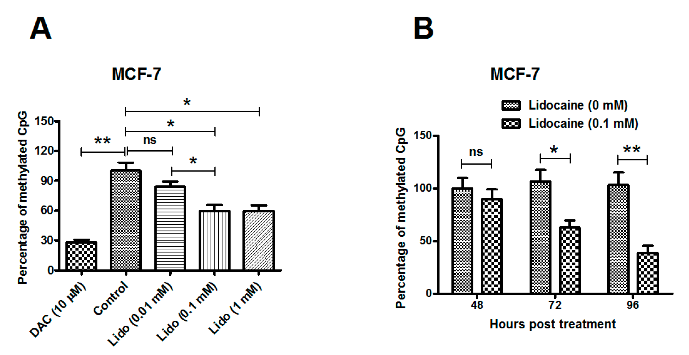
2.1. Lidocaine Promotes Global Genomic Demethylation of the CpG Island in Human Breast Cancer Lines

Two human breast cancer lines, MCF-7 and MDA-MB-231 cells, were treated with various concentrations of lidocaine (0.01, 0.1 or 1 mM) for 72 h and the global DNA methylation of 5' CpG islands before and after each treatment was measured by sodium bisulfite DNA sequencing. As shown in Figure 1A (column 1 and 2), 10 μM 5-aza-2'-deoxycytidine (DAC, a demethylation agent as positive control) treatment significantly reduced the global methylation of the CpG island in MCF-7 cells, compared to the control MCF-7 cells (p ˂ 0.01). The lidocaine treatment with 0.1 or 1 mM also significantly promoted global genomic CpG island demethylation (Figure 1A, column 4 and 5; both p ˂ 0.05). To confirm the demethylation promotion by lidocaine, MCF-7 cells post 0.1 mM lidocaine for various hours were examined for global genomic methylation. Figure 1B demonstrates that the methylation level was significantly reduced by the 0.1 mM lidocaine treatment for either 72 or 96 h (p ˂ 0.05 for 72 h, and p ˂ 0.01 for 96 h), rather than at 48 h post treatment.
We then evaluated the global DNA methylation induced by lidocaine in MDA-MB-231 cells. Figure 1C indicates that the treatment with 0.1 or 1 mM lidocaine for 72 h also promoted global DNA demethylation (p ˂ 0.05 for 0.1 mM and p ˂ 0.01 for 1 mM), with a dose-dependence (either p ˂ 0.05 between the 0.01 and 0.1 mM treatment, or between the 0.01 and 0.1 mM treatment). Moreover, the promotion in MDA-MB-231 cells developed from 48 h post treatment, earlier than in MCF-7 cells (Figure 1D). The time-dependence of the promotion was also significant (p ˂ 0.01 or p ˂ 0.001), and the demethylation difference between the lidocaine and control groups increased with treatment time. Taken together, lidocaine promotes global genomic demethylation of the CpG islands in human breast cancer MCF-7 and MDA-MB-231 cells.
Figure 1. Global genomic demethylation of CpG islands promoted by lidocaine. (A) DNA methylation levels in MCF-7 breast cancer cells treated for 72 h with 0.01, 0.1 or 1 mM lidocaine treatment, or with 10 μM DAC treatment, respectively; (B) DNA methylation levels in MCF-7 cells treated with or without 0.1 mM lidocaine, for 48, 72 or 96 h; (C,D) DNA methylation levels in MDA-MB-231 breast cancer cells post above-mentioned treatment respectively. Results were expressed as mean ± SD for three independent experiments. Statistical significance was assessed by the unpaired Student’s t-test (* p ˂ 0.05, ** p ˂ 0.01, or *** p ˂ 0.001, ns: no significance).
Ijms 15 23519 g001aIjms 15 23519 g001b

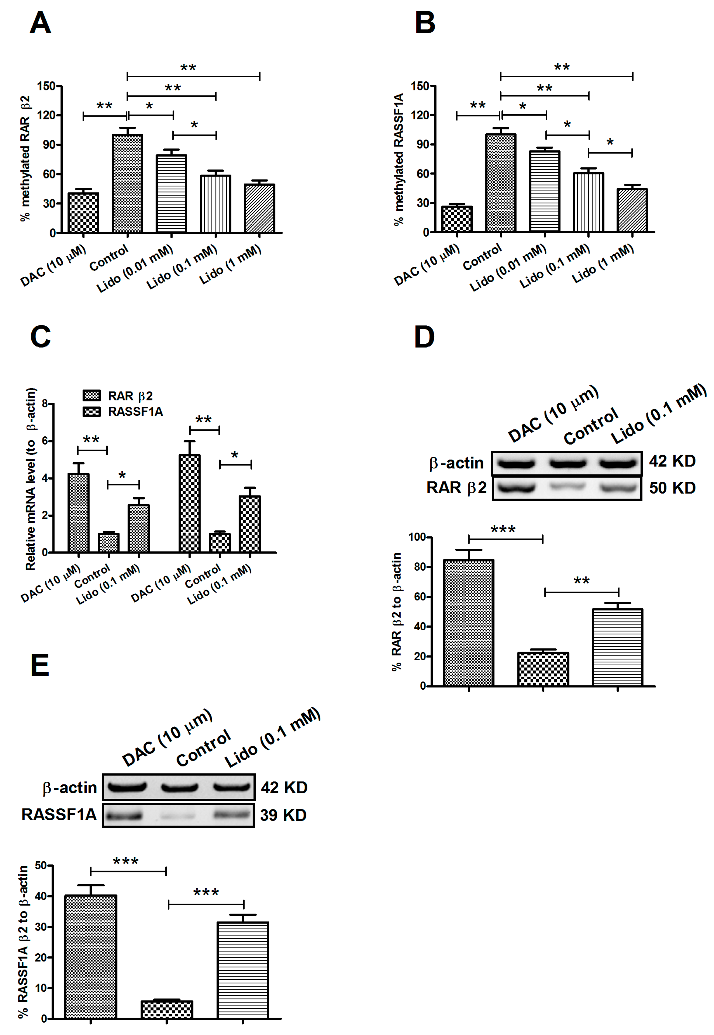
2.2. Lidocaine Ameliorates the Expression of RARβ2 and RASSF1A Genes in MCF-7 and MDA-MB-231 Cells

The tumor suppressive genes, Retinoic acid receptor β (RARβ2) [27] and Ras Association Domain Family 1A (RASSF1A) [28,29] have been shown to be silenced by their promoter methylation in breast cancers. We evaluated the methylation levels of 5' CpG islands in these two TSG promoters, in MCF-7 cells with lidocaine treatment. Figure 2A,B indicates a similar methylation level in both genes to that of the global genomic CpG islands in MCF-7 cells; there was a significant dose-dependent demethylation by lidocaine of both RARβ2 and RASSF1A genes (p ˂ 0.05 or p ˂ 0.01). To examine the influence of the promoter demethylation on the expression of RARβ2 and RASSF1A, we then determined the mRNA and protein levels of both molecules by real-time quantitative PCR (RT-qPCR) and western blot assay. As shown in Figure 2C, there was a significant promotion to RARβ2 and RASSF1A mRNA levels either by 10 μM DAC (p ˂ 0.01 respectively), or by 0.1 mM lidocaine treatment for 72 h (p ˂ 0.05 respectively). Furthermore, the Western blot assay of both molecules reconfirmed the up-regulation of RARβ2 and RASSF1A in protein level by the demethylation, which was promoted by 0.1 mM lidocaine treatment for 96 h (Figure 2D,E; p ˂ 0.01 or p ˂ 0.001). Therefore, the demethylation of RARβ2 and RASSF1A promotes the expression of both genes.

2.3. Regulation of Lidocaine on the Viability of MCF-7 and MDA-MB-231 Cells

RARβ2 has been recognized as a potent tumor suppressor. Expression of RARβ2 in RARβ2-negative cancer cells restored retinoic acid-induced growth inhibition and caused decreased tumorigenicity [30]. Exogenous expression of RARβ2 results both in RA-dependent and RA-independent breast cancer cell apoptosis [31,32]. The other TSG, RASSF1A-encoded protein was found to interact with such molecules as CNK1 [33], Nore1 [34], MDM2 [35], MAP1S [36] to induce cell death. Methylation of both TSGs has been reported to silence their expression and promote the tumorigenesis of breast cancers [27,28,29]. Given the significant demethylation effect of TSGs in breast cancer cells, we supposed a possible anti-tumor effect of lidocaine in the breast cancer cells. Firstly, we examined the viability of both MCF-7 and MDA-MB-231 cells post lidocaine treatment with a range of concentrations. Figure 3A (column 2 compared to column 1, or column 6 compared to column 5) shows that 0.1 mM lidocaine had no impact on the viability of both cell lines, 72 h post treatment. Also, either 0.01 or 1 mM lidocaine had an impact on cell viability (data not shown). Interestingly, the 0.1 mM lidocaine treatment decreased the cell viability reduction which was promoted by 0.2 μM cisplatin in both cell lines, 72 h post treatment (Figure 3A, column 4 or 8 compared to column 3 or 7; both p ˂ 0.01). To further determine the deterioration of cell viability induced by lidocaine, we re-evaluated the cell viability post lidocaine treatment, in the presence of 0.2 μM cisplatin. The cell viability reduction had been reconfirmed in the dosage of 0.1 or 0.5 rather than 0.02 mM (p ˂ 0.01 or p ˂ 0.001; Figure 3B). Figure 3C,D demonstrated that the cell viability reduction appeared significant from 48 h, and lasted to at least 72 h post treatment (p ˂ 0.05 or p ˂ 0.01 for MCF-7 cells and p ˂ 0.05 for MDA-MB-231 cells). Therefore, lidocaine reduced cell viability, in the presence of cisplatin, though its direct regulation was not observed in this study.
Figure 2. Lidocaine-promoted RARβ2 and RASSF1A demethylation up-regulates the expression of both genes. Methylation levels of RARβ2 (A) or RASSF1A (B) in MCF-7 cells treated for 72 h with 0.01, 0.1 or 1 mM lidocaine, or with 10 μM DAC, respectively; (C) mRNA level of RARβ2 or RASSF1A in MCF-7 cells post treatment with 0.1 mM lidocaine or with 10 μM DAC for 72 h; Western blot assay for protein level of RARβ2 (D) or RASSF1A (E) in MCF-7 cells with or without 0.1 mM lidocaine treatment for 96 h. Each value was expressed as mean ± SD for three independent tests. Statistical significance was assessed by the unpaired Student’s t-test (* p ˂ 0.05, ** p ˂ 0.01, or *** p ˂ 0.001).
Ijms 15 23519 g002
Figure 3. Lidocaine reduces the viability of MCF-7 and MDA-MB-231 cells in the presence of cisplatin. (A) Relative viability of MCF-7 and MDA-MB-231 cells treated with 0.2 μM cisplatin or (and) 0.1 mM lidocaine for 72 h; (B) Influence of 0.02, 0.1 or 0.5 mM lidocaine on the viability of 0.2 μM cisplatin-treated MCF-7 cells for 72 h; Relative viability of MCF-7 (C) or MDA-MB-231 (D) cells treated with 0.2 μM cisplatin or (and) 0.1 mM for 36, 48 or 72 h. All results were averaged for triple independent experiments. And each statistical significance was considered when p ˂ 0.05 or less. * p ˂ 0.05, ** p ˂ 0.01, or *** p ˂ 0.001, ns: no significance.
Ijms 15 23519 g003

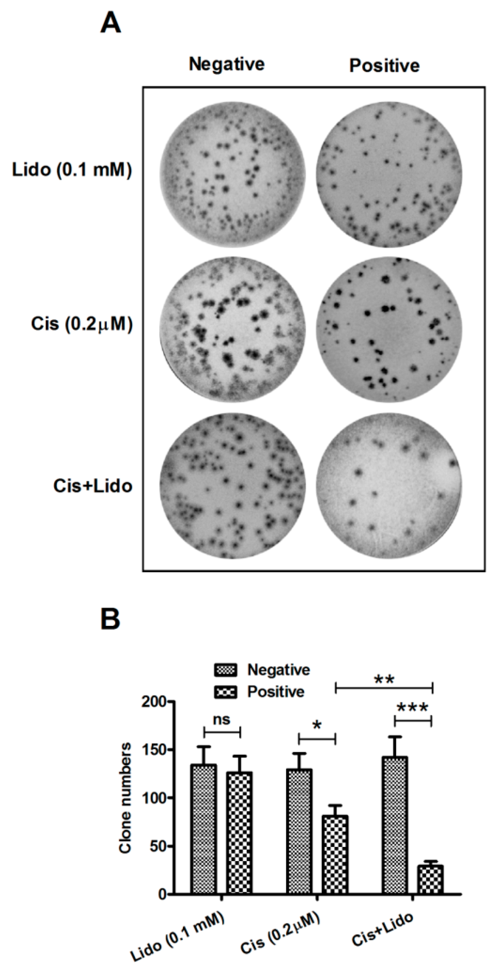
2.4. Lidocaine Enhances the Cytotoxicity of Cisplatin against MCF-7 Cells

Cisplatin has been clinically utilized for breast cancer treatment for decades [37,38], and its anti-proliferation effect has been confirmed to be abrogated [39] or sensitized [40] by other molecules. To investigate the regulation of lidocaine on the cisplatin-mediated anti-tumor effect, we evaluated the proliferation of MCF-7 cells, by clone assay, post treatment with 0.1 mM lidocaine, 0.2 μM cisplatin, or both agents. It was shown in Figure 4A that MCF-7 cells with or without the above-mentioned treatment formed clones after 96 h culture at 37 °C, and there was no obvious morphological difference of MCF-7 cells among these groups. Then the clone numbers in each group were calculated, and as shown in Figure 4B, the singular 0.1 mM lidocaine treatment did not significantly regulate clone forming (p > 0.05), whereas clone forming of MCF-7 cells was reduced in the presence of 0.2 μM cisplatin (p ˂ 0.001), and the reduction in clone number was more significant than the singular 0.2 μM cisplatin treatment (p ˂ 0.01). Taken together, lidocaine inhibits the MCF-7 cell proliferation in the presence of 0.2 μM cisplatin, and thus enhances the cytotoxicity of cisplatin against breast cancer cells.
Figure 4. Colony formation of MCF-7 cells treated with lidocaine and/or cisplatin. (A) Colony formation of MCF-7 cells post the transfection with 0.2 μM cisplatin, 0.1 mM lidocaine or with both agents for 96 h; (B) The colony number in groups with or without the treatment of 0.2 μM cisplatin and/or 0.1 mM lidocaine. All experiments were performed independently in triplicate. Statistical significance was shown as * p ˂ 0.05, ** p ˂ 0.01, or *** p ˂ 0.001, ns: no significance.
Ijms 15 23519 g004

2.5. Lidocaine Regulation of Apoptosis Promotion by Cisplatin in MCF-7 Cells

Cisplatin induces apoptosis in breast cancer cells [41,42], and the induction may be suppressed [43] or be sensitized [44]. To confirm whether cisplatin cytotoxicity enhancement by lidocaine is associated with cisplatin-induced apoptosis, we compared apoptosis-induction differences among singular treatment of cisplatin, of lidocaine and the combined treatment with both agents. Firstly, we measured the apoptosis level of MCF-7 cells, post treatment with 0.1 mM lidocaine, with 0.2 μM cisplatin, or with both agents, via flow cytometric analysis. It was shown that when subjected to 0.1 mM lidocaine for 36, 48 or 60 h, less than 10% MCF-7 cells underwent apoptosis, and there was no significant difference among them (Figure 5A; Figure S1). However, significantly more cells post treatment with 0.2 μM cisplatin underwent apoptosis (p < 0.05 for 36 h, p < 0.01 for 48 or 60 h; Figure 1A). Moreover, 0.1 mM lidocaine sensitized apoptosis promotion by cisplatin; the treatment with both agents promoted significantly higher levels of apoptosis in MCF-7 cells (p < 0.01 or p < 0.001 for 36, 48 or 60 h, compared to the lidocaine group; and p < 0.05 for 48 or 60 h, compared to the cisplatin group). Secondly, we analyzed by Western blot analysis apoptosis-associated molecules, such as procaspase 3, which is cleaved in apoptosis and performs as an apoptosis executor, PARPPARP, which is cleaved by activated caspase 3, and cytochrome c, which is released from mitochondria in apoptosis. Figure 5B–E demonstrates that singular treatment with 0.1 mM lidocaine had no significant regulation on procaspase 3 activation, PARP cleavage and cytochrome c release, all of which were promoted by 0.2 μM cisplatin. Moreover, the cisplatin-mediated promotion was sensitized by lidocaine; treatment with both agents promoted higher levels of the activation of above-mentioned molecules than singular cisplatin treatment (p < 0.01 or p < 0.001, compared to the cisplatin group). Finally, we examined the caspase 3 activity with the fluorophore 7-amino-4-methylcoumarin (AMC)-conjuncting kit. Figure 5F confirms the sensitization of lidocaine on the cisplatin-promoted caspase 3 activity; treatment with both agents promoted a significantly higher level of AMC fluorescence intensity than singular lidocaine or cisplatin treatment (p < 0.05 to p < 0.0001). Therefore, lidocaine sensitizes the apoptosis promotion by cisplatin in MCF-7 cells.

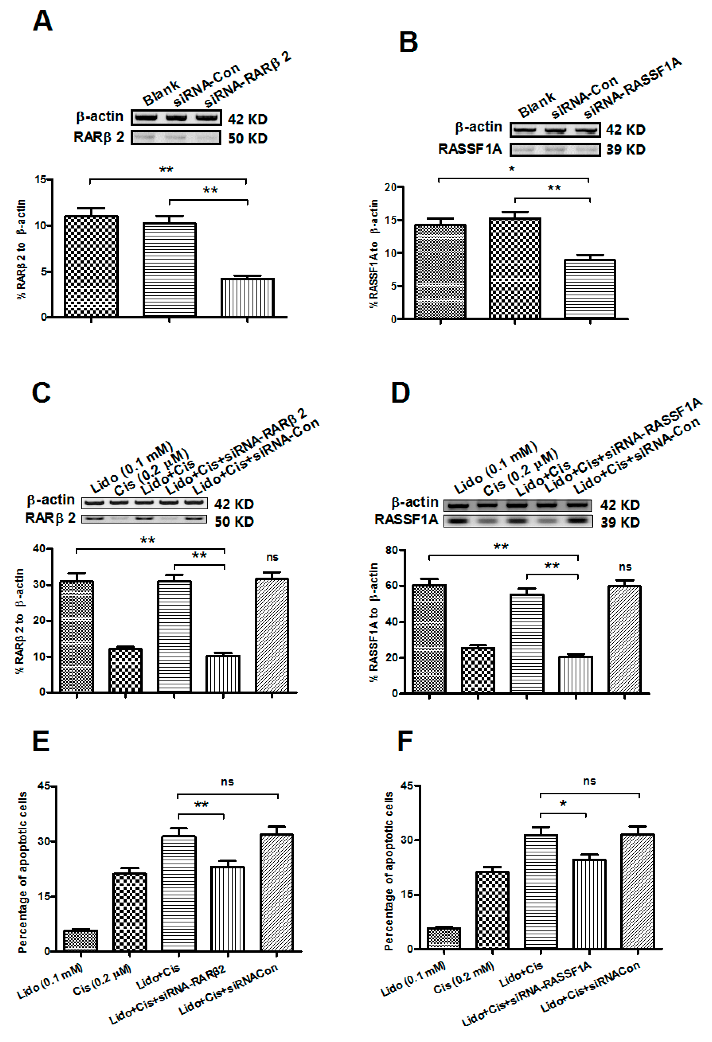
2.6. Abrogation of the RARβ2 and RASSF1A Genes Blocks the Apoptosis Promotion by Lidocaine and Cisplatin in MCF-7 Cells

To further investigate the association of the lidocaine-promoted expression of RARβ2 or RASSF1A with the MCF-7 cell apoptosis, we abrogated the expression of RARβ2 or RASSF1A with the siRNA targeting either gene, and then determine the influence on the MCF-7 cell apoptosis, which was caused by the combined treatment with lidocaine and cisplatin. As shown in Figure 6A,B, the expression of RARβ2 or RASSF1A was significantly down-regulated, 48 h post the siRNA–RARβ2 or siRNA–RASSF1A transfection, in MCF-7 cells. Moreover, the donw-regulation of the RARβ2 or RASSF1A expression was also confirmed in in the lidocaine- and cisplatin-treated MCF-7 cells, compared to the siRNA–control (Figure 6C,D; either p < 0.01). We then evaluated the influence of RARβ2 or RASSF1A abrogation on the lidocaine- and cisplatin-induced apoptosis of MCF-7 cell cells. Figure 6E,F indicated that either siRNA–RARβ2 or siRNA–RASSF1A transfection significantly inhibited the apoptosis of the MCF-7 cells, subject to 0.1 mM lidocaine and 0.2 μM cisplatin, for 48 h (p < 0.05 or p < 0.01). Therefore, the up-regulated expression of RARβ2 or RASSF1A by the lidocaine-mediated demethylation of either gene contributed to the lidocaine- and cisplatin-induced apoptosis. In addition, to exclude a possibly different influence of siRNA transfection on cell apoptosis, we evaluated apoptosis induction of MCF-7 cells post-transfection with siRNA–RASSF1A, siRNA–RARβ2, or with siRNA control. No significant difference was found among the three groups (Figure S2).
Figure 5. Lidocaine enhances cisplatin-induced apoptosis in MCF-7 cells. (A) Apoptotic cells induced by 0.2 μM cisplatin, or (and) 0.1 mM lidocaine for 36, 48 or 60 h; (B) Western blot analysis of activated caspase 3, cleaved PARP by activated caspase 3 and cytochrome c released from mitochondria in MCF-7 cells treated with 0.2 μM cisplatin, and/or 0.1 mM lidocaine for 60 h; (CE) Relative levels of activated caspase 3, PARP cleavage and cytochrome c release to β-actin in MCF-7 cells treated with lidocaine, with cisplatin or with both agents; (F) Caspase 3 activity in lidocaine-, cisplatin- or both agents-treated MCF-7 cells, revealing by AMC Caspase Profiling Kit. Each value was averaged for three independent experiment results, and statistical significance was considered when p ˂ 0.05 or less, * p ˂ 0.05, ** p ˂ 0.01, *** p ˂ 0.001, **** p ˂ 0.0001, ns: no significance.
Ijms 15 23519 g005
Figure 6. Transfection of RARβ2- or RASSF1A-specific siRNA inhibits the lidocaine and cisplatin-induced apoptosis in MCF-7 cells. (AD) Western blot analysis of RARβ2 (A,C) or RASSF1A (B,D) expression in MCF-7 cells post-transfection of the RARβ2- or RASSF1A-specific siRNA, without (A,B) or with (C,D) the lidocane and/or cisplatin treatment; (E,F) Apoptotic cells induced by the combined treatment with 0.1 mM lidocaine and 0.2 μM cisplatin for 48 h, post-transfection of RARβ2- (E) or RASSF1A-specific siRNA (F) * p ˂ 0.05, ** p ˂ 0.01, ns: no significance.
Ijms 15 23519 g006

3. Discussion

Studies have indicated that silencing of TSGs by promoter methylation contributes to tumor development [17]. TSG silencing by DNA methylation induces mechanisms such as uncontrolled cell growth, metastasis, reduced apoptosis or maintainence of angiogenesis, all of which promote tumor development [17,19]. Moreover, DNA methylation may facilitate the mutation of TSGs, such as mutated p53 [20], or contribute directly to tumorigenesis, such as the hypermethylation in APC-1A, CDKN2 and RASSF1A genes in colorectal cancer [21], or the hypermethylation in MTHFR gene in lung cancer [22]. On the other side, promotion of DNA demethylation decreases or inhibits the incidence or the development of cancers [45,46,47,48]; DNA demethylation agent 5-aza-2'-deoxycytidine decreases the incidence and inhibits the growth of gastric cancers [45,48]; miRNA-34b inhibits prostate cancer through demethylation, activating chromatin modifications [46]; and DNA demethylation by 5-aza-2-deoxycytidine abrogates 17 β-estradiol-induced cell growth of human breast cancer cells [49]. However, the mechanisms of the anti-tumor effects of DNA demethylation agents still need to be clarified.
Lidocaine has recently been indicated to promote DNA demethylation in breast cancer cell lines [25]. The present study has confirmed lidocaine promotion of global genomic demethylation of CpG islands in MCF-7 and MDA-MB-231 cells; treatment with 0.1 or 1 mM lidocaine for 72 h promoted global DNA methylation in both cell lines with time-dependent and dose-dependent repeatability. The retinoic acid receptor β (RARβ2) was shown to mediate the growth-inhibitory effect of retinoic acid by promoting apoptosis in human breast cancer cells [32] and to be silenced by promoter methylation in breast cancer [27]. RARβ2 over-expression restored retinoic acid-induced growth arrest and apoptosis in breast cancer cells [50]. Ras Association Domain Family 1A (RASSF1A), a putative tumor suppressor gene from the 3p21.3 locus, is another well-recognized tumor suppressive gene in various types of tumors [51,52,53]. The hypermethylation of CpG Islands in RASSF1A occurs in a large percentage of human breast cancers [54]. Results here indicate that hypermethylation in both genes was reduced significantly by lidocaine treatment, and that the demethylation induced by lidocaine ameliorates the reduced expression of RARβ2 and RASSF1A, with a significant increase in both mRNA and protein levels in MCF-7 and MDA-MB-231 cells. However, we did not observe significant regulation by lidocaine on viability of either cell line. Interestingly, lidocaine decreased the reduction in cell viability promoted by 0.2 μM cisplatin in both cell lines, with time- and dose-dependence. The sensitization effect of lidocaine on the anti-tumor effect of cisplatin was confirmed by clone assay; combined treatment with 0.1 mM lidocaine and 0.2 μM cisplatin significantly reduced the clone formation of MCF-7 cells compared to the singular 0.1 mM lidocaine treatment.
Taken together, lidocaine sensitizes the cytotoxicity of cisplatin against breast cancer cells. Cisplatin has been clinically utilized for breast cancer treatment for decades [37,38], and it mainly induces apoptotic death of breast cancer cells [41,43,55]. Cisplatin-induced apoptosis has shown to be sensitized by Mdm2 antagonists [56], by the expression of Siva-1 protein [57], or by theophylline [58]. Here, we demonstrate that lidocaine sensitized apoptosis promotion by cisplatin in MCF-7 cells. Though lidocaine did not directly induce apoptosis in MCF-7 cells, it enhanced cisplatin-induced apoptosis; treatments with both agents promoted significantly higher levels of apoptosis in MCF-7 cells. Western blot analysis confirmed sensitization by lidocaine. Treatment with both agents promoted higher expression or activation level of apoptosis-associated molecules, such as activated caspase 3, cleaved PARP and released cytochrome c. Moreover, apoptosis promotion by lidocaine and cisplatin treatment was inhibited by abrogation of the RARβ2 or RASSF1A gene. Thus, we demonstrate here lidocaine sensitization to the cytotoxicity of cisplatin in MCF-7 breast cancer cells. However, further investigation is needed to clarify the exact mechanisms of sensitization to cisplatin-induced apoptosis in breast cancer cells through lidocaine-induced demethylation of RARβ2 and RASSF1A genes.

4. Materials and Methods

4.1. Cell Culture and Treatment with Reagents

MDA-MB-231 and MCF-7 breast cancer cell lines were provided by the cell resource center of the Chinese Academy of Medical Sciences (Beijing, China). MDA-MB-231 cells were grown in DMEM/F12 (HyClone, Logan, UT, USA), supplemented with 10% fetal bovine serum (FBS; Gibco, Rockville, MD, USA). MCF-7 breast cells were maintained in Dulbecco’s modified Eagle’s medium (Invitrogen, Carlsbad, CA, USA), supplemented with 10% FBS (Gibco, Rockville, MD, USA). Cells were cultured at 37 °C in 5% CO2 to 80%–90% confluence and were subjected to DMEM/F12 or DMEM supplemented with 2% FBS, containing 0.01, 0.02, 0.1, 0.5 or 1 mM lidocaine (Sigma–Aldrich, St. Louis, MO, USA), 10 μM 5-aza-2'-deoxycytidine (DAC) (Sigma–Aldrich) or (and) 0.2 μM cisplatin (Sigma–Aldrich for various hours for the DNA methylation sequencing, cell viability or cell apoptosis assay, and cell colony forming assay. DAC or cisplatin was utilized to as a positive demethylation agent or as an apoptosis inducer. To abrogate the expression of RARβ2 or RASSF1A, siRNA–RARβ2 (5'-CAGC UGAG UUGG ACGA UCU-3'), siRNA–RASSF1A (5'-GAC CUC UGU GGC GAC UUCA-3') or siRNA control (5'-AGCG AATT AGCT TGCC GTG-3') was synthesized by GenePharma Technology (Shanghai, China) and was transfected by lipofectamine 2000 (Thermo Fisher Scientific, Waltham, MA, USA) with a concentration of 50 nM according to the manufacurer’s guidance.

4.2. Methylation Analysis of Global Genomics and TSGs, RARβ2 and RASSF1A

To analyze the effects of 0.01, 0.1, or 1 mM lidocaine, with 10 mM DAC as positive demethylation agent, we extracted genomic DNA from MDA-MB-231 or MCF-7 cells post various treatment using DNeasy tissue kit (Qiagen, Hilden, Germany) according to the manufacturer’ manual. Sodium bisulfite conversion of genomic DNA in each sample was performed as described [59]. The DNA methylation was analyzed using MethyLight method [60], to quantify the methylation level of RARβ2 and RASSF1A, both of which have reported to be hypermethylated in breast cancer [27,38]. The methylation status of individual CpG sites in each gene within the promoter region was determined by the sodium bisulfite-sequencing assay as described previously [61,62].

4.3. RNA Extraction and RT-qPCR

Total cellular mRNA samples were extracted from cultured cells using the RNeasy Mini Kit (Qiagen, Valencia, CA, USA). Quantification of RARβ2 and RASSF1A expression was conducted using the QuantiTect SYBR Green PCR Kit (Qiagen, Hilden, Germany) in the LightCycle 2.0 (Roche, Mannheim, Germany). The sequences of the primers for RARβ2 are as following, forward primer: 5'-TGAG TCCT GGGC AAAT CCTG-3', reverse primer: 5'-TTGA GAGC TTTC TCCT GGAG-3'. And the sequences of the primers for RASSF1A are as following, forward primer: 5'-AGCC TGAG CTCA TTGA GCTG-3', reverse primer: 5'-ACCA GCTG CCGT GTGG-3'. All mRNA expression levels were normalized to β-actin (forward primer: 5'-GATG AGAT TGGC ATGG CTTT-3', revserse primer: 5'-GTCA CCTT CACC GTTC CAGT-3'), and ∆∆Ct method was used for relative quantification [63].

4.4. Protein Isolation and Western Blot Analysis

Total cellular protein samples were prepared with a cell lysis reagent (Sigma–Aldrich) according to the manual and supplemented with a protease inhibitor cocktail (Pierce, Rockford, IL, USA). For the Cyt C release assay, the cytoplasmic protein was isolated by the Mitochondria/Cytosol Fractionation Kit (Abcam, Cambridge, UK). Each protein sample was separated by 10% SDS-PAGE gel and was transferred to a nitrocellulose membrane. Then the rabbit polyclonal antibody to RARβ2 (Santa Cruz Biotechnology, Santa Cruz, CA, USA), RASSF1A (Abcam, Cambridge, UK), caspase 3 (Sino Biological, Beijing, China), cleaved-PARP (Cell Signaling Technology Inc., Danvers, MA, USA), Cytochrome c (Santa Cruz Biotechnology, Santa Cruz, CA, USA) or β-actin (Sino Biological, Beijing, China) was used to detect the protein level of each molecule. Goat anti-rabbit IgG conjugated to horseradish peroxidase (Pierce) and ECL detection systems (Super Signal West Femto; Pierce) were used for detection.

4.5. MTT Assay and Cell Colony Formation Assay

Cell viability was determined by MTT cell viability assay kit (Biotium Inc., Beijing, China). MCF-7 or MDA-MB-231 cells were seeded in 96-well plates and were incubated at 37 °C for 24 h to approximately 85% confluence, and then cells were treated with 0.1 mM lidocaine, 0.2 μM cisplatin or with both agents for various hours. Then cells were incubated with 10 μL MTT and were incubated at 37 °C for 4 h. Post addition of 200 μL DMSO into each well to dissolve the formazan, the absorbance was measured on an ELISA plate reader with a test wavelength of 570 nm and a reference wavelength of 630 nm to obtain sample signal (OD570–OD630). For cell colony formation assay, 4 × 10² cells were incubated in 6-well plates at 37 °C containing 5% CO2, and were treated with 0.1 mM lidocaine, 0.2 μM cisplatin or with both agents. 96 h post treatment, cells were stained with crystal violet (0.005%) for 30 min and colony numbers were counted.

4.6. Cell Apoptosis Assay and Fluorometric Analysis of Caspase 3 Activity

Apoptosis of MCF-7 or MDA-MB-231 cells was examined with an annexin V/FITC apoptosis detection kit (Abcam, Cambridge, UK). Briefly, approximate 6 × 10⁵ cells post treatment were stained with annexin V-FITC and propidium iodide and detected by a FACScan flow cytometer (BD Biosciences, San Jose, CA, USA). The apoptosis was evaluated by a percentage of apoptotic cells to total cells. The caspase 3 activity was examined with an AMC Caspase Profiling Kit (for caspase 3) (AnaSpec, Fremont, CA, USA) according to the manual. MCF-7 cells (105 cells) post treatment were collected, and the cell pellets were washed with phosphate-buffered saline, then caspase-3-like activity was determined by assessment of Asp-Glu-Val-Asp (DEVD)-AMC cleavage. Briefly, pellets were transferred to a microtiter plate and were resuspended in 100 μL (final volume) of a caspase buffer solution supplemented with the fluorogenic peptide substrate Ac-DEVD-AMC. The cleavage was monitored over a 30 min period at 37 °C in a Fluoroscan II plate reader using an excitation wavelength of 390 nm and an emission wavelength of 460 nm. And the activity was expressed as a relative value of fluorescence intensity of AMC to control.

5. Conclusions

In conclusion, we have confirmed the DNA demethylation effects of lidocaine in breast cancer cells, and demonstrated the lidocaine-sensitized cytotoxicity of cisplatin against MCF-7 cells via enhancement of cisplatin-induced apoptosis.

Supplementary Information

The supplementary figures can be found at https://0-www-mdpi-com.brum.beds.ac.uk/1422-0067/15/12/23519/s1.

Acknowledgments

The present study was supported by a grant from The First Affiliated Hospital of Henan Science and Technology University (S2012004).

Author Contributions

Kehan Li and Jianxue Yang designed the study; Kehan Li, Jianxue Yang and Xuechang Han performed the experiments; Kehan Li and Xuechang Han wrote the paper.

Conflicts of Interest

The authors declare no conflict of interest.

References

  1. Lambert, L.A.; Lambert, D.H.; Strichartz, G.R. Irreversible conduction block in isolated nerve by high concentrations of local anesthetics. Anesthesiology 1994, 80, 1082–1093. [Google Scholar] [CrossRef] [PubMed]
  2. Rigler, M.L.; Drasner, K.; Krejcie, T.C.; Yelich, S.J.; Scholnick, F.T.; DeFontes, J.; Bohner, D. Cauda equina syndrome after continuous spinal anesthesia. Anesth. Analg. 1991, 72, 275–281. [Google Scholar] [CrossRef] [PubMed]
  3. Kasaba, T.; Onizuka, S.; Takasaki, M. Procaine and mepivacaine have less toxicity in vitro than other clinically used local anesthetics. Anesth. Analg. 2003, 97, 85–90. [Google Scholar] [CrossRef] [PubMed]
  4. Onizuka, S.; Takasaki, M.; Syed, N.I. Long-term exposure to local but not inhalation anesthetics affects neurite regeneration and synapse formation between identified lymnaea neurons. Anesthesiology 2005, 102, 353–363. [Google Scholar] [CrossRef] [PubMed]
  5. Werdehausen, R.; Braun, S.; Essmann, F.; Schulze-Osthoff, K.; Walczak, H.; Lipfert, P.; Stevens, M.F. Lidocaine induces apoptosis via the mitochondrial pathway independently of death receptor signaling. Anesthesiology 2007, 107, 136–143. [Google Scholar] [CrossRef] [PubMed]
  6. Johnson, M.E.; Uhl, C.B.; Spittler, K.H.; Wang, H.; Gores, G.J. Mitochondrial injury and caspase activation by the local anesthetic lidocaine. Anesthesiology 2004, 101, 1184–1194. [Google Scholar] [CrossRef] [PubMed]
  7. Onizuka, S.; Tamura, R.; Hosokawa, N.; Kawasaki, Y.; Tsuneyoshi, I. Local anesthetics depolarize mitochondrial membrane potential by intracellular alkalization in rat dorsal root ganglion neurons. Anesth. Analg. 2010, 111, 775–783. [Google Scholar] [CrossRef] [PubMed]
  8. Onizuka, S.; Yonaha, T.; Tamura, R.; Kasiwada, M.; Shirasaka, T.; Tsuneyoshi, I. Lidocaine depolarizes the mitochondrial membrane potential by intracellular alkalization in rat dorsal root ganglion neurons. J. Anesth. 2011, 25, 229–239. [Google Scholar] [CrossRef] [PubMed]
  9. Chang, Y.C.; Liu, C.L.; Chen, M.J.; Hsu, Y.W.; Chen, S.N.; Lin, C.H.; Chen, C.M.; Yang, F.M.; Hu, M.C. Local anesthetics induce apoptosis in human breast tumor cells. Anesth. Analg. 2014, 118, 116–124. [Google Scholar] [CrossRef] [PubMed]
  10. Yagiela, J.A.; Benoit, P.W.; Fort, N.F. Mechanism of epinephrine enhancement of lidocaine-induced skeletal muscle necrosis. J. Dent. Res. 1982, 61, 686–690. [Google Scholar] [CrossRef] [PubMed]
  11. Lawrence, V.S.; Marte, E.; Brown, B.W.; van Bergen, F.H. Lidocaine, 2-chlorprocaine and hepatic necrosis. Anesth. Analg. 1966, 45, 55–58. [Google Scholar] [PubMed]
  12. Onizuka, S.; Tamura, R.; Yonaha, T.; Oda, N.; Kawasaki, Y.; Shirasaka, T.; Shiraishi, S.; Tsuneyoshi, I. Clinical dose of lidocaine destroys the cell membrane and induces both necrosis and apoptosis in an identified Lymnaea neuron. J. Anesth. 2012, 26, 54–61. [Google Scholar] [CrossRef] [PubMed]
  13. Arrebola, F.; Zabiti, S.; Canizares, F.J.; Cubero, M.A.; Crespo, P.V.; Fernandez-Segura, E. Changes in intracellular sodium, chlorine, and potassium concentrations in staurosporine-induced apoptosis. J. Cell Physiol. 2005, 204, 500–507. [Google Scholar] [CrossRef] [PubMed]
  14. Belaud-Rotureau, M.A.; Leducq, N.; Macouillard, P.D.G.F.; Diolez, P.; Lacoste, L.; Lacombe, F.; Bernard, P.; Belloc, F. Early transitory rise in intracellular pH leads to Bax conformation change during ceramide-induced apoptosis. Apoptosis 2000, 5, 551–560. [Google Scholar] [CrossRef] [PubMed]
  15. Karpie, J.C.; Chu, C.R. Lidocaine exhibits dose- and time-dependent cytotoxic effects on bovine articular chondrocytes in vitro. Am. J. Sports Med. 2007, 35, 1621–1627. [Google Scholar] [CrossRef] [PubMed]
  16. Rademaker, A.W.; Kellen, J.; Tam, Y.K.; Wyse, D.G. Character of adverse effects of prophylactic lidocaine in the coronary care unit. Clin. Pharmacol. Ther. 1986, 40, 71–80. [Google Scholar] [CrossRef] [PubMed]
  17. Lewandowska, J.; Bartoszek, A. DNA methylation in cancer development, diagnosis and therapy—Multiple opportunities for genotoxic agents to act as methylome disruptors or remediators. Mutagenesis 2011, 26, 475–487. [Google Scholar] [CrossRef] [PubMed]
  18. Kubicek, S.; Schotta, G.; Lachner, M.; Sengupta, R.; Kohlmaier, A.; Perez-Burgos, L.; Linderson, Y.; Martens, J.H.; OʼSullivan, R.J.; Fodor, B.D.; et al. The role of histone modifications in epigenetic transitions during normal and perturbed development. Ernst Scher. Res. Found. Workshop 2006, 57, 1–27. [Google Scholar]
  19. Ushijima, T. Detection and interpretation of altered methylation patterns in cancer cells. Nat. Rev. Cancer 2005, 5, 223–231. [Google Scholar] [CrossRef] [PubMed]
  20. Singal, R.; Ginder, G.D. DNA methylation. Blood 1999, 93, 4059–4070. [Google Scholar] [PubMed]
  21. Van Engeland, M.; Weijenberg, M.P.; Roemen, G.M.; Brink, M.; de Bruine, A.P.; Goldbohm, R.A.; van den Brandt, P.A.; Baylin, S.B.; de Goeij, A.F.; Herman, J.G. Effects of dietary folate and alcohol intake on promoter methylation in sporadic colorectal cancer: The Netherlands cohort study on diet and cancer. Cancer Res. 2003, 63, 3133–3137. [Google Scholar] [PubMed]
  22. Vaissiere, T.; Hung, R.J.; Zaridze, D.; Moukeria, A.; Cuenin, C.; Fasolo, V.; Ferro, G.; Paliwal, A.; Hainaut, P.; Brennan, P. Quantitative analysis of DNA methylation profiles in lung cancer identifies aberrant DNA methylation of specific genes and its association with gender and cancer risk factors. Cancer Res. 2009, 69, 243–252. [Google Scholar] [CrossRef] [PubMed]
  23. Villar-Garea, A.; Fraga, M.F.; Espada, J.; Esteller, M. Procaine is a DNA-demethylating agent with growth-inhibitory effects in human cancer cells. Cancer Res. 2003, 63, 4984–4989. [Google Scholar] [PubMed]
  24. Tada, M.; Imazeki, F.; Fukai, K.; Sakamoto, A.; Arai, M.; Mikata, R.; Tokuhisa, T.; Yokosuka, O. Procaine inhibits the proliferation and DNA methylation in human hepatoma cells. Hepatol. Int. 2007, 1, 355–364. [Google Scholar] [CrossRef] [PubMed]
  25. Lirk, P.; Berger, R.; Hollmann, M.W.; Fiegl, H. Lidocaine time- and dose-dependently demethylates deoxyribonucleic acid in breast cancer cell lines in vitro. Br. J. Anaesth. 2012, 109, 200–207. [Google Scholar] [CrossRef] [PubMed]
  26. Hong, D.Y.; Kwon, K.; Lee, K.R.; Choi, Y.J.; Goo, T.W.; Yu, K.; Kim, S.W.; Kwon, O.Y. Lidocaine induces endoplasmic reticulum stress-associated apoptosis in vitro and in vivo. Int. J. Mol. Sci. 2011, 12, 7652–7661. [Google Scholar] [CrossRef] [PubMed]
  27. Sirchia, S.M.; Ren, M.; Pili, R.; Sironi, E.; Somenzi, G.; Ghidoni, R.; Toma, S.; Nicolò, G.; Sacchi, N. Endogenous reactivation of the RARbeta2 tumor suppressor gene epigenetically silenced in breast cancer. Cancer Res. 2002, 62, 2455–2461. [Google Scholar] [PubMed]
  28. Burbee, D.G.; Forgacs, E.; Zochbauer-Muller, S.; Shivakumar, L.; Fong, K.; Gao, B.; Randle, D.; Kondo, M.; Virmani, A.; Bader, S.; et al. Epigenetic inactivation of RASSF1A in lung and breast cancers and malignant phenotype suppression. J. Natl. Cancer Inst. 2011, 93, 691–699. [Google Scholar] [CrossRef]
  29. Agathanggelou, A.; Honorio, S.; Macartney, D.P.; Martinez, A.; Dallol, A.; Rader, J.; Fullwood, P.; Chauhan, A.; Walker, R.; Shaw, J.A.; et al. Methylation associated inactivation of RASSF1A from region 3p21.3 in lung, breast and ovarian tumours. Oncogene 2001, 20, 1509–1518. [Google Scholar] [CrossRef] [PubMed]
  30. Si, S.P.; Lee, X.; Tsou, H.C.; Buchsbaum, R.; Tibaduiza, E.; Peacocke, M. RAR beta 2-mediated growth inhibition in HeLa cells. Exp. Cell Res. 1996, 223, 102–111. [Google Scholar] [CrossRef] [PubMed]
  31. Swisshelm, K.; Ryan, K.; Lee, X.; Tsou, H.C.; Peacocke, M.; Sager, R. Down-regulation of retinoic acid receptor beta in mammary carcinoma cell lines and its up-regulation in senescing normal mammary epithelial cells. Cell Growth Differ. 1994, 5, 133–141. [Google Scholar] [PubMed]
  32. Liu, Y.; Lee, M.O.; Wang, H.G.; Li, Y.; Hashimoto, Y.; Klaus, M.; Reed, J.C.; Zhang, X. Retinoic acid receptor beta mediates the growth-inhibitory effect of retinoic acid by promoting apoptosis in human breast cancer cells. Mol. Cell. Biol. 1996, 16, 1138–1149. [Google Scholar] [PubMed]
  33. Rabizadeh, S.; Xavier, R.J.; Ishiguro, K.; Bernabeortiz, J.; Lopez-Ilasaca, M.; Khokhlatchev, A.; Mollahan, P.; Pfeifer, G.P.; Avruch, J.; Seed, B. The scaffold protein CNK1 interacts with the tumor suppressor RASSF1A and augments RASSF1A-induced cell death. J. Biol. Chem. 2004, 279, 29247–29254. [Google Scholar] [CrossRef] [PubMed]
  34. Ortiz-Vega, S.; Khokhlatchev, A.; Nedwidek, M.; Zhang, X.F.; Dammann, R.; Pfeifer, G.P.; Avruch, J. The putative tumor suppressor RASSF1A homodimerizes and heterodimerizes with the Ras-GTP binding protein Nore1. Oncogene 2002, 21, 1381–1390. [Google Scholar] [CrossRef] [PubMed]
  35. Song, M.S.; Song, S.J.; Kim, S.Y.; Oh, H.J.; Lim, D.S. The tumour suppressor RASSF1A promotes MDM2 self-ubiquitination by disrupting the MDM2-DAXX-HAUSP complex. EMBO J. 2008, 27, 1863–1874. [Google Scholar] [CrossRef] [PubMed]
  36. Liu, L.; Vo, A.; McKeehan, W.L. Specificity of the methylation-suppressed A isoform of candidate tumor suppressor RASSF1 for microtubule hyperstabilization is determined by cell death inducer C19ORF5. Cancer Res. 2005, 65, 1830–1838. [Google Scholar] [CrossRef] [PubMed]
  37. Vassilomanolakis, M.; Koumakis, G.; Barbounis, V.; Demiri, M.; Pateras, H.; Efremidis, A.P. Vinorelbine and cisplatin in metastatic breast cancer patients previously treated with anthracyclines. Ann. Oncol. 2000, 11, 1155–1160. [Google Scholar] [CrossRef] [PubMed]
  38. Nole, F.; Munzone, E.; Mandala, M.; Catania, C.; Orlando, L.; Zampino, M.G.; Minchella, I.; Colleoni, M.; Peruzzotti, G.; Marrocco, E.; et al. Vinorelbine, cisplatin and continuous infusion of 5-fluorouracil (ViFuP) in metastatic breast cancer patients: A phase II study. Ann. Oncol. 2001, 12, 95–100. [Google Scholar] [CrossRef] [PubMed]
  39. Dixit, M.; Yang, J.L.; Poirier, M.C.; Price, J.O.; Andrews, P.A.; Arteaga, C.L. Abrogation of cisplatin-induced programmed cell death in human breast cancer cells by epidermal growth factor antisense RNA. J. Natl. Cancer Inst. 1997, 89, 365–373. [Google Scholar] [CrossRef] [PubMed]
  40. Sakakura, C.; Sweeney, E.A.; Shirahama, T.; Igarashi, Y.; Hakomori, S.; Tsujimoto, H.; Imanishi, T.; Ogaki, M.; Ohyama, T.; Yamazaki, J.; et al. Overexpression of bax sensitizes breast cancer MCF-7 cells to cisplatin and etoposide. Surg. Today 1997, 27, 676–679. [Google Scholar] [CrossRef] [PubMed]
  41. Blanc, C.; Deveraux, Q.L.; Krajewski, S.; Janicke, R.U.; Porter, A.G.; Reed, J.C.; Jaggi, R.; Marti, A. Caspase-3 is essential for procaspase-9 processing and cisplatin-induced apoptosis of MCF-7 breast cancer cells. Cancer Res. 2000, 60, 4386–4390. [Google Scholar] [PubMed]
  42. Thomadaki, H.; Scorilas, A. Breast cancer cells response to the antineoplastic agents cisplatin, carboplatin, and doxorubicin at the mRNA expression levels of distinct apoptosis-related genes, including the new member, BCL2L12. Ann. N. Y. Acad. Sci. 2007, 1095, 35–44. [Google Scholar] [CrossRef] [PubMed]
  43. Yang, M.; Yuan, F.; Li, P.; Chen, Z.; Chen, A.; Li, S.; Hu, C. Interferon regulatory factor 4 binding protein is a novel p53 target gene and suppresses cisplatin-induced apoptosis of breast cancer cells. Mol. Cancer 2012, 11, 54. [Google Scholar] [CrossRef] [PubMed]
  44. Shiu, L.Y.; Chang, L.C.; Liang, C.H.; Huang, Y.S.; Sheu, H.M.; Kuo, K.W. Solamargine induces apoptosis and sensitizes breast cancer cells to cisplatin. Food Chem. Toxicol. 2007, 45, 2155–2164. [Google Scholar] [CrossRef] [PubMed]
  45. Schneider, B.G.; Peek, R.J. Gastric cancer prevention by demethylation. Cancer Prev. Res. 2013, 6, 253–256. [Google Scholar] [CrossRef]
  46. Majid, S.; Dar, A.A.; Saini, S.; Shahryari, V.; Arora, S.; Zaman, M.S.; Chang, I.; Yamamura, S.; Tanaka, Y.; Chiyomaru, T. miRNA-34b inhibits prostate cancer through demethylation, active chromatin modifications, and AKT pathways. Clin. Cancer Res. 2013, 19, 73–84. [Google Scholar] [CrossRef] [PubMed]
  47. Shin, H.; Kim, J.H.; Lee, Y.S.; Lee, Y.C. Change in gene expression profiles of secreted frizzled-related proteins (SFRPs) by sodium butyrate in gastric cancers: Induction of promoter demethylation and histone modification causing inhibition of Wnt signaling. Int. J. Oncol. 2012, 40, 1533–1542. [Google Scholar] [PubMed]
  48. Liu, J.; Xie, Y.S.; Wang, F.L.; Zhang, L.J.; Zhang, Y.; Luo, H.S. Cytotoxicity of 5-aza-2'-deoxycytidine against gastric cancer involves DNA damage in an ATM-P53 dependent signaling pathway and demethylation of P16(INK4A). Biomed. Pharmacother. 2013, 67, 78–87. [Google Scholar] [CrossRef] [PubMed]
  49. Singh, K.P.; Treas, J.; Tyagi, T.; Gao, W. DNA demethylation by 5-aza-2-deoxycytidine treatment abrogates 17 beta-estradiol-induced cell growth and restores expression of DNA repair genes in human breast cancer cells. Cancer Lett. 2012, 316, 62–69. [Google Scholar] [CrossRef] [PubMed]
  50. Seewaldt, V.L.; Johnson, B.S.; Parker, M.B.; Collins, S.J.; Swisshelm, K. Expression of retinoic acid receptor beta mediates retinoic acid-induced growth arrest and apoptosis in breast cancer cells. Cell Growth Differ. 1995, 6, 1077–1088. [Google Scholar] [PubMed]
  51. Levesley, J.; Lusher, M.E.; Lindsey, J.C.; Clifford, S.C.; Grundy, R.; Coyle, B. RASSF1A and the BH3-only mimetic ABT-737 promote apoptosis in pediatric medulloblastoma cell lines. Neuro Oncol. 2011, 13, 1265–1276. [Google Scholar] [CrossRef] [PubMed]
  52. Yi, M.; Yang, J.; Chen, X.; Li, J.; Li, X.; Wang, L.; Tan, Y.; Xiong, W.; Zhou, M.; McCarthy, J.B.; et al. RASSF1A suppresses melanoma development by modulating apoptosis and cell-cycle progression. J. Cell Physiol. 2011, 226, 2360–2369. [Google Scholar] [CrossRef] [PubMed]
  53. Matallanas, D.; Romano, D.; Yee, K.; Meissl, K.; Kucerova, L.; Piazzolla, D.; Baccarini, M.; Vass, J.K.; Kolch, W.; OʼNeill, E. RASSF1A elicits apoptosis through an MST2 pathway directing proapoptotic transcription by the p73 tumor suppressor protein. Mol. Cell 2007, 27, 962–975. [Google Scholar] [CrossRef] [PubMed]
  54. Dammann, R.; Yang, G.; Pfeifer, G.P. Hypermethylation of the cpG island of Ras association domain family 1A (RASSF1A), a putative tumor suppressor gene from the 3p21.3 locus, occurs in a large percentage of human breast cancers. Cancer Res. 2011, 61, 3105–3109. [Google Scholar]
  55. Niknafs, B. Induction of apoptosis and non-apoptosis in human breast cancer cell line (MCF-7) by cisplatin and caffeine. Iran. Biomed. J. 2011, 15, 130–133. [Google Scholar] [PubMed]
  56. Mir, R.; Tortosa, A.; Martinez-Soler, F.; Vidal, A.; Condom, E.; Perez-Perarnau, A.; Ruiz-Larroya, T.; Gil, J.; Giménez-Bonafé, P. Mdm2 antagonists induce apoptosis and synergize with cisplatin overcoming chemoresistance in TP53 wild-type ovarian cancer cells. Int. J. Cancer 2013, 132, 1525–1536. [Google Scholar] [CrossRef] [PubMed]
  57. Chu, F.; Barkinge, J.; Hawkins, S.; Gudi, R.; Salgia, R.; Kanteti, P.V. Expression of Siva-1 protein or its putative amphipathic helical region enhances cisplatin-induced apoptosis in breast cancer cells: Effect of elevated levels of BCL-2. Cancer Res. 2005, 65, 5301–5309. [Google Scholar] [CrossRef] [PubMed]
  58. Yoshida, Y.; Hosokawa, K.; Dantes, A.; Tajima, K.; Kotsuji, F.; Amsterdam, A. Theophylline and cisplatin synergize in down regulation of BCL-2 induction of apoptosis in human granulosa cells transformed by a mutated p53 (p53 val135) and Ha-ras oncogene. Int. J. Oncol. 2000, 17, 227–235. [Google Scholar] [PubMed]
  59. Cho, N.Y.; Kim, B.H.; Choi, M.; Yoo, E.J.; Moon, K.C.; Cho, Y.M.; Kim, D.; Kang, G.H. Hypermethylation of CpG island loci and hypomethylation of LINE-1 and Alu repeats in prostate adenocarcinoma and their relationship to clinicopathological features. J. Pathol. 2007, 211, 269–277. [Google Scholar] [CrossRef] [PubMed]
  60. Eads, C.A.; Danenberg, K.D.; Kawakami, K.; Saltz, L.B.; Blake, C.; Shibata, D.; Danenberg, P.V.; Laird, P.W. MethyLight: A high-throughput assay to measure DNA methylation. Nucleic Acids Res. 2000, 28, E32. [Google Scholar] [CrossRef] [PubMed]
  61. Farias, E.F; Arapshian, A.; Bleiweiss, I.J.; Waxman, S.; Zelent, A.; Mira-Y-Lopez, R. Retinoic acid receptor alpha2 is a growth suppressor epigenetically silenced in MCF-7 human breast cancer cells. Cell Growth Differ. 2002, 13, 335–341. [Google Scholar] [PubMed]
  62. Yan, P.S.; Shi, H.; Rahmatpanah, F.; Hsiau, T.H.; Hsiau, A.H.; Leu, Y.W.; Liu, J.C.; Huang, T.H. Differential distribution of DNA methylation within the RASSF1A CpG island in breast cancer. Cancer Res. 2003, 63, 6178–6186. [Google Scholar] [PubMed]
  63. Livak, K.J.; Schmittgen, T.D. Analysis of relative gene expression data using real-time quantitative PCR and the 2(-Delta Delta C(T)) Method. Methods 2001, 25, 402–408. [Google Scholar] [CrossRef] [PubMed]

