
Developments in FTICR-MS and Its Potential for Body Fluid Signatures

Abstract
:1. Introduction
2. FTICR-MS Instrumental Developments
2.1. FTICR-MS Components
2.2. ICR Cell Ion Trapping and MS/MS
2.3. ICR Cell Improvements
3. State-of-the-Art FTICR-MS Body Fluid Peptide and Protein Signatures
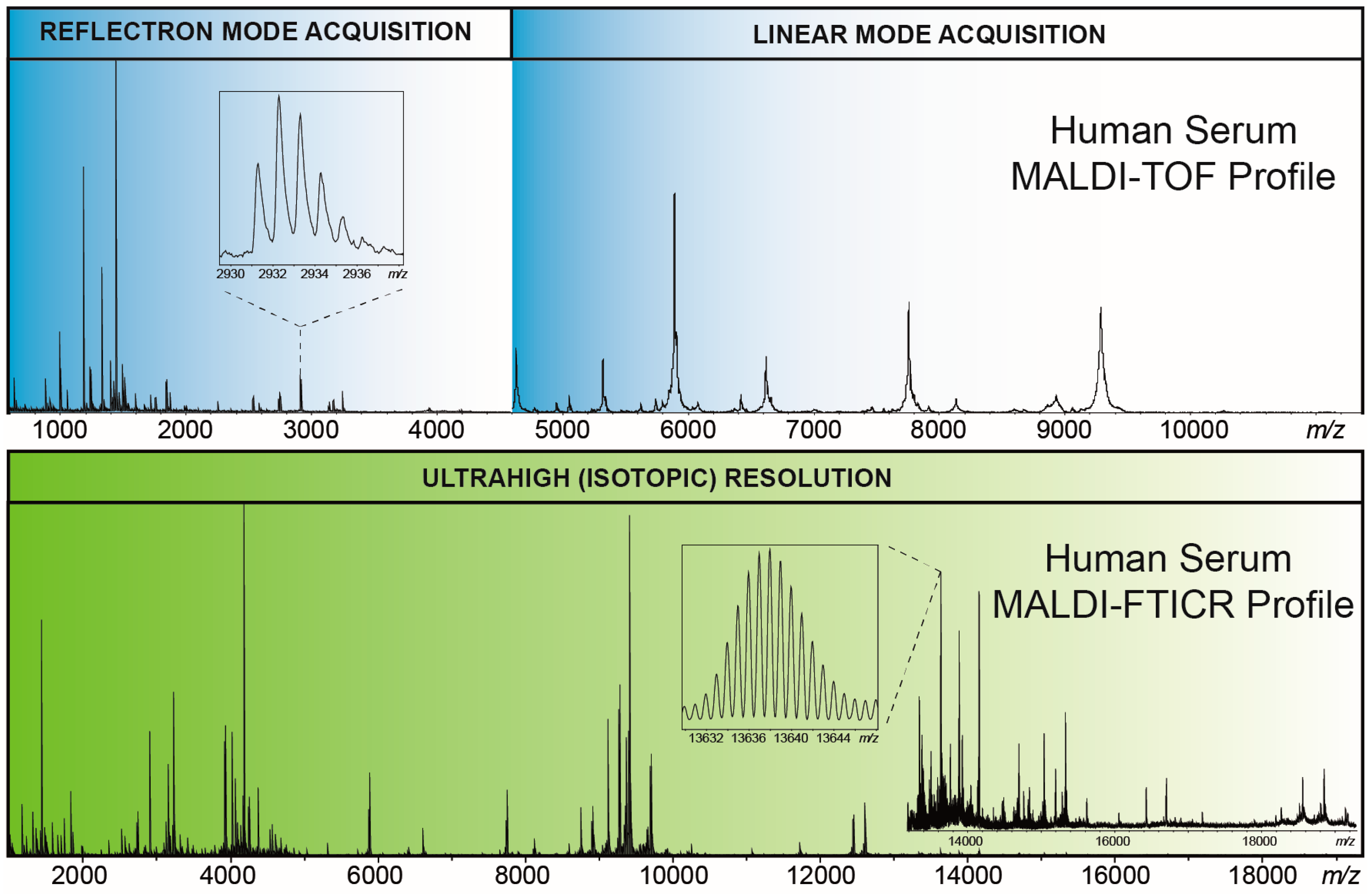
3.1. Example 1: MALDI-FTICR-MS Profiling of Serum Peptides and Proteins

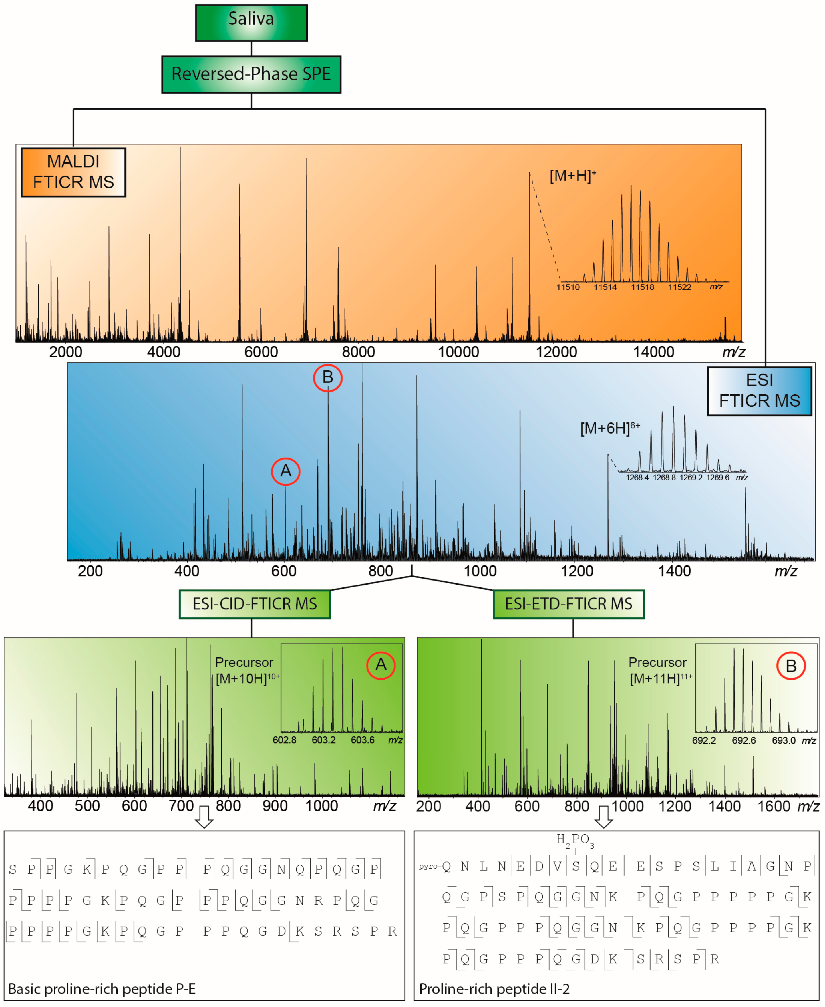
3.2. Example 2: MALDI-FTICR-MS Profiling of Salivary Peptides and Proteins

4. Conclusions and Future Outlook
Author Contributions
Conflicts of Interest
References
- Marshall, A.G. Milestones in Fourier transform ion cyclotron resonance mass spectrometry technique development. Int. J. Mass Spectrom. 2000, 200, 331–356. [Google Scholar] [CrossRef]
- Makarov, A. Electrostatic axially harmonic orbital trapping: A high-performance technique of mass analysis. Anal. Chem. 2000, 72, 1156–1162. [Google Scholar] [CrossRef] [PubMed]
- Zubarev, R.A.; Makarov, A.A. Orbitrap mass spectrometry. Anal. Chem. 2013, 85, 5288–5296. [Google Scholar] [CrossRef] [PubMed]
- Hughey, C.A.; Rodgers, R.P.; Marshall, A.G. Resolution of 11000 compositionally distinct components in a single electrospray ionization Fourier transform ion cyclotron resonance mass spectrum of crude oil. Anal. Chem. 2002, 74, 4145–4149. [Google Scholar] [CrossRef] [PubMed]
- Marshall, A.G.; Rodgers, R.P. Petroleomics: Chemistry of the underworld. Proc. Natl. Acad. Sci. USA 2008, 105, 18090–18095. [Google Scholar] [CrossRef] [PubMed]
- Cho, Y.; Ahmed, A.; Islam, A.; Kim, S. Developments in FT-ICR instrumentation, ionization techniques, and data interpretation methods for petroleomics. Mass Spectrom. Rev. 2015, 34, 248–263. [Google Scholar] [CrossRef] [PubMed]
- Bogdanov, B.; Smith, R.D. Proteomics by FTICR mass spectrometry: Top down and bottom up. Mass Spectrom. Rev. 2005, 24, 168–200. [Google Scholar] [CrossRef] [PubMed]
- Johansson, Å.; Enroth, S.; Palmblad, M.; Deelder, A.M.; Bergquist, J.; Gyllensten, U. Identification of genetic variants influencing the human plasma proteome. Proc. Natl. Acad. Sci. USA 2013, 110, 4673–4378. [Google Scholar] [CrossRef] [PubMed]
- Kalli, A.; Sweredoski, M.J.; Hess, S. Data-dependent middle-down nano-liquid chromatography-electron capture dissociation-tandem mass spectrometry: An application for the analysis of unfractionated histones. Anal. Chem. 2013, 85, 3501–3507. [Google Scholar] [CrossRef] [PubMed]
- Comisarow, M.B.; Marshall, A.G. Fourier transform ion cyclotron resonance spectroscopy. Chem. Phys. Lett. 1974, 25, 282–283. [Google Scholar] [CrossRef]
- Fenn, J.B.; Mann, M.; Meng, C.K.; Wong, S.F.; Whitehouse, C.M. Electrospray ionization for mass spectrometry of large biomolecules. Science 1989, 246, 64–71. [Google Scholar] [CrossRef] [PubMed]
- Karas, M.; Bachmann, D.; Hillenkamp, F. Influence of the wavelength in high-irradiance ultraviolet laser desorption mass spectrometry of organic molecules. Anal. Chem. 1985, 57, 2935–2939. [Google Scholar] [CrossRef]
- Cancilla, M.T.; Penn, S.G.; Carroll, J.A.; Lebrilla, C.B. Coordination of alkali metals to oligosaccharides dictates fragmentation behavior in matrix assisted laser desorption ionization Fourier transform mass spectrometry. J. Am. Chem. Soc. 1996, 118, 6736–6745. [Google Scholar] [CrossRef]
- Nicolardi, S.; Palmblad, M.; Hensbergen, P.J.; Tollenaar, R.A.E.M.; Deelder, A.M.; van der Burgt, Y.E.M. Precision profiling and identification of human serum peptides using Fourier transform ion cyclotron resonance mass spectrometry. Rapid Commun. Mass Spectrom. 2011, 25, 3457–3463. [Google Scholar] [CrossRef] [PubMed]
- Nicolardi, S.; van der Burgt, Y.E.M.; Wuhrer, M.; Deelder, A.M. Mapping O-glycosylation of apolipoprotein C-III in MALDI-FT-ICR protein profiles. Proteomics 2013, 13, 992–1001. [Google Scholar] [CrossRef] [PubMed]
- Nicolardi, S.; Switzar, L.; Deelder, A.M.; Palmblad, M.; van der Burgt, Y.E.M. Top-down MALDI-in-source decay-FTICR mass spectrometry of isotopically resolved proteins. Anal. Chem. 2015, 87, 3429–3437. [Google Scholar] [CrossRef] [PubMed]
- Shaffer, S.A.; Tang, K.; Anderson, G.A.; Prior, D.C.; Udseth, H.R.; Smith, R.D. A novel ion funnel for focusing ions at elevated pressure using electrospray ionization mass spectrometry. Rapid Commun. Mass Spectrom. 1997, 11, 1813–1817. [Google Scholar] [CrossRef]
- Hossain, M.; Kaleta, D.T.; Robinson, E.W.; Liu, T.; Zhao, R.; Page, J.S.; Kelly, R.T.; Moore, R.J.; Tang, K.; Camp, D.G., II; et al. Enhanced sensitivity for selected reaction monitoring mass spectrometry-based targeted proteomics using a dual stage electrodynamic ion funnel interface. Mol. Cell. Proteom. 2011, 10. [Google Scholar] [CrossRef] [PubMed]
- Ibrahim, Y.; Tang, K.; Tolmachev, A.V.; Shvartsburg, A.A.; Smith, R.D. Improving mass spectrometer sensitivity using a high-pressure electrodynamic ion funnel interface. J. Am. Soc. Mass Spectrom. 2006, 17, 1299–1305. [Google Scholar] [CrossRef] [PubMed]
- Ibrahim, Y.; Belov, M.E.; Tolmachev, A.V.; Prior, D.C.; Smith, R.D. Ion funnel trap interface for orthogonal time-of-flight mass spectrometry. Anal. Chem. 2007, 79, 7845–7852. [Google Scholar] [CrossRef] [PubMed]
- Harkewicz, R.; Belov, M.E.; Anderson, G.A.; Paša-Tolić, L.; Masselon, C.D.; Prior, D.C.; Udseth, H.R.; Smith, R.D. ESI-FTICR Mass spectrometry employing data-dependent external ion selection and accumulation. J. Am. Soc. Mass Spectrom. 2002, 13, 144–154. [Google Scholar] [CrossRef]
- Syka, J.E.; Marto, J.A.; Bai, D.L.; Horning, S.; Senko, M.W.; Schwartz, J.C.; Ueberheide, B.; Garcia, B.; Busby, S.; Muratore, T.; et al. Novel linear quadrupole ion trap/FT mass spectrometer: Performance characterization and use in the comparative analysis of histone H3 post-translational modifications. J. Prot. Res. 2004, 3, 621–626. [Google Scholar] [CrossRef]
- Belov, M.E.; Anderson, G.A.; Angell, N.H.; Shen, Y.; Tolić, N.; Udseth, H.R.; Smith, R.D. Dynamic range expansion applied to mass spectrometry based on data-dependent selective ion ejection in capillary liquid chromatography Fourier transform ion cyclotron resonance for enhanced proteome characterization. Anal. Chem. 2001, 73, 5052–5060. [Google Scholar] [CrossRef] [PubMed]
- Wilcox, B.E.; Hendrickson, C.L.; Marshall, A.G. Improved ion extraction from a linear octopole ion trap: SIMION analysis and experimental demonstration. J. Am. Soc. Mass Spectrom. 2002, 13, 1304–1312. [Google Scholar] [CrossRef]
- Kaiser, N.K.; Quinn, J.P.; Blakney, G.T.; Hendrickson, C.L.; Marshall1, A.G. A Novel 9.4 Tesla FTICR mass spectrometer with improved sensitivity, mass resolution, and mass range. J. Am. Soc. Mass Spectrom. 2011, 22, 1343–1351. [Google Scholar] [CrossRef] [PubMed]
- Kaiser, N.K.; Skulason, G.E.; Weisbrod, C.R.; Wu, S.; Zhang, K.; Prior, D.C.; Buschbach, M.A.; Anderson, G.A.; Bruce, J.E. Restrained ion population transfer: A novel ion transfer method for mass spectrometry. Rapid Commun. Mass Spectrom. 2008, 22, 1955–1964. [Google Scholar] [CrossRef] [PubMed]
- Chan, T.-W.D.; Duan, L.; Sze, T.-P.E. Accurate mass measurements for peptide and protein mixtures by using matrix-assisted laser desorption/ionization Fourier transform mass spectrometry. Anal. Chem. 2002, 74, 5282–5289. [Google Scholar] [CrossRef] [PubMed]
- McIver, R.T.; Hunter, R.L.; Bowers, W.D. Coupling a quadrupole mass spectrometer and a Fourier transform mass spectrometer. Int. J. Mass Spectrom. Ion Proc. 1985, 64, 67–77. [Google Scholar] [CrossRef]
- McIver, R.T. Trajectory calculations for axial injection of ions into a magnetic field: Overcoming the magnetic mirror effect with an rf quadrupole lens. Int. J. Mass Spectrom. Ion Proc. 1990, 98, 35–50. [Google Scholar] [CrossRef]
- Beu, S.C.; Hendrickson, C.L.; Marshall, A.G. Excitation of radial ion motion in an rf-only multipole ion guide immersed in a strong magnetic field gradient. J. Am. Soc. Mass Spectrom. 2011, 22, 591–601. [Google Scholar] [CrossRef] [PubMed]
- Williams, D.K.; Muddiman, D.C. Parts-per-billion mass measurement accuracy achieved through the combination of multiple linear regression and automatic gain control in a Fourier transform ion cyclotron resonance mass spectrometer. Anal. Chem. 2007, 79, 5058–5063. [Google Scholar] [CrossRef] [PubMed]
- Savory, J.J.; Kaiser, N.K.; McKenna, A.M.; Xian, F.; Blakney, G.T.; Rodgers, R.P.; Hendrickson, C.L.; Marshall, A.G. Parts-per-billion Fourier transform ion cyclotron resonance mass measurement accuracy with a “walking” calibration equation. Anal. Chem. 2011, 83, 1732–1736. [Google Scholar] [CrossRef] [PubMed]
- Williams, D.K.; Kovach, A.L.; Muddiman, D.C.; Hanck, K.W. Utilizing artificial neural networks in MATLAB to achieve parts-per-billion mass measurement accuracy with a Fourier transform ion cyclotron resonance mass spectrometer. J. Am. Soc. Mass Spectrom. 2009, 20, 1303–1310. [Google Scholar] [CrossRef] [PubMed]
- Xian, F.; Hendrickson, C.L.; Blakney, G.T.; Beu, S.C.; Marshall, A.G. Automated broadband phase correction of Fourier transform ion cyclotron resonance mass spectra. Anal. Chem. 2010, 82, 8807–8812. [Google Scholar] [CrossRef] [PubMed]
- Qian, Y.; O’Connor, P.B. Data processing in Fourier transform ion cyclotron resonance mass spectrometry. Mass Spectrom. Rev. 2014, 33, 333–352. [Google Scholar] [CrossRef] [PubMed]
- Kilgour, D.P.A.; Wills, R.; Qi, Y.; O’Connor, P.B. Autophaser: An algorithm for automated generation of absorption mode spectra for FT-ICR MS. Anal. Chem. 2013, 85, 3903–3911. [Google Scholar] [CrossRef] [PubMed]
- Nagornov, K.O.; Gorshkov, M.V.; Kozhinov, A.N.; Tsybin, Y.O. High-resolution Fourier transform ion cyclotron resonance mass spectrometry with increased throughput for biomolecular analysis. Anal. Chem. 2014, 86, 9020–9028. [Google Scholar] [CrossRef] [PubMed]
- Senko, M.W.; Hendrickson, C.L.; Emmett, M.R.; Shi, S.D.-H.; Marshall, A.G. External accumulation of ions for enhanced electrospray ionization Fourier transform ion cyclotron resonance mass spectrometry. J. Am. Soc. Mass Spectrom. 1997, 8, 970–976. [Google Scholar] [CrossRef]
- Caravatti, P.U.S. Method and Apparatus for the Accumulation of Ions in a Trap of an Ion Cyclotron Resonance Spectrometer, by Transferring the Kinetic Energy of the Motion Parallel to the Magnetic Field into Directions Perpendicular to the Magnetic Field. Patent 4,924,089, 8 May 1990. [Google Scholar]
- Alford, J.M.; Williams, P.E.; Trevor, D.J.; Smalley, R.E. Metal cluster ion cyclotron resonance. Combining supersonic metal cluster beam technology with FT-ICR. Int. J. Mass Spectrom. Ion Proc. 1986, 72, 33–51. [Google Scholar] [CrossRef]
- Nikolaev, E.N.; Kostyukevich, Y.I.; Vladimirov, G.N. Fourier transform ion cyclotron resonance (FT ICR) mass spectrometry: Theory and simulations. Mass Spectrom. Rev. 2014. [Google Scholar] [CrossRef] [PubMed]
- Nikolaev, E.N. Some notes about FT ICR mass spectrometry. Int. J. Mass Spectrom. 2015, 377, 421–431. [Google Scholar] [CrossRef]
- De Koning, L.J.; Nibbering, N.M.M.; van Orden, S.L.; Laukien, F.H. Mass selection of ions in a Fourier transform ion cyclotron resonance trap using correlated harmonic excitation fields (CHEF). Int. J. Mass Spectrom. Ion Proc. 1997, 165–166, 209–219. [Google Scholar] [CrossRef]
- Guan, S.; Burlingame, A.L. High mass selectivity for top-down proteomics by application of SWIFT technology. J. Am. Soc. Mass Spectrom. 2010, 21, 455–459. [Google Scholar] [CrossRef] [PubMed]
- Freitas, M.A.; Hendrickson, C.L.; Emmett, M.R.; Marshall, A.G. Gas-phase bovine ubiquitin cation conformations resolved by gas-phase hydrogen/deuterium exchange rate and extend. Int. J. Mass Spectrom. 1999, 185, 565–575. [Google Scholar] [CrossRef]
- Gauthier, J.W.; Trautman, T.R.; Jaconson, D.B. Sustained off-resonance irradiation for collision-activated dissociation involving Fourier transform mass spectrometry. Collision-activated dissociation technique that emulates infrared multiphoton dissociation. Anal. Chim. Acta 1991, 246, 211–225. [Google Scholar] [CrossRef]
- Zubarev, R.A.; Kelleher, N.L.; McLafferty, F.W. Electron capture dissociation of multiply charge protein cations: A nonergodic process. J. Am. Chem. Soc. 1998, 120, 3265–3266. [Google Scholar] [CrossRef]
- Little, D.P.; Speir, J.P.; Senko, M.W.; O’Connor, P.B.; McLafferty, F.W. Infrared multiphoton dissociation of large multiply charged ions for biomolecule sequencing. Anal. Chem. 1994, 66, 2809–2815. [Google Scholar] [CrossRef] [PubMed]
- Bowers, W.D.; Delbert, S.S.; Hunter, R.L.; McIver, R.T. Fragmentation of oligopeptide ions using ultraviolet laser radiation and Fourier transform mass spectrometry. J. Am. Chem. Soc. 1984, 106, 7288–7289. [Google Scholar] [CrossRef]
- Laskin, J.; Denisov, E.V.; Shukla, A.K.; Barlow, S.E.; Futrell, J.H. Surface-induced dissociation in a Fourier transform ion cyclotron resonance mass spectrometer: Instrument design and evaluation. Anal. Chem. 2002, 74, 3255–3261. [Google Scholar] [CrossRef] [PubMed]
- Marshall, A.G.; Roe, D.C. Theory of Fourier transform ion cyclotron resonance mass spectroscopy: Response to frequency-sweep excitation. J. Chem. Phys. 1980, 73, 1581–1590. [Google Scholar] [CrossRef]
- Guan, S.; Marshall, A.G. Stored waveform inverse Fourier transform (SWIFT) axial excitation/ejection for quadrupole ion trap mass spectrometry. Anal. Chem. 1993, 65, 1288–1294. [Google Scholar] [CrossRef] [PubMed]
- Tolmachev, A.V.; Robinson, E.W.; Wu, S.; Kang, H.; Lourette, N.M.; Paša-Tolić, L.; Smith, R.D. Trapped-ion cell with improved DC potential harmonicity for FT-ICR MS. J. Am. Soc. Mass Spectrom. 2008, 19, 586–597. [Google Scholar] [CrossRef] [PubMed]
- Kaiser, N.K.; Bruce, J.E. Reduction of ion magnetron motion and space charge using radial electric field modulation. Int. J. Mass Spectrom. 2007, 265, 271–280. [Google Scholar] [CrossRef]
- Mathur, R.; Knepper, R.W.; O’Connor, P.B. A low-noise, wideband preamplifier for a Fourier-transform ion cyclotron resonance mass spectrometer. J. Am. Soc. Mass Spectrom. 2007, 18, 2233–2241. [Google Scholar] [CrossRef] [PubMed]
- Kaiser, N.K.; Skulason, G.E.; Weisbrod, C.R.; Bruce, J.E. A novel Fourier transform ion cyclotron resonance mass spectrometer with improved ion trapping and detection capabilities. J. Am. Soc. Mass Spectrom. 2009, 20, 755–762. [Google Scholar] [CrossRef] [PubMed]
- Hendrickson, C.L.; Quinn, J.P.; Kaiser, N.K.; Smith, D.F.; Blakney, G.T.; Chen, T.; Marshall, A.G.; Weisbrod, C.R.; Beu, S.C. 21 tesla Fourier transform ion cyclotron resonance mass spectrometer: A national resource for ultrahigh resolution mass analysis. J. Am. Soc. Mass Spectrom. 2015, 26, 1626–1632. [Google Scholar] [CrossRef] [PubMed]
- BREAKING NEWS! EMSL’s 21 T mass spectrometer at field. Available online: http://www.emsl.pnl.gov/emslweb/news/breaking-news-emsl%E2%80%99s-21-t-mass-spectrometer-field (accessed on 6 November 2015).
- Marshall, A.G.; Hendrickson, C.L.; Jackson, G.S. Fourier transform ion cyclotron resonance mass spectrometry: A primer. Mass Spectrom. Rev. 1998, 17, 1–35. [Google Scholar] [CrossRef]
- Comisarow, M.B.; Marshall, A.G. Fourier Transform Ion Cyclotron Resonance Spectroscopy Method and Apparatus. USA Patent No. 3,937,955, 14 February 1976. [Google Scholar]
- Knight, R.D. The general form of the quadrupole ion trap potential. Int. J. Mass Spectrom. Ion Proc. 1983, 51, 515–518. [Google Scholar] [CrossRef]
- Jackson, G.S.; White, F.M.; Guan, S.; Marshall, A.G. Matrix-shimmed ion cyclotron resonance ion trap simultaneously optimized for excitation, detection, quadrupolar axialization, and trapping. J. Am. Soc. Mass Spectrom. 1999, 10, 759–769. [Google Scholar] [CrossRef]
- Beu, S.C.; Laude, D.A. Open trapped ion cell geometries for FT/ICR/MS. Int. J. Mass Spectrom. Ion Proc. 1992, 112, 215–230. [Google Scholar] [CrossRef]
- Beu, S.C.; Laude, D.A. Elimination of axial ejection during excitation with a capacitively coupled open trapped cyclotron cell for Fourier transform ion cyclotron resonance mass spectrometry. Anal. Chem. 1992, 64, 177–180. [Google Scholar] [CrossRef]
- Littlejohn, D.P.; Ghaderi, S. Mass spectrometer and method. USA Patent No. 4,581,533, 8 April 1986. [Google Scholar]
- Gorshkov, M.V.; Paša-Tolić, L.; Bruce, J.E.; Anderson, G.A.; Smith, R.D. A dual-trap design and its applications in electrospray ionization FTICR mass spectrometry. Anal. Chem. 1997, 69, 1307–1314. [Google Scholar] [CrossRef] [PubMed]
- Guoa, X.; Duursma, M.; Ahmed Al-Khalili, A.A.; McDonnell, L.A.; Heeren, R.M.A. Design and performance of a new FT-ICR cell operating at a temperature range of 77–438 K. Int. J. Mass Spectrom. 2004, 231, 37–45. [Google Scholar] [CrossRef]
- Brustkern, A.M.; Rempel, D.L.; Gross, M.L. An electrically compensated trap designed to eighth order for FT-ICR mass spectrometry. J. Am. Soc. Mass Spectrom. 2008, 19, 1281–1285. [Google Scholar] [CrossRef] [PubMed]
- Wieghaus, A.; Fröhlich, U.; Malek, R.; Horning, S. The grid cell: A new cell design for reduced z-axis ejection in fourier transform ion cyclotron mass spectrometry. In Proceedings of the 54th ASMS Conference on Mass Spectrometry and Allied Topics, Seattle, WA, USA, 28 May–1 June 2006.
- Bruce, J.E.; Anderson, G.A.; Lin, C.-Y.; Gorshkov, M.; Rockwood, A.L.; Smith, R.D. A novel high-performance Fourier transform ion cyclotron resonance cell for improved biopolymer characterization. J. Mass Spectrom. 2000, 35, 85–94. [Google Scholar] [CrossRef]
- Weisbrod, C.R.; Kaiser, N.K.; Skulason, G.E.; Bruce, J.E. Trapping ring electrode cell: A FTICR mass spectrometer cell for improved signal-to-noise and resolving power. Anal. Chem. 2008, 80, 6545–6553. [Google Scholar] [CrossRef] [PubMed]
- Misharin, A.S.; Zubarev, R.A.; Doroshenko, V.M. Fourier transform ion cyclotron resonance mass spectrometer with coaxial multi-electrode cell (“O-trap”): First experimental demonstration. Rapid Commun. Mass Spectrom. 2010, 24, 1931–1940. [Google Scholar] [CrossRef] [PubMed]
- Nikolaev, E.N.; Boldin, I.A.; Jertz, R.; Baykut, G. Initial experimental characterization of a new ultra-high resolution FTICR cell with dynamic harmonization. J. Am. Soc. Mass Spectrom. 2011, 22, 1125–1133. [Google Scholar] [CrossRef] [PubMed]
- Nagornov, K.O.; Kozhinov, A.N.; Tsybin, O.Y.; Tsybin, Y.O. Ion trap with narrow aperture detection electrodes for Fourier transform ion cyclotron resonance mass spectrometry. J. Am. Soc. Mass Spectrom. 2015, 26, 741–751. [Google Scholar] [CrossRef] [PubMed]
- Boldin, I.A.; Nikolaev, E.N. Fourier transform ion cyclotron resonance cell with dynamic harmonization of the electric field in the whole volume by shaping of the excitation and detection electrode assembly. Rapid Commun. Mass Spectrom. 2011, 25, 122–126. [Google Scholar] [CrossRef] [PubMed]
- Kostyukevich, Y.I.; Vladimirov, G.N.; Nikolaev, E.N. Dynamically harmonized FT-ICR cell with specially shaped electrodes for compensation of inhomogeneity of the magnetic field. Computer simulations of the electric field and ion motion dynamics. J. Am. Soc. Mass Spectrom. 2012, 23, 2198–2207. [Google Scholar] [CrossRef] [PubMed]
- Qi, Y.; Witt, M.; Jertz, R.; Baykut, G.; Barrow, M.P.; Nikolaev, E.N.; O’Connor, P.B. Absorption-mode spectra on the dynamically harmonized Fourier transform ion cyclotron resonance cell. Rapid Commun. Mass Spectrom. 2012, 26, 2021–2026. [Google Scholar] [CrossRef] [PubMed]
- Nikolaev, E.N.; Jertz, R.; Grigoryev, A.; Baykut, G. Fine structure in isotopic peak distributions measured using a dynamically harmonized Fourier transform ion cyclotron resonance cell at 7 T. Anal. Chem. 2012, 84, 2275–2283. [Google Scholar] [CrossRef] [PubMed]
- Bruker. Available online: https://www.bruker.com/products/mass-spectrometry-and-separations/esimaldi-ftms/solarix/overview.html (accessed on 9 November 2015).
- Bladergroen, M.R.; Derks, R.J.; Nicolardi, S.; de Visser, B.; van Berloo, S.; van der Burgt, Y.E.M.; Deelder, A.M. Standardized and automated solid-phase extraction procedures for high-throughput proteomics of body fluids. J Proteom. 2012, 77, 144–153. [Google Scholar] [CrossRef] [PubMed]
- Nicolardi, S.; Velstra, B.; Mertens, B.J.; Bonsing, B.; Mesker, W.E.; Tollenaar, R.A.E.M.; Deelder, A.M.; van der Burgt, Y.E.M. Ultrahigh resolution profiles lead to more detailed serum peptidome signatures of pancreatic cancer. Transl. Proteom. 2014, 2, 39–51. [Google Scholar] [CrossRef]
- Nicolardi, S.; Bladergroen, M.R.; Deelder, A.M.; Tollenaar, R.A.E.M.; Palmblad, M.; Mesker, W.E.; van der Burgt, Y.E.M. SPE-MALDI profiling of serum peptides and proteins by ultrahigh resolution FTICR-MS. Chromatographia 2015, 78, 445–449. [Google Scholar] [CrossRef]
- Van den Broek, I.; Romijn, F.P.; Smit, N.P.; van der Laarse, A.; Drijfhout, J.W.; van der Burgt, Y.E.M.; Cobbaert, C.M. Quantifying protein measurands by peptide measurements: Where do errors arise? J. Prot. Res. 2015, 14, 928–942. [Google Scholar] [CrossRef] [PubMed]
- Bladergroen, M.R.; Reiding, K.R.; Hipgrave Ederveen, A.L.; Vreeker, G.C.; Clerc, F.; Holst, S.; Bondt, A.; Wuhrer, M.; van der Burgt, Y.E.M. Automation of high-throughput mass spectrometry-based plasma N-glycome analysis with linkage-specific sialic acid esterification. J. Prot. Res. 2015, 14, 1480–1486. [Google Scholar] [CrossRef] [PubMed]
- Velstra, B.; Vonk, M.A.; Bonsing, B.; Mertens, B.J.; Nicolardi, S.; Huijbers, A.; Vasen, H.; Deelder, A.M.; Mesker, W.E.; van der Burgt, Y.E.M.; et al. Serum peptide signatures for pancreatic cancer based on mass spectrometry: A comparison to CA19–9 levels and routine imaging techniques. J. Cancer Res. Clin. Oncol. 2014, 141, 531–541. [Google Scholar] [CrossRef] [PubMed]
- Marko-Varga, G.; Lindberg, H.; Löfdahl, C.G.; Jonsson, P.; Hansson, L.; Dahlback, M.; Lindquist, E.; Johansson, L.; Foster, M.; Fehniger, T.E. Discovery of biomarker candidates within disease by protein profiling: Principles and concepts. J. Prot. Res. 2005, 4, 1200–1212. [Google Scholar] [CrossRef] [PubMed]
- Hortin, G.L. The MALDI-TOF mass spectrometric view of the plasma proteome and peptidome. Clin. Chem. 2006, 52, 1223–1237. [Google Scholar] [CrossRef] [PubMed]
- Palmblad, M.; Tiss, A.; Cramer, R. Mass spectrometry in clinical proteomics—From the present to the future. Proteom. Clin. Appl. 2009, 3, 6–17. [Google Scholar] [CrossRef] [PubMed]
- Magni, F.; van der Burgt, Y.E.M.; Chinello, C.; Mainini, V.; Gianazza, E.; Squeo, V.; Deelder, A.M.; Kienle, M.G. Biomarkers discovery by peptide and protein profiling in biological fluids based on functionalized magnetic beads purification and mass spectrometry. Blood Transfus. 2010, 8, 92–97. [Google Scholar]
- Albrethsen, J. The first decade of MALDI protein profiling: A lesson in translational biomarker research. J. Proteom. 2011, 74, 765–773. [Google Scholar] [CrossRef] [PubMed]
- Huijbers, A.; Mesker, W.E.; Mertens, B.J.; Bladergroen, M.R.; Deelder, A.M.; van der Burgt, Y.E.M.; Tollenaar, R.A.E.M. Case-controlled identification of colorectal cancer based on proteomic profiles and the potential for screening. Colorectal Dis. 2014, 16, 907–913. [Google Scholar] [CrossRef] [PubMed]
- Taban, I.M.; Altelaar, A.F.; van der Burgt, Y.E.M.; McDonnell, L.A.; Heeren, R.M.; Fuchser, J.; Baykut, G. Imaging of peptides in the rat brain using MALDI-FTICR mass spectrometry. J. Am. Soc. Mass Spectrom. 2007, 18, 145–151. [Google Scholar] [CrossRef] [PubMed]
- Spraggins, J.M.; Rizzo, D.G.; Moore, J.L.; Rose, K.L.; Hammer, N.D.; Skaar, E.P.; Caprioli, R.M. MALDI FTICR IMS of intact proteins: Using mass accuracy to link protein images with proteomics data. J. Am. Soc. Mass Spectrom. 2015, 26, 974–985. [Google Scholar] [CrossRef] [PubMed]
- Belov, M.E.; Damoc, E.; Denisov, E.; Compton, P.D.; Horning, S.; Makarov, A.A.; Kelleher, N.L. From protein complexes to subunit backbone fragments: A multi-stage approach to native mass spectrometry. Anal. Chem. 2013, 85, 11163–11173. [Google Scholar] [CrossRef] [PubMed]
- Snijder, J.; van de Waterbeemd, M.; Damoc, E.; Denisov, E.; Grinfeld, D.; Bennett, A.; Agbandje-McKenna, M.; Makarov, A.; Heck, A.J. Defining the stoichiometry and cargo load of viral and bacterial nanoparticles by Orbitrap mass spectrometry. J. Am. Chem. Soc. 2014, 136, 295–299. [Google Scholar] [CrossRef] [PubMed]
- Nakabayashi, R.; Sawada, Y.; Yamada, Y.; Suzuki, M.; Hirai, M.Y.; Sakurai, T.; Saito, K. Combination of liquid chromatography-Fourier transform ion cyclotron resonance-mass spectrometry with 13C-labeling for chemical assignment of sulfur-containing metabolites in onion bulbs. Anal. Chem. 2013, 85, 1310–1315. [Google Scholar] [CrossRef] [PubMed]
© 2015 by the authors; licensee MDPI, Basel, Switzerland. This article is an open access article distributed under the terms and conditions of the Creative Commons by Attribution (CC-BY) license (http://creativecommons.org/licenses/by/4.0/).
Share and Cite
Nicolardi, S.; Bogdanov, B.; Deelder, A.M.; Palmblad, M.; Van der Burgt, Y.E.M. Developments in FTICR-MS and Its Potential for Body Fluid Signatures. Int. J. Mol. Sci. 2015, 16, 27133-27144. https://0-doi-org.brum.beds.ac.uk/10.3390/ijms161126012
Nicolardi S, Bogdanov B, Deelder AM, Palmblad M, Van der Burgt YEM. Developments in FTICR-MS and Its Potential for Body Fluid Signatures. International Journal of Molecular Sciences. 2015; 16(11):27133-27144. https://0-doi-org.brum.beds.ac.uk/10.3390/ijms161126012
Chicago/Turabian StyleNicolardi, Simone, Bogdan Bogdanov, André M. Deelder, Magnus Palmblad, and Yuri E. M. Van der Burgt. 2015. "Developments in FTICR-MS and Its Potential for Body Fluid Signatures" International Journal of Molecular Sciences 16, no. 11: 27133-27144. https://0-doi-org.brum.beds.ac.uk/10.3390/ijms161126012