Article

Free Radical Scavenging and Cellular Antioxidant Properties of Astaxanthin

1
Institute of Human Nutrition and Food Science, University of Kiel, Hermann-Rodewald-Straße 6, D-24118 Kiel, Germany
2
School of Natural System, Kanazawa University, Kakuma-machi, Kanazawa 920-1192, Japan
3
Medical Photonics Research Center, Hamamatsu University School of Medicine, Handamachi 1-20-1, Higashi-ku, Hamamatsu, Shizuoka 431-3192, Japan
4
DSM Nutritional Products, P.O. Box 2676, 4002 Basel, Switzerland
*
Author to whom correspondence should be addressed.
Int. J. Mol. Sci. 2016, 17(1), 103; https://0-doi-org.brum.beds.ac.uk/10.3390/ijms17010103
Submission received: 12 October 2015 / Revised: 5 January 2016 / Accepted: 8 January 2016 / Published: 14 January 2016
(This article belongs to the Special Issue Macro- and Micro-nutrient Antioxidants)

Abstract

:
Astaxanthin is a coloring agent which is used as a feed additive in aquaculture nutrition. Recently, potential health benefits of astaxanthin have been discussed which may be partly related to its free radical scavenging and antioxidant properties. Our electron spin resonance (ESR) and spin trapping data suggest that synthetic astaxanthin is a potent free radical scavenger in terms of diphenylpicryl-hydrazyl (DPPH) and galvinoxyl free radicals. Furthermore, astaxanthin dose-dependently quenched singlet oxygen as determined by photon counting. In addition to free radical scavenging and singlet oxygen quenching properties, astaxanthin induced the antioxidant enzyme paroxoanase-1, enhanced glutathione concentrations and prevented lipid peroxidation in cultured hepatocytes. Present results suggest that, beyond its coloring properties, synthetic astaxanthin exhibits free radical scavenging, singlet oxygen quenching, and antioxidant activities which could probably positively affect animal and human health.

Graphical Abstract

1. Introduction

Astaxanthin (3,3′-dihydroxy-β,β′-carotene-4,4′-dione, for chemical structure see Figure 1) is a xanthophyll carotenoid [1] which naturally occurs in algae, krill, trout, crayfish, and salmon. Astaxanthin is widely used in aquaculture nutrition as a coloring agent [2]. In addition to its coloring properties, astaxanthin may also affect immune status and reproduction [3,4].
Figure 1. Chemical structure of astaxanthin.
Ijms 17 00103 g001
Astaxanthin has two chiral centers at positions 3 and 3'. The astaxanthin stereoisomer -3S,3S′- is the main form found in wild salmon [5]. Most astaxanthin used in aquaculture nutrition is produced synthetically which yields three different stereoisomers, including 3S, 3′S; 3R, 3′S; and 3R, 3′R [1]. In 1981, Widmer et al. [6] synthesized astaxanthin from the educt 6-oxo-isophorone (3,5,5-trimethyl-2-cyclohexene-1,4-dione) via a seven-step synthesis. Over a Wittig reaction of two equivalents C15-phosphonium salt with C10-dialdehyde, astaxanthin was obtained with yields up to 50% yields [6].
Astaxanthin has also significant applications in the nutraceutical industry [7]. Various health benefits, including cardiovascular disease and cataract prevention, have been associated with astaxanthin consumption [3,8,9,10].
Although it has been suggested that potential health benefits of astaxanthin are, at least partly, mediated by its free radical scavenging [11], antioxidant [12,13], and gene-regulatory properties [14,15], systematic studies are scarce.
In previous studies, free radical scavenging activity of astaxanthin has been determined by FRAP (ferric reducing antioxidant power) [16], TEAC (trolox equivalent antioxidant capacity) [17], and ORAC (oxygen radical absorbance capacity) [18] assays, respectively. Furthermore, the decolorization of the 2,2-diphenyl-1-picrylhydrazyl (DPPH) radical may be used to assess the free radical scavenging activity of antioxidants, taking into account that alcohols as solvents may lead to an overestimation of its free radical scavenging activity [19,20,21]. The different antioxidant test systems, as described in the literature, differ in terms of underlying methodology, reagents, wavelength, pH, etc. It is suggested that a combination of various assays should be used in assessing antioxidant activities in vitro [22].
In addition to these rather indirect methods, electron spin resonance spectroscopy (ESR) combined with spin trapping has been established as a robust method for the direct assessment of free radical reactions [23]. Therefore, in this study, we applied ESR, as well as photon counting methods, to determine the free radical scavenging and singlet oxygen quenching activity of astaxanthin.
Furthermore, we studied the cellular antioxidant activity of astaxanthin in cultured hepatocytes. We addressed the question, if and to what extend astaxanthin may induce enzymatic antioxidant defense mechanisms, including paraoxoanase-1. Furthermore, we determined cellular glutathione levels in response to an astaxanthin treatment, since glutathione is the most important endogenous cytosolic antioxidant centrally involved in redox signaling and stress response.
Overall, this manuscript aims at providing comprehensive information in terms of the free radical scavenging, antioxidant, and cell signaling modifying properties of astaxanthin.

2. Results and Discussion

We applied ESR and spin trapping in order to directly quantify the free radical scavenging activity of astaxanthin. Astaxanthin dose-dependently scavenged DPPH (Figure 2A) and galvinoxyl (Figure 2B) free radicals. However, astaxanthin did not scavenge superoxide anion free radicals (data not shown). Accordingly, astaxanthin did not inhibit xanthine oxidase, which generates superoxide anion free radicals (Figure 2C). When singlet oxygen is relaxed to the ground state, oxygen photon emission is observed. Singlet oxygen quenching activity of astaxanthin as a function of emission spectrum, concentration, and decay curve is given in Figure 2D. As shown in Figure 2D, astaxanthin dose-dependently quenched singlet oxygen as determined by photon counting.
Figure 2. Scavenging effects of astaxanthin on DPPH radical (A), galvinoxyl radical (B), xanthine oxidase inhibition (C), and quenching of singlet oxygen (D). (A) The reaction mixture contained 500 μM DPPH and the given concentration of astaxanthin. All values are means + SD (experiments performed in triplicate); (B) Various astaxanthin concentrations were mixed with 500 µM galvinoxyl. Changes in the radical signal intensity are shown on the right side of the figure. All values are means + SD (three independent experiments performed in triplicate); (C) The reaction mixture contained 5 U/mL xanthine oxidase in 50 mM potassium phosphate buffer and the given astaxanthin concentrations. Allopurinol was used as a positive control. All values are means + SD (three independent experiments performed in triplicate); (D) Singlet oxygen quenching activity of astaxanthin as a function of concentration (5.4 × 10⁻⁷, 1.1 × 10⁻⁶, 2.1 × 10⁻⁶, 4.2 × 10⁻⁶, and 8.1 × 10⁻⁶ M astaxanthin), wavelength (left), and time (right). Photo emission was determined by a photon complex after laser irradiation at 532 nm in the presence of the test sample. Two independent experiments performed in duplicate.
Ijms 17 00103 g002aIjms 17 00103 g002b
In our cell culture studies, astaxanthin did not exhibit any significant cytotoxicity in concentrations of up to 20 µM in Huh7, PON1-Huh7, and HepG2 as summarized in Figure 3. TritonX was used as a control to induce cytotoxicity as determined by the neutral red assay.
Figure 3. Effects of astaxanthin on cell viability in Huh7, PON1-Huh7, and HepG2 cells after 24 h incubation. Data are means + SD of at least two experiments performed in triplicate.
Ijms 17 00103 g003
Supplementation of cultured PON1-Huh7 cells with 20 µM synthetic astaxanthin resulted in a moderate induction of paraoxoanse-1 (PON1) (Figure 4A). Furthermore, synthetic astaxanthin dose-dependently increased cellular glutathione (GSH) levels in HepG2 cells (Figure 4B). The increase in cellular GSH was not accompanied by an increase in Nrf2 transactivation as shown in Figure 4C. Curcumin and resveratrol were used as positive controls, respectively.
Figure 4. Effects of astaxanthin on transactivation of paraoxonase-1 (PON1) (A), cellular glutathione (GSH) levels (B) and transactivation of Nrf2 (C) in cultured hepatocytes. (A) PON1-Huh7 cells were seeded at a density of 0.15 × 10⁶ cells/well into 24 well plates and incubated for 24 h at 37 °C. Cells were treated with 1 and 20 µM astaxanthin. PON1 transactivation was measured after 48 h incubation of the cells with synthetic astaxanthin. Curcumin (Curc; 20 µM) was used as a positive control; (B) HepG2 cells were seeded at a density of 0.15 × 10⁶ cells/well into 24 well plates and incubated for 24 h. Cells were treated with 1 and 5 µM astaxanthin and incubated for an additional 24 h. Resveratrol (Res; 25 µM) was used as a positive control; (C) Huh7 cells were seeded at a density of 0.15 × 10⁶ cells/well into 24 well plates and incubated for 24 h at 37 °C. Cells were transfected for 24 h. Nrf2 transactivation was measured after 24 h incubation of the cells with 1 and 20 µM astaxanthin. Curcumin (Curc; 20 µM) was used as a positive control. All values are means + SEM (two independent experiments performed in triplicate), statistical significant differences between THF-control cells and astaxanthin supplemented cells are indicated as * p < 0.05, *** p < 0.001; one-way ANOVA LSD (Figure 4A) and Dunnett-T (Figure 4B).
Ijms 17 00103 g004
Additionally, mRNA levels of Gclc (glutamate-cysteine ligase, catalytic subunit) and other Nrf2-target genes, including Gsta (glutathione S-transferase alpha 1), Gpx (glutathione peroxidase), Hmxo1 (heme oxygenase 1), Nqo1 (NAD(P)H dehydrogenase [quinone] 1), Sod1 (superoxide dismutase), and Sod2, were not significantly changed due to the astaxanthin treatment (Table 1).
Table 1. Nrf2-target gene expression in astaxanthin treated Huh7 cells (THF control cells were set to be 1.0).
GeneAstaxanthin [µM]
120
Gclc0.91 ± 0.081.03 ± 0.26
Gsta1.01 ± 0.150.99 ± 0.18
Gpx11.34 ± 0.241.12 ± 0.30
Hmxo10.40 ± 0.040.85 ± 0.32
Nqo11.21 ± 0.181.32 ± 0.21
Sod11.22 ± 0.210.99 ± 0.18
Sod21.35 ± 0.181.39 ± 0.54
HepG2 cells were stressed with cumene hydroperoxide/hemin to provoke lipid peroxidation. We applied the BODIPY assay in order to determine lipid peroxidation in our HepG2 hepatocytes following the astaxanthin treatment. The BODIPY probe exhibits a change in fluorescence after interaction with peroxyl groups. Under the conditions investigated, supplementation of HepG2 cells with 10 and 20 µM astaxanthin significantly decreased lipid peroxidation in HepG2 cells (Figure 5).
Figure 5. Impact of astaxanthin on the ability of human HepG2 cells to prevent cumene hydroperoxide/hemin stimulated lipid peroxidation. HepG2 cells were seeded at a density of 0.1 × 10⁶ cells/well into a 96-well plate and incubated for 24 h. Cells were treated with two concentrations of astaxanthin (10 and 20 µM) for an additional 24 h. Cells were rinsed and loaded with C11-BODIPY for 30 min and then exposed to cumene hydroperoxide (8 mM)/hemin (8 µM) for 1 h. Control cells were treated with cell culture medium only. Simultaneously, basal levels of lipid peroxidation were assessed for each treatment concentration (data not shown) and control. Lipid peroxidation was determined by examining the ratio of green (oxidized) and the sum of green and red (oxidized + non-oxidized) emissions using a fluorometric plate reader. Data are means + SD of at least two independent experiments performed in duplicate. ** p < 0.01, one-way ANOVA Games–Howell.
Ijms 17 00103 g005
In the present study, we observed a dose-dependent free radical scavenging activity of astaxanthin in terms of DPPH and galvinoxyl free radicals as determined by ESR and spin trapping. Current ESR data regarding the free radical scavenging activity of astaxanthin in terms of DPPH free radicals are in line with literature data [24,25], where different photometric assay systems were applied, e.g., DPPH, ferric reducing antioxidant power and hydroxyl radical scavenging.
The free radical scavenging activity of astaxanthin is suggested to be mediated by electron transfer, radical adduct formation and hydrogen atom transfer [26]. Singlet oxygen quenching of carotenoids, including astaxanthin is mediated by energy transfer between the electrophilic singlet oxygen and the polyene backbone. In general, the singlet oxygen quenching activity of carotenoids increases with increasing length of conjugation [27]. Furthermore, our BODIPY data suggest that astaxanthin significantly prevents lipid peroxidation. The prevention of lipid peroxidation due to astaxanthin may improve the sensory quality and shelve life of fish which warrants further investigation. However, it needs to be taken into account that the astaxanthin concentrations, as administered in our cell culture assays, may be higher than astaxanthin concentrations in fish plasma [28].
We did not observe any inhibition of xanthine oxidase in response to the astaxanthin treatment. Thus, unlike other antioxidants including flavonoids [29,30], astaxanthin does not seem to mediate antioxidant activity via xanthine oxidase inhibition.
For our cell studies, astaxanthin was dissolved in tetrahydrofuran (THF). We used THF since it has been previously shown by our group to be non-cytotoxic to HepG2 cells [31]. Furthermore, astaxanthin dissolved in THF exhibits an appropriate stability in the cell culture medium [31] and is effectively taken up by HepG2 and HT29 cultured cells [31,32].
Current cell culture data clearly suggest that synthetic astaxanthin does not only work as a free radical scavenger, but also as an inductor of cellular antioxidant defense mechanisms, including PON1 and reduced glutathione. PON1 is a hepatic enzyme which prevents and/or delays the oxidation of LDL [33]. Thus, the prevention of plasma lipid peroxidation in humans due to astaxanthin, as reported by Barlic and co-workers [34], may be partly mediated by an induction of paraoxonase-1. Recently, it has been shown that PON1 also counteracts oxidative stress induced by mercury chloride, thereby offering a beneficial strategy against HgCl2 toxicity [35] which may be of particular interest for fish nutrition. We also determined the cellular activity of natural astaxanthin which was isolated from Haematococcus pluvialis (data not shown). Importantly, natural source astaxanthin did not exhibit a superior bioactivity in any of our test systems as also stated by others [36]. In terms of PON1, a transactivation occurred only in response to synthetic astaxanthin.
Synthetic astaxanthin dose-dependently increased cellular GSH levels in HepG2 cells which is in line with findings by Saw and co-workers who found an induction of cellular GSH due to both astaxanthin and omega-3 fatty acids [13]. Interestingly, in our study the increase in cellular GSH in HepG2 cells was, however, not accompanied by an increase in Nrf2 transactivation. Furthermore, Gclc, the rate limiting enzyme of GSH synthesis, was not induced by astaxanthin. Additionally, steady state levels of other Nrf2 target genes, including Gsta, Gpx, Hmxo, Nqo1, Sod1, and Sod2 were not induced in response to the astaxanthin treatment. Thus, it is suggested that the increase in cellular GSH due to astaxanthin was not mediated by an Nrf2 dependent signal transduction pathway. It is worth mentioning that GSH synthesis is not only regulated by Nrf2, but also by other transcription factors, including activator protein 1 (AP-1) [37,38], specificity protein 1 (Sp1) [39,40], nuclear respiratory factor 1 (Nrf1) [41,42], and nuclear factor kappa-light-chain-enhancer of activated B cells (NFκB) [42,43]. Based on the present data, it is suggested that elevated GSH concentration may be a sign of lower oxidative stress levels as a result of the astaxanthin treatment.

3. Experimental Section

3.1. Radical Scavenging Measured by Electron Spin Resonance Spectroscopy (ESR)

ESR measurements were performed using a JEOL JES-FR30EX free radical monitor (JEOL Ltd., Akishima, Japan). The measurement conditions were as follows: magnetic field, 337.394 ± 7.5 mT; power, 4 mW; sweep time, 1 min; sweep width, 7.5; modulation, 100 kHz, 0.32 mT; amplitude, 400; and time constant, 0.3 s. Signal intensity was compared based on the ratio against the magnetic Mn2+ marker and was represented by relative height ratio. The ESR spectra were measured three times.
Preparation of the astaxanthin stock solution. Astaxanthin (DSM, Kaiseraugst, Switzerland) was dissolved in 21.8% tetrahydrofuran (THF) (Wako Chemicals, Osaka, Japan) and 78.2% ethanol (Nacalai Tesque, Kyoto, Japan). The actual concentration of astaxanthin was determined by diluting the stock solution with THF using the molar coefficient of astaxanthin (69,600 M−1·cm−1 at 477 nm) [44]. Based on this value, the stock solution of astaxanthin was determined as 30.56 mM. The astaxanthin stock solution was further diluted with tert-butyl alcohol/THF (89.3:10.7, v/v).
DPPH radical scavenging experiments. To a reaction mixture containing 80 µL distilled water, 20 µL 500 µM DPPH (in methanol; Nacalai Tesque, Kyoto, Japan) and 100 µL astaxanthin (0, 50, 100, 200, 300, 400, 500, 600, 800, 1000, 1200, and 1500 µM) were added and stirred for a few seconds. tert-Butyl alcohol (containing 10.7% THF) was used as a control. After 1 min incubation the ESR spectra were measured.
Galvinoxyl radical scavenging experiments. To a reaction mixture containing 80 µL distilled water, 20 µL 500 µM galvinoxyl (in methanol; Nacalai Tesque, Kyoto, Japan) and 100 µL astaxanthin (0, 10, 20, 50, 100, 200, 300, 400, 500 µM) were added and stirred for a few seconds. tert-Butyl alcohol (containing 10.7% THF) was used as a control. After 1 min incubation the ESR spectra were measured.
Superoxide radical scavenging experiments. To the reaction mixture of 30 µL of 5 mM hypoxanthine (dissolved in NaOH and then diluted with PBS (pH 7.4)), 40 µL 4 M 5,5-dimethyl-1-pyrroline N-oxide (DMPO), 25 µL ultrapure water, and 10 µL of astaxanthin (0, 1, 7.5, 15 mM) were added to 5 µL of 1 U/mL xanthine oxidase in 200 mM PBS buffer solution (pH 7.4). After 1 min incubation at 25 °C the ESR spectra were measured. Three independent experiments were performed in triplicate.

3.2. Inhibition of Xanthine Oxidase

The inhibition of xanthine oxidase was measured according to Bräunlich et al. 2013 [45] with some modifications. An enzyme solution consisting of 400 µL xanthine oxidase (5 U/mL) in 7.6 mL 50 mM potassium phosphate buffer (pH = 7.5) was prepared immediately before use. One hundred and fifty microliters of the prepared enzyme solution was added to each well of a 96 well microtiterplate. Astaxanthin (4 g/L in THF) was serially diluted in the wells (dilution factor 2). THF was used as blank sample. Allopurinol (500 µM; Sigma, Darmstadt, Germany) served as positive control and was serially diluted in the wells (dilution factor 2). The substrate hypoxanthine (40 µg/mL; dissolved in NaOH and then in distilled water) was prepared daily and 80 µL were injected at cycle 10. The absorption was measured at 290 nm (microplate reader Tecan infinite 200Pro, Crailsheim, Germany) over 110 cycles (≈150 s) at 28 °C. At least three independent experiments were performed in triplicate.

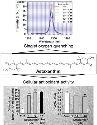
3.3. Quenching Experiments of Near-Infrared Emission Spectra of 1O2 by Astaxanthin

The singlet oxygen scavenging activity of astaxanthin was determined by photon counting according to Shimizu et al. (2010) [46] with some modifications.
Ten millimolar astaxanthin was prepared in THF/ethanol (20/80; v/v). After preparing 10 mM astaxanthin, 1 mM astaxanthin was prepared in THF/ethanol (10.7/89.3; v/v). The final concentration of astaxanthin was confirmed by measuring the solution of 2 mL ethanol and 80 μL astaxanthin solution (1 mM) by UV–Vis spectrometer (Hitachi, Tokyo, Japan) using the molar coefficient of astaxanthin (69,600 M−1·cm−1 at λ = 477 nm).
The 1O2 formation was directly measured by the near-infrared luminescence at around 1270 nm from deactivated 1O2 which corresponds to the 1O2 (1Δg)–3O2 (3Σg) transition. The emission from 1O2 was measured using an apparatus based on a commercially available apparatus and improved for high-sensitivity detection (NIR-PII System, Hamamatsu Photonics K.K., Hamamatsu, Japan). The excitation pulse was obtained using a dye laser (CL-EGC USHO Optical Systems Co., Ltd., Osaka, Japan) excited by an Nd-YAG Laser (NL 240/TH EKSPLA Lithuania). The excitation wavelength was 530 nm, pulse width and intensity were approximately 7 ns and 17 mW, respectively, and the repetition rate was 500 Hz. Emission of 1O2 was monitored using an infrared-gated image intensifier (NIR-PII, Hamamatsu Photonics) after passage through a polychromator (250 is, Chromex Inc., Albuquerque, NM, USA). Measurements started at 1 μs after application of the excitation pulse and the exposure time was 50 μs. Signals were accumulated by repeated detection (500 Hz, 10 s). The emission decay was detected by the NIR-PII system (Hamamatsu Photonics) after passing through the bandpass filter (1270 nm) and recorded by the multichannel scalar (NanoHarp 250, PicoQuant GmbH, Berlin, Germany). The measuring time was set at 16 ns/bin for ca. 260 μs and accumulated for 2 min at 500 Hz. The solution studies consist of 2 mL of 5 μM Rose Bengal (in ethanol) as a photosensitizer. The quenching of the singlet oxygen decay was examined by adding 109 μM astaxanthin as follows: addition of 10 μL, 10 μL (final 20 μL), 20 μL (final 40 μL), 40 μL (final 80 μL), and 80 μL (final 160 μL). The decay curve and reaction rate of astaxanthin were calculated using the decay curve and quenching ratio of the duplicate experiments.

3.4. Cell Culture

Human Huh7 hepatic cellular carcinoma cells (Institute of Applied Cell Culture, Munich, Germany) were cultivated in high glucose (4.5 g/L) Dulbecco’s modified Eagle’s medium (DMEM, with sodium pyruvate, l-glutamine and 3.7 g/L NaHCO3 supplemented with 10% (v/v) fetal bovine serum (FBS), 100 U/mL penicillin and 100 µg/mL streptomycin (all PAN-Biotech, Aidenbach, Germany) at 37 °C in a 5% CO2 setting. For the preparation of PON1-Huh7 cells, Huh7 cells were stably transfected with a 1009 bp [−1013, −4] fragment of the human PON1 promoter (kindly provided by X. Coumoul and R. Barouki, INSERM UMR-S, Paris, France [47]). PON1-Huh7 cells were cultured in high glucose (4.5 g/L) DMEM (without l-glutamine) supplemented with 10% heat-inactivated FBS, 2 mmol/L glutamine, 100 U/mL penicillin, 100 µg/mL streptomycin, and 100 µg/mL G418 sulfate (PAN-Biotech, Aidenbach, Germany) in a humidified atmosphere of 5% CO2 at 37 °C. Human HepG2 hepatocellular carcinoma cells (Institute of Applied Cell Culture, Munich, Germany) were cultured in RPMI 1640 (with l-glutamine and 2.0 g/L NaHCO3; PAN-Biotech, Aidenbach, Germany) containing 10% FBS, 100 U/mL penicillin and 100 µg/mL streptomycin in an incubator (95% air, 5% CO2) at 37 °C. At least two independent experiments were performed in triplicate. All cell culture experiments were conducted at non-cytotoxic concentrations of astaxanthin.

3.5. Neutral Red Cell Viability Assay

PON1-Huh7, Huh7, and HepG2 cells were seeded at a density of 1.5 × 10⁵ cells per well in 24-well plates. Cells were treated with 1–20 µM astaxanthin for 48 (PON1-Huh7) or 24 h (Huh7, HepG2). Afterwards, the culture medium was replaced with neutral red solution (50 μg/mL, prepared in cell culture medium) and incubated for 2 h at 37 °C. Cells were extracted with ethanol:water:glacial acetic acid (50:49:1, v/v/v; glacial acetic acid purchased from Carl Roth, Karlsruhe, Germany) and incubated at room temperature for 15 min under continuous shaking. The absorbance was measured at 540 nm in a plate reader (Labsystems iEMS Reader, Helsinki, Finland). At least two independent experiments were performed in triplicate.

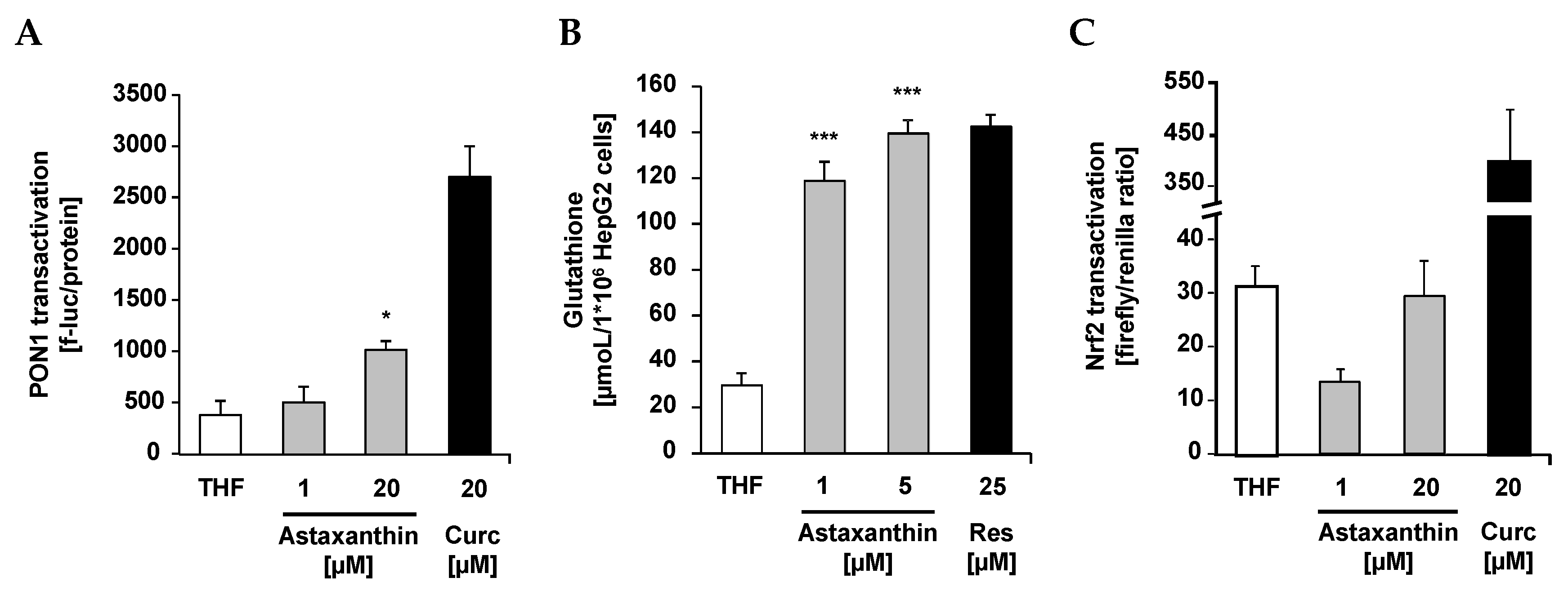
3.6. Paraoxonase Activity

PON1-Huh7 cells were seeded at a density of 0.15 × 10⁶ cells per well in 24-well plates and incubated for 24 h. Subsequently, cells were treated with 1 and 20 µM synthetic (DSM, Kaiseraugst, Switzerland) versus algae-based (BioAstin, Cyanotech Corporation, Kailua-Kona, HI, USA) astaxanthin. Curcumin (20 µM) served as positive control. After 48 h incubation, cells were washed with PBS and lysed with cell culture lysis reagent (1:5, v/v; Promega, Mannheim, Germany). Luciferase activity (Luciferase Assay System; Promega, Mannheim, Germany) was measured by luminescence reading (Infinite 200 microplate reader, Tecan, Crailsheim, Germany). Results were normalized to total protein content, determined by the BCA assay (Pierce, Bonn, Germany) according to the manufacturer’s instructions. At least two independent experiments were performed in triplicate.

3.7. Glutathione Assay

HepG2 cells (0.15 × 10⁶ cells/well) were incubated for 24 h at 37 °C. Cells were treated with 1 and 5 µM astaxanthin, respectively and incubated for an additional 24 h. Glutathione measurements were performed according to Esatbeyoglu et al. (2014) [48] and Vandeputte et al. (1994) [49].

3.8. Nrf2 Transactivation

Huh7 cells (0.15 × 10⁶ cells per well) were incubated in 24-well plates for 24 h. Cells were transiently transfected with pARE_GIGPx_Luc according to Wagner et al. (2010) [50]. After 24 h-transfection, cells were incubated with 1 and 20 µM astaxanthin for further 24 h. Curcumin (20 µM) served as positive control. Cell lysis and measurement of Nrf2 transactivation were conducted as reported previously [50]. At least two independent experiments were performed in triplicate.

3.9. RNA Isolation and qRT-PCR

Cells were harvested with TriFAST (VWR International GmbH, Erlangen, Germany) and RNA was isolated according to the manufacturer’s protocol. RNA concentration and quality were determined using the NanoDrop ND-2000 spectrophotometer (VWR International GmbH, Erlangen, Germany). Aliquots of RNA samples were stored at −80 °C until PCR analyses. mRNA expression levels of glutathione peroxidase 1 (Gpx1), superoxide dismutase 1, soluble (Sod1), superoxide dismutase 2, mitochondrial (Sod2), heme oxygenase 1 (Ho-1), NAD(P)H dehydrogenase [quinone] 1, (Nqo1), glutamate-cysteine ligase, catalytic subunit (Gclc), glutathione S-transferase alpha 1 (Gsta), and glyceraldehyde-3-phosphate dehydrogenase (Gapdh) were determined using target-specific primers. Primer3 Input software version 0.4.0 was used for primer design (http://bioinfo.ut.ee/primer3-0.4.0/primer3/). Primer sequences are depicted in Table 2. Primers were purchased from Eurofins MWG (Ebersberg, Germany). qRT-PCR was carried out as a one-step procedure using the SensiFAST™ SYBR No-ROX One-Step Kit (Bioline, Luckenwalde, Germany) with SYBR Green detection on a Rotorgene 6000 cycler (Corbett Life Science, Sydney, Australia). Calculation of results was done by an external standard curve. Transcription levels of target genes were normalized by the transcription of Gapdh which served as housekeeping gene in qRT-PCR analyses.

3.10. Lipid Peroxidation Determined by BODIPY Assay

C11-BODIPY (581/591) (Life technologies, Darmstadt, Germany) fluorescent dye served as a lipid peroxidation sensor. C11-BODIPY is a lipophilic fatty acid analog which incorporates into biomembranes and shifts its fluorescent properties from reduced (λex = 540 nm/λem = 595 nm) to oxidized (λex = 480 nm/λem = 520 nm) status. Adjusted index of BODIPY oxidation was calculated as follows:
Adjusted Index = EmOx/(EmOx + EmRed)
HepG2 cells (density of 0.15 × 10⁶ cells per well in 24-well plates and incubated for 24 h) were incubated with 10 and 20 µM astaxanthin for 24 h. Cells were treated with 10 µM C11-BODIPY (581/591) in medium without FBS for 30 min. BODIPY was removed and cells were stressed with 80 µM cumene hydroperoxide/80 nM hemin in PBS for 1 h. Afterwards, PBS was refreshed and fluorescence was measured in adherent cells.

3.11. Statistical Analysis

Statistical analysis was conducted using SPSS software version 23.0 (SPSS Inc., Munich, Germany). All cell culture data were tested for normal distribution (Kolmogorow–Smirnow). One-way ANOVA was applied. In case of homogeneous variances, LSD and Dunnett-T post hoc test and, in case of heterogeneous variances, Games–Howell post hoc test was conducted to test significant differences between the control group and the test groups. All data are expressed as the mean + SD or SEM. Significance was accepted at p < 0.05.
Table 2. Nucleotide sequences and annealing temperatures of primers used in qRT-PCR analyses in cultured Huh7 cells.
GeneGene-IDDescriptionPrimer, Forward (5′–3′)Primer, Reverse (5′–3′)Annealing (°C)
Gpx12876glutathione peroxidase 1ACACCCAGATGAACGAGCTGCCGGACGTACTTGAGGGAAT58
Sod16647superoxide dismutase 1, solubleGGGGAAGCATTAAAGGACTGCAACATGCCTCTCTTCATCC55
Sod26648superoxide dismutase 2, mitochondrialGCACTAGCAGCATGTTGAGCGATCTGCGCGTTGATGTG55
Hmox13162heme oxygenase 1CCA GGC AGA GAA TGC TGA GTGTA GAC AGG GGC GAA GAC TG59
Nqo11728NAD(P)H dehydrogenase [quinone] 1CTG ATC GTA CTG GCT CAC TCGAA CAG ACT CGG CAG GAT AC58
Gclc2729glutamate-cysteine ligase, catalytic subunitTTT GGT CAG GGA GTT TCC AGTGA ACA GGC CAT GTC AAC TG59
Gst2938glutathione S-transferase alpha 1CGTATGTCCACCTGAGGAAAGCCAACAAGGTAGTCTTGTCC60
Gapdh2597glyceraldehyde-3-phosphate dehydrogenaseCAATGACCCCTTCATTGACCGATCTCGCTCCTGGAAGATG58

4. Conclusions

Collectively, our data suggest that, beyond its coloring properties, synthetic astaxanthin exhibits important free radical scavenging, oxygen quenching, and antioxidant activities. The direct free radical scavenging activity of astaxanthin was determined by ESR and spin trapping and its oxygen quenching activity by photon counting. The cellular antioxidant activity of astaxanthin was examined in various bioassays (e.g., transactivation of PON1, cellular GSH levels, transactivation of Nrf2 and expression of its target genes, lipid peroxidation) in cultured cell lines. Since we worked with cell lines, our findings can not be generalized to primary cells and animal models, respectively, and should therefore be verified in appropriate in vivo models in the future. Additionally, it would be interesting to test whether there are synergistic interactions between astaxanthin and other lipid (e.g., vitamin E) and water soluble antioxidants (e.g., ascorbic acid, flavonoids) in terms of their free radical scavenging, antioxidant and gene-regulatory activity.

Acknowledgments

We thank Christoph Plieth (Center of Biochemistry and Molecular Biology, University of Kiel) for providing the microplate reader (Tecan infinite 200Pro, Crailsheim, Germany) and for the fruitful discussions. We also thank Vivien Schmuck for excellent technical assistance.

Author Contributions

Janina Dose, Gerald Rimbach, and Tuba Esatbeyoglu designed the study and wrote the manuscript. Seiichi Matsugo, Haruka Yokokawa, and Yutaro Koshida conducted the electron spin resonance spectroscopy experiments. Shigetoshi Okazaki conducted the singlet oxygen quenching activity experiments. Ulrike Seidel performed the BODIPY assay. All other experiments were conducted by Janina Dose and Tuba Esatbeyoglu. All authors read, edited and gave feedback for the final manuscript.

Conflicts of Interest

Manfred Eggersdorfer is an employee of DSM.

References

  1. Ambati, R.R.; Phang, S.M.; Ravi, S.; Aswathanarayana, R.G. Astaxanthin: Sources, extraction, stability, biological activities and its commercial applications—A review. Mar. Drugs 2014, 12, 128–152. [Google Scholar] [CrossRef] [PubMed]
  2. Jiao, G.; Hui, J.P.M.; Burton, I.W.; Thibault, M.-H.; Pelletier, C.; Boudreau, J.; Tchoukanova, N.; Subramanian, B.; Djaoued, Y.; Ewart, S.; et al. Characterization of shrimp oil from Pandalus borealis by high performance liquid chromatography and high resolution mass spectrometry. Mar. Drugs 2015, 13, 3849–3876. [Google Scholar] [CrossRef] [PubMed]
  3. Higuera-Ciapara, I.; Félix-Valenzuela, L.; Goycoolea, F.M. Astaxanthin: A review of its chemistry and applications. Crit. Rev. Food Sci. Nutr. 2006, 46, 185–196. [Google Scholar] [CrossRef] [PubMed]
  4. Otton, R.; Marin, D.P.; Bolin, A.P.; de Cássia Santos Macedo, R.; Campoio, T.R.; Fineto, C.; Guerra, B.A.; Leite, J.R.; Barros, M.P.; Mattei, R. Combined fish oil and astaxanthin supplementation modulates rat lymphocyte function. Eur. J. Nutr. 2012, 51, 707–718. [Google Scholar] [CrossRef] [PubMed]
  5. Megdal, P.A.; Craft, N.A.; Handelman, G.J. A simplified method to distinguish farmed (Salmo salar) from wild salmon: Fatty acid ratios versus astaxanthin chiral isomers. Lipids 2009, 44, 569–576. [Google Scholar] [CrossRef] [PubMed]
  6. Widmer, E.; Zell, R.; Broger, E.A.; Crameri, Y.; Wagner, H.P.; Dinkel, J.; Schlageter, M.; Lukáč, T. Technische Verfahren zur Synthese von Carotinoiden und verwandten Verbindungen aus 6-Oxo-isophoron. II. Ein neues Konzept für die Synthese von (3RS, 3´RS)-Astaxanthin. Helv. Chim. Acta 1981, 64, 2436–2446. (In German) [Google Scholar] [CrossRef]
  7. Guerin, M.; Huntley, M.E.; Olaizola, M. Haematococcus astaxanthin: Applications for human health and nutrition. Trends Biotechnol. 2003, 21, 210–216. [Google Scholar] [CrossRef]
  8. Fassett, R.G.; Coombes, J.S. Astaxanthin: A potential therapeutic agent in cardiovascular disease. Mar. Drugs 2011, 9, 447–465. [Google Scholar] [CrossRef] [PubMed]
  9. Hashimoto, H.; Arai, K.; Hayashi, S.; Okamoto, H.; Takahashi, J.; Chikuda, M.; Obara, Y. Effects of astaxanthin on antioxidation in human aqueous humor. J. Clin. Biochem. Nutr. 2013, 53, 1–7. [Google Scholar] [CrossRef] [PubMed]
  10. Fassett, R.G.; Healy, H.; Driver, R.; Robertson, I.K.; Geraghty, D.P.; Sharman, J.E.; Coombes, J.S. Astaxanthin vs placebo on arterial stiffness, oxidative stress and inflammation in renal transplant patients (Xanthin): A randomised controlled trial. BMC Nephrol. 2008, 9, 17. [Google Scholar] [CrossRef] [PubMed]
  11. Rodrigues, E.; Mariutti, L.R.B.; Mercadante, A.Z. Scavenging capacity of marine carotenoids against reactive oxygen and nitrogen species in a membrane-mimicking system. Mar. Drugs 2012, 10, 1784–1798. [Google Scholar] [CrossRef] [PubMed]
  12. Rao, A.R.; Sarada, R.; Shylaja, M.D.; Ravishankar, G.A. Evaluation of hepatoprotective and antioxidant activity of astaxanthin and astaxanthin esters from microalga-Haematococcus pluvialis. J. Food Sci. Technol. 2015, 52, 6703–6710. [Google Scholar] [CrossRef] [PubMed]
  13. Saw, C.L.L.; Yang, A.Y.; Guo, Y.; Kong, A.N.T. Astaxanthin and omega-3 fatty acids individually and in combination protect against oxidative stress via the Nrf2-ARE pathway. Food Chem. Toxicol. 2013, 62, 869–875. [Google Scholar] [CrossRef] [PubMed]
  14. Kavitha, K.; Kowshik, J.; Kishore, T.K.K.; Baba, A.B.; Nagini, S. Astaxanthin inhibits NF-κB and Wnt/β-catenin signaling pathways via inactivation of Erk/MAPK and PI3K/Akt to induce intrinsic apoptosis in a hamster model of oral cancer. Biochim. Biophys. Acta 2013, 1830, 4433–4444. [Google Scholar] [CrossRef] [PubMed]
  15. Lee, S.-J.; Bai, S.-K.; Lee, K.-S.; Namkoong, S.; Na, H.-J.; Ha, K.-S.; Han, J.-A.; Yim, S.-V.; Chang, K.; Kwon, Y.-G.; et al. Astaxanthin inhibits nitric oxide production and inflammatory gene expression by suppressing IκB kinase-dependent NF-κB activation. Mol. Cells 2003, 16, 97–105. [Google Scholar] [PubMed]
  16. Polotow, T.G.; Vardaris, C.V.; Mihaliuc, A.R.; Gonçalves, M.S.; Pereira, B.; Ganini, D.; Barros, M.P. Astaxanthin supplementation delays physical exhaustion and prevents redox imbalances in plasma and soleus muscles of Wistar rats. Nutrients 2014, 6, 5819–5838. [Google Scholar] [CrossRef] [PubMed]
  17. Régnier, P.; Bastias, J.; Rodriguez-Ruiz, V.; Caballero-Casero, N.; Caballo, C.; Sicilia, D.; Fuentes, A.; Maire, M.; Crepin, M.; Letourneur, D.; et al. Astaxanthin from Haematococcus pluvialis prevents oxidative stress on human endothelial cells without toxicity. Mar. Drugs 2015, 13, 2857–2874. [Google Scholar] [CrossRef] [PubMed]
  18. Sueishi, Y.; Ishikawa, M.; Yoshioka, D.; Endoh, N.; Oowada, S.; Shimmei, M.; Fujii, H.; Kotake, Y. Oxygen radical absorbance capacity (ORAC) of cyclodextrin-solubilized flavonoids, resveratrol and astaxanthin as measured with the ORAC-EPR method. J. Clin. Biochem. Nutr. 2012, 50, 127–132. [Google Scholar] [CrossRef] [PubMed]
  19. Litwinienko, G.; Ingold, K.U. Abnormal solvent effects on hydrogen atom abstractions. 1. The reactions of phenols with 2,2-diphenyl-1-picrylhydrazyl (dpph*) in alcohols. J. Org. Chem. 2003, 68, 3433–3438. [Google Scholar] [CrossRef] [PubMed]
  20. Litwinienko, G.; Ingold, K.U. Abnormal solvent effects on hydrogen atom abstraction. 2. Resolution of the curcumin antioxidant controversy. The role of sequential proton loss electron transfer. J. Org. Chem. 2004, 69, 5888–5896. [Google Scholar] [CrossRef] [PubMed]
  21. Litwinienko, G.; Ingold, K.U. Solvent effects on the rates and mechanisms of reaction of phenols with free radicals. Acc. Chem. Res. 2007, 40, 222–230. [Google Scholar] [CrossRef] [PubMed]
  22. Aruoma, O.I. Methodological considerations for characterizing potential antioxidant actions of bioactive components in plant foods. Mutat. Res. 2003, 523–524, 9–20. [Google Scholar] [CrossRef]
  23. Rimbach, G.; Höhler, D.; Fischer, A.; Roy, S.; Virgili, F.; Pallauf, J.; Packer, L. Methods to assess free radicals and oxidative stress in biological systems. Arch. Tierernahr. 1999, 52, 203–222. [Google Scholar] [CrossRef] [PubMed]
  24. Dong, S.; Huang, Y.; Zhang, R.; Wang, S.; Liu, Y. Four different methods comparison for extraction of astaxanthin from green alga Haematococcus pluvialis. Sci. World J. 2014, 2014, 694305. [Google Scholar] [CrossRef] [PubMed]
  25. Yuan, C.; Du, L.; Jin, Z.; Xu, X. Storage stability and antioxidant activity of complex of astaxanthin with hydroxypropyl-β-cyclodextrin. Carbohydr. Polym. 2013, 91, 385–389. [Google Scholar] [CrossRef] [PubMed]
  26. Zhang, J.; Sun, Z.; Sun, P.; Chen, T.; Chen, F. Microalgal carotenoids: Beneficial effects and potential in human health. Food Funct. 2014, 5, 413–425. [Google Scholar] [CrossRef] [PubMed]
  27. Papa, T.B.R.; Pinho, V.D.; do Nascimento, E.S.P.; Santos, W.G.; Burtoloso, A.C.B.; Skibsted, L.H.; Cardoso, D.R. Astaxanthin diferulate as a bifunctional antioxidant. Free Radic. Res. 2015, 49, 102–111. [Google Scholar] [CrossRef] [PubMed]
  28. Ytrestøyl, T.; Bjerkeng, B. Intraperitoneal and dietary administration of astaxanthin in rainbow trout (Oncorhynchus mykiss)—Plasma uptake and tissue distribution of geometrical E/Z isomers. Comp. Biochem. Physiol. B 2007, 147, 250–259. [Google Scholar] [CrossRef] [PubMed]
  29. Mladěnka, P.; Zatloukalová, L.; Filipskỳ, T.; Hrdina, R. Cardiovascular effects of flavonoids are not caused only by direct antioxidant activity. Free Radic. Biol. Med. 2010, 49, 963–975. [Google Scholar] [CrossRef] [PubMed]
  30. Moini, H.; Guo, Q.; Packer, L. Xanthine oxidase and xanthine dehydrogenase inhibition by the procyanidin-rich French maritime pine bark extract, pycnogenol: A protein binding effect. Adv. Exp. Med. Biol. 2002, 505, 141–149. [Google Scholar] [PubMed]
  31. Boesch-Saadatmandi, C.; Rimbach, G.; Jungblut, A.; Frank, J. Comparison of tetrahydrofuran, fetal calf serum, and Tween 40 for the delivery of astaxanthin and canthaxanthin to HepG2 cells. Cytotechnology 2011, 63, 89–97. [Google Scholar] [CrossRef] [PubMed]
  32. Briviba, K.; Bornemann, R.; Lemmer, U. Visualization of astaxanthin localization in HT29 human colon adenocarcinoma cells by combined confocal resonance Raman and fluorescence microspectroscopy. Mol. Nutr. Food Res. 2006, 50, 991–995. [Google Scholar] [CrossRef] [PubMed]
  33. Schrader, C.; Rimbach, G. Determinants of paraoxonase 1 status: Genes, drugs and nutrition. Curr. Med. Chem. 2011, 18, 5624–5643. [Google Scholar] [CrossRef] [PubMed]
  34. Baralic, I.; Djordjevic, B.; Dikic, N.; Kotur-Stevuljevic, J.; Spasic, S.; Jelic-Ivanovic, Z.; Radivojevic, N.; Andjelkovic, M.; Pejic, S. Effect of astaxanthin supplementation on paraoxonase 1 activities and oxidative stress status in young soccer players. Phytother. Res. 2013, 27, 1536–1542. [Google Scholar] [CrossRef] [PubMed]
  35. Rao, A.R.; Sindhuja, H.N.; Dharmesh, S.M.; Sankar, K.U.; Sarada, R.; Ravishankar, G.A. Effective inhibition of skin cancer, tyrosinase, and antioxidative properties by astaxanthin and astaxanthin esters from the green alga Haematococcus pluvialis. J. Agric. Food Chem. 2013, 61, 3842–3851. [Google Scholar] [CrossRef] [PubMed]
  36. Capelli, B.; Bagchi, D.; Cysewski, G.R. Synthetic astaxanthin is significantly inferior to algal-based astaxanthin as an antioxidant and may not be suitable as a human nutraceutical supplement. Nutrafoods 2014, 12, 145–152. [Google Scholar] [CrossRef]
  37. Morales, A.; García-Ruiz, C.; Miranda, M.; Marí, M.; Colell, A.; Ardite, E.; Fernández-Checa, J.C. Tumor necrosis factor increases hepatocellular glutathione by transcriptional regulation of the heavy subunit chain of γ-glutamylcysteine synthetase. J. Biol. Chem. 1997, 272, 30371–30379. [Google Scholar] [CrossRef] [PubMed]
  38. Morales, A.; Miranda, M.; Sanchez-Reyes, A.; Colell, A.; Biete, A.; Fernández-Checa, J.C. Transcriptional regulation of the heavy subunit chain of γ-glutamylcysteine synthetase by ionizing radiation. FEBS Lett. 1998, 427, 15–20. [Google Scholar] [CrossRef]
  39. Moffat, G.J.; McLaren, A.W.; Wolf, C.R. Sp1-mediated transcriptional activation of the human Pi class glutathione S-transferase promoter. J. Biol. Chem. 1996, 271, 1054–1060. [Google Scholar] [CrossRef] [PubMed]
  40. Ye, Q.; Zhang, X.; Huang, B.; Zhu, Y.; Chen, X. Astaxanthin suppresses MPP+-induced oxidative damage in PC12 cells through a Sp1/NR1 signaling pathway. Mar. Drugs 2013, 11, 1019–1034. [Google Scholar] [CrossRef] [PubMed]
  41. Myhrstad, M.C.; Husberg, C.; Murphy, P.; Nordström, O.; Blomhoff, R.; Moskaug, J.O.; Kolstø, A.B. TCF11/Nrf1 overexpression increases the intracellular glutathione level and can transactivate the γ-glutamylcysteine synthetase (GCS) heavy subunit promoter. Biochim. Biophys. Acta 2001, 1517, 212–219. [Google Scholar] [CrossRef]
  42. Yang, H.; Magilnick, N.; Lee, C.; Kalmaz, D.; Ou, X.; Chan, J.Y.; Lu, S.C. Nrf1 and Nrf2 regulate rat glutamate-cysteine ligase catalytic subunit transcription indirectly via NF-κB and AP-1. Mol. Cell. Biol. 2005, 25, 5933–5946. [Google Scholar] [CrossRef] [PubMed]
  43. Kurozumi, R.; Kojima, S. Increase of intracellular glutathione by low-level NO mediated by transcription factor NF-κB in RAW 264.7 cells. Biochim. Biophys. Acta 2005, 1744, 58–67. [Google Scholar] [CrossRef] [PubMed]
  44. Lababpour, A.; Lee, C.-G. Simultaneous measurement of chlorophyll and astaxanthin in Haematococcus pluvialis cells by first-order derivative ultraviolet-visible spectrophotometry. J. Biosci. Bioeng. 2006, 101, 104–110. [Google Scholar] [CrossRef] [PubMed]
  45. Bräunlich, M.; Slimestad, R.; Wangensteen, H.; Brede, C.; Malterud, K.E.; Barsett, H. Extracts, anthocyanins and procyanidins from Aronia melanocarpa as radical scavengers and enzyme inhibitors. Nutrients 2013, 5, 663–678. [Google Scholar] [CrossRef] [PubMed]
  46. Shimizu, T.; Nakanishi, Y.; Nakahara, M.; Wada, N.; Moro-oka, Y. Radical on structure effect antioxidant activity of catecholamines toward singlet oxygen and other reactive oxygen species in vitro. J. Clin. Biochem. Nutr. 2010, 47, 181–190. [Google Scholar] [CrossRef] [PubMed]
  47. Boesch-Saadatmandi, C.; Egert, S.; Schrader, C.; Coumoul, X.; Barouki, R.; Muller, M.J.; Wolffram, S.; Rimbach, G. Effect of quercetin on paraoxonase 1 activity-studies in cultured cells, mice and humans. J. Physiol. Pharmacol. 2010, 61, 99–105. [Google Scholar] [PubMed]
  48. Esatbeyoglu, T.; Wagner, A.E.; Motafakkerazad, R.; Nakajima, Y.; Matsugo, S.; Rimbach, G. Free radical scavenging and antioxidant activity of betanin: Electron spinresonance spectroscopy studies and studies in cultured cells. Food Chem. Toxicol. 2014, 73, 119–126. [Google Scholar] [CrossRef] [PubMed]
  49. Vandeputte, C.; Guizon, I.; Genestie-Denis, I.; Vannier, B.; Lorenzon, G. A microtiter plate assay for total glutathione and glutathione disulfide contents in cultured/isolated cells: Performance study of a new miniaturized protocol. Cell Biol. Toxicol. 1994, 10, 415–421. [Google Scholar] [CrossRef] [PubMed]
  50. Wagner, A.E.; Ernst, I.; Iori, R.; Desel, C.; Rimbach, G. Sulforaphane but not ascorbigen, indole-3-carbinole and ascorbic acid activates the transcription factor Nrf2 and induces phase-2 and antioxidant enzymes in human keratinocytes in culture. Exp. Dermatol. 2010, 19, 137–144. [Google Scholar] [CrossRef] [PubMed]

