Article

Biodegradation of Single-Walled Carbon Nanotubes in Macrophages through Respiratory Burst Modulation

1
State Key Laboratory of Environmental Chemistry and Ecotoxicology, Research Center for Eco-Environmental Sciences, Chinese Academy of Sciences, Beijing 100085, China
2
Institute of Environment and Health, Jianghan University, Wuhan 430056, China
*
Authors to whom correspondence should be addressed.
Int. J. Mol. Sci. 2016, 17(3), 409; https://0-doi-org.brum.beds.ac.uk/10.3390/ijms17030409
Submission received: 22 February 2016 / Revised: 14 March 2016 / Accepted: 14 March 2016 / Published: 22 March 2016
(This article belongs to the Special Issue Inorganic Nanostructures in Biological Systems)

Abstract

:
The biodegradation of carbon nanotubes (CNTs) may be one of major determinants of the toxic outcomes in exposed individuals. In this study, we employed a macrophage/monocyte model, Raw264.7, to investigate the feasibility of regulating the biodegradation of three types of single-walled carbon nanotubes (SWCNTs) (pristine, ox-, and OH-SWCNTs) by respiratory burst modulation. An artificial fluid mimicking the enzymatic reactions of respiratory burst was constituted to reveal the role of respiratory burst played in SWCNT biodegradation. The biodegradation of SWCNTs were characterized by Raman, ultraviolet-visible-near-infrared spectroscopy, and transmission electron microscopy. Our results showed significantly accelerated biodegradation of ox-SWCNTs and OH-SWCNTs in macrophages activated by phorbol myristate acetate (PMA), which could be prevented by N-acetyl-l-cysteine (NAC), whereas p-SWCNTs were resistant to biodegradation. Similar tendencies were observed by using the in vitro enzymatic system, and the degradation rates of these SWCNTs are in the order of OH-SWCNTs > ox-SWCNTs >> p-SWCNTs, suggesting a pivotal role of respiratory burst in accelerating the biodegradation of SWCNTs and that defect sites on SWCNTs might be a prerequisite for the biodegradation to occur. Our findings might provide invaluable clues on the development of intervention measurements for relieving the side effects of SWCNTs and would help to design safer SWCNT products with higher biodegradability and less toxicity.

Graphical Abstract

1. Introduction

Carbon nanotubes (CNTs) are one of the most important and widely used nanomaterials in research fields and consumer industrials [1,2]. Among them, functionalized single-walled carbon nanotubes (f-SWCNTs) are particularly attracting in biomedical applications [3,4], such as biomedical imaging [5], nanoinjectors [6], nanoneuroengineering [7], gene therapy [8] and drug delivery [4,9,10]. Human exposure to CNTs can potentially occur throughout the CNTs lifecycle, ranging from laboratory research, manufacturing, incorporation of CNTs into products, manipulation of CNT containing products, and finally through disposal and recycling of CNTs [11,12,13]. With increasing production and applications of SWCNTs, one can envision an increase of the chance of human exposure to SWCNTs and great concerns are raised on the side effects of CNTs.
Imaging and spectroscopy information unequivocally support the fact that SWCNTs can be taken up, retained, excreted or degraded within nucleated cells [14,15,16]. Study showed that pristine SWCNTs exposed to mice remained in the lungs for over three months [17]. Once inside organisms, the physical and chemical properties of nanomaterials usually undergo a series of alterations that either increase or reduce their toxicity. The internalized CNTs are cytotoxic [18,19,20,21], and subsequently cause pulmonary inflammation, granuloma formation and fibrosis [17,22,23,24] in vivo, mostly by inducing oxidative stress [25,26]. Reactive oxygen species (ROS)-mediated cytotoxicity can be induced by the metal impurities present in CNTs, which can be removed through CNT oxidation [27,28]. Carbon nanotubes were once considered to be resistant to chemical damage due to their rigid and perfect chemical structure, which rendered them immune to biodegradation. However, functionalized SWCNTs carrying defect sites on the sidewall and tips of the carbon nanotubes have been demonstrated to be degraded by plant-derived horse radish peroxidase (HRP) [29,30]. Thorough disintegration of the carbon nanotubes was achieved in 10 days, and oxidized aromatic fragments were detected in the course of nanotube degradation, which eventually evolved to carbon dioxide [31,32]. A study investigated the degradation of SWCNTs and MWCNTs in artificial fluids mimicking phagolysosome content and found that carboxylated SWCNTs were degraded over time along with the fracture of CNTs and disaggregation of bundles, further proving the feasibility of biodegradation of CNTs in mammalian cells [33,34]. Indeed, neutrophil myeloperoxidase (MPO) and eosinophil peroxidase (EPO) were both demonstrated to be able to degrade CNTs after supplement of exogenous H2O2 [35,36]. Reactive intermediates of the peroxidase-mediated reactions were found to play an important role in the process of oxidative biodegradation of nanotubes [35,36,37]. Also nanotubes can be degraded by the lignin peroxidase [38] or bacteria [39], even the neutrophil extracellular traps (NETs) [40]. Most importantly, biodegraded SWCNTs did not generate an inflammatory response when aspirated into the lungs of mice [35]. Therefore, the capability of regulating the biodegradation of CNTs is of significance in modulating their toxicity.
In mammalian immune systems, polymorphonuclear neutrophils (PMN) have relatively higher level of MPO than macrophages do, and thus are more efficient in degrading CNTs [35,36]. However, it is macrophages instead of PMN cells that are more practically relevant in digesting CNTs due to: (1) PMN recruitment after SWCNT exposure is a transient rather than persistent inflammatory response, and there is no strong evidence for SWCNT phagocytosis by PMN; (2) macrophages are distributed almost throughout the body, scrutinizing for foreign particles, including nanomaterials, so that they have better chance of encountering and engulfing CNTs; (3) there are multiple evidences showing that CNTs resident in tissues including lung and brain were engulfed by macrophages [41,42,43]. Fortunately, monocytes/macrophages can be activated as they come into contact with bacteria or fungi, and the oxygen consumption increases significantly along with the rapid generation of reactive oxygen species (superoxide radicals and hydrogen peroxide) [35,38] to degrade internalized particles and bacteria, which is defined as respiratory burst [44]. The whole reaction is initiated with the activation of nicotinamide adenine dinucleotide phosphate (NADPH)-oxidase that reduces O2 to superoxide anion (O2*), which is then catalyzed by superoxide dismutase (SOD) to form H2O2. Myeloperoxidase (MPO) further combines H2O2 with Cl to produce hypochlorite, which plays a role in destroying bacteria as well as internalized particles such as SWCNTs [35,44,45]. Chemicals such as lipopolysaccharide (LPS), PMA, are known to initiate the respiratory burst in macrophages, and thus provide a meaningful way to modulate the biodegradation of CNTs.
In this study, we employed a macrophage/monocyte model, Raw264.7 cells, to investigate the feasibility of controlling the biodegradation of SWCNTs, by either respiratory burst inducer or the anti-oxidant treatment. In addition, an artificial fluid was constructed to simulate the enzymatic reactions of respiratory burst to reveal the role of respiratory burst played in the SWCNT biodegradation. Simultaneously, different SWCNTs (pristine, acid oxidized and hydroxylated SWCNTs) were used to study the influence of functional surface groups on the biodegradation of SWCNTs. The results showed that stimulation of respiratory bursts in macrophages could accelerate the degradation of SWCNTs significantly, while anti-oxidant treatment inhibited the process. Pristine SWCNTs were resistant to degradation, whereas acid oxidized and hydroxylated SWCNTs were degraded upon respiratory bursts, indicating that functionalization of SWCNTs introducing defect sites is a prerequisite for CNT degradation.

2. Results

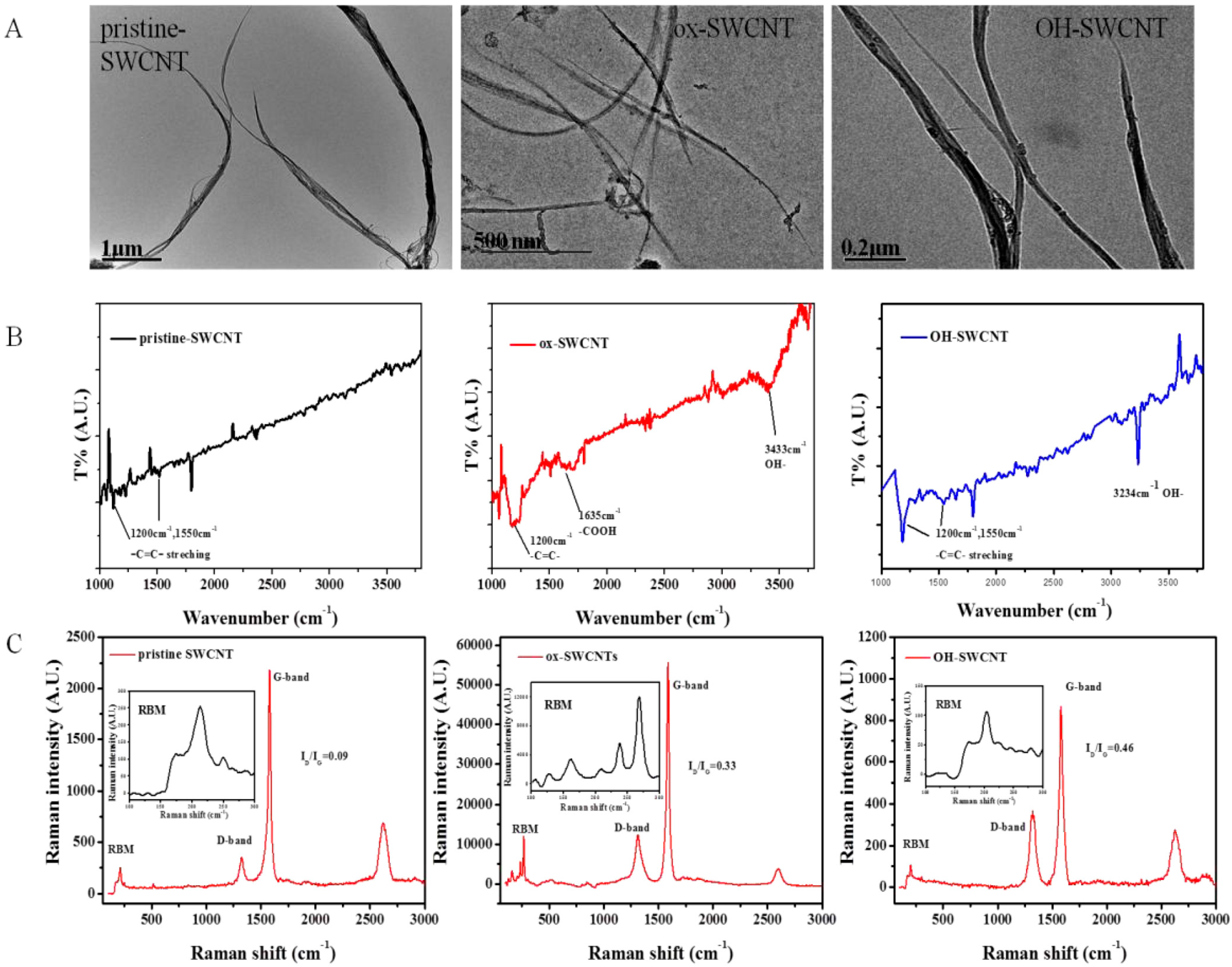
2.1. Characterization of Single-Walled Carbon Nanotubes (SWCNTs)

The characterization of SWCNTs by transmission electron microscopy (TEM) showed that pristine SWCNTs (p-SWCNTs), acid-oxidized SWCNTs (ox-SWCNTs), and hydroxylated SWCNTs (OH-SWCNTs) all presented as fibrillar shape and retained the structural integrity of carbon nanotubes (Figure 1A). Fourier transform infrared spectroscopy (FT-IR) analyses on these SWCNTs showed the presence of carboxyl groups (υ = 1635 cm−1, –COOH) and hydroxyl groups (υ = 3433 cm−1, –OH) on the surface of ox-SWCNTs, while only hydroxyl groups (υ = 3234 cm−1, –OH) were present on the surface of OH-SWCNTs and no functional group was found on the surface of p-SWCNTs (Figure 1B). In the Raman spectrum, the two characteristic peaks: disorder induced D-band at ~1340 cm−1 (±10) and tangential mode G-band at ~1580 cm−1 (±10) were identified and ID/IG ratios were shown as insets (Figure 1C) in the order of: OH-SWCNTs (ID/IG = 0.46) > ox-SWCNTs (0.33) > p-SWCNTs (0.09). The diameters and length of these SWCNTs were listed in supplementary Table S1.

2.2. Biodegradation of SWCNTs in Macrophages

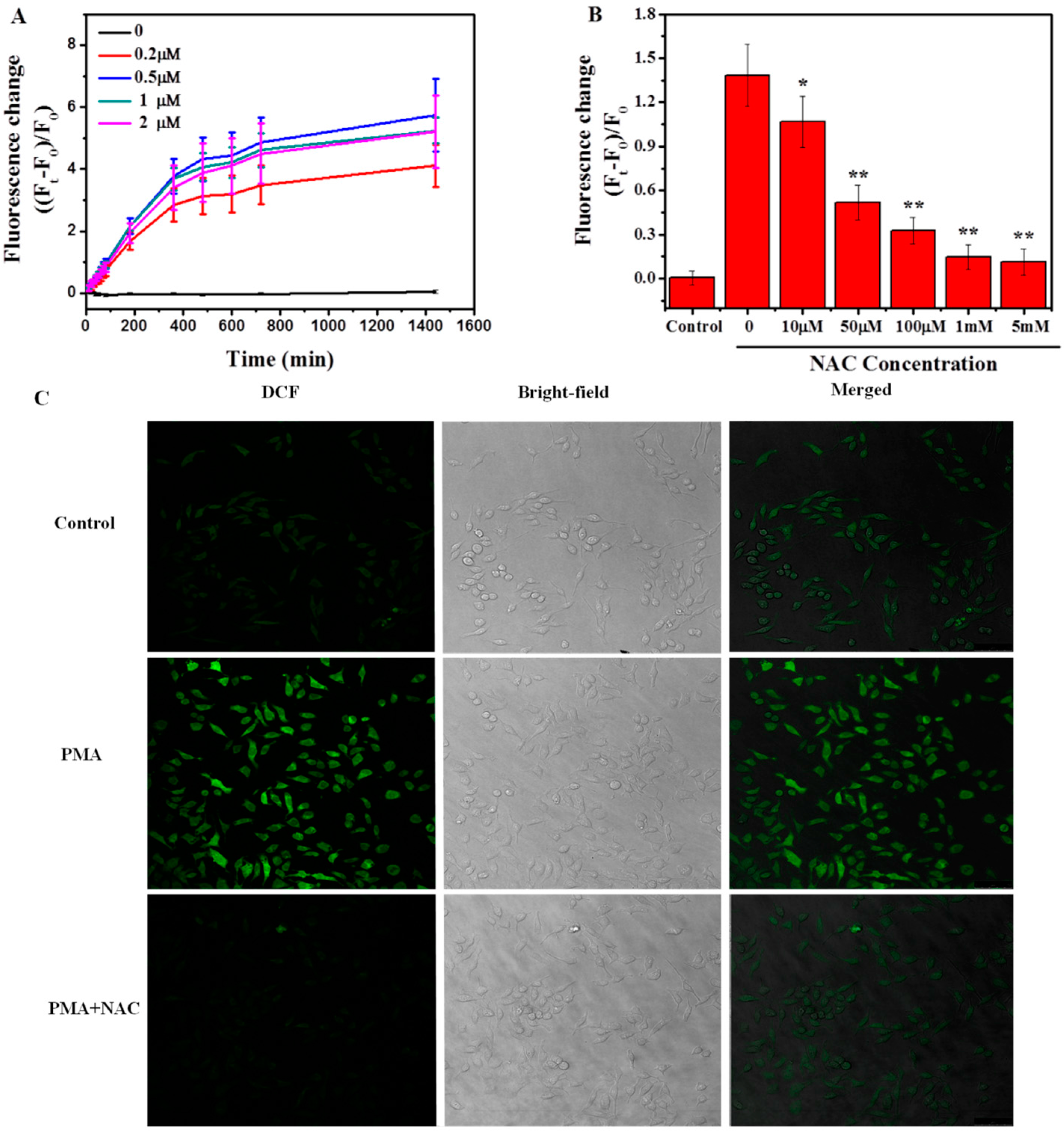
2.2.1. Induction of Respiratory Burst in Macrophages

The respiratory burst can be activated through the interaction between macrophages and microorganisms or by other stimuli including chemotactic factors and phorbol esters such asphorbol myristate acetate (PMA) [49,50,51]. In this study, we used PMA as the activator to induce respiratory burst in macrophages, and the anti-oxidant, N-acetyl-l-cysteine (NAC), as the inhibitor of the respiratory burst. The respiratory burst was evaluated through the measurement of reactive oxygen species (ROS) generation by using a 2′,7′-dichlorofluorescein diacetate (DCF-DA) assay. When cells were treated with different concentrations of PMA (0–2 μM), the fluorescence of intracellular DCF increased over time and 0.5 μM PMA has the highest intensity of DCF fluorescence, indicating the maximum generation of ROS and respiratory burst in macrophages under this treatment (Figure 2A). We further optimized the concentration of NAC pretreatment for inhibiting the respiratory burst. The results showed that NAC pretreatment could significantly inhibit the ROS production in a concentration-dependent manner (Figure 2B). One mM NAC pretreatment was selected for further experiments. In addition, the effects of PMA and NAC on the generation of ROS in macrophages were observed by direct imaging of DCF fluorescence signals in cells and are shown in Figure 2C.

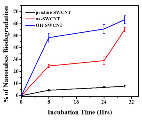
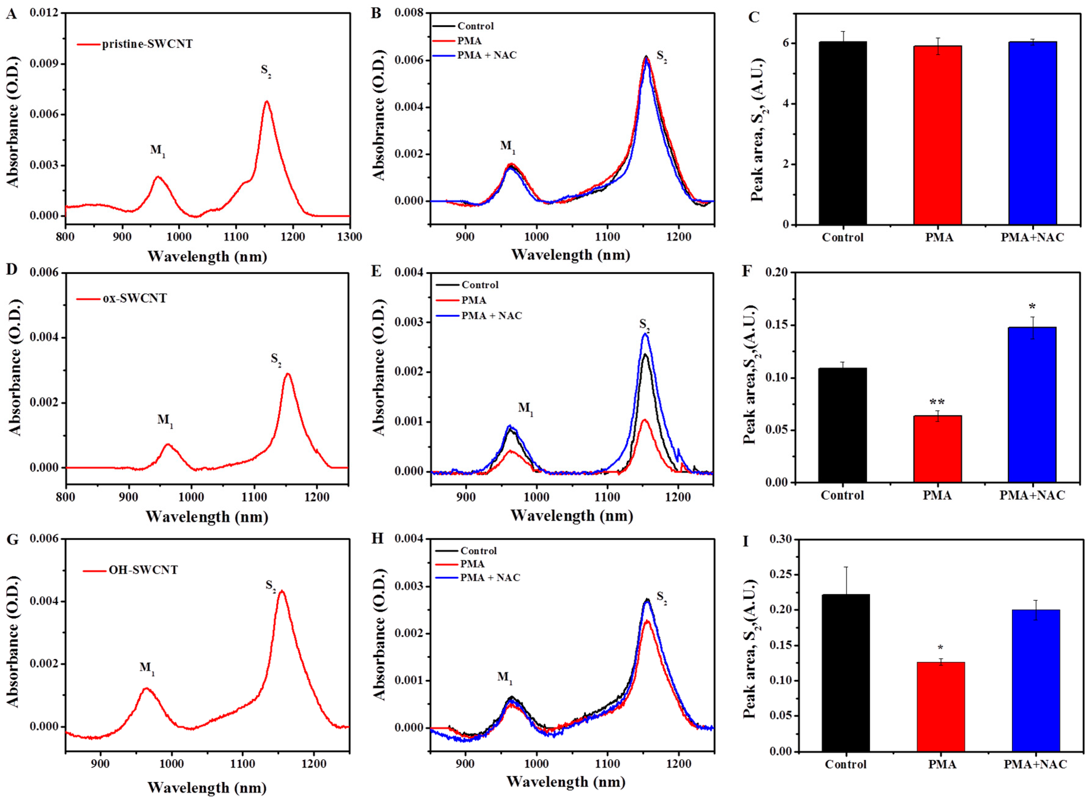
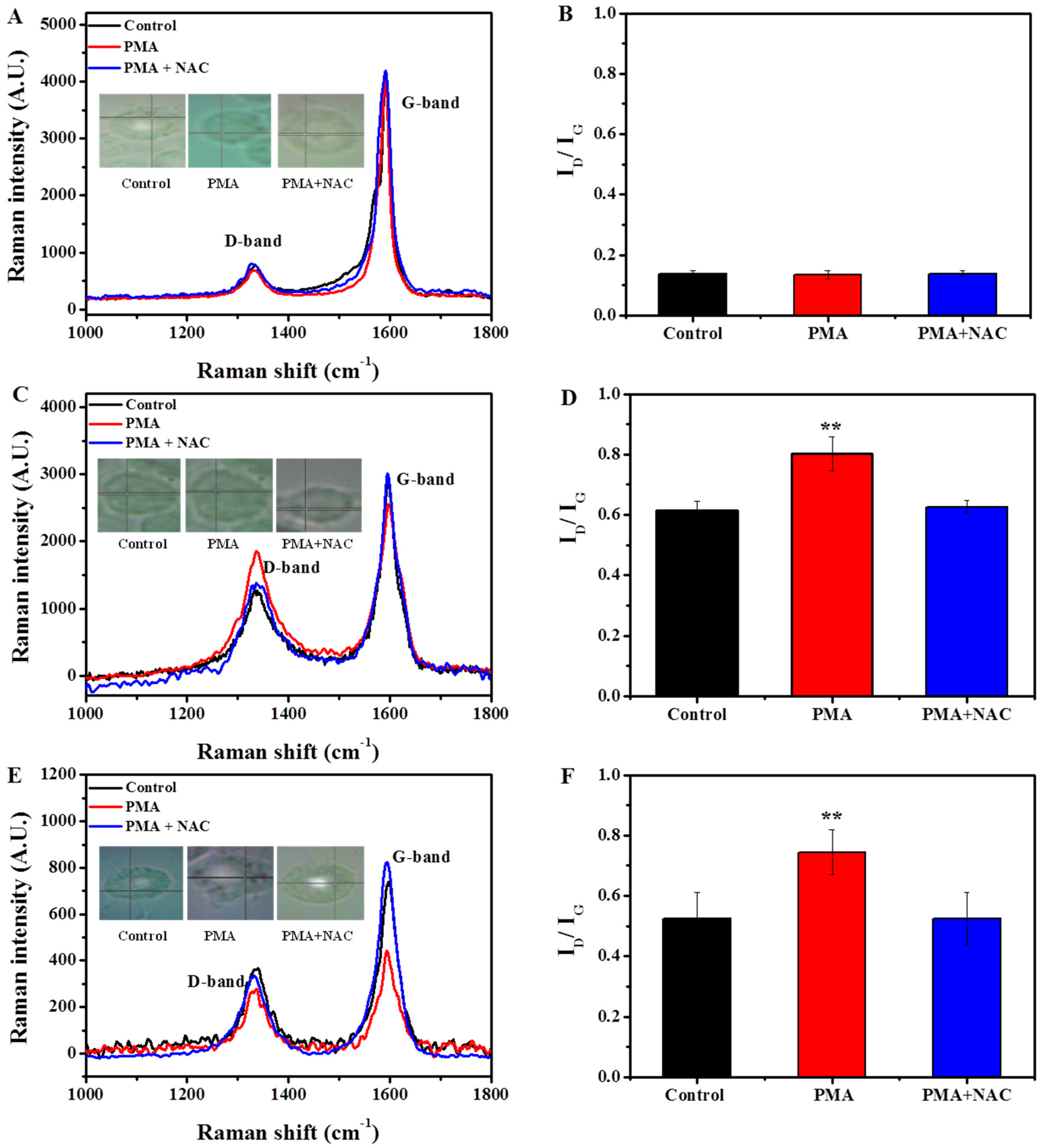
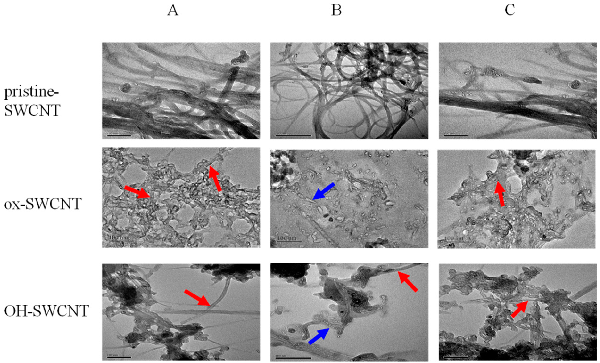
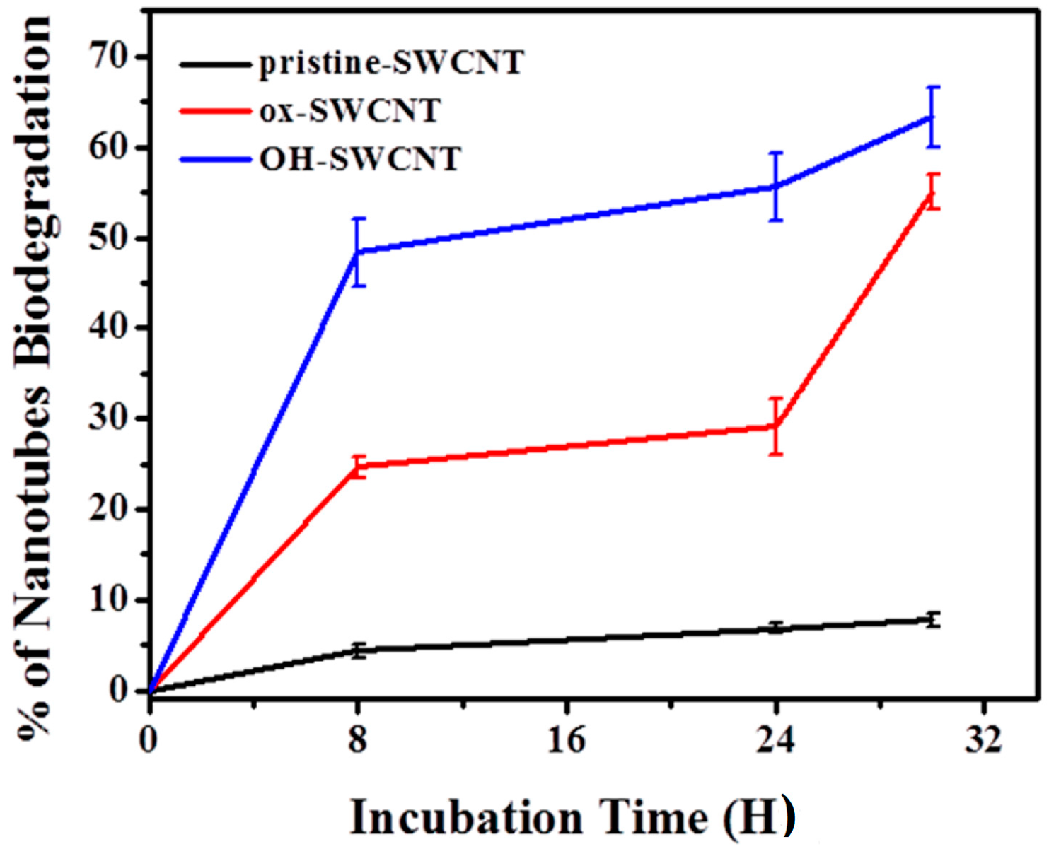
2.2.2. Characterization of SWCNTs Degradation in Macrophages

On the basis of the respiratory burst model, we exposed the macrophages to different types of SWCNTs and observed the degradation of these SWCNTs by using transmission electron microscopy (TEM), ultraviolet-visible-near-infrared (UV-vis-NIR) spectroscopy, and Raman spectroscopy [31]. The optical absorption of carbon nanotubes is a quick and facile method to obtain the information of nanotubes [52]. Because the SWCNTs are usually synthesized in the format of mixtures with various chiralities and diameters, they possess metallic and semiconducting properties. Since respiratory burst takes place mainly inside of cells, CNTs must be internalized first before degradation occurs, as evidenced in the present study (Figure S1). The treatment of p-SWCNTs, ox-SWCNTs, and OH-SWCNTs (5 µg/mL) in the presence or absence of PMA (0.5 µM) did not cause cell viability loss (Figure S2). In this study, we observed the typical UV-vis-NIR spectra of carbon nanotubes, showing the characteristic metallic band (M1, ~900–1000 nm) and semiconducting transition absorbing band (S2, ~1100–1200 nm) [53] (Figure 3A,D,G). The transitions related to the sp2- carbon framework of the SWCNTs will lessen when the oxidation of the graphitic structure occurs, and decreased absorbance of the band would indicate the degradation of carbon nanotubes [32]. We found a decrease in the absorbance of the metallic, especially the semiconducting band of SWCNTs in macrophages treated with PMA (Figure 3E,H, red lines), compared to those in untreated macrophages (black lines). After normalization against baseline and through the peak area calculation, we quantitatively detected the significant decrease in S2 band of ox- and OH-SWCNTs in PMA-treated macrophages (Figure 3F,I), suggesting different degrees of degradation of both SWCNTs. Interestingly, when cells were pretreated with 1 mM NAC, followed by PMA stimulation, the decreased absorbance of S2 band was alleviated significantly for OH-SWCNTs, and the even the absorbance was increased for ox-SWCNTs (Figure 3E,H, blue line), suggesting that anti-oxidant could inhibit the degradation of SWCNTs. No increase or decrease of either M1 or S2 band absorbance was observed for p-SWCNTs (Figure 3B,C), suggesting that p-SWCNTs are resistant to the biodegradation by cells, even when macrophages were in respiratory burst.
The damage of SWCNT structure was also characterized by Raman microscopy (Figure 4), which allows monitoring the evolution of the oxidation of the SWCNTs [33]. Compared to their corresponding original SWCNTs, ox-SWCNTs and OH-SWCNTs showed increased intensity of disorder induced D-band at ~1340 cm−1 (±10) whereas the tangential mode G-band at ~1580 cm−1 (±10) was decreased in untreated macrophages, indicating the oxidation damage on the graphene walls and the resulting of ID/IG ratios were increased from 0.33 to 0.63 (±0.02) for ox-SWCNTs, and from 0.46 to 0.53 (±0.05) for OH-SWCNTs (Figure 1C vs. Figure 4D; Figure 1C vs. Figure 4F). These data suggested that macrophages could normally degrade ox- and OH-SWCNTs. When macrophages were stimulated with PMA, the increase of ID/IG ratios was even prominent after 24 h, from 0.33 to 0.81 (±0.04) for ox-SWCNTs (Figure 1C and Figure 4D) and from 0.46 to 0.73 (±0.07) for OH-SWCNTs (Figure 1C and Figure 4F). The results suggested that PMA-induced respiratory burst in macrophages could accelerate the biodegradation of ox- and OH-SWCNTs, inducing more defect sites on SWCNTs. Meanwhile, we found that NAC pretreatment of cells could significantly attenuate the biodegradation of ox-SWCNTs and OH-SWCNTs, with the ID/IG ratios returned to control levels (Figure 4D,F). No above-mentioned effect was observed for p-SWCNTs. Taken together, our data support that macrophages can normally degrade ox-SWCNTs and OH-SWCNTs and the process can be speeded up significantly by PMA-induced respiratory burst in macrophages, probably through the generation of large amount of ROS, and thus anti-oxidant treatment with NAC can efficiently block the process.

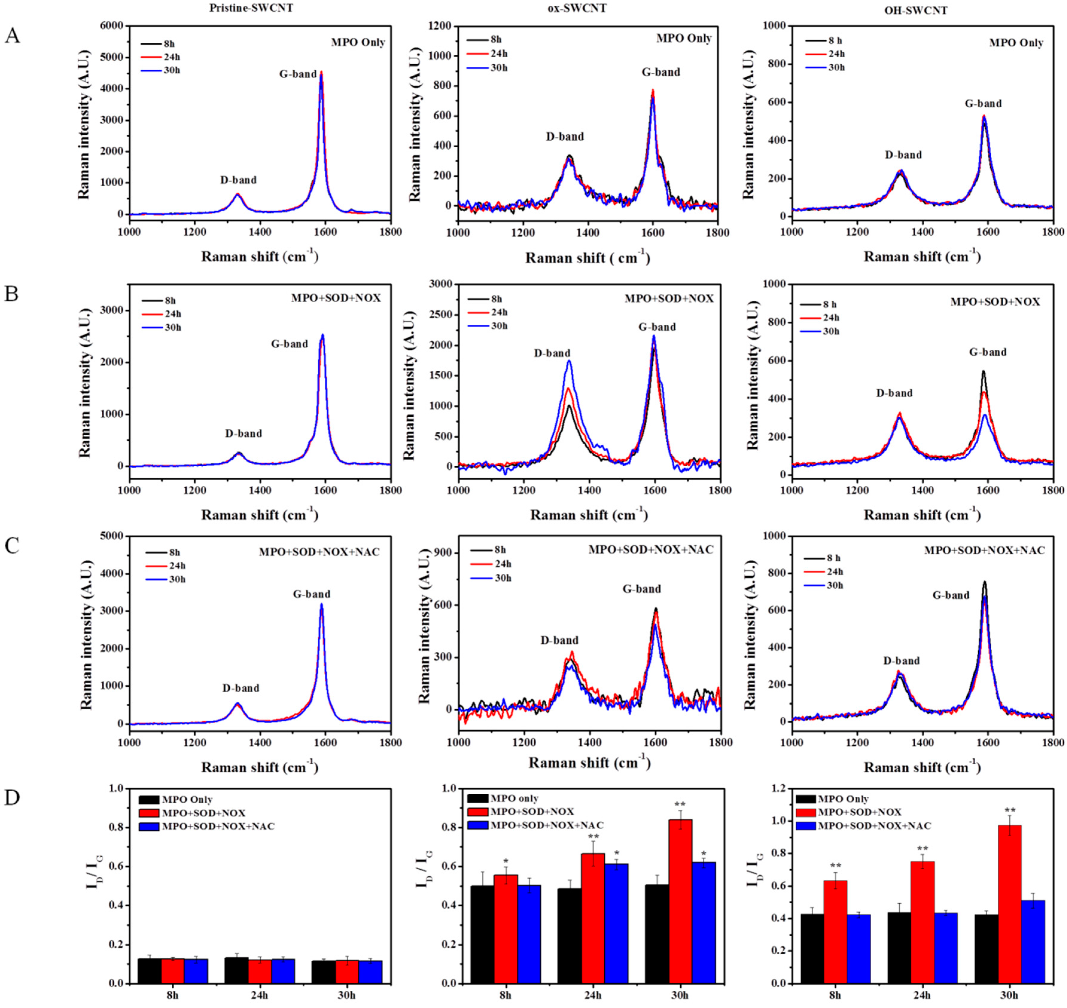
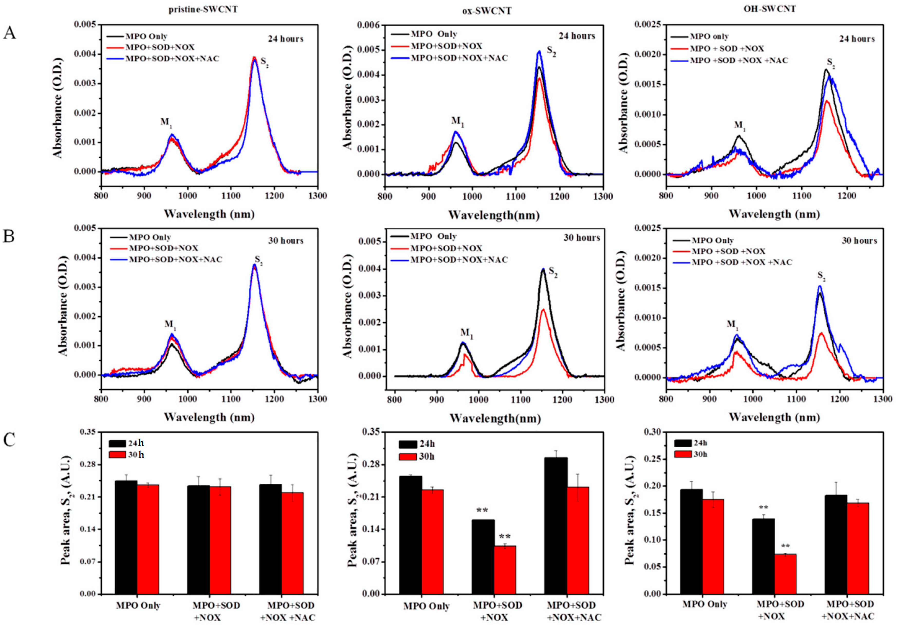
2.3. Biodegradation of SWCNTs in Respiratory Burst Enzymatic System in Vitro

To further confirm the role of respiratory burst in the degradation of SWCNTs, we constructed an enzymatic system to simulate the respiratory burst in vitro [54,55,56]. Respiratory burst is initiated by the activation of NOX (NADPH Oxidase), which is a complex enzyme consisting of five components including p40PHOX (PHOX for Phagocyte oxidase), p47PHOX, p67PHOX, p22PHOX and gp91PHOX. Once the cell is activated, the cytosolic subunits migrate to the membrane, where they bind to cytochrome b558 to assemble the active oxidase [57,58,59]. NOX catalyzes the generation of O2* from NADPH by transferring an electron to oxygen. Then the O2* is converted to H2O2 by superoxide dismutase (SOD). And the H2O2 serves as the substrate for peroxidase such as MPO in oxidating halides to generate hypohalous acids [44,60]. First, the construction of in vitro respiratory burst enzymatic reaction was evaluated by the measurement of O2* to test the activity of NOX. The NOX solution itself could not generate detectable amount of O2*, manifested as constant low level of the absorption of ferrocytochrome C at 550 nm (Figure 5A). However, the absorption of ferrocytochrome C increased over time after the addition of NADPH into the NOX solution, and the increase could be significantly inhibited by SOD (100 U/mL), suggesting that NOX was viable to produce O2* after supplement of NADPH (Figure 5A). Next, we optimized the concentration of NADPH by measuring the production of H2O2 in the NOX + SOD system supplemented with different concentrations of NADPH, and results showed that addition of 5 mM NADPH generated the maximum amount of H2O2 (Figure 5B). The data indicated that the in vitro respiratory burst enzymatic system was constructed successfully and could generate required substrates (H2O2) for MPO.
The nanotubes were previously shown to be degraded by hMPO in the presence of H2O2 [35,37,61]. hMPO can catalyze the production of even stronger oxidizing substances such as hypohalous acids in the presence of sodium chloride (NaCl) to degrade internalized particles and bacteria [44]. We therefore incubated SWCNTs in the in vitro enzymatic system with the addition of NaCl, and at different time points (8, 24, and 30 h) we sampled the SWCNTs to track the degradation process of SWCNTs. As shown in Figure 6, for p-SWCNTs, the Raman spectra have little or no change in the D-band and G-band intensity in MPO only, full enzyme solution (MPO + SOD + NOX) and NAC (MPO + SOD + NOX + NAC) groups, which means there is nearly no oxidation on p-SWCNTs. As for ox-SWCNTs and OH-SWCNTs, MPO alone caused no change in the Raman spectra, but in full enzyme solution, both types of SWCNTs exhibited the tendency of degradation as ID/IG ratios increased with time (Figure 6, ox-SWCNTs, OH-SWCNTs), to 0.67 (±0.05) and 0.85 (±0.04) for ox-SWCNTs at 24 and 30 h, respectively, and to 0.76 (±0.04) and 0.93 (±0.06) for OH-SWCNTs at 24 and 30 h, respectively, suggesting that defect sites of SWCNTs increased and the structure of nanotubes were damaged. However, the degradation process could be suppressed by the addition of NAC to the full enzyme solution, as shown in Figure 6 (MPO + SOD + NOX + NAC group), proving that SWCNT biodegradation induced by respiratory burst could be completely inhibited (OH-SWCNTs) or alleviated (ox-SWCNTs) by anti-oxidants that scavenge the ROS.
We further used UV-vis-NIR spectroscopy to assess the degradation of SWCNTs after 24 and 30 h incubation with enzyme solutions (Figure 7). Very similar to the results obtained from Raman spectrum analyses, we found essentially no change in the absorbance of S2 band of p-SWCNTs (Figure 7, pristine-SWCNTs). However, both ox-SWCNTs and OH-SWCNTs showed a significant decrease of the absorbance of S2 band in a time-dependent manner after being incubated with full enzyme solution, whereas the addition of NAC significantly inhibited the degradation process, with the peak area of S2 band back to the level comparable to that of MPO alone group (Figure 7C).
The relatively simple composition of an in vitro enzymatic system allows us to examine the morphological change of SWCNTs as a result of enzymatic degradation by using transmission electron microscopy (TEM) (Figure 8). Before incubation in the enzymatic system, all original SWCNTs samples appeared to have a fiber-like shape, as shown in Figure 1. After 24 h incubation, no morphological change was observed for p-SWCNTs, which still exist as long-fiber shaped tubes (Figure 8, pristine-SWCNTs). For ox-SWCNTs and OH-SWCNTs, MPO treatment alone did not induce apparent change of SWCNTs, whereas in full enzyme solution, both SWCNTs underwent serious structural damage, resulting in fragmentation and loss of fibrous structure. Most of the tubular structure of SWCNTs became undefined. In addition, a small amount of debris and pieces of nanotubes were observed (Figure 8B). Taken together, our data obtained using an in vitro respiratory burst enzymatic system suggests that MPO alone could barely degrade SWCNTs; full respiratory burst enzymatic system could only degrade the ox-SWCNTs and OH-SWCNTs but not p-SWCNTs. More importantly, the degradation process can be modulated by NAC.

3. Discussion

The biodegradability of carbon nanotubes may be a major determinant of the toxic outcomes in exposed individuals [35]. Peroxidases such as MPO and EPO have been demonstrated to be able to degrade SWCNTs [35,36], due to their capability of converting H2O2 and oxidating halides to generate hypohalous acids [59,60,62]. In mammalian systems, macrophages are ubiquitous, patrolling for invading pathogens, dying cells and particles. Most often CNTs exposed to individuals were found engulfed by macrophages [41]. Thus, macrophages constitute the potential target cells for modulating CNTs biodegradation. The respiratory burst in macrophages activates a series of enzymatic reactions involving the enzyme NADPH oxidase, SOD (Cu/ZnSOD and MnSOD), and MPO that ultimately produce hypochlorous acid (HClO). Therefore, activation of respiratory bursts in macrophages by PMA would accelerate the biodegradation of internalized SWCNTs, as shown in this study. More prominently, our results also demonstrated that due to the intrinsic property of ROS production during the respiratory burst, an anti-oxidant treatment (such as NAC) of cells could efficiently suppress the degradation process.
Recently, the reactive intermediate produced by respiratory burst, O2*, was also shown to react with nitric oxide (NO*) to generate a potent oxidant-peroxynitrite (ONOO), which plays an important role in macrophage biodegradation of SWCNTs [63]. Therefore, PMA-induced respiratory burst in macrophages may also activate the peroxynitrite-driven biodegradation of SWCNTs. To further confirm the role of respiratory burst in the biodegradation of SWCNTs, we constructed an in vitro simulating enzymatic fluid of respiratory burst to rule out the interference of peroxynitrite. The simulating fluid could generate sufficient amounts of reactive intermediates including O2* and H2O2 (Figure 5), to provide substrates for MPO. Very similar to the results obtained from PMA-activated macrophages, the in vitro system containing full enzymes could considerably degrade ox-SWCNTs and OH-SWCNTs, manifested as increased ID/IG ratios and decrease of S2 band. In addition, NAC supplemented to the system could cause apparent inhibition of the biodegradation of SWCNTs. These data suggested that respiratory burst played an important role in the biodegradation of SWCNTs, and the in vitro enzymatic system might be a useful tool to simulate the respiratory burst for other applications.
Previous studies showed a close relationship between the surface chemistry and the biodegradation of CNTs, and it seems that carboxylation on CNT surface is a prerequisite for CNT degradation [35]. The possible reason is because the carboxylation on CNTs might break the π-π interactions of CNTs and create two reactive sites that are potential sites for oxidative attack [64]. In this study, we compared three types of SWCNTs with different surface modifications, including p-SWCNTs, ox-SWCNTs, and OH-SWCNTs. The infrared spectra assessments revealed about 51% of OH-SWCNTs and 25% of ox-SWCNTs were degraded after 8 h incubation with an in vitro enzymatic system. After 30 h incubation, 63% of OH-SWCNTs and 53% of ox-SWCNTs were degraded. During the whole incubation, only negligible degradation (<5%) of pristine SWCNTs was observed (Figure 9). This suggests that except for p-SWCNTs carrying negligible defect sites, both ox-SWCNTs and OH-SWCNTs underwent serious structural damage and OH-SWCNTs were more susceptible to the oxidative attack than ox-SWCNTs. During the preparation of ox-SWCNTs, the acid oxidation of SWCNTs introduced both carboxyl and hydroxyl groups on the surface of SWCNTs, while OH-SWCNTs possess hydroxyl groups only (Figure 1B). It is difficult to differentiate the contribution of the carboxyl and hydroxyl groups to the biodegradation of SWCNTs. However, our results at least illustrated that oxygen-functionalized SWCNTs were biodegradable. Alternatively, the higher biodegradation of OH-SWCNTs may result from the fact that OH-SWCNTs originally possess higher level of defect sites than ox-SWCNTs do (Figure 1C), and thus provide more points for oxidative attack.
We observed 27% degradation of ox-SWCNTs after 24 h incubation with the in vitro enzymatic system (Figure 9). In another in vitro hMPO system reported by Kagan et al. [35], the degradation of IgG-coated oxidized SWCNTs seems to be much higher (solution turned translucent in 24 h), probably due to the constant higher concentrations of H2O2 kept throughout their experiments by frequent (every 5 h) replenishment of hMPO and H2O2. We envision that by increasing the frequency and concentration of NADPH and MPO added, higher degradation efficiency of the in vitro enzymatic system would be achieved. Interestingly, based on the absorbance of S2 band, the 24 h degradation of ox-SWCNTs in untreated macrophages is very similar to that reported by Kagan et al. [35] (25.8% in this study vs. ~20%). Compared with ox-SWCNTs, 35% of OH-SWCNTs were degraded under the same condition, further confirming that OH-SWCNTs were more biodegradable than ox-SWCNTs. With PMA treatment, macrophages degraded up to 67% of ox-SWCNTs in 24 h (Figure 3F), which is comparable to the efficiency of neutrophils that were shown to degrade ~30% of ox-SWCNTs (no IgG coating) within 12 h [35], suggesting that through proper stimulation, the biodegradation of SWCNTs by macrophages would be elevated to that by neutrophils.
Taken together, we have demonstrated both ways of accelerating and slowing down the biodegradation of SWCNTs in macrophages by modulating the respiratory burst. In addition, SWCNTs with defect sites were demonstrated to be more prone to be biodegraded by macrophages. Therefore, by careful functionalization of SWCNTs with proper coating that modulates the macrophage response, one can design better SWCNTs products with controlled retention time and toxicity. Alternatively, for drug delivery, modulating the biodegradation of nanocarriers may facilitate the release of the payloads as needed.

4. Materials and Methods

4.1. Materials

Pristine SWCNTs (p-SWCNTs, CNTs purity > 95%, SWCNT purity > 90%, ash < 5 weight %) and hydroxylated SWCNTs (OH-SWCNTs, purity > 90%) synthesized by chemical vapor deposition (CVD) method were originally obtained from Cheng du Organic Chemicals Co. (Cheng du, China). The following reagents were all purchased from Sigma (St. Louis, MO, USA): Phorbol myristate acetate (PMA), 2′,7′-dichlorofluorescein diacetate (DCF-DA), ferricytochrome c, β-nicotinamide adenine dinucleotide 2′-phosphate reduced tetrasodium salt hydrate(NADPH), bovine erythrocyte superoxide dismutase (SOD), EGTA, Flavin adenine dinucleotide disodium salt hydrate (FAD), Myeloperoxidase (MPO), N-acetyl-l-cysteine (NAC).

4.2. Preparation of SWCNTs Solutions and Characterization

Acid-oxidized SWCNTs (ox-SWCNTs) were prepared according to the procedure described previously [65,66,67]. In detail, 10 mg of SWCNTs were suspended in 40 mL of a 3:1 mixture of concentrated H2SO4/HNO3 in a 100 mL test tube and sonicated in a water bath (KQ-500DV, 100 kHz, 60%) for 24 h at 40–50 °C. The resultant suspension was then diluted with 200 mL deionized water and filtered through a membrane (pore size 0.22 µm), followed by wash with 50 mL deionized water on the membrane. The acid-oxidized nanotubes were re-suspended in deionized water at a concentration of 1 mg/mL with brief sonication (KQ-50DE, 40 kHz, 2 min). The resultant ox-SWCNT suspension was black, well dispersed and had a neutral pH. The stock solutions of 1 mg/mL pristine SWCNTs (p-SWCNTs) and hydroxylated SWCNTs (OH-SWCNTs) were prepared separately by mixing SWCNTs with cell culture medium, followed by sonication for 5 min on ice using a ultrasonic probe tip sonicator (JY92-IIN, Ningbo Scientz Biotechnology Co., Ltd. Ningbo, China).
For transmission electron microscopy (TEM) characterization, SWCNTs were diluted to 0.01 mg/mL and precipitated onto a copper net and dried for imaging with a Hitachi H-7500 transmission electron microscopy at 80 kv (Tokyo, Japan). In addition, the infrared spectra of SWCNTs were collected by using a FT-IR spectrometer (JASCO, Inc., Easton, MD, USA). Raman spectroscopy analyses were performed on an inVia Raman spectroscopy (Renishaw Plc., Gloucestershire, UK) with a 633 nm wavelength laser source.

4.3. Cell Culture and Treatment

The murine monocytic cell line Raw264.7 was obtained from American Type Culture Collection (ATCC: TIB-71) and cultured in a humidified incubator with 95% air and 5% CO2 at 37 °C in complete culture medium(cRPMI) containing RPMI-1640 fundamental medium and 100 U · mL−1 penicillin/streptomycin, supplement with 10% heat deactivated fetal bovine serum (FBS). All ingredients for the media were purchased from Hyclone Inc. (Waltham, MA, USA). For optimization of respiratory burst induction in macrophages, cells were seeded in a 96-well plate at a density of 2 × 10⁵ cells/mL and allowed to attach for overnight. Next, cells were loaded with 10 μM of 2′,7′-dichlorofluorescein diacetate (DCF-DA, a probe for intracellular ROS detection) at 37 °C for 30 min in dark. After wash with PBS twice, cells were treated with various concentrations (0–2 µM) of phorbol myristate acetate (PMA, a respiratory burst inducer [51,68]), and the fluorescence of DCF in cells was measured on a Thermo Varioskan Flash microplate reader (Winooski, VT, USA) at the excitation/emission wavelength 488 nm/530 nm very 10 s for the first 90 min and at time points of 3, 6, 9, 12, and 24 h. To optimize the concentration of N-acetyl-l-cysteine (NAC) for the inhibition of respiratory burst, cells were first pretreated with various concentrations (0–5 mM) of NAC for 1 h and then loaded with DCF-DA. After, cells were treated by PMA (0.5 µM) for 3 h in the presence of NAC (0–5 mM) and fluorescence detection was performed on the Varioskan Flash microplate reader. Furthermore, the DCF fluorescence in cells was also imaged with a Leica TCS SP5 confocal laser-scanning microscope (Mannheim, Germany) with a laser excitation at 488 nm and emission at 530 nm. For SWCNT treatment, cells (2 × 10⁵ cells/mL) were seeded in culture dishes (60 mm in diameter) and allowed to attach for overnight. Then, 15 µg of SWCNTs (p-SWCNTs, ox-SWCNTs, and OH-SWCNTs) were added to the cell culture along with PMA (0.5 µM). Cells with NAC pretreatment (1 mM), followed by the addition of PMA (0.5 µM) were set to investigate the inhibitory effect of antioxidant.

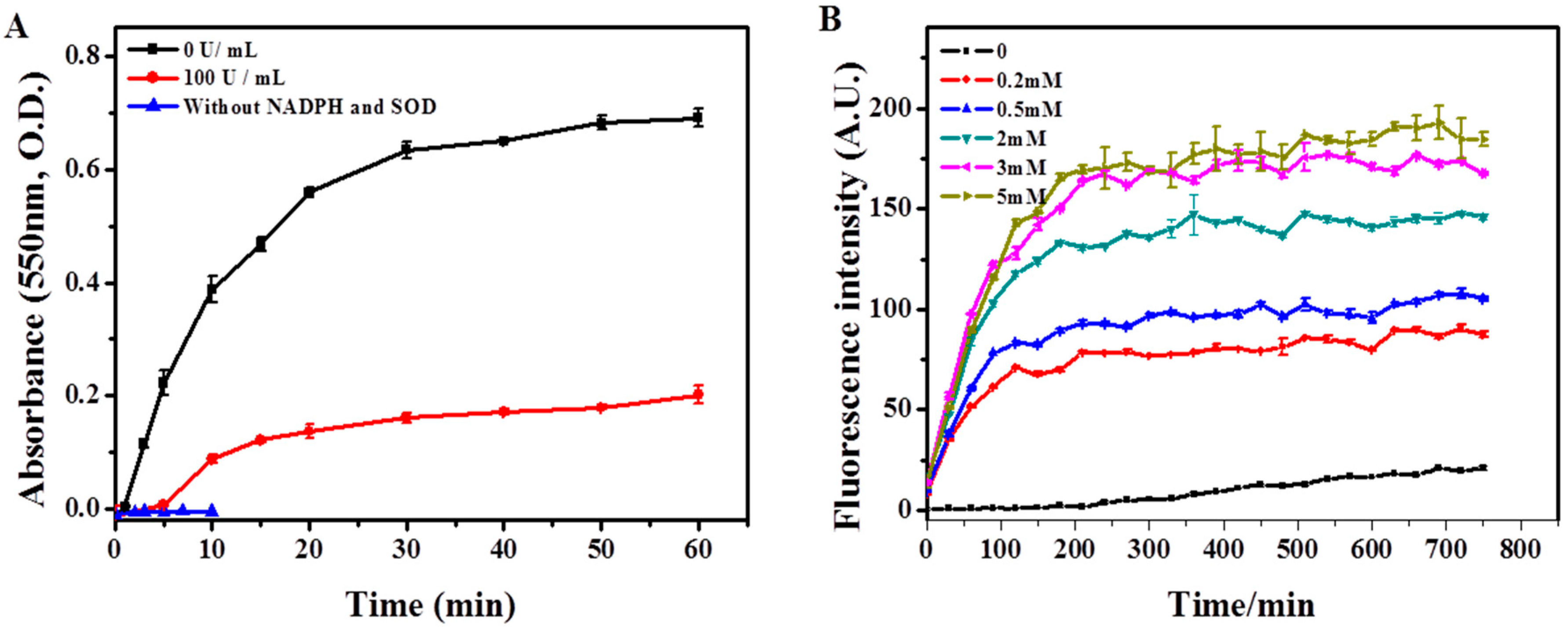
4.4. Construction of in Vitro Respiratory Burst Enzymatic System and Verification

First, NADPH oxidase (NOX) was isolated according to the method described previously [54,55,56]. In detail, the macrophages were suspended to a concentration of 0.5–1.5 × 10⁸ cells/mL in a phosphate-sucrose buffer (1× PBS buffer, pH 7.0, and 340 mM sucrose). Cell disruption was performed by 5 min sonication using an ultrasonic probe tip sonicator (JY92-IIN, Ningbo Scientz Biotechnology Co., Ltd., Ningbo, China) in a 1.5 mL tube on ice. Then, the homogenate was centrifuged at 800× g for 10 min at 4 °C to remove unbroken cells, nuclei, and large debris. The supernatant supplemented with EGTA (1 mM), and flavin adenine dinucleotide disodium salt hydrate (FAD, 0.1 mM) was used as the NADPH Oxidase (NOX). Thereafter, the second enzyme, SOD (300 U/mL) was added into the mixture, followed by the addition of NADPH (5 mM), NaCl (140 mM) and MPO (1 U). The full enzyme solution (total volume 500 µL) was then incubated at 37 °C with shaking (80 rpm) and replenishment of MPO (1 U) and NADPH (40 mM) every 8 h in dark. For SWCNTs incubation, 8 µg of SWCNTs per sample were mixed with the enzyme solution for indicated time, and then the samples were washed by centrifugation at 3400 rpm for 1 h twice and re-suspended in deionized water before Raman, UV-vis-NIR and TEM characterization.
The efficiency of this enzymatic system was evaluated by monitoring the generation of O2* and H2O2 following the presence of NOX and SOD in the system, respectively. O2* was measured by the method of ferricytochrome c reduction, as described by Bellavite et al. [69]. In detail, the 60 µL NOX solution were mixed with 0.94 mL phosphate-sucrose buffer containing ferricytochrome c (0.1 mM) and freshly prepared NADPH solution (0.2 mM). The absorption of ferrocytochrome C, the product of ferricytochrome C reduction, was measured at 550 nm on a UV-visible spectrophotometer (Agilent 8453, Santa Clara, CA, USA). The H2O2 was detected by using a Fluorimetric Hydrogen Peroxide Assay Kit (Sigma, St. Louis, MO, USA), according to manufacturer’s instruction.

4.5. Assessment of SWCNT Degradation

Raman spectroscopy. For cell samples, macrophages incubated with SWCNTs were fixed using 4% paraformaldehyde (4% PFA) onto a glass bottom cell culture dish (Nest Scientific, Rahway, NJ, USA). For samples from in vitro enzymatic system, 30 µL of solution were dropped on a monocrystalline silicon wafer and allowed to dry overnight. An inVia Raman microscope spectrometer (Renishaw Plc., Gloucestershire, UK) with a 633 nm laser source was used for all samples, spectrum was obtained over the range of 800 to 1800 cm−1 to visualize D and G band intensity changes throughout the degradation process. All Raman spectra were recorded for 10 s, 10% laser power, using a 50 L objective. For each sample, at least 20 spectra were recorded.
Infrared spectroscopy (UV-vis-NIR). For cell samples, macrophages incubated with SWCNTs were harvested together with the incubation medium. The cell suspensions were centrifuged (3400 rpm, 1 h) and resuspended in deionized water. Samples were further subjected to sonication for 20 min using a ultrasonic probe tip sonicator (JY92-IIN, Ningbo Scientz Biotechnology Co., Ltd. Zhejiang, China), and washed twice with PBS by centrifugation at 3400 rpm for 1 h to remove the cellular components before assessment of SWCNT degradation. The UV-vis-NIR spectra were acquired by using a Cary 5000 UV-Vis-NIR spectrophotometer (Varian, Palo Alto, USA).
Transmission Electron Microscopy (TEM). SWCNTs were mixed with the enzyme solution for 24 h, and the samples then were washed by centrifugation at 3400 rpm for 1 h twice and resuspended in deionised water. Five µL of prepared sample were precipitated onto a copper net and dried for imaging with a Hitachi H-7500 transmission electron microscopy at 80 kv (Tokyo, Japan).

4.6. Statistical Analysis

The data were expressed as the mean ± S.D., and the difference between groups was evaluated using Student’s t-test, with the significance level set at * p < 0.05 or ** p < 0.01.

5. Conclusions

In the present study, we explored and compared ways of modulating the biodegradation of three types of SWCNTs (p-, ox-, and OH-SWCNTs) in macrophages and in an in vitro enzymatic system simulating respiratory burst. The results demonstrated that: (1) SWCNTs carrying defect sites (ox-SWCNTs and OH-SWCNTs) could be degraded by macrophages, whereas p-SWCNTs were resistant to biodegradation probably due to the lack of reactive sites for oxidative attack; the order of degradation rate is OH-SWCNTs > ox-SWCNTs >> p-SWCNTs; (2) The biodegradation of SWCNTs could be significantly accelerated by PMA-activation in macrophages, or inhibited by an anti-oxidant, NAC; (3) Respiratory burst played an important role in accelerating the biodegradation of SWCNTs, as revealed by using the in vitro enzymatic system simulating respiratory burst. In addition, the in vitro enzymatic system constructed in the present study was proven to be able to simulate the enzymatic reaction cycles of respiratory burst for the effective degradation of SWCNTs. In principle, the enzymatic system can be expanded to study the degradation of other nanomaterials. Our findings on the biodegradation of functionalized SWCNTs would help to design safer SWCNT consumer products that are biodegradable and less toxic to living organisms, as well as drug carriers with controllable release of its payload. The ways of modulating the biodegradation of SWCNTs as we revealed might also provide invaluable clues on the development of intervention measurements for relieving the side effects of SWCNTs.

Supplementary Materials

Supplementary materials can be found at https://0-www-mdpi-com.brum.beds.ac.uk/1422-0067/17/3/409/s1.

Acknowledgments

The Chinese Academy of Sciences (XDB14040101) and the National Natural Science Foundation of China (21477146, 21277158, 91543203, 21321004, 21377142, and 21407168) supported this research.

Author Contributions

All authors read and approved the final manuscript. Bin Wan and Liang-Hong Guo conceived and designed the experiments; Jie Hou performed the experiments and analyzed the data; Yu Yang and Xiao-Min Ren contributed reagents/materials/analysis tools; Bin Wan and Jie Hou wrote the paper. Jing-Fu Liu helped with the Raman detection.

Conflicts of Interest

The authors declare no conflict of interest.

Abbreviations

SWCNT
Single-walled carbon nanotube
HRP
Horse radish peroxidase
MPO
Myeloperoxidase
EPO
Eosinophil peroxidase
NETs
Neutrophil extracellular traps
PMN
Polymorphonuclear neutrophils
O2*
Superoxide anion
NOX
Nicotinamide adenine dinucleotide phosphate (NADPH)-oxidase
SOD
Superoxide dismutase
PMA
Phorbol myristate acetate
LPS
Lipopolysaccharide
NAC
N-acetyl-l-cysteine
ROS
Reactive oxygen species
DCF-DA
2′,7′-Dichlorofluorescein diacetate
TEM
Transmission electron microscopy
UV-vis-NIR
Ultraviolet-visible-near-infrared
FAD
Flavin adenine dinucleotide disodium salt hydrate

References

  1. De Volder, M.F.L.; Tawfick, S.H.; Baughman, R.H.; Hart, A.J. Carbon nanotubes: Present and future commercial applications. Science 2013, 339, 535–539. [Google Scholar] [CrossRef] [PubMed]
  2. Marchesan, S.; Kostarelos, K.; Bianco, A.; Prato, M. The winding road for carbon nanotubes in nanomedicine. Mater. Today 2015, 18, 12–19. [Google Scholar] [CrossRef]
  3. Marchesan, S.; Melchionna, M.; Prato, M. Wire up on carbon nanostructures! How to play a winning game. ACS Nano 2015, 9, 9441–9450. [Google Scholar] [CrossRef] [PubMed] [Green Version]
  4. Karimi, M.; Solati, N.; Amiri, M.; Mirshekari, H.; Mohamed, E.; Taheri, M.; Hashemkhani, M.; Saeidi, A.; Estiar, M.A.; Kiani, P.; et al. Carbon nanotubes part I: Preparation of a novel and versatile drug-delivery vehicle. Expert Opin. Drug Deliv. 2015, 12, 1071–1087. [Google Scholar] [CrossRef] [PubMed]
  5. Martincic, M.; Tobias, G. Filled carbon nanotubes in biomedical imaging and drug delivery. Expert Opin. Drug Deliv. 2015, 12, 563–581. [Google Scholar] [CrossRef] [PubMed]
  6. Chen, X.; Kis, A.; Zettl, A.; Bertozzi, C.R. A cell nanoinjector based on carbon nanotubes. Proc. Natl. Acad. Sci. USA 2007, 104, 8218–8222. [Google Scholar] [CrossRef] [PubMed]
  7. Malarkey, E.B.; Parpura, V. Applications of carbon nanotubes in neurobiology. Neurodegener. Dis. 2007, 4, 292–299. [Google Scholar] [CrossRef] [PubMed]
  8. Karimi, M.; Solati, N.; Ghasemi, A.; Estiar, M.A.; Hashemkhani, M.; Kiani, P.; Mohamed, E.; Saeidi, A.; Taheri, M.; Avci, P.; et al. Carbon nanotubes part II: A remarkable carrier for drug and gene delivery. Expert Opin. Drug Deliv. 2015, 12, 1089–1105. [Google Scholar] [CrossRef] [PubMed]
  9. Prato, M.; Kostarelos, K.; Bianco, A. Functionalized carbon nanotubes in drug design and discovery. Acc. Chem. Res. 2008, 41, 60–68. [Google Scholar] [CrossRef] [PubMed]
  10. Wong, B.S.; Yoong, S.L.; Jagusiak, A.; Panczyk, T.; Ho, H.K.; Ang, W.H.; Pastorin, G. Carbon nanotubes for delivery of small molecule drugs. Adv. Drug Deliv. Rev. 2013, 65, 1964–2015. [Google Scholar] [CrossRef] [PubMed]
  11. Lamberti, M.; Zappavigna, S.; Sannolo, N.; Porto, S.; Caraglia, M. Advantages and risks of nanotechnologies in cancer patients and occupationally exposed workers. Expert Opin. Drug Deliv. 2014, 11, 1087–1101. [Google Scholar] [CrossRef] [PubMed]
  12. Schulte, P.A.; Kuempel, E.D.; Zumwalde, R.D.; Geraci, C.L.; Schubauer-Berigan, M.K.; Castranova, V.; Hodson, L.; Murashov, V.; Dahm, M.M.; Ellenbecker, M. Focused actions to protect carbon nanotube workers. Am. J. Ind. Med. 2012, 55, 395–411. [Google Scholar] [CrossRef] [PubMed]
  13. Maynard, A.D.; Kuempel, E.D. Airborne nanostructured particles and occupational health. J. Nanopart. Res. 2005, 7, 587–614. [Google Scholar] [CrossRef]
  14. Porter, A.E.; Gass, M.; Muller, K.; Skepper, J.N.; Midgley, P.A.; Welland, M. Direct imaging of single-walled carbon nanotubes in cells. Nat. Nanotechnol. 2007, 2, 713–717. [Google Scholar] [CrossRef] [PubMed]
  15. Tong, L.; Liu, Y.; Dolash, B.D.; Jung, Y.; Slipchenko, M.N.; Bergstrom, D.E.; Cheng, J.X. Label-free imaging of semiconducting and metallic carbon nanotubes in cells and mice using transient absorption microscopy. Nat. Nanotechnol. 2012, 7, 56–61. [Google Scholar] [CrossRef] [PubMed]
  16. Al-Jamal, K.T.; Nunes, A.; Methven, L.; Ali-Boucetta, H.; Li, S.; Toma, F.M.; Herrero, M.A.; Al-Jamal, W.T.; ten Eikelder, H.M.M.; Foster, J.; Mather, S.; Prato, M.; et al. Degree of chemical functionalization of carbon nanotubes determines tissue distribution and excretion profile. Angew. Chem. Int. Ed. 2012, 51, 6389–6393. [Google Scholar] [CrossRef] [PubMed]
  17. Lam, C.W.; James, J.T.; McCluskey, R.; Hunter, R.L. Pulmonary toxicity of single-wall carbon nanotubes in mice 7 and 90 days after intratracheal instillation. Toxicol. Sci. 2004, 77, 126–134. [Google Scholar] [CrossRef] [PubMed]
  18. Shi, X.H.; von dem Bussche, A.; Hurt, R.H.; Kane, A.B.; Gao, H.J. Cell entry of one-dimensional nanomaterials occurs by tip recognition and rotation. Nat. Nanotechnol. 2011, 6, 714–719. [Google Scholar] [CrossRef] [PubMed]
  19. Jia, G.; Wang, H.F.; Yan, L.; Wang, X.; Pei, R.J.; Yan, T.; Zhao, Y.L.; Guo, X.B. Cytotoxicity of carbon nanomaterials: Single-wall nanotube, multi-wall nanotube, and fullerene. Environ. Sci. Technol. 2005, 39, 1378–1383. [Google Scholar] [CrossRef] [PubMed]
  20. Magrez, A.; Kasas, S.; Salicio, V.; Pasquier, N.; Seo, J.W.; Celio, M.; Catsicas, S.; Schwaller, B.; Forro, L. Cellular toxicity of carbon-based nanomaterials. Nano Lett. 2006, 6, 1121–1125. [Google Scholar] [CrossRef] [PubMed]
  21. Firme, C.P.; Bandaru, P.R. Toxicity issues in the application of carbon nanotubes to biological systems. Nanomed. Nanotechnol. Biol. Med. 2010, 6, 245–256. [Google Scholar] [CrossRef] [PubMed]
  22. Warheit, D.B.; Laurence, B.R.; Reed, K.L.; Roach, D.H.; Reynolds, G.A.M.; Webb, T.R. Comparative pulmonary toxicity assessment of single-wall carbon nanotubes in rats. Toxicol. Sci. 2004, 77, 117–125. [Google Scholar] [CrossRef] [PubMed]
  23. Shvedova, A.A.; Kisin, E.; Murray, A.R.; Johnson, V.J.; Gorelik, O.; Arepalli, S.; Hubbs, A.F.; Mercer, R.R.; Keohavong, P.; Sussman, N.; et al. Inhalation vs. aspiration of single-walled carbon nanotubes in C57BL/6 mice: Inflammation, fibrosis, oxidative stress, and mutagenesis. Am. J. Physiol. Lung Cell. Mol. Physiol. 2008, 295, L552–L565. [Google Scholar] [CrossRef] [PubMed]
  24. Ali-Boucetta, H.; Nunes, A.; Sainz, R.; Herrero, M.A.; Tian, B.; Prato, M.; Bianco, A.; Kostarelos, K. Asbestos-like pathogenicity of long carbon nanotubes alleviated by chemical functionalization. Angew. Chem. Int. Ed. 2013, 52, 2274–2278. [Google Scholar] [CrossRef] [PubMed]
  25. Kagan, V.E.; Tyurina, Y.Y.; Tyurin, V.A.; Konduru, N.V.; Potapovich, A.I.; Osipov, A.N.; Kisin, E.R.; Schwegler-Berry, D.; Mercer, R.; Castranova, V.; et al. Direct and indirect effects of single walled carbon nanotubes on raw 264.7 macrophages: Role of iron. Toxicol. Lett. 2006, 165, 88–100. [Google Scholar] [CrossRef] [PubMed]
  26. Kisin, E.R.; Murray, A.R.; Keane, M.J.; Shi, X.C.; Schwegler-Berry, D.; Gorelik, O.; Arepalli, S.; Castranova, V.; Wallace, W.E.; Kagan, V.E.; et al. Single-walled carbon nanotubes: Geno- and cytotoxic effects in lung fibroblast v79 cells. J. Toxicol. Environ. Health A 2007, 70, 2071–2079. [Google Scholar] [CrossRef] [PubMed]
  27. Jain, S.; Thakare, V.S.; Das, M.; Godugu, C.; Jain, A.K.; Mathur, R.; Chuttani, K.; Mishra, A.K. Toxicity of multiwalled carbon nanotubes with end defects critically depends on their functionalization density. Chem. Res. Toxicol. 2011, 24, 2028–2039. [Google Scholar] [CrossRef] [PubMed]
  28. Ji, Z.; Zhang, D.; Li, L.; Shen, X.; Deng, X.; Dong, L.; Wu, M.; Liu, Y. The hepattoxicity of multi-walled carbon nanotubes in mice. Nanotechnology 2009, 20. [Google Scholar] [CrossRef] [PubMed]
  29. Sureshbabu, A.R.; Kurapati, R.; Russier, J.; Menard-Moyon, C.; Bartolini, I.; Meneghetti, M.; Kostarelos, K.; Bianco, A. Degradation-by-design: Surface modification with functional substrates that enhance the enzymatic degradation of carbon nanotubes. Biomaterials 2015, 72, 20–28. [Google Scholar] [CrossRef] [PubMed]
  30. Kotchey, G.P.; Zhao, Y.; Kagan, V.E.; Star, A. Peroxidase-mediated biodegradation of carbon nanotubes in vitro and in vivo. Adv. Drug Deliv. Rev. 2013, 65, 1921–1932. [Google Scholar] [CrossRef] [PubMed]
  31. Allen, B.L.; Kichambare, P.D.; Gou, P.; Vlasova, I.I.; Kapralov, A.A.; Konduru, N.; Kagan, V.E.; Star, A. Biodegradation of single-walled carbon nanotubes through enzymatic catalysis. Nano Lett. 2008, 8, 3899–3903. [Google Scholar] [CrossRef] [PubMed]
  32. Allen, B.L.; Kotchey, G.P.; Chen, Y.; Yanamala, N.V.K.; Klein-Seetharaman, J.; Kagan, V.E.; Star, A. Mechanistic investigations of horseradish peroxidase-catalyzed degradation of single-walled carbon nanotubes. J. Am. Chem. Soc. 2009, 131, 17194–17205. [Google Scholar] [CrossRef] [PubMed]
  33. Russier, J.; Menard-Moyon, C.; Venturelli, E.; Gravel, E.; Marcolongo, G.; Meneghetti, M.; Doris, E.; Bianco, A. Oxidative biodegradation of single- and multi-walled carbon nanotubes. Nanoscale 2011, 3, 893–896. [Google Scholar] [CrossRef] [PubMed]
  34. Liu, X.Y.; Hurt, R.H.; Kane, A.B. Biodurability of single-walled carbon nanotubes depends on surface functionalization. Carbon 2010, 48, 1961–1969. [Google Scholar] [CrossRef] [PubMed]
  35. Kagan, V.E.; Konduru, N.V.; Feng, W.; Allen, B.L.; Conroy, J.; Volkov, Y.; Vlasova, I.I.; Belikova, N.A.; Yanamala, N.; Kapralov, A.; et al. Carbon nanotubes degraded by neutrophil myeloperoxidase induce less pulmonary inflammation. Nat. Nanotechnol. 2010, 5, 354–359. [Google Scholar] [CrossRef] [PubMed]
  36. Andon, F.T.; Kapralov, A.A.; Yanamala, N.; Feng, W.; Baygan, A.; Chambers, B.J.; Hultenby, K.; Ye, F.; Toprak, M.S.; Brandner, B.D.; et al. Biodegradation of single-walled carbon nanotubes by eosinophil peroxidase. Small 2013, 9, 2721–2729. [Google Scholar] [CrossRef] [PubMed]
  37. Vlasova, I.I.; Sokolov, A.V.; Chekanov, A.V.; Kostevich, V.A.; Vasilyev, V.B. Myeloperoxidase-induced biodegradation of single-walled carbon nanotubes is mediated by hypochlorite. Russ. J. Bioorg. Chem. 2011, 37, 453–463. [Google Scholar] [CrossRef]
  38. Chandrasekaran, G.; Choi, S.K.; Lee, Y.C.; Kim, G.J.; Shin, H.J. Oxidative biodegradation of single-walled carbon nanotubes by partially purified lignin peroxidase from Sparassis latifolia mushroom. J. Ind. Eng. Chem. 2013, 20, 3367–3374. [Google Scholar] [CrossRef]
  39. Zhang, L.W.; Petersen, E.J.; Habteselassie, M.Y.; Mao, L.; Huang, Q.G. Degradation of multiwall carbon nanotubes by bacteria. Environ. Pollut. 2013, 181, 335–339. [Google Scholar] [CrossRef] [PubMed]
  40. Farrera, C.; Bhattacharya, K.; Lazzaretto, B.; Andon, F.T.; Hultenby, K.; Kotchey, G.P.; Star, A.; Fadeel, B. Extracellular entrapment and degradation of single-walled carbon nanotubes. Nanoscale 2014, 6, 6974–6983. [Google Scholar] [CrossRef] [PubMed]
  41. Teeguarden, J.G.; Webb-Robertson, B.J.; Waters, K.M.; Murray, A.R.; Kisin, E.R.; Varnum, S.M.; Jacobs, J.M.; Pounds, J.G.; Zanger, R.C.; Shvedova, A.A. Comparative proteomics and pulmonary toxicity of instilled single-walled carbon nanotubes, crocidolite asbestos, and ultrafine carbon black in mice. Toxicol. Sci. 2011, 120, 123–135. [Google Scholar] [CrossRef] [PubMed]
  42. Elgrabli, D.; Floriani, M.; Abella-Gallart, S.; Meunier, L.; Gamez, C.; Delalain, P.; Rogerieux, F.; Boczkowski, J.; Lacroix, G. Biodistribution and clearance of instilled carbon nanotubes in rat lung. Part. Fibre Toxicol. 2008, 5. [Google Scholar] [CrossRef] [PubMed]
  43. Nunes, A.; Bussy, C.; Gherardini, L.; Meneghetti, M.; Herrero, M.A.; Bianco, A.; Prato, M.; Pizzorusso, T.; Al-Jamal, K.T.; Kostarelos, K. In vivo degradation of functionalized carbon nanotubes after stereotactic administration in the brain cortex. Nanomedicine 2012, 7, 1485–1494. [Google Scholar] [CrossRef] [PubMed]
  44. Babior, B.M.; Lambeth, J.D.; Nauseef, W. The neutrophil NADPH oxidase. Arch. Biochem. Biophys. 2002, 397, 342–344. [Google Scholar] [CrossRef] [PubMed]
  45. Inoguchi, T.; Sonta, T.; Tsubouchi, H.; Etoh, T.; Kakimoto, M.; Sonoda, N.; Sato, N.; Sekiguchi, N.; Kobayashi, K.; Sumimoto, H.; et al. Protein kinase c-dependent increase in reactive oxygen species (ROS) production in vascular tissues of diabetes: Role of vascular NAD(P)H oxidase. J. Am. Soc. Nephrol. 2003, 14, S227–S232. [Google Scholar] [CrossRef] [PubMed]
  46. Curulli, A.; Cesaro, S.N.; Coppe, A.; Silvestri, C.; Palleschi, G. Functionalization and dissolution of single-walled carbon nanotubes by chemical-physical and electrochemical treatments. Microchim. Acta 2006, 152, 225–232. [Google Scholar] [CrossRef]
  47. Ahmad, M.N.; Xie, J.Y.; Ma, Y.H.; Yang, W.T. Surface functionalization of single-walled carbon nanotubes using photolysis for enhanced dispersion in an organic solvent. New Carbon Mater. 2010, 25, 134–140. [Google Scholar] [CrossRef]
  48. Zhang, L.; Kiny, V.U.; Peng, H.Q.; Zhu, J.; Lobo, R.F.M.; Margrave, J.L.; Khabashesku, V.N. Sidewall functionalization of single-walled carbon nanotubes with hydroxyl group-terminated moieties. Chem. Mater. 2004, 16, 2055–2061. [Google Scholar] [CrossRef]
  49. Octavia, Y.; Brunner-La Rocca, H.P.; Moens, A.L. NADPH oxidase-dependent oxidative stress in the failing heart: From pathogenic roles to therapeutic approach. Free Radic. Biol. Med. 2012, 52, 291–297. [Google Scholar] [CrossRef] [PubMed]
  50. Morel, F.; Doussiere, J.; Vignais, P.V. The superoxide-generating oxidase of phagocytic-cells—Physiological, molecular and pathological aspects. Eur. J. Biochem. 1991, 201, 523–546. [Google Scholar] [CrossRef] [PubMed]
  51. Green, S.P.; Phillips, W.A. Activation of the macrophage respiratory burst by phorbol-myristate acetate—Evidence for both tyrosine kinase-dependent and kinase-independent pathways. Biochim. Biophys. Acta Mol. Cell Res. 1994, 1222, 241–248. [Google Scholar] [CrossRef]
  52. Decker, J.E.; Hight Walker, A.R.; Bosnick, K.; Clifford, C.A.; Dai, L.; Fagan, J.; Hooker, S.; Jakubek, Z.J.; Kingston, C.; Makar, J.; et al. Sample preparation protocols for realization of reproducible characterization of single-wall carbon nanotubes. Metrologia 2009, 46, 682–692. [Google Scholar] [CrossRef]
  53. Hamon, M.A.; Itkis, M.E.; Niyogi, S.; Alvaraez, T.; Kuper, C.; Menon, M.; Haddon, R.C. Effect of rehybridization on the electronic structure of single-walled carbon nanotubes. J. Am. Chem. Soc. 2001, 123, 11292–11293. [Google Scholar] [CrossRef] [PubMed]
  54. Bromberg, Y.; Pick, E. Activation of NADPH-dependent superoxide production in a cell-free system by sodium dodecyl-sulfate. J. Biol. Chem. 1985, 260, 3539–3545. [Google Scholar]
  55. Green, T.R.; Schaefer, R.E. Intrinsic dichlorophenolindophenol reductase-activity associated with the superoxide-generating oxidoreductase of human-granulocytes. Biochemistry 1981, 20, 7483–7487. [Google Scholar] [CrossRef] [PubMed]
  56. Gabig, T.G.; English, D.; Akard, L.P.; Schell, M.J. Regulation of neutrophil NADPH oxidase activation in a cell-free system by guanine nucleotides and fluoride. Evidence for participation of a pertussis and cholera toxin-insensitive g protein. J. Biol. Chem. 1987, 262, 1685–1690. [Google Scholar] [PubMed]
  57. Babior, B.M. NADPH oxidase: An update. Blood 1999, 93, 1464–1476. [Google Scholar] [PubMed]
  58. Nauseef, W.M. Nox enzymes in immune cells. Semin. Immunopathol. 2008, 30, 195–208. [Google Scholar] [CrossRef] [PubMed]
  59. Bedard, K.; Krause, K.H. The nox family of ROS-generating NADPH oxidaes: Physiology and pathophysiology. Physiol. Rev. 2007, 87, 245–313. [Google Scholar] [CrossRef] [PubMed]
  60. Babior, B.M. NADPH oxidase. Curr. Opin. Immunol 2004, 16, 42–47. [Google Scholar] [CrossRef] [PubMed]
  61. Kotchey, G.P.; Gaugler, J.A.; Kapralov, A.A.; Kagan, V.E.; Star, A. Effect of antioxidants on enzyme-catalysed biodegradation of carbon nanotubes. J. Mat. Chem. B 2013, 1, 302–309. [Google Scholar] [CrossRef] [PubMed]
  62. Paravicini, T.M.; Touyz, R.M. NADPH oxidases, reactive oxygen species, and hypertension: Clinical implications and therapeutic possibilities. Diabetes Care 2008, 31, S170–S180. [Google Scholar] [CrossRef] [PubMed]
  63. Kagan, V.E.; Kapralov, A.A.; St Croix, C.M.; Watkins, S.C.; Kisin, E.R.; Kotchey, G.P.; Balasubramanian, K.; Vlasova, I.I.; Yu, J.; Kim, K.; et al. Lung macrophages “digest” carbon nanotubes using a superoxide/peroxynitrite oxidative pathway. ACS Nano 2014, 8, 5610–5621. [Google Scholar] [CrossRef] [PubMed]
  64. Zhao, Y.; Allen, B.L.; Star, A. Enzymatic degradation of multiwalled carbon nanotubes. J. Phys. Chem. A 2011, 115, 9536–9544. [Google Scholar] [CrossRef] [PubMed]
  65. Liu, J.; Rinzler, A.G.; Dai, H.J.; Hafner, J.H.; Bradley, R.K.; Boul, P.J.; Lu, A.; Iverson, T.; Shelimov, K.; Huffman, C.B.; et al. Fullerene pipes. Science 1998, 280, 1253–1256. [Google Scholar] [CrossRef] [PubMed]
  66. Dong, P.X.; Wan, B.; Guo, L.H. In vitro toxicity of acid-functionalized single-walled carbon nanotubes: Effects on murine macrophages and gene expression profiling. Nanotoxicology 2012, 6, 288–303. [Google Scholar] [CrossRef] [PubMed]
  67. Wan, B.; Wang, Z.X.; Lv, Q.Y.; Dong, P.X.; Zhao, L.X.; Yang, Y.; Guo, L.H. Single-walled carbon nanotubes and graphene oxides induce autophagosome accumulation and lysosome impairment in primarily cultured murine peritoneal macrophages. Toxicol. Lett. 2013, 221, 118–127. [Google Scholar] [CrossRef] [PubMed]
  68. Hu, T.H.; Liu, Z.X.; Shen, X. Roles of phospholipase d in phorbol myristate acetate-stimulated neutrophil respiratory burst. J. Cell. Mol. Med. 2011, 15, 647–653. [Google Scholar] [CrossRef] [PubMed]
  69. Bellavite, P.; Berton, G.; Dri, P.; Soranzo, M.R. Enzymatic basis of the respiratory burst of guinea-pig resident peritoneal-macrophages. J. Reticuloendothel. Soc. 1981, 29, 47–60. [Google Scholar] [PubMed]
Figure 1. Characterization of three types of SWCNTs: (A) TEM images of (from left to right) pristine-SWCNTs, ox-SWCNTs, and OH-SWCNTs; (B) FT-IR spectra of (from left to right) pristine-SWCNTs, ox-SWCNTs, and OH-SWCNTs, showing the presence of –C=C– stretching (υ = 1200 and 1550 cm−1), –COOH (υ = 1635 cm−1) and –OH (υ = 3200–3600 cm−1) [46,47,48]; (C) Raman spectra of (from left to right) pristine-SWCNTs, ox-SWCNTs, and OH-SWCNTs. G-band and D-band were indicated. ID/IG ratios were shown as insets.
Ijms 17 00409 g001
Figure 2. Induction and inhibition of respiratory burst in macrophages by PMA and NAC, respectively, evaluated through the generation of ROS. (A) Fluorescence intensity change of DCF over time in macrophages under the treatment of 0–2 μM PMA; (B) Fluorescence intensity change of DCF in macrophages with different concentrations of NAC pretreatment, followed by 0.5 μM PMA stimulation for 3 h (Ft); (C) Representative confocal images of macrophages treated in (B) with PMA concentration 0.5 μM, and NAC 1 mM. Green fluorescence represents DCF signal. The control group was cells without any treatment. Scale bar = 50 μM. (* p < 0.05; ** p < 0.01).
Ijms 17 00409 g002
Figure 3. Biodegradation of SWCNTs in macrophages for 24 h with or without PMA activation, as evaluated by the infrared (NIR) spectra, normalized against baseline. (A,D,G) Typical UV-vis-NIR spectra of p-SWCNTs, ox-SWCNTs and OH-SWCNTs, respectively; (B,E,H) Influence of PMA and NAC treatment on the absorbance of M1 and S2 bands of p-SWCNTs, ox-SWCNTs and OH-SWCNTs in macropahges, respectively. Black line: control macrophages (untreated); Red line: PMA-activated macrophages; Blue line: PMA-activated macrophages with NAC pretreatment; (C,F,I) Quantification of the peak area of characteristic semiconducting transition band (S2) of (C) p-SWCNTs; (F) ox-SWCNTs; and (I) OH-SWCNTs in macrophages. Results were from at least three independent experiments and expressed as mean ± S.D. (* p < 0.05; ** p < 0.01).
Ijms 17 00409 g003
Figure 4. Biodegradation of SWCNTs by macrophages for 24 h, as characterized with Raman spectra. (A,C,E) Representative Raman spectra of p-SWCNTs, ox-SWCNTs, and OH-SWCNTs incubated with un-stimulated macrophages (control, black line), PMA-activated macrophages (PMA, red line), and PMA-activated macrophages with NAC treatment (PMA + NAC, blue line) for 24 h. Corresponding cell images and sampling sites were shown as insets; (B,D,F) Quantitative ID/IG ratios of p-SWCNTs, ox-SWCNTs, and OH-SWCNTs treated in (A,C,E), respectively. The results were obtained from at least 20 spectra and expressed as mean ± S.D. (standard deviation). (** p < 0.01).
Ijms 17 00409 g004
Figure 5. Construction of the in vitro respiratory burst enzymatic system, as evaluated through the generation of O2* and H2O2. (A) The generation of O2* by NOX solution over time in the presence or absence of SOD, as measured by ferrocytochrome c absorbance at 550 nm; (B) The production of H2O2 over time by NOX and SOD catalyses, in the presence of different concentrations (0–5 mM) of NADPH, as measured by the fluorimetric hydrogen peroxide assay.The data demonstrated that NOX was viable to produce O2*, which was then converted to H2O2 by SOD.
Ijms 17 00409 g005
Figure 6. Degradation of SWCNTs by in vitro respiratory burst enzymatic system, as measured by Raman spectra. Column from left to right: pristine-SWCNTs, ox-SWCNTs and OH-SWCNTs (8 µg per sample). (A) Raman spectra of SWCNTs incubated with MPO alone (containing 140 mM NaCl, 1 U MPO, and 1 mM EGTA in PBS buffer) for the indicated time; (B) Raman spectra of SWCNTs incubated in full enzyme solution (labeled as MPO + SOD + NOX) for indicated time; (C) Raman spectra of SWCNTs in full enzyme solution supplemented with 1 mM NAC (labeled as MPO + SOD + NOX + NAC); (D) Corresponding quantitative measurement of ID/IG ratios of SWCNTs incubated in (AC). Results shown are the mean values ± S.D. of at least 20 spectra. Significance of differences by comparing with MPO only group was indicated with asterisks (* p < 0.05;** p < 0.01).
Ijms 17 00409 g006
Figure 7. Degradation of SWCNTs by in vitro respiratory burst enzymatic system, as measured by UV-vis-NIR, normalized against baseline. Column from left to right: pristine-SWCNTs, ox-SWCNTs and OH-SWCNTs. The UV-vis-NIR spectra of SWCNTs incubated for 24 h (A) and 30 h (B) with different solutions were shown; (C) The peak area of S2 spectra of SWCNT. The reduced intensity of the S2 absorbance spectra indicating the degradation of SWCNTs. Results were from at least three samples and expressed as mean ± S.D. Significance of differences by comparing with the MPO only group was indicated with asterisks (** p < 0.01).
Ijms 17 00409 g007
Figure 8. TEM imaging of SWCNTs (pristine-SWCNT, ox-SWCNT, OH-SWCNT) after 24 h incubation with in vitro respiratory burst enzymatic system. (A) MPO only; (B) Full enzyme solution (MPO + SOD + NOX); (C) Full enzyme solution supplemented with 1 mM NAC. Red arrows point to SWCNTs with long fiber-like shape; Blue arrows point to degraded fragments of SWCNTs. Scale bar = 100 nm.
Ijms 17 00409 g008
Figure 9. Biodegradation of SWCNTs by in vitro enzymatic system over time. The level of biodegradation at different time points is plotted as percentage assessed by the intensity magnitudes of the characteristic semi-conducting (S2) peaks of SWCNTs present in the recorded Vis-NIR spectra.
Ijms 17 00409 g009

