Article

Cryopreservation Differentially Alters the Proteome of Epididymal and Ejaculated Pig Spermatozoa

by
Cristina Perez-Patiño
1,
Isabel Barranco
1,2,
Junwei Li
1,3,
Lorena Padilla
1,
Emilio A. Martinez
1,
Heriberto Rodriguez-Martinez
4,
Jordi Roca
1,* and
Inmaculada Parrilla
1
1
Department of Medicine and Animal Surgery, Veterinary Science, University of Murcia, 30100 Murcia, Spain
2
Department of Biology, Faculty of Sciences, University of Girona, 17003 Girona, Spain
3
School of Veterinary Medicine, Yangzhou University, Yangzhou 225009, China
4
Department of Clinical and Experimental Medicine, Faculty of Medicine and Health Sciences, Linköping University, SE-58185 Linköping, Sweden
*
Author to whom correspondence should be addressed.
Int. J. Mol. Sci. 2019, 20(7), 1791; https://0-doi-org.brum.beds.ac.uk/10.3390/ijms20071791
Submission received: 26 March 2019 / Accepted: 10 April 2019 / Published: 11 April 2019
(This article belongs to the Special Issue Molecular Biology of Spermatozoa)

Abstract

:
Cryopreservation induces differential remodeling of the proteome in mammalian spermatozoa. How these proteome changes relate to the loss of sperm function during cryopreservation remains unsolved. The present study aimed to clarify this issue evaluating differential changes in the proteome of fresh and frozen-thawed pig spermatozoa retrieved from the cauda epididymis and the ejaculate of the same boars, with clear differences in cryotolerance. Spermatozoa were collected from 10 healthy, sexually mature, and fertile boars, and cryopreserved using a standard 0.5 mL-straw protocol. Total and progressive motility, viability, and mitochondria membrane potential were higher and membrane fluidity and reactive oxygen species generation lower in frozen-thawed (FT) epididymal than ejaculated spermatozoa. Quantitative proteomics of fresh and FT spermatozoa were analyzed using a LC-ESI-MS/MS-based Sequential Window Acquisition of All Theoretical Spectra approach. Cryopreservation quantitatively altered more proteins in ejaculated than cauda epididymal spermatozoa. Differential protein–protein networks highlighted a set of proteins quantitatively altered in ejaculated spermatozoa, directly involved in mitochondrial functionality which would explain why ejaculated spermatozoa deteriorate during cryopreservation.

Graphical Abstract

1. Introduction

Freezing is the best procedure for long-term sperm preservation. Use of frozen-thawed (FT) spermatozoa is currently the best way to (i) overcome some infertility issues in men [1,2], (ii) accelerate genetic progress in livestock species [3], and (iii) contribute to the survival of endangered species [4]. Despite improvements made in cryopreservation procedures and its inherent value, FT-semen is yet not routinely used neither for most livestock species [5,6] nor humans [7,8]. The main drawback of FT is the lower fertility rates after artificial insemination (AI), compared to natural mating or AI with unfrozen semen [9,10]. Therefore, improving both sperm cryosurvival and fertility outcomes for FT-semen in most mammal species remains a major challenge [11].
Cryopreservation involves sudden osmotic and temperature changes that induce molecular disarrangements in both sperm lipids and proteins [11,12]. A thorough understanding of these sperm molecular disarrangements during the process of cryopreservation is a necessary step to optimize current freezing protocols [13,14]. Regarding proteins, earlier studies evidenced that cryopreservation altered the profile of specific proteins involved in key sperm functions, such as motility, capacitation, oocyte binding ability, and the acrosome reaction [15,16,17,18,19]. These findings encouraged further studies focused on revealing the totality of sperm proteins affected by cryopreservation. The advent of quantitative proteomics has allowed the identification of sets of sperm proteins quantitatively affected by cryopreservation in several mammalian species, including human [20,21], ovine [22,23], and porcine [24]. Though a question remains: how can these sperm proteome changes during cryopreservation explain the loss of functional performance of FT spermatozoa? The present study attempts to clarify this issue using pig spermatozoa as cell model.
In addition to their worldwide relevance for human food industry, pigs are a valuable animal model for biomedical research [25] and are particularly interesting for research involving spermatozoa [26]. Boar sires housed in AI centers are subjected to regular programs of ejaculate collection, delivering copious ejaculations containing large numbers of viable, highly functional spermatozoa for AI. The sires are slaughtered when still healthy and fertile, owing to genetic replacement reasons, thus making possible the collection of cauda epididymal mature spermatozoa at the abbatoir. Interestingly, cauda epididymal mature spermatozoa cryopreserve better than ejaculated pig spermatozoa [27] and their cryosurvival rates are more consistent among boars than those of ejaculated spermatozoa [27,28]; whereas substantial differences are registered among AI boars [29,30,31]. Quantitative differences in some sperm proteins have been directly related to the variability of ejaculate spermatozoa among AI boars to overcome cryopreservation [32,33,34,35].
With this background in mind, the first objective of this study was to evaluate the changes sustained in the proteome of cauda epididymal and ejaculated pig spermatozoa during cryopreservation, comparing unfrozen and FT spermatozoa, where absence/presence of seminal plasma (SP) contact is the major difference. Assuming such changes occur, the second objective was to determine how they would explain differences in cryotolerance between these sperm sources.

2. Results

2.1. Fresh and Post-Thaw Sperm Parameters

Fresh semen samples met the requirements for AI programs, and quality sperm parameters did not statistically differ between ejaculated and epididymal samples (Table S1). Table 1 summarizes sperm parameters measured at 30 min post-thawing in the cauda epididymal and ejaculate samples. Percentage of motile (total and progressive) and viable spermatozoa were higher (p ≤ 0.001) in epididymal than ejaculated samples. Epididymal samples also showed higher (p ≤ 0.001) percentage of viable spermatozoa with high mitochondria membrane potential. In contrast, ejaculated samples showed higher (p ≤ 0.001) percentage of viable spermatozoa with high plasma membrane fluidity. The intracellular generation of reactive oxygen species (ROS) was highest (p ≤ 0.001) in ejaculated viable spermatozoa.

2.2. Protein Repertory

The raw protein dataset of pig spermatozoa used in the present study was generated from a pool of 15 samples of mature spermatozoa derived from cauda epididymis or the ejaculate. A total of 1210 proteins with a cutoff of unused prot score > 1.3 (corresponding to a confidence limit of 95% and a false discovery rate (FDR) < 1%) were identified. The dataset was deposited to the ProteomeXchange Consortium (http://www.proteomexchange.org/) via the PRIDE partner repository with the identifier PXD012984.
The sequential window acquisition of all theoretical spectra (SWATH) approach allowed the quantification of 1065 proteins, all of them present in fresh and FT spermatozoa of both cauda epididymis and ejaculated samples. The relative abundance of the quantified proteins in each sperm source is shown in Table S2. The two-dimensional principal component analysis (PCA), used to evaluate whether the abundance of the quantified proteins differed among sperm sources, explained 55.6% of the total variance. PC1, explaining 32.6% of the variance, discriminated between epididymal and ejaculated sperm samples, regardless of whether they were fresh or FT. PC2, explaining 20.1% of the variance, discriminated between fresh and FT spermatozoa, regardless of whether they were from epididymis or ejaculate (Figure 1). The heat maps (Figure S1) showed that the three technical replicates of each sperm source formed clearly separated own clusters. The dendrograms of the heat maps also showed that the technical replicates of each sperm source merged into a close cluster, reporting the robustness of the proteomics analysis carried out.

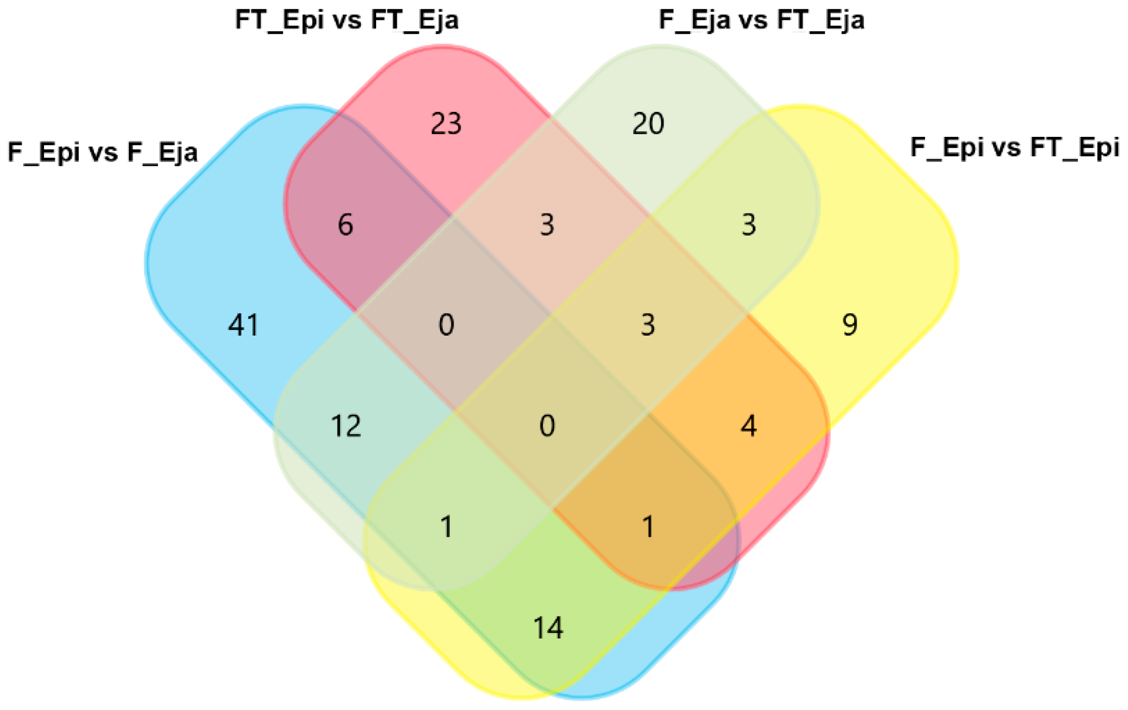
2.3. Differentially Abundant Proteins
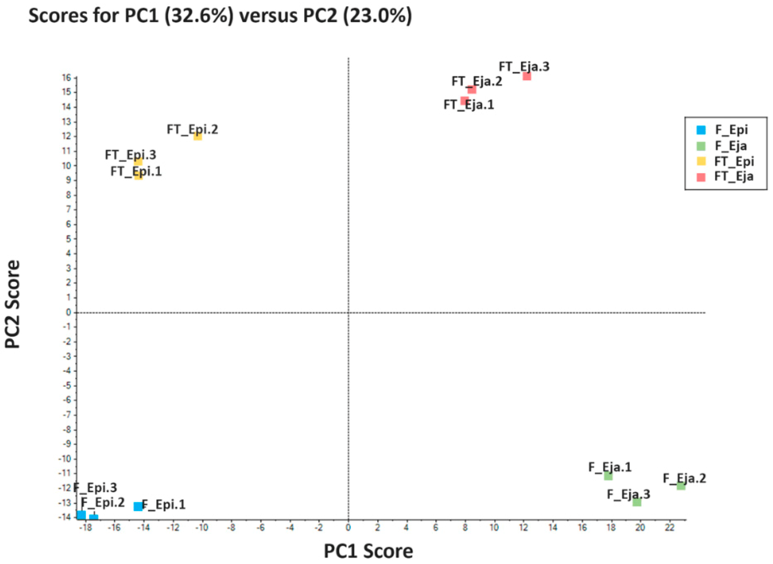
A total of 140 proteins showed quantitative differences among the sperm sources, 55 of them showing post-translational modifications (PTM). The inventory of these differentially abundant proteins, including organism, encoding genome, and relative amount quantified per sperm source and of the PTM, is shown in Table S3. The Venn diagram in Figure 2 shows how these quantitative different proteins were distributed among the sperm sources. A total of 44 differentially abundant proteins were overlapped among the four sperm sources. Looking at the nonoverlapped differentially abundant proteins, it was noticeable that the changes in abundance shown by 41 proteins were uniquely related to the transit of the spermatozoa from the cauda epididymis to the ejaculate (Table 2). Thirty-one of these 41 proteins were more abundant in fresh cauda epididymal spermatozoa; the other 10 were more abundant in fresh ejaculated spermatozoa. Focusing on the influence of the cryopreservation process, 32 sperm proteins experienced abundance change uniquely related to cryopreservation (Table 3). Three of these 32 proteins were less abundant in FT spermatozoa from both epididymal and ejaculated samples, compared to fresh samples. Another nine proteins changed uniquely between fresh and FT cauda epididymal spermatozoa, all of them being more abundant in fresh spermatozoa. The remaining 20 proteins showed abundance changes between fresh and FT but only for ejaculated spermatozoa. Ten of these 20 proteins were more abundant in the fresh spermatozoa whereas the remaining 10 were more abundant in the FT spermatozoa. Another 23 proteins showed differences in abundance only between FT spermatozoa of the cauda epididymis and those of the ejaculate (Table 4). Specifically, six of these proteins were more abundant in the FT spermatozoa from the cauda epididymis, and the other 17 in those from the ejaculate.

2.4. Bioinformatics

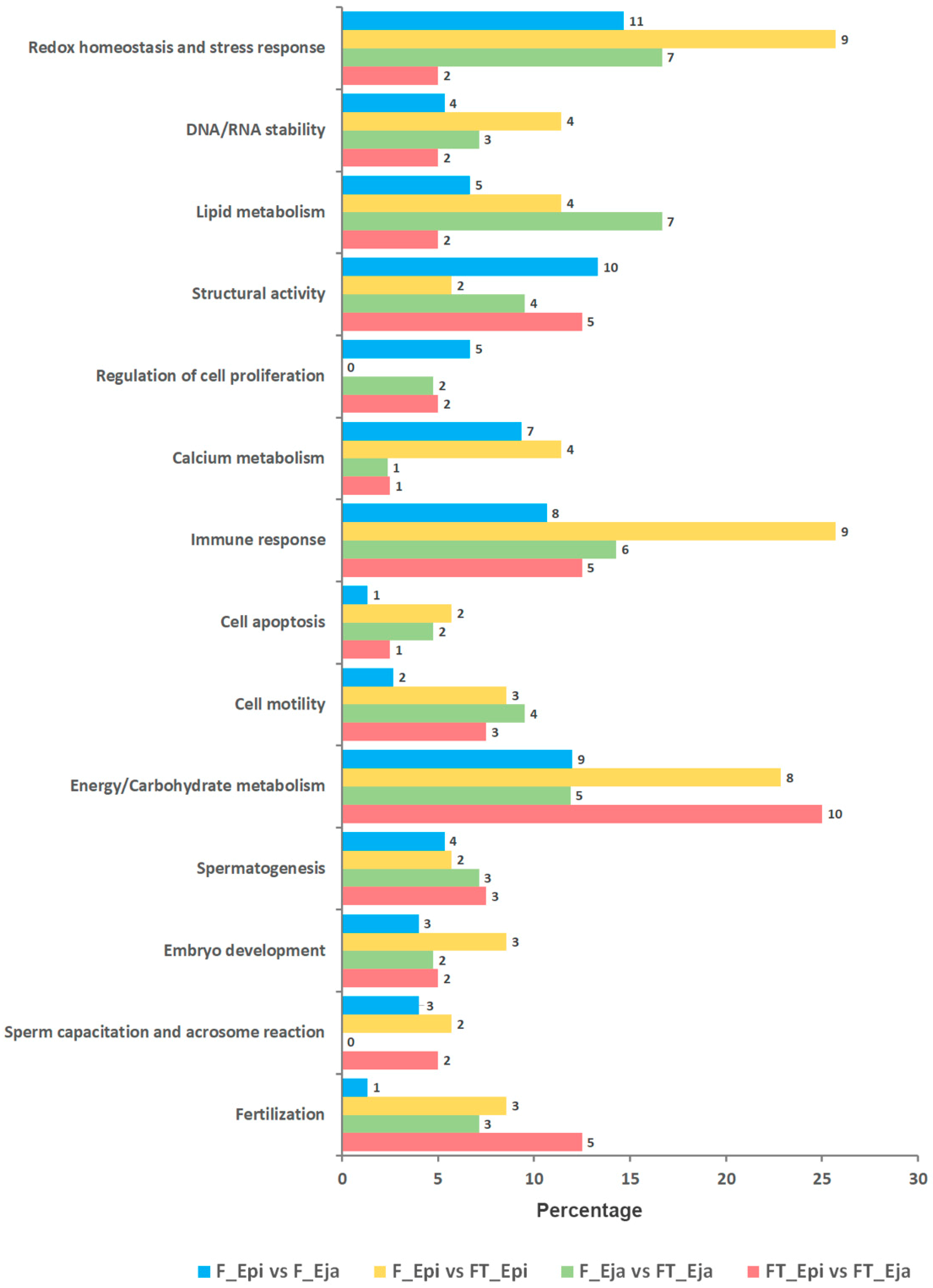
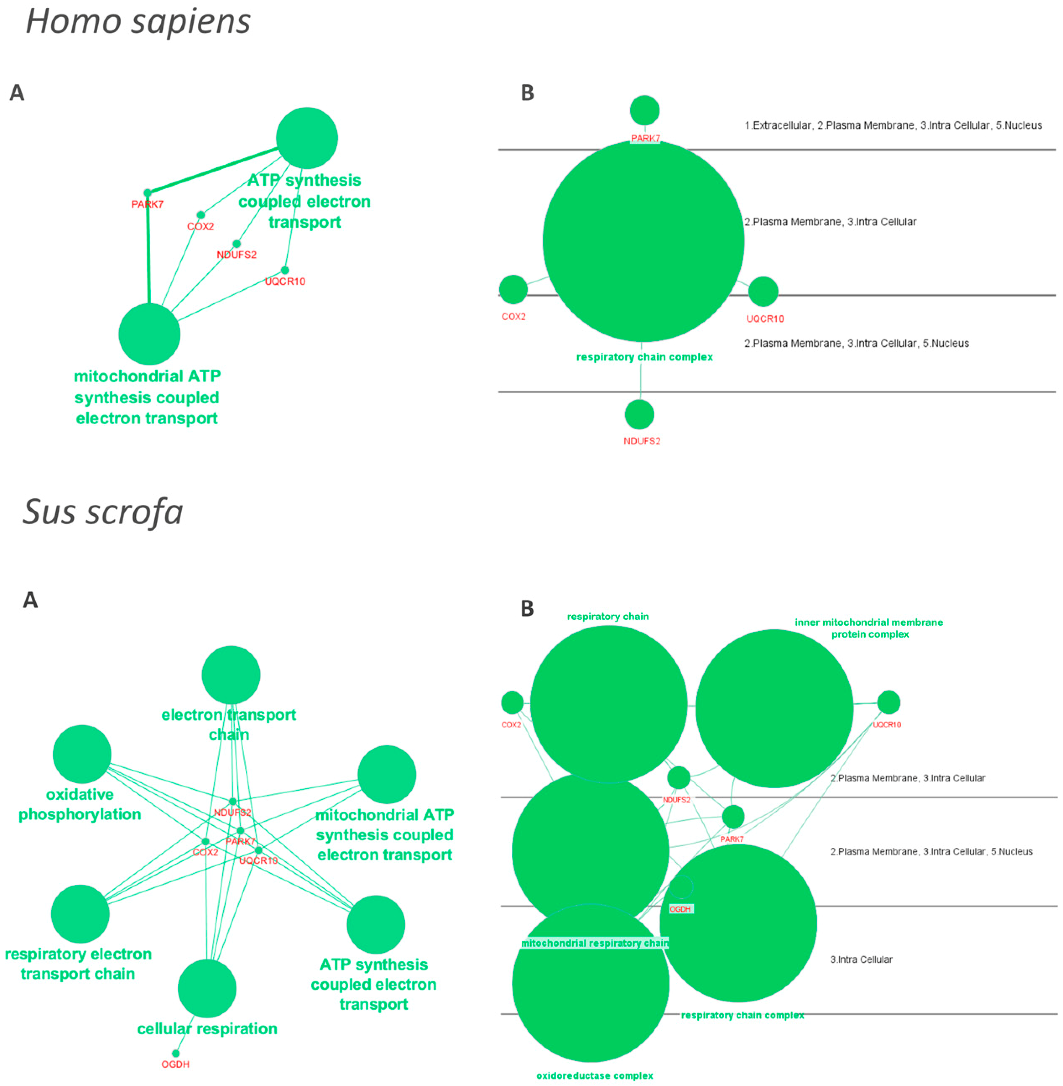
2.4.1. Gene Ontology Enrichment Analysis

The differentially abundant proteins among the four pairwise sperm comparisons did not show differences in the distribution pattern among the functional Gene Ontology (GO) categories specifically related to sperm and reproductive functions included in UniProt KB and DAVID databases (Figure 3). The GO categories with the largest protein number were redox homeostasis and stress response, immune response, and energy/carbohydrate metabolism.

2.4.2. Protein–Protein Interaction Networks

The Cytoscape software with ClueGO plugin were used to examine the interaction networks of differentially abundant proteins between fresh and FT spermatozoa of both epididymal and ejaculate sources. Protein databases of Homo sapiens and Sus scrofa were separately explored, allowing functional analysis of the 90% and 70% of the differentially abundant proteins, respectively. The generated networks did not include any differentially abundant proteins between fresh and FT epididymal spermatozoa. In contrast, they included five proteins of those found as differentially abundant between fresh and FT ejaculated spermatozoa, four of them coincident between Homo sapiens and Sus scrofa. Based on the degree of connectivity, these five proteins, so-called DJ-1, cytochrome C oxidase subunit 2 (Cox2), NADH:ubiquinone oxidoreductase core subunit S2 (Ndufs2), Ubiquinol-cytochrome c reductase complex O (Uqcr10), and mitochondrial 2-oxoglutarate dehydrogenase (Ogdh), were involved in pathways related to mitochondrial activity, redox reaction, energy generation and cellular respiration (Figure 4). DJ-1 was the only of the five proteins exhibiting higher abundance in fresh than in FT spermatozoa. The other four proteins were more abundant in FT spermatozoa.

3. Discussion

This study is the first one in mammals comparing the proteome of fresh and FT spermatozoa derived from cauda epididymis and ejaculate of the same sire. The results showed that cryopreservation causes quantitative changes in the boar sperm proteome and that many of the altered proteins differ between both cauda epididymis and ejaculated spermatozoa.

3.1. Post-Thaw Sperm Quality and Functionality

Data of post-thaw sperm quality and functionality agree with previous studies, showing that mature boar spermatozoa collected from the cauda epididymis cryosurvives better than those from the ejaculate [27,28]. In addition to lower post-thaw recovery rates of motile and viable spermatozoa in the ejaculate samples, the results also showed that the ejaculate cryosurvival sperm population was less functional than that from the cauda epididymis; which would be linked to increased sensitivity of ejaculated spermatozoa to cryostress [11]. These results clearly indicated that cryopreservation affects distinctly epididymal and ejaculated pig spermatozoa, attributed to the protective effect of epididymal fluid to epididymal spermatozoa and the negative subsequent interaction of these spermatozoa with seminal plasma (SP) during ejaculation, an interaction that would impair the freezability of ejaculated spermatozoa. These putative opposite effects of epididymal fluid and SP on sperm freezability would be linked to quantitative differences in protein composition between both fluids [36]. The proteomic results of this study would support this hypothesis, as we argument for next.

3.2. Quantitative Changes in Sperm Proteins

The first proteomics finding of the present study confirmed previously reported proteome quantitative differences between fresh pig spermatozoa from cauda epididymal and ejaculate [35], as 41 sperm proteins showed quantitative differences uniquely related to the transit of mature spermatozoa from the cauda epididymis to the ejaculate. Studies comparing features of epididymal and ejaculated fresh spermatozoa are uncommon, maybe due to the difficulty of collecting mature spermatozoa from both sources from a reproductive healthy male. To the best of our knowledge, only two studies, one in ovine [37] and our own above mentioned in pig [35], compared the proteome of cauda epididymal and ejaculated fresh spermatozoa. In agreement, the present study corroborated the absence of qualitative proteome differences between the spermatozoa of both sources. Moreover, it clearly showed that the documented quantitative differences in the sperm proteome relate to the interaction of spermatozoa with the secretions of the male reproductive accessory glands building the SP during ejaculation. These secretions would play a double and opposite role on the ejaculated spermatozoa. On one hand, they would sequentially wash the sperm surface facilitating the detachment of epididymal adsorbed proteins during sperm maturation and cauda storage and, on the other hand, they would transfer some of its components to the spermatozoa, including SP proteins. Some of the proteins that increased in abundance in fresh ejaculated spermatozoa were identified in the pig SP [38]. Similarly, Pini et al. [37] also showed that the increase in the amount of some proteins in ejaculated ovine spermatozoa with respect to epididymal spermatozoa would be due to exposure of epididymal spermatozoa to SP during ejaculation. Remarkable is the finding that many of the proteins whose abundance shifted between epididymal and ejaculated fresh spermatozoa were involved in redox homeostasis and stress response, structural activity, energy and carbohydrate metabolisms, immune response, and calcium metabolism, which would indicate that their change in abundance could influence sperm performance as previously demonstrated by Perez-Patiño et al. [35].
The second proteomics finding of the present study was that cryopreservation remodelled the proteome of pig spermatozoa. This finding was previously evidenced in ejaculated spermatozoa from several mammalian species, as bovine [39], human [20,21], ovine [22,23], porcine [24], and also in bovine epididymal spermatozoa [40]. All these studies found quantitative changes in a significant number of proteins between fresh and FT spermatozoa, although few of these differentially abundant proteins were common across studies [11]. Focusing in pig, Chen et al. [24] identified 41 differentially abundant proteins between fresh and FT spermatozoa, 35 of them showing greater quantity in FT spermatozoa. To clarify the reasons for these quantitative changes in a relatively large number of sperm proteins during cryopreservation remains a challenge. Mature spermatozoa are both transcriptionally and translationally silent [41]. Therefore, modifications in the sperm proteome are mainly quantitative and probably, depending on the interchange of sperm proteins, either adsorbed or structural. These quantitative modifications are normally related to PTMs or to the interaction of spermatozoa with the surrounding environment [42,43]. Although many of the differentially abundant proteins between fresh and FT spermatozoa found in the present study can undergo PTMs, the relevance of such modifications for explaining the results of the present study should be minimal, since PTMs have been contemplated in the proteomic analysis carried out. Regarding interaction with the surrounding environment, cauda epididymal and ejaculated fresh spermatozoa remained stored overnight with its own fluid (i.e., cauda epididymal fluid and SP, respectively) before freezing, facilitating sperm–fluid interaction. In addition, many spermatozoa had undergone membrane damage during cryopreservation, as evidenced by propidium iodide (PI) labeling, a DNA fluorescent label that gets into cells with compromised membrane integrity. This loss of membrane integrity could promote efflux of intracellular components, such as cytoplasmic proteins, which could explain the decrease in abundance experienced by some sperm proteins during the cryopreservation process [21]. The increase in abundance experienced by some sperm proteins during cryopreservation is more difficult to explain. Excluding the existing PTMs, because they were considered in the proteomic analysis carried out in the present study; the other possible hypothesized causes would either be protein degradation or the secondary or tertiary structure changes experienced by some proteins during cryopreservation [21]. In this sense, the efflux of cellular components experienced by the relative high number of spermatozoa dying during cryopreservation could lead to the degradation of proteins of living spermatozoa. Gurupriya et al. [44] showed that cryopreservation changed the activity of proteases and protease-inhibitors of buffalo and bovine semen. Another deleterious cause would be the interaction of spermatozoa with the cryoprotectant. Yoon et al. [40] showed that the usual prefreezing incubation of spermatozoa with extenders containing glycerol induced changes in the proteome of bull epididymal spermatozoa. The influence of the cryoprotectant on the sperm proteome was also demonstrated in human spermatozoa by Bogle et al. [21]. These results and those of the present study clearly highlighted the relationship between the alterations in the sperm proteome and the loss of sperm quality and functionality during cryopreservation. Some of the above studies emphasized that sperm proteins quantitatively altered during cryopreservation could be potential biomarkers of sperm cryo-stress [22,40]. However, the above-cited studies missed to look for direct links between altered proteins and functional changes experienced by spermatozoa during cryopreservation. Regarding distribution into GO categories, the majority of the sperm proteins that changed in abundance during cryopreservation in the above studies had both intracellular and extracellular localization, with a significant percentage of them being membrane proteins. Regarding function, they were involved in a wide range of functions directly or indirectly related to sperm performance, which agree with the results achieved in our study (data no shown).
The third and most novel proteomics finding of the present study was the demonstration that the quantitative changes in the proteome caused by cryopreservation differed between cauda epididymal and ejaculated spermatozoa. Certainly, only three out of the 32 proteins that changed in abundance between fresh and FT spermatozoa were common for epididymal and ejaculated spermatozoa. Of the remaining 29, the abundance of nine proteins changed between fresh and FT epididymal spermatozoa and the other 20 between fresh and FT ejaculated spermatozoa. This peculiar distribution of the altered proteins would indicate that cryopreservation affected differently epididymal and ejaculated porcine spermatozoa, the latter being the most affected. This fact could explain that quality and functionality post-thawing were worse in spermatozoa from the ejaculate than in those from the cauda epididymis. To test this hypothesis, we first identified the functional allocation of the differentially abundant protein into GO categories directly or indirectly related to sperm and reproductive function. The distribution was relatively similar between cauda epididymal and ejaculated spermatozoa, making it difficult to find any sound relationship between the differentially altered proteins during cryopreservation and the differences in post-thaw sperm quality and functionality between the sperm sources. Therefore, the use of more stringent filters allowed us to test how the differentially abundant proteins between fresh and FT spermatozoa of either source (cauda epididymal or ejaculate) were grouped into known protein–protein network topologies for Homo sapiens (largest) and Sus scrofa (still limited). The results were striking because the generated networks only included proteins differentially abundant between fresh and FT ejaculated spermatozoa, those most affected by cryopreservation. The network highlighted five of the differentially abundant proteins between fresh and FT spermatozoa from the ejaculate, all of them related to mitochondrial functionality, either involved directly in energy requirements or oxidoreductase activity. Three of them, Ndufs2, Uqcr10, and Cox2, are key mitochondrial electron transport complex proteins specifically involved in complexes I, III, and IV, respectively [45]. DJ-1—a protein deglycase—contributes in the regulation of mitochondrial complex I and it would be involved in the control of oxidative stress [46,47]. Finally, Ogdh is an enzyme of the Krebs cycle that would also be involved in mitochondrial functionality, specifically in ROS generation [48]. Hence changes in the abundance of the above proteins would have a direct influence on sperm mitochondrial functionality, including ATP synthesis and ROS generation. Therefore, the altered abundance of these proteins among ejaculated spermatozoa during cryopreservation could explain the lower mitochondrial membrane potential and the higher ROS affecting motility in ejaculated compared to epididymal FT spermatozoa.
To conclude, this study showed that the quantitative changes in the proteome experienced by porcine mature spermatozoa during cryopreservation differs between those retrieved from the cauda epididymis and those from the ejaculate from the same individuals; those ejaculated showing a greater number of affected proteins. Among these affected proteins, those involved in mitochondrial electron transport complex would explain why FT spermatozoa derived from the ejaculate produced more ROS and had worse mitochondrial membrane potential and motility than FT spermatozoa from the cauda epididymis. In addition to improve our current knowledge of molecular disarrangements experienced by spermatozoa during cryopreservation, the results of this study can be of value to improve the efficiency of cryopreservation extenders for pig spermatozoa by inclusion of mitochondrial protective agents in their composition.

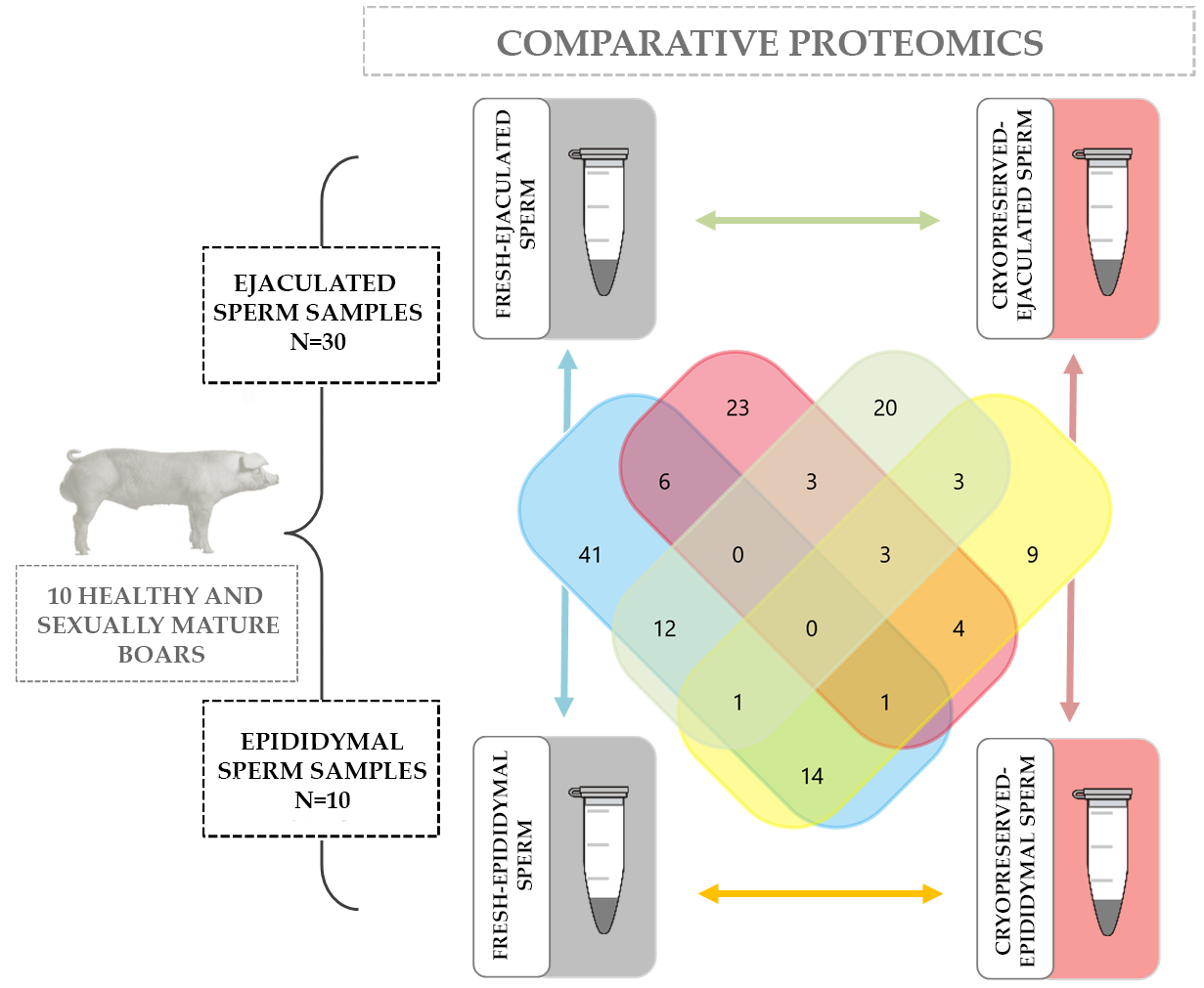
4. Materials and Methods

The procedures implying boar handling and collection of sperm samples were conducted following international guidelines (Directive 2010/63/EU) and previously approved by the Bioethics Committee of the University of Murcia (research code: 639/2012). Chemicals used for preparing sperm extenders, and fluorescent probes for checking sperm quality and functionality were from Sigma Aldrich Co. (St. Louis, MO, USA).

4.1. Boars, Epididymis, and Ejaculates

Ejaculated and epididymal mature sperm samples were collected from 10 healthy, sexually mature (12 to 24 months old) and fertile boars housed under controlled environmental conditions (15–25 °C and 16 h of light) in a Spanish commercial AI center (Topigs Norsvin España, Madrid, Spain). Entire ejaculates (a total of 30, 3 per boar) were collected using the semiautomatic collection procedure Collectis® (IMV Technologies, L’Aigle, France). The boars were slaughtered when still healthy and fertile (Slaughterhouse of Mercamurcia, Murcia, Spain) for reasons of genetic replacement. Their scrotal contents were collected immediately after slaughter and kept in isolated containers (5 °C) until the cauda epididymal content was retrieved at the laboratory through retrograde infusion of air in the ductus deferens [27,35]. All sperm samples, either from the cauda epididymis or the ejaculate, were assessed for morphology and total and progressive motility by using procedures listed below.

4.2. Experimental Workflow

The samples from the cauda epididymis (10 samples, one per boar) and ejaculate (30 samples, 3 per boar) were split in two aliquots immediately after collection. The first aliquot, used as fresh sperm source, was extended (1:3, v/v) in phosphate-buffered saline (PBS) and centrifuged twice (1500× g at room temperature (RT) during 10 min; Megafuge 1.0 R, Heraeus, Hanau, Germany). The resulting sperm pellets were extended in PBS (1000 × 106 sperm/mL), dispensed in cryotubes (2 mL Cryogenic vial, Fisher Scientific, Madrid, Spain) and stored at −80 °C (Ultra Low Freezer; Haier, Schomberg, Ontario, Canada). The second aliquot, used as FT sperm source, was extended (1:1, v/v) in Beltsville Thawing Solution (BTS, 205 mM glucose, 20.4 mM sodium citrate, 10.0 mM KCl, 15.0 mM NaHCO3, and 3.6 mM EDTA), stored at 17 °C overnight and thereafter frozen using the below cryopreservation protocol. Once thawed, the content of three straws for each of the 30 FT sperm samples was pooled and centrifuged at 600× g for 20 min at RT on a Percoll monolayer gradient (45% in PBS, v/v) to separate the spermatozoa from other putative cells, debris, and egg yolk remnants. The resulting sperm pellets were extended in PBS and stored at −80 °C following the same protocol as used for fresh sperm samples. To carry out the proteomics analysis, the sperm samples were thawed at RT, and each of the 4 sperm sources (fresh cauda epididymal spermatozoa, fresh ejaculate spermatozoa, FT epididymal spermatozoa, and FT ejaculated spermatozoa) were pooled generating four single sperm samples, one for each sperm source. Each of the 4 sperm pools was split into three aliquots generating three technical replicates for sperm source. Thereby, proteomics was analyzed in a total of 12 samples.

4.3. Sperm Cryopreservation

The sperm samples were centrifuged at 17 °C for 3 min at 2400× g, and the resulting sperm pellets were frozen using a standard 0.5 mL-straw freezing protocol described by Alkmin et al. [27]. Briefly, sperm pellets were extended to 1.5 × 10⁹ sperm/mL in a Tris-citric acid-glucose extender (111 mM Trizma Base, 31.4 mM monohydrate citric acid, 185 mM glucose) supplemented with egg yolk (80%/20%, v/v). Then, cooled to 5 °C for 150 min and re-extended to 1.0 × 10⁹ sperm/mL with the same extender (89.5%, v/v) plus glycerol (9%, v/v) and Equex STM (1.5%, v/v, Nova Chemical Sales, Scituate, MA, USA). The extended sperm samples were packed (0.5 mL polyvinyl chloride French straws, Minitüb, Tiefenbach, Germany) and frozen at −40 °C/min using a controlled rate freezer (IceCube 1810, Minitüb, Germany). The straws, stored in liquid nitrogen tanks (GT40, Air Liquide, Paris, France) for at least two weeks, were thawed in a circulating water bath at 37 °C for 20 s.

4.4. Quality and Sperm Functionality Assessments

Fresh sperm samples were evaluated within 2 h after collection and FT sperm samples at 30 min after thawing. Sperm quality was evaluated in terms of morphology (only fresh sperm samples), total and progressive motility and viability. The functionality of viable FT spermatozoa was evaluated in terms of intracellular generation of ROS, fluidity of the plasma membrane and the potential of mitochondrial membrane.
Sperm morphology was evaluated under a phase contrast microscope from sperm samples fixed in buffered 2% formaldehyde solution and recorded as the proportion of spermatozoa with normal morphology. Sperm motility was objectively evaluated using a computer-assisted analysis system (ISASV1®, Proiser R + D, Valencia, Spain) operating up to 100 video frames per second. The sperm samples (5 µL containing 100,000 spermatozoa) were loaded on Makler counting chambers (Sefi Medical Instruments, Haifa, Israel) and a minimum of 400 spermatozoa per sperm sample were analyzed. Sperm motility was recorded as the percentages of total motile spermatozoa (average path velocity ≥ 20 µm s−1) and the proportion of motile spermatozoa showing rapid and progressive movement (straightness of the average path ≥ 40%).
Viability and functionality of viable FT spermatozoa were cytometrically evaluated (BD FACSCanto II flow cytometer, Becton, Dickinson, CA, USA) using the settings, software, and procedures described by Alkmin et al. [49]. The cytometry workflow used is shown in Figure S2. Sperm viability, in terms of both plasma and acrosome membrane integrity, was evaluated in sperm samples (100 µL aliquots containing 2 × 10⁶ sperm) incubated (37 °C for 10 min in the dark) with 3 µL of Hoechst 33342 (H-42, 0.05 mg/mL in PBS), 2 µL of PI (0.5 mg/mL stock solution in PBS), and 2 µL of fluorescein-conjugated peanut agglutinin (PNA-FITC, 100 µg/mL stock solution in PBS). Spermatozoa positive to H-42 and negative to PI and PNA-FITC were recorded as viable. The intracellular generation of ROS was measured in terms of hydrogen peroxide (H2O2) generation in FT-sperm samples (1.5 × 10⁶ sperm in 1 mL of PBS) incubated (38 °C for 30 min in the dark) with 1.5 µL of H-42 (0.05 mg/mL in PBS), 1 µL of 2′,7′-dichlorodihydrofluorescein diacetate (H2DCFDA, 1 mM in DMSO), and 1 µL of PI (1 mg/mL in PBS). The mean fluorescence intensity of dichlorofluorescein (DCF; H2DCF is oxidized by H2O2 into DCF) emitted by viable spermatozoa (PI negative) was recorded in the results as fluorescence units (FU) per 106 viable spermatozoa. The fluidity of the plasma membrane was evaluated in FT-sperm samples (1.5 × 10⁶ sperm in 1 mL of PBS) incubated (38 °C for 10 min in the dark) with 1.5 µL of H-42 (0.05 mg/mL in PBS), 1 µL of Yo-Pro-1 (25 µM in DMSO), and 2.6 µL of Merocyanine 540 (M-540, 1 mM in DMSO). Spermatozoa positive to H-42 and M-540 and negative to Yo-Pro-1 were recorded as viable and with high plasma membrane fluidity. Mitochondria membrane potential was evaluated in FT-sperm samples (100 µL containing 3 × 10⁶ sperm) incubated (38 °C for 15 min in the dark) with 3 µL of H-42 (0.05 mg/mL in PBS), 2 µL of PI (0.5 mg/mL in PBS), and 0.5 µL of Mitotracker Deep Red 633 (Mitotracker, 20 µM in PBS of a stock solution of 1 mM in DMSO). The spermatozoa positive to H-42 and Mitotracker and negative to PI were recorded as viable with high mitochondria membrane potential.

4.5. Sperm Proteomics

The proteomics analyses were carried out in the Proteomics Unit of the University of Valencia (Valencia, Spain), a member of the PRB2-ISCIII ProteoRed Proteomics Platform.

4.5.1. Protein Extraction

Protein extraction of sperm samples was conducted as described previously by Perez-Patiño et al. [50]. Briefly, sperm samples were centrifuged (14,000× g for 10 min; Eppendorf 5424R, Eppendorf AG, Hamburg, Germany) and the total protein of the resulting sperm pellets were extracted using UTC buffer (7 M Urea, 2 M thiourea, 4% 3-((3-cholamidopropyl) dimethylammonio)-1-propanesulfonate (CHAPS)), supplemented with protease inhibitor cocktail (1%, v/v). The RC_DC Lowry (Bio-Rad, Richmond, CA, USA) assay was used to determine the concentration of extracted protein.

4.5.2. SDS-PAGE and In-Gel Digestion

Thirty micrograms of protein per sample was mixed (4:1) with 4× Laemmli sample buffer (Bio-Rad, Hercules, CA, USA) were loaded on one dimensional sodium dodecyl sulfate-polyacrylamide gel electrophoresis (1D SDS-PAGE) to remove the remaining UTC and other interferences before tandem mass spectrometry (MS/MS) analysis. The proteins contained in the 1D SDS-PAGE were in-gel digested following the protocol used by Shevchenko et al. [51], which included (i) trypsin digestion stopped with 10% trifluoroacetic (TFA), (ii) peptide extraction by incubation in pure acetonitrile (ACN) at 37 °C in a shaker for 15 min, and (iii) suspension of resulting peptide mixture in of 2% ACN and 0.1% TFA.

4.5.3. Sequential Window Acquisition of All Theoretical Spectra (SWATH) Analysis

A SWATH analysis was performed by liquid chromatography (LC) using a NanoLC Ultra 1D plus Eksigent (Eksigent Technologies, Dublin, CA, USA) connected to an AB SCIEX TripleTOF 5600 mass spectrometer (AB SCIEX, Framingham, MA, USA) in direct injection mode following the procedure described by Perez-Patiño et al. [50]. Firstly, for the spectral library acquisition, 5 µL of a mixture of the twelve digested samples (2 µL per sample) was loaded on a trap NanoLC precolumn (3-μm particle size C18-CL, 350 µm diameter × 0.5 mm long; Eksigent Technologies, Dublin, CA, USA) and desalted with 0.1% TFA at 3 µL/min for 5 min. Then, the peptides were separated using an analytical LC column (3-μm particle size C18-CL, 75 µm diameter × 12 cm long, Nikkyo Technos Co®, Tokyo, Japan) equilibrated in 5% ACN and 0.1% formic acid (FA) (Fisher Scientific). Thereafter, a linear gradient from 5% to 35% of ACN containing 0.1% FA at a constant flow rate of 300 nL/min over 180 min was applied to peptide elution. The TripleTOF operated in data-dependent mode with a time-of-flight (TOF) MS scan from 350 to 1250 m/z, accumulated for 250 ms TOF followed by 150 ms TOF with the same scan range for MS. The 25 most abundant multiply charged (2+, 3+, 4+, or 5+) precursor peptide ions were automatically selected. Ions with 1+ and unassigned charge states were rejected from the MS/MS analysis. The rolling collision energy equations were automatically set by the instrument according to the equation |CE| = (slope)x(m/z) + (intercept) with Charge = 2; Slope = 0.0575 and Intercept = 9.
Then, for a label-free quantification using SWATH analysis, each sperm sample was individually analyzed configuring the TripleTOF 5600 (AB SCIEX, Framingham, MA, USA) as described by Gillet et al. [52] for SWATH-MS-based experiments. In this way, 5 µL of one of the three technical replicates from each sample was randomly loaded onto a 3 µL C18-CL trap column (dimensions: 75 μm × 15 cm; NanoLC Column, Eksigent Technologies) and flushed for 5 min with 0.1% trifluoroacetic (TFA) at 3 µL/min to remove salts. Peptides were resolved over a 90 min gradient of 5 to 40% acetonitrile (ACN) at a flow rate of 300 nL/min using an analytical 3-µm C18-CL column equilibrated in 5% ACN and 0.1% FA. The analysis of eluted peptides was carried out in the spectrometer nanoESI qQTOF (AB SCIEX TripleTOF 5600) and the TripleTOF (AB SCIEX) operated in SWATH mode, in which a 0.050 s TOF MS scan from 350 to 1250 m/z was performed, followed by 0.080 s product ion scans from 230 to 1800 m/z split into 37 overlapping windows (3.05 sec/cycle). The collision energy for each window was calculated for 2+ charged ion at the center of each SWATH block with a collision energy spread of 15 eV. The MS was always operated in high sensitivity mode.

4.5.4. Protein Quantification

The wiff files obtained from SWATH experiment were analyzed by PeakView® (v2.1, AB SCIEX) and peaks from SWATH were extracted with a peptide confidence threshold of 95%. A FDR less than 1% and 6 transitions per peptide were required to quantify one peptide. The extracted ions chromatograms were integrated, and the peak areas were normalized by total sum, and the sum of all areas was equalized for all the samples.

4.5.5. Gene Ontology and Bioinformatics Analysis

A GO enrichment analysis was performed for the differentially expressed sperm-proteins using the online bioinformatics tool UniProt KB and DAVID. The UniProt KB database (www.uniprot.org), downloaded 23/07/2018, contains 120,243,849 total entries with 48,936 of them encoded in Sus scrofa taxonomy. The DAVID Database for Annotation, Visualization and Integrated Discovery (DAVID Bioinformatics Resources 6.8; https://david.ncifcrf.gov/ [53,54]) integrates numerous public sources of protein annotation and, consequently, contains information of more than 1.5 million genes from more than 65,000 species. The PTMs were explored using Unimod website (http://www.unimod.org/modifications_list.php). Peptides showing exclusively artificial modifications introduced by the sample preparation procedure were manually removed from the list. The functional allocation of the differentially abundant proteins was manually performed and restricted to the GO categories directly or indirectly related to sperm and reproductive functions contemplated in UniProt KB and DAVID databases. GO enrichment and network visualization and analysis were performed with Cytoscape v3.2.1 [55] and ClueGO plugin v2.2.3 [56] using the enrichment/depletion multiple two-sided hypergeometric test with Bonferroni step down correction and a Kappa score (term/pathway connectivity) of 0.4. Only enriched functions with a p-value < 0.05 were considered. The other ClueGO parameters were kept to default settings.

4.6. Statistical Analysis

The influence of sperm source (cauda epididymis vs. ejaculate) in post-thaw sperm quality and functionality was evaluated using a mixed ANOVA test (IBM SPSS v24.0 software, IBM Spain, Madrid) including boar and ejaculate as random effects. In proteomics, the quantitative data processed by PeakView® were further exported to MarkerView® (v1.2, AB SCIEX) for quantitative and statistical analysis. The data across the runs were normalized using total area sum. Then, the Multiexperiment Viewer (MeV) software (version 4.8) (http://www.tm4.org) was used to identify quantitative differently abundant sperm proteins applying a Student’s t-test. Differences in protein abundance were considered with an adjusted p-value ≤ 0.01, and only those with a fold change (FC) ≥ ±1.5 after log2 transformation were recorded. The explanatory ability of different abundant proteins was illustrated by mean of heat maps after z-score normalization, using Euclidean distances. The online software ‘FunRich’ (http://www.funrichweb.org) was used to show how the differently abundant proteins were distributed among the sperm sources. Differences of differentially abundant proteins into the functional GO categories among sperm sources were analyzed using a chi-squared analysis with an adjusted p-value ≤ 0.05.

Supplementary Materials

Supplementary materials can be found at https://0-www-mdpi-com.brum.beds.ac.uk/1422-0067/20/7/1791/s1, Figure S1: Heat maps including dendrograms depicting the differentially abundant proteins between sperm sources. Specifically, between epididymal and fresh ejaculate spermatozoa, fresh and frozen-thawed epididymal spermatozoa, fresh and frozen-thawed ejaculate spermatozoa, and between epididymal and ejaculate frozen-thawed spermatozoa. The hierarchical clustering tree of sperm samples is shown at the top. The relative abundance level of each protein is shown on a color scale from green (lowest level) to red (highest level); Figure S2: Flow cytometric gating strategy applied during assessment of the quality and functionality of frozen-thawed pig spermatozoa; Table S1: Quality parameters of fresh pig spermatozoa collected from cauda epididymis and ejaculate; Table S2: The relative abundance of the quantified proteins in fresh and frozen-thawed pig spermatozoa from cauda epididymis and ejaculate; Table S3: The relative abundance of differentially quantified proteins, including those sensitive to post-translational modifications (PTMs), among the four pig sperm sources (fresh and frozen-thawed spermatozoa from the cauda epididymis and ejaculate).

Author Contributions

Conceptualization, H.R.-M., J.R. and I.P.; Formal Analysis, C.P.-P.; Funding Acquisition, E.A.M., H.R.-M. and J.R.; Investigation, C.P.-P., I.B., J.L. and L.P.; Methodology, J.R. and I.P.; Software, C.P.-P. and I.B.; Supervision, J.R. and I.P.; Writing—Original Draft, C.P.-P. and I.B.; Writing—Review & Editing, E.A.M., H.R.-M. and J.R. All authors approved the final manuscript and the version to be published.

Funding

The study was supported by MINECO (Spain), FEDER funds (EU) (AGL2015-69738-R), the Seneca Foundation Murcia, Spain (19892/GERM-15), FORSS (745971), and the Swedish Research Council FORMAS (2017-00946), Stockholm, Sweden. C. Perez-Patiño and J. Li were financially supported by Seneca Foundation (Murcia, Spain) and the China Scholarship Council, respectively.

Acknowledgments

The authors thank AIM Iberica (Topigs Norsvin Iberica) for supplying the ejaculates and reproductive tracts of boars.

Conflicts of Interest

The authors declare no conflict of interest.

References

  1. Abram McBride, J.; Lipshultz, L.I. Male Fertility Preservation. Curr. Urol. Rep. 2018, 19, 49. [Google Scholar] [CrossRef]
  2. Kawai, K.; Nishiyama, H. Preservation of fertility of adult male cancer patients treated with chemotherapy. Int. J. Clin. Oncol. 2018, in press. [Google Scholar] [CrossRef]
  3. Morrell, J.M.; Mayer, I. Reproduction biotechnologies in germplasm banking of livestock species: A review. Zygote 2017, 25, 545–557. [Google Scholar] [CrossRef]
  4. Comizzoli, P. Biobanking efforts and new advances in male fertility preservation for rare and endangered species. Asian J. Androl. 2015, 17, 640–645. [Google Scholar] [CrossRef] [PubMed]
  5. Alvarenga, M.A.; Papa, F.O.; Ramires Neto, C. Advances in Stallion Semen Cryopreservation. Vet. Clin. N. Am. Equine Pract. 2016, 32, 521–530. [Google Scholar] [CrossRef] [PubMed]
  6. Yeste, M.; Rodríguez-Gil, J.E.; Bonet, S. Artificial insemination with frozen-thawed boar sperm. Mol. Reprod. Dev. 2017, 84, 802–813. [Google Scholar] [CrossRef] [PubMed] [Green Version]
  7. Rozati, H.; Handley, T.; Jayasena, C.N. Process and Pitfalls of Sperm Cryopreservation. J. Clin. Med. 2017, 6, 89. [Google Scholar] [CrossRef]
  8. Rosenbusch, B. To What Extent Are Cryopreserved Sperm and Testicular Biopsy Samples Used in Assisted Reproduction? J. Reprod. Infertil. 2018, 19, 115–118. [Google Scholar]
  9. Sharma, R.; Kattoor, A.J.; Ghulmiyyah, J.; Agarwal, A. Effect of sperm storage and selection techniques on sperm parameters. Syst. Biol. Reprod. Med. 2015, 61, 1–12. [Google Scholar] [CrossRef]
  10. Knox, R.V. The Fertility of Frozen Boar Sperm When used for Artificial Insemination. Reprod. Domest. Anim. 2015, 50, 90–97. [Google Scholar] [CrossRef] [Green Version]
  11. Pini, T.; Leahy, T.; de Graaf, S.P. Sublethal sperm freezing damage: Manifestations and solutions. Theriogenology 2018, 118, 172–181. [Google Scholar] [CrossRef]
  12. Morris, G.J.; Acton, E.; Murray, B.J.; Fonseca, F. Freezing injury: The special case of the sperm cell. Cryobiology 2012, 64, 71–80. [Google Scholar] [CrossRef]
  13. Li, C.J.; Wang, D.; Zhou, X. Sperm proteome and reproductive technologies in mammals. Anim. Reprod. Sci. 2016, 173, 1–7. [Google Scholar] [CrossRef] [PubMed]
  14. Hezavehei, M.; Sharafi, M.; Kouchesfahani, H.M.; Henkel, R.; Agarwal, A.; Esmaeili, V.; Shahverdi, A. Sperm cryopreservation: A review on current molecular cryobiology and advanced approaches. Reprod. Biomed. Online 2018, 37, 327–339. [Google Scholar] [CrossRef] [PubMed]
  15. Huang, S.Y.; Kuo, Y.H.; Lee, W.C.; Tsou, H.L.; Lee, Y.P.; Chang, H.L.; Wu, J.J.; Yang, P.C. Substantial decrease of heat-shock protein 90 precedes the decline of sperm motility during cooling of boar spermatozoa. Theriogenology 1999, 51, 1007–1016. [Google Scholar] [CrossRef]
  16. Lessard, C.; Parent, S.; Leclerc, P.; Bailey, J.L.; Sullivan, R. Cryopreservation alters the levels of the bull sperm surface protein P25b. J. Androl. 2000, 21, 700–707. [Google Scholar] [PubMed]
  17. Chatterjee, S.; de Lamirande, E.; Gagnon, C. Cryopreservation alters membrane sulfhydryl status of bull spermatozoa: Protection by oxidized glutathione. Mol. Reprod. Dev. 2001, 60, 498–506. [Google Scholar] [CrossRef]
  18. Casas, I.; Sancho, S.; Ballester, J.; Briz, M.; Pinart, E.; Bussalleu, E.; Yeste, M.; Fàbrega, A.; Rodríguez-Gil, J.E.; Bonet, S. The HSP90AA1 sperm content and the prediction of the boar ejaculate freezability. Theriogenology 2010, 74, 940–950. [Google Scholar] [CrossRef]
  19. Flores, E.; Fernández-Novell, J.M.; Peña, A.; Rigau, T.; Rodríguez-Gil, J.E. Cryopreservation-induced alterations in boar spermatozoa mitochondrial function are related to changes in the expression and location of midpiece mitofusin-2 and actin network. Theriogenology 2010, 74, 354–363. [Google Scholar] [CrossRef]
  20. Wang, S.; Wang, W.; Xu, Y.; Tang, M.; Fang, J.; Sun, H.; Sun, Y.; Gu, M.; Liu, Z.; Zhang, Z.; et al. Proteomic characteristics of human sperm cryopreservation. Proteomics 2014, 14, 298–310. [Google Scholar] [CrossRef]
  21. Bogle, O.A.; Kumar, K.; Attardo-Parrinello, C.; Lewis, S.E.M.; Estanyol, J.M.; Ballesca, J.L.; Oliva, R. Identification of protein changes in human spermatozoa throughout the cryopreservation process. Andrology 2017, 5, 10–22. [Google Scholar] [CrossRef]
  22. He, Y.; Wang, K.; Zhao, X.; Zhang, Y.; Ma, Y.; Hu, J. Differential proteome association study of freeze-thaw damage in ram sperm. Cryobiology 2016, 72, 60–68. [Google Scholar] [CrossRef]
  23. Pini, T.; Rickard, J.P.; Leahy, T.; Crossett, B.; Druart, X.; de Graaf, S.P. Cryopreservation and egg yolk medium alter the proteome of ram spermatozoa. J. Proteom. 2018, 181, 73–83. [Google Scholar] [CrossRef]
  24. Chen, X.; Zhu, H.; Hu, C.; Hao, H.; Zhang, J.; Li, K.; Zhao, X.; Qin, T.; Zhao, K.; Zhu, H.; et al. Identification of differentially expressed proteins in fresh and frozenethawed boar spermatozoa by iTRAQcoupled 2D LCeMS/MS. Reproduction 2014, 147, 321–330. [Google Scholar] [CrossRef]
  25. Kuzmuk, K.N.; Schook, L.B. Pigs as a model of biochemical science. In The Genetics of the Pig, 2nd ed.; Rothschild, M.F., Ruvinsky, A., Eds.; CAB International: Oxfordshire, UK, 2011; pp. 426–444. [Google Scholar]
  26. Roca, J.; Martinez-Alborcia, M.J.; Gil, M.A.; Parrilla, I.; Martinez, E.A. Dead spermatozoa in raw semen samples impair in vitro fertilization outcomes of frozen-thawed spermatozoa. Fertil. Steril. 2013, 100, 875–881. [Google Scholar] [CrossRef]
  27. Alkmin, D.V.; Perez-Patiño, C.; Barranco, I.; Parrilla, I.; Vazquez, J.M.; Martinez, E.A.; Rodriguez-Martinez, H.; Roca, J. Boar sperm cryosurvival is better after exposure to seminal plasma from selected fractions than to those from entire ejaculate. Cryobiology 2014, 69, 203–210. [Google Scholar] [CrossRef] [Green Version]
  28. Rath, D.; Niemann, H. In vitro fertilization of porcine oocytes with fresh and frozen-thawed ejaculated or frozen-thawed epididymal semen obtained from identical boars. Theriogenology 1997, 47, 785–793. [Google Scholar] [CrossRef]
  29. Holt, W.V.; Medrano, A.; Thurston, L.M.; Watson, P.F. The significance of cooling rates and animal variability for boar sperm cryopreservation: Insights from the cryomicroscope. Theriogenology 2005, 63, 370–382. [Google Scholar] [CrossRef]
  30. Roca, J.; Hernández, M.; Carvajal, G.; Vázquez, J.M.; Martínez, E.A. Factors influencing boar sperm cryosurvival. J. Anim. Sci. 2006, 84, 2692–2699. [Google Scholar] [CrossRef]
  31. Yeste, M. Sperm cryopreservation update: Cryodamage, markers, and factors affecting the sperm freezability in pigs. Theriogenology 2016, 85, 47–64. [Google Scholar] [CrossRef]
  32. Vilagran, I.; Castillo, J.; Bonet, S.; Sancho, S.; Yeste, M.; Estanyol, J.M.; Oliva, R. Acrosin-binding protein (ACRBP) and triosephosphate isomerase (TPI) are good markers to predict boar sperm freezing capacity. Theriogenology 2013, 80, 443–450. [Google Scholar] [CrossRef]
  33. Vilagran, I.; Yeste, M.; Sancho, S.; Casas, I.; Rivera del Álamo, M.M.; Bonet, S. Relationship of sperm small heat-shock protein 10 and voltage-dependent anion channel 2 with semen freezability in boars. Theriogenology 2014, 82, 418–426. [Google Scholar] [CrossRef]
  34. Guimarães, D.B.; Barros, T.B.; van Tilburg, M.F.; Martins, J.A.M.; Moura, A.A.; Moreno, F.B.; Monteiro-Moreira, A.C.; Moreira, R.A.; Toniolli, R. Sperm membrane proteins associated with the boar semen cryopreservation. Anim. Reprod. Sci. 2017, 183, 27–38. [Google Scholar] [CrossRef]
  35. Perez-Patiño, C.; Parrilla, I.; Li, J.; Barranco, I.; Martinez, E.A.; Rodriguez-Martinez, H.; Roca, J. The proteome of pig spermatozoa is remodeled during ejaculation. Mol. Cell. Proteom. 2019, 18, 41–50. [Google Scholar] [CrossRef]
  36. Rodriguez-Martinez, H.; Kvist, U.; Ernerudh, J.; Sanz, L.; Calvete, J.J. Seminal plasma proteins: What role do they play? Am. J. Reprod. Immunol. 2011, 66, 11–22. [Google Scholar] [CrossRef]
  37. Pini, T.; Leahy, T.; Soleilhavoup, C.; Tsikis, G.; Labas, V.; Combes-Soia, L.; Harichaux, G.; Rickard, J.P.; Druart, X.; de Graaf, S.P. Proteomic Investigation of Ram Spermatozoa and the Proteins Conferred by Seminal Plasma. J. Proteome Res. 2016, 15, 3700–3711. [Google Scholar] [CrossRef]
  38. Perez-Patiño, C.; Barranco, I.; Parrilla, I.; Valero, M.L.; Martinez, E.A.; Rodriguez-Martinez, H.; Roca, J. Characterization of the porcine seminal plasma proteome comparing ejaculate portions. J. Proteom. 2016, 142, 15–23. [Google Scholar] [CrossRef] [Green Version]
  39. Westfalewicz, B.; Dietrich, M.A.; Ciereszko, A. Impact of cryopreservation on bull () semen proteome. J. Anim. Sci. 2015, 93, 5240–5253. [Google Scholar] [CrossRef]
  40. Yoon, S.J.; Rahman, M.S.; Kwon, W.S.; Ryu, D.Y.; Park, Y.J.; Pang, M.G. Proteomic identification of cryostress in epididymal spermatozoa. J. Anim. Sci. Biotechnol. 2016, 7, 67. [Google Scholar] [CrossRef]
  41. Brewis, I.A.; Gadella, B.M. Sperm surface proteomics: From protein lists to biological function. Mol. Hum. Reprod. 2010, 16, 68–79. [Google Scholar] [CrossRef]
  42. Aitken, R.J.; Baker, M.A. The role of proteomics in understanding sperm cell biology. Int. J. Androl. 2008, 31, 295–302. [Google Scholar] [CrossRef]
  43. Druart, X.; de Graaf, S. Seminal plasma proteomes and sperm fertility. Anim. Reprod. Sci. 2018, 194, 33–40. [Google Scholar] [CrossRef]
  44. Gurupriya, V.S.; Divyashree, B.C.; Roy, S.C. Cryogenic changes in proteases and antiprotease activities of buffalo (Bubalus bubalis) and cattle (Bos taurus) semen. Theriogenology 2014, 81, 396–402. [Google Scholar] [CrossRef]
  45. Rich, P.R.; Maréchal, A. The mitochondrial respiratory chain. Essays Biochem. 2010, 47, 1–23. [Google Scholar] [CrossRef] [Green Version]
  46. Yasuda, T.; Kaji, Y.; Agatsuma, T.; Niki, T.; Arisawa, M.; Shuto, S.; Ariga, H.; Iguchi-Ariga, S.M. DJ-1 cooperates with PYCR1 in cell protection against oxidative stress. Biochem. Biophys. Res. Commun. 2013, 436, 289–294. [Google Scholar] [CrossRef]
  47. Wang, Y.; Sun, Y.; Zhao, X.; Yuan, R.; Jiang, H.; Pu, X. Downregulation of DJ-1 Fails to Protect Mitochondrial Complex I Subunit NDUFS3 in the Testes and Contributes to the Asthenozoospermia. Mediat. Inflamm. 2018, 2018, 6136075. [Google Scholar] [CrossRef]
  48. Moraes, C.R.; Meyers, S. The sperm mitochondrion: Organelle of many functions. Anim. Reprod. Sci. 2018, 194, 71–80. [Google Scholar] [CrossRef]
  49. Alkmin, D.V.; Parrilla, I.; Tarantini, T.; Del Olmo, D.; Vazquez, J.M.; Martinez, E.A.; Roca, J. Seminal plasma affects sperm sorting in boars. Reprod. Fertil. Dev. 2016, 28, 556–564. [Google Scholar] [CrossRef]
  50. Pérez-Patiño, C.; Li, J.; Barranco, I.; Martínez, E.A.; Rodriguez-Martínez, H.; Roca, J.; Parrilla, I. The proteome of frozen-thawed pig spermatozoa is dependent on the ejaculate fraction source. Sci. Rep. 2019, 9, 705. [Google Scholar] [CrossRef]
  51. Shevchenko, A.; Jensen, O.N.; Podtelejnikov, A.V.; Sagliocco, F.; Wilm, M.; Vorm, O.; Mortensen, P.; Shevchenko, A.; Boucherie, H.; Mann, M. Linking genome and proteome by mass spectrometry: Large-scale identification of yeast proteins from two dimensional gels. Proc. Natl. Acad. Sci. USA 1996, 93, 14440–14445. [Google Scholar] [CrossRef] [Green Version]
  52. Gillet, L.C.; Navarro, P.; Tate, S.; Röst, H.; Selevsek, N.; Reiter, L.; Bonner, R.; Aebersold, R. Targeted data extraction of the MS/MS spectra generated by data-independent acquisition: A new concept for consistent and accurate proteome analysis. Mol. Cell. Proteom. 2012, 11. [Google Scholar] [CrossRef]
  53. Huang, D.W.; Sherman, B.T.; Lempicki, R.A. Systematic and integrative analysis of large gene lists using DAVID Bioinformatics Resources. Nat. Protoc. 2009, 4, 44–57. [Google Scholar] [CrossRef]
  54. Huang, D.W.; Sherman, B.T.; Lempicki, R.A. Bioinformatics enrichment tools: Paths toward the comprehensive functional analysis of large gene lists. Nucleic Acids Res. 2009, 37, 1–13. [Google Scholar] [CrossRef]
  55. Shannon, P.; Markiel, A.; Ozier, O.; Baliga, N.S.; Wang, J.T.; Ramage, D.; Amin, N.; Schwikowski, B.; Ideker, T. Cytoscape: A software environment for integrated models of biomolecular interaction networks. Genome Res. 2003, 13, 2498–2504. [Google Scholar] [CrossRef]
  56. Bindea, G.; Mlecnik, B.; Hackl, H.; Charoentong, P.; Tosolini, M.; Kirilovsky, A.; Fridman, W.H.; Pagès, F.; Trajanoski, Z.; Galon, J. ClueGO: A Cytoscape plug-in to decipher functionally grouped gene ontology and pathway annotation networks. Bioinformatics 2009, 25, 1091–1093. [Google Scholar] [CrossRef]
Figure 1. Two-dimensional principal component analysis (PCA) evaluating differences on the quantified proteins among sperm sources. PC1 discriminates between epididymal (Epi) and ejaculated (Eja) spermatozoa, whereas PC2 between fresh (F) and frozen-thawed (FT) spermatozoa. The squares represent the three technical replicates for each sperm source, based on the relative amounts of protein quantified in each one of them.
Ijms 20 01791 g001
Figure 2. Venn diagram depicting the distribution of differentially abundant proteins among fresh (F) and frozen-thawed (FT) spermatozoa collected from cauda epididymis (Epi) and the ejaculate (Eja).
Ijms 20 01791 g002
Figure 3. Bar chart depicting the Gene Ontology (GO) distribution of the differentially expressed sperm proteins according to their annotated function in UniProt KB and DAVID databases. Only categories directly or indirectly related to sperm and reproductive functions were considered. Fresh (F) or frozen-thawed (FT) mature boar spermatozoa collected from cauda epididymis (Epi) or ejaculate (Eja). Bars show the percentage of proteins in each functional group and the number in front of each bar indicates the number of proteins.
Ijms 20 01791 g003
Figure 4. Networks highlighting the most relevant protein–protein interactions among those differentially abundant pig sperm proteins, comparing fresh and frozen-thawed ejaculated spermatozoa. The network was generated using Cytoscape and the ClueGO plug-in considering the database proteins of Homo sapiens or Sus scrofa. The networks were generated for functional (A) and subcellular localization (B) categories with the terms represented as nodes linked based on their κ-score level (≥0.4) is shown. The node size represents the relevance of enrichment.
Ijms 20 01791 g004
Table 1. Post-thawing parameters (mean ± SEM) of mature pig spermatozoa collected from the cauda epididymis and ejaculates of 10 boars.
Sperm Parameters 1Sperm Source
Cauda EpididymisEjaculate
Total motility (%)60.50 ± 0.63 a46.83 ± 1.04 b
Progressive motility (%)43.60 ± 0.70 a36.23 ± 1.03 b
Viability (%)62.19 ± 0.85 a48.40 ± 0.84 b
Viable sperm with high plasma membrane fluidity (%)3.44 ± 0.33 a7.83 ± 0.36 b
Viable sperm with high mitochondria membrane potential (%)52.71 ± 0.88 a47.99 ± 0.90 b
Intracellular ROS2 generation in viable sperm (fluorescent units per 106 viable sperm)1826.71 ± 102.04 a2778.35 ± 116.84 b
1 Measured at 30 min post-thawing. 2 ROS: Reactive oxygen species. a,b p ≤ 0.001.
Table 2. Proteins differentially abundant between fresh pig spermatozoa collected from cauda epididymis (Epi) and ejaculate (Eja).
Entry NameProtein NameGene NameFC* log2
Epi vs. Eja
M3YIY8_MUSPFSolute carrier family 25 member 3SLC25A35.61
Q1RLJ4_PIGPrepro-beta-defensin 129 proteinDEFB1294.15
F1S086_PIGSolute carrier family 25 (Aspartate/glutamate carrier), member 12SLC25A122.93
A0A286ZQW4_PIGNucleoporin 210 likeNUP210L2.83
F1RRA6_PIGSolute carrier family 25 member 31SLC25A312.66
W5P5P6_SHEEPCalcium regulated heat stable protein 1CARHSP12.63
L8ISZ2_9CETAUncharacterized proteinM91_059572.51
G1PY44_MYOLUSaccharopine dehydrogenase (putative)SCCPDH2.46
H9H013_HORSESolute carrier family 25 member 1SLC25A12.34
U3CNV1_CALJAADP/ATP translocase 3SLC25A62.31
I3LBP3_PIGMaestro heat like repeat family member 2BMROH2B2.25
I3LR09_PIGCell division cycle 14BCDC14B2.22
F1RLC5_PIGFerritinFTMT2.18
A0A287BD08_PIGUncharacterized protein_2.06
F1SKK4_PIGTransmembrane protein 89TMEM891.95
F1S8P1_PIGUncharacterized proteinSCCPDH1.9
W5PLN5_SHEEPUncharacterized protein_1.9
F1SAS2_PIGAcyl-CoA synthetase short chain family member 1ACSS11.86
F1RX00_PIGAmine oxidaseMAOA1.84
A0A287AN57_PIGLipocalin 12LCN121.83
I3LPR1_PIGCancer/testis antigen 83CT831.81
A0A287ATC1_PIGUncharacterized protein_1.75
A0A287BJG7_PIGUncharacterized protein_1.74
Q2I373_PIGFascinFSCN31.73
F1RVC1_PIGGlycerol kinase 2GK21.71
H0V2U0_CAVPOFrataxinFXN1.66
A0A287A9I6_PIGSAMM50 sorting and assembly machinery componentSAMM501.65
A0A287BSC3_PIGAlkaline phosphataseALPL1.62
F1S7L6_PIGUBX domain protein 6UBXN61.62
F1SN95_PIGNucleoporin 155NUP1551.6
M3VZT4_FELCADynein axonemal intermediate chain 1DNAI11.57
A0A287D4X0_ICTTRDynein light chain roadblockDYNLRB2−2.02
I6R469_PIGCalcium binding tyrosine-(Y)-phosphorylation regulated transcript variant 3CABYR−2.17
F1RG35_PIGSTIP1 homology and U-box containing protein 1STUB1−2.42
A0A287BNS1_PIGChromosome 10 open reading frame 82C10orf82−2.45
F1S137_PIGPro-epidermal growth factorEGF−3.7
A0A2C9F3H7_PIGDipeptidyl peptidase 4DPP4−3.83
I3LH70_PIGCollagen type XVIII alpha 1 chainCOL18A1−3.95
Q8WNW8_PIGNexin-1PN-1−6.99
I7HJH6_PIGSeminal plasma sperm motility inhibitorAQN-3 SPMI−7.01
A0A286ZY95_PIGFibronectin 1FN1−9.54
* FC: Fold change.
Table 3. Proteins differentially abundant between fresh (F) and frozen-thawed (FT) pig spermatozoa from cauda epididymis (Epi) and ejaculate (Eja).
Entry NameProtein NameGene NameFC* log2
F vs. FT_EpiF vs. FT_Eja
K7GMV8_PIGEctonucleotide pyrophosphatase/phosphodiesterase 3ENPP33.74_
M3W2V1_FELCAThioredoxinTXN3.6_
E1CAJ5_PIGProtein disulfide-isomerasegrp-583.15_
D0G6X8_PIGBeta-hexosaminidaseHEXB2.9_
W5P708_SHEEPActinin alpha 4ACTN42.87_
G9F6X8_PIGProtein disulfide-isomeraseP4HB2.74_
F7HXH1_CALJAPhosphoglycerate mutasePGAM22.27_
V9HWB4_HUMANEpididymis secretory sperm binding protein Li 89nHEL-S-89n2.23_
K7GRY0_PIGUbiquitin like modifier activating enzyme 1UBA12.08_
M3WST2_FELCAGlutaminyl-peptide cyclotransferaseQPCT3.262.6
A0A287A5M7_PIGAldose reductaseAKR1B11.741.97
F1SAT2_PIGCystatinCST111.521.58
MSMB_PIGBeta-microseminoproteinMSMB PSP94_2.85
A0A287AXJ7_PIGInositol-3-phosphate synthase 1ISYNA1_2.82
F7E460_HORSEUncharacterized proteinRELCH_2.64
A0A287BM88_PIGLipocalin 8LCN8_2.32
Q307R2_RABITPeptidyl-prolyl cis-trans isomerase (Fragment)PPIA_2.23
A0A1B2TT55_PIGAspartate aminotransferaseGOT1_1.97
U3F9K8_CALJA26S proteasome non-ATPase regulatory subunit 1 isoform 1PSMD1_1.83
Q0R678_PIGDJ-1 proteinPARK7_1.71
Q8WNR3_PIGArylsulfatase AAS-A_1.52
A0A287ARR1_PIGFerritinFTH1_1.51
Q2EN79_PIGUbiquinol-cytochrome c reductase complex OUQCR10_−1.69
F1SDZ2_PIGUncharacterized proteinGALC_−1.77
A0A0K0KW08_PIGL-lactate dehydrogenaseLDHAL6B_−1.81
F1S1A8_PIGNADH:ubiquinone oxidoreductase core subunit S2NDUFS2_−1.92
F1RWZ8_PIGDual specificity phosphatase 21DUSP21_−1.97
I3LNF2_PIGDynein axonemal heavy chain 1DNAH1_−2.2
F1S0P1_PIGRegulator of G protein signaling 22RGS22_−2.22
K9IVI1_PIG2-oxoglutarate dehydrogenase, mitochondrialOGDH_−2.45
V5KX18_PIGCytochrome c oxidase subunit 2COX2_−2.51
G3QF37_GORGOChromosome 9 open reading frame 84C9ORF84_−3.8
* FC: Fold change.
Table 4. Proteins differentially abundant between frozen-thawed pig spermatozoa from cauda epididymis (Epi) and ejaculate (Eja).
Entry NameProtein NameGene NameFC* log2
Epi vs. Eja
A0A287BPZ5_PIGUncharacterized proteinIQCN3.08
A0A2I3T063_PANTRUncharacterized protein_2.68
A0A287BCS4_PIGPitrilysin metallopeptidase 1PITRM12.63
I3LJA4_PIGUncharacterized protein_2.15
A0A286ZN09_PIGUncharacterized protein_1.9
A0A287AI93_PIGUncharacterized proteinIQCN1.8
W5Q086_SHEEPcAMp-dependent protein kinase type I-alpha regulatory subunitPRKAR1A−1.6
ENPL_PIGEndoplasminHSP90B1−1.61
G1M5S6_AILMECytochrome b-c1 complex subunit 6LOC100469420−1.77
F1S1R1_PIGCylicin 1 OS = Sus scrofaCYLC1−2.22
I3LNH3_PIGNeutral alpha-glucosidase ABGANAB−2.28
A7VK02_PIGTransmembrane protease, serine 2TMPRSS2−2.32
W5Q770_SHEEPUncharacterized protein_−2.42
W5QAX3_SHEEPActin gamma 1ACTG1−2.74
A0A286XIX4_CAVPOFibronectin 1FN1−3.29
A0A250Y012_CASCNGlia-derived nexinSERPINE2−3.3
A0A287B423_PIGUncharacterized proteinSPESP1−3.52
M3Z880_MUSPFPeptidyl-prolyl cis-trans isomerasePPIA−3.64
Q29057_PIGGlutathione S-transferaseGST−3.67
F1PLM3_CANLFTubulin alpha chainTUBA1A−4.18
F1RT83_PIGUncharacterized proteinSDCBP−4.91
Q4R0H6_PIGSpermadhesin PSP-IPSP-I−5.72
A0A0A8IK66_PIGHeparin-binding protein WGA16WGA16−6.58
* FC: Fold change.

