Dielectrophoresis of Amyloid-Beta Proteins as a Microfluidic Template for Alzheimer’s Research

 , , , ,
  ,
,
Abstract
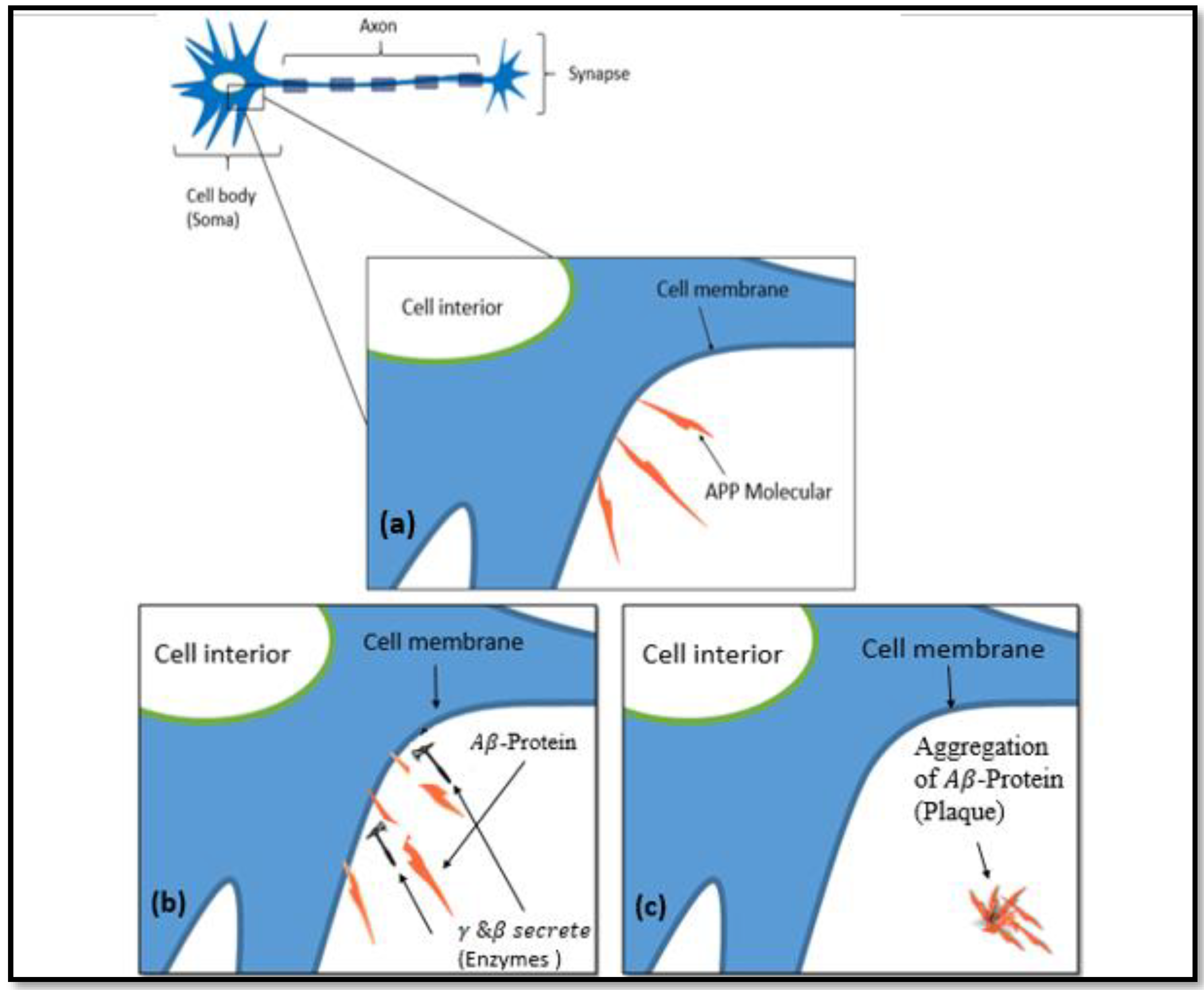
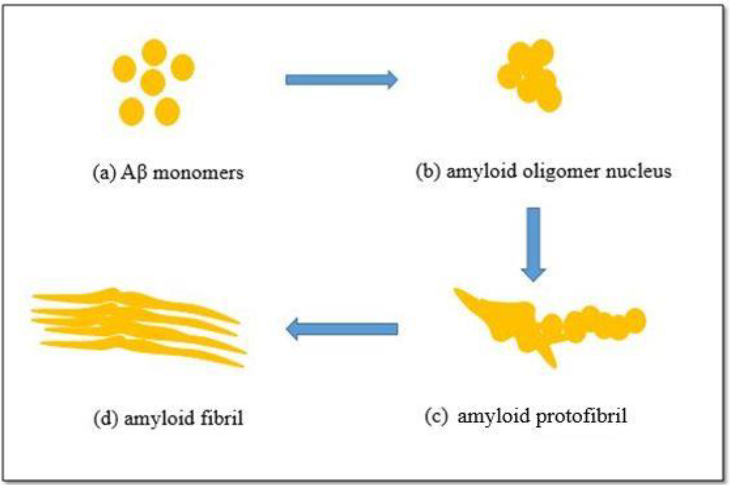
:1. Introduction
Basic Principle of Dielectrophoresis
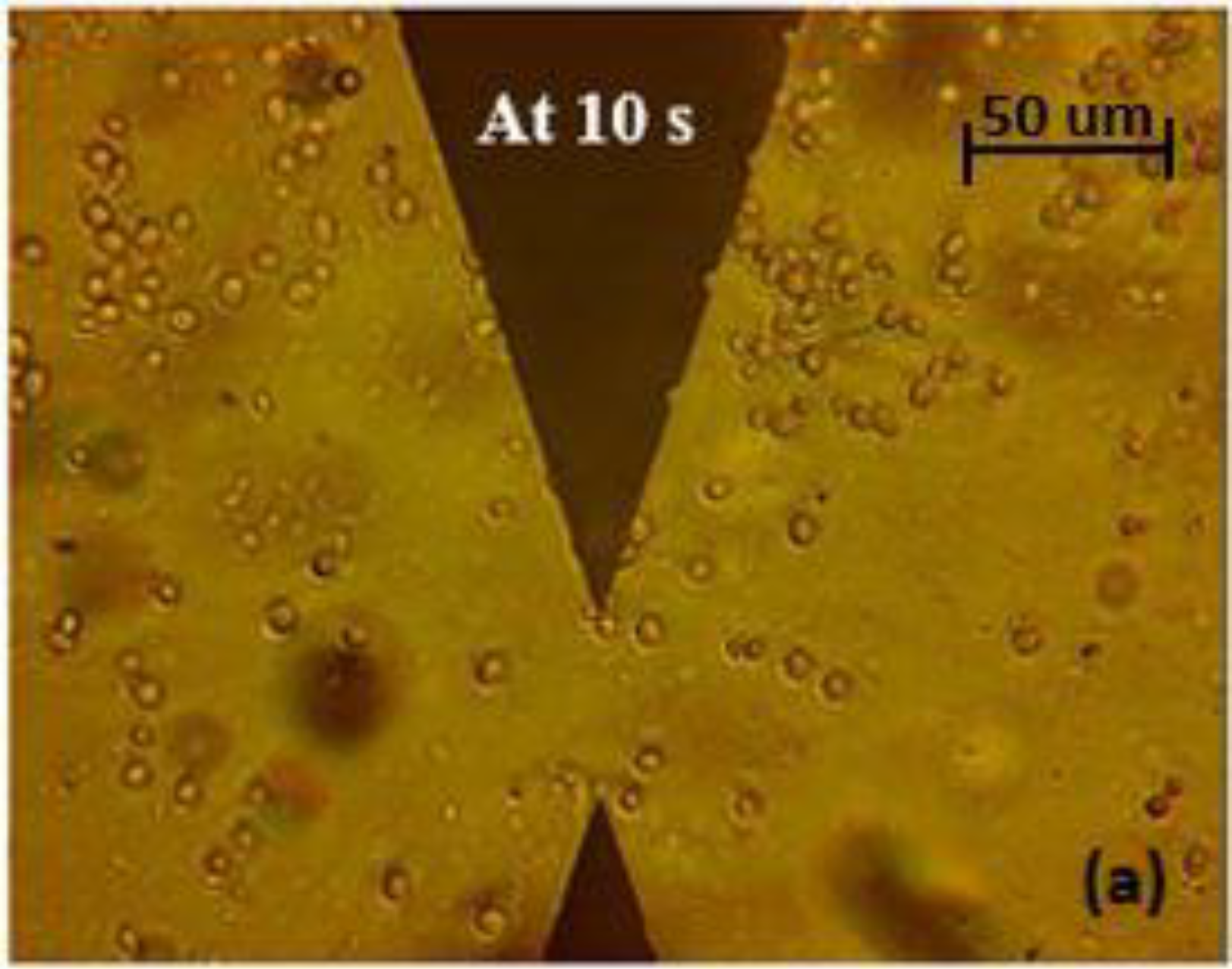
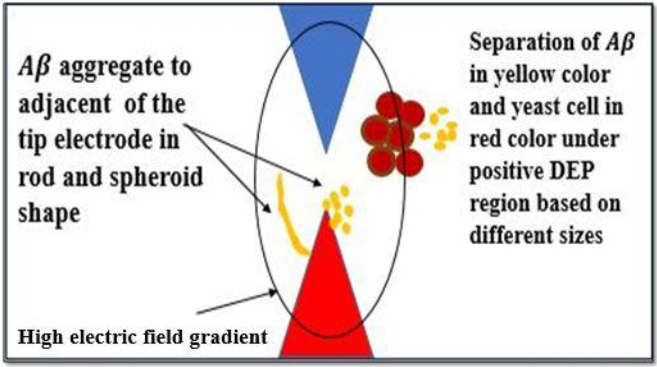
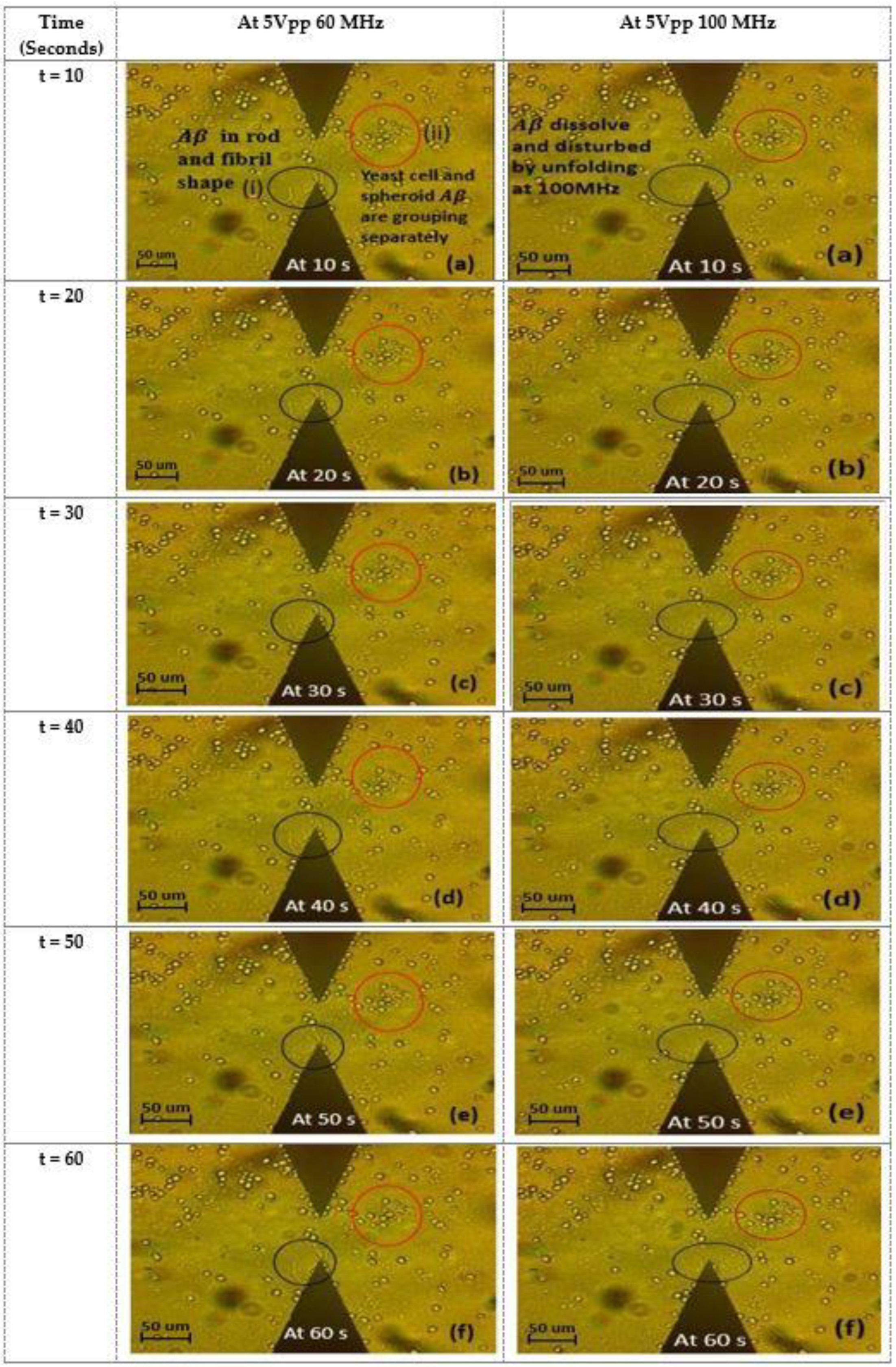
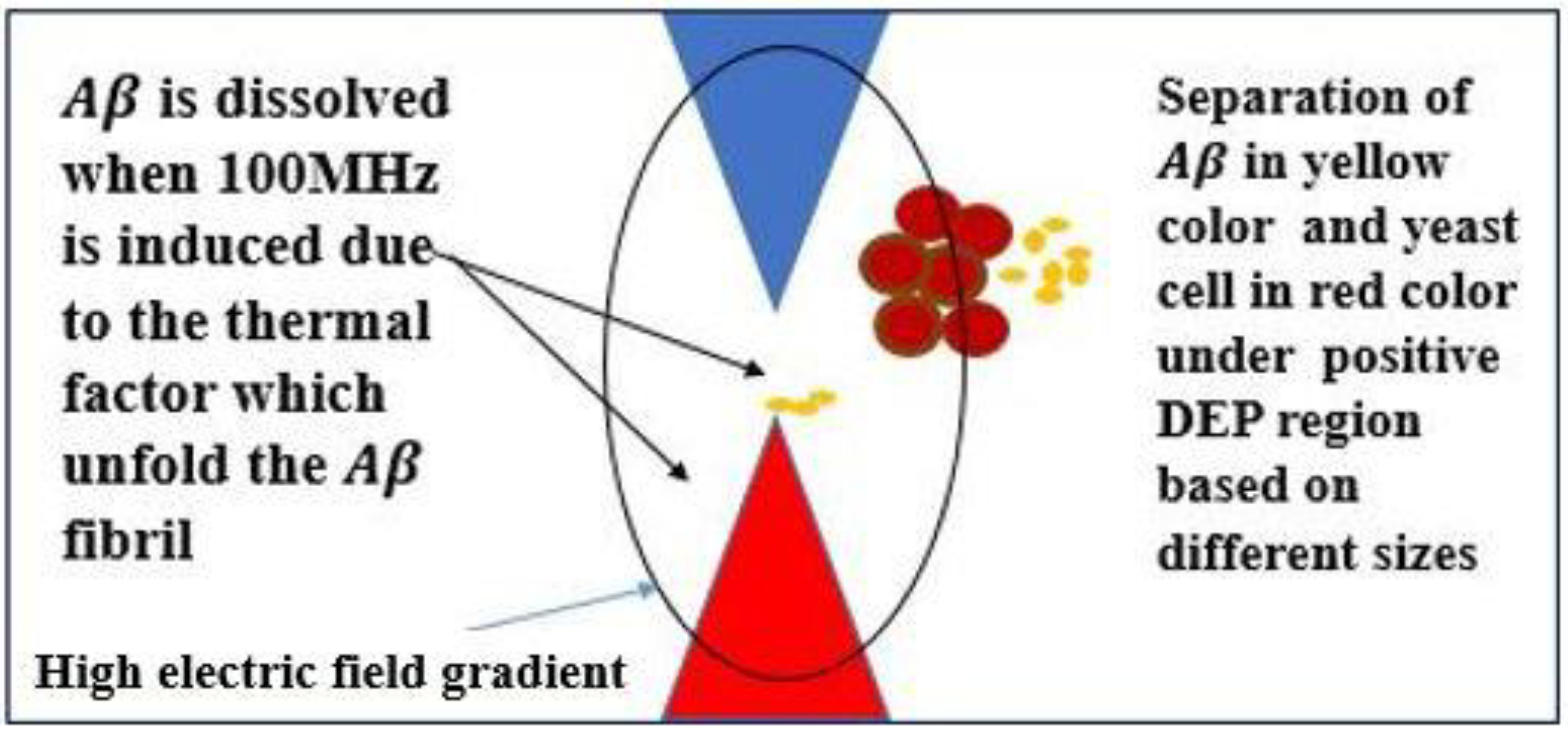
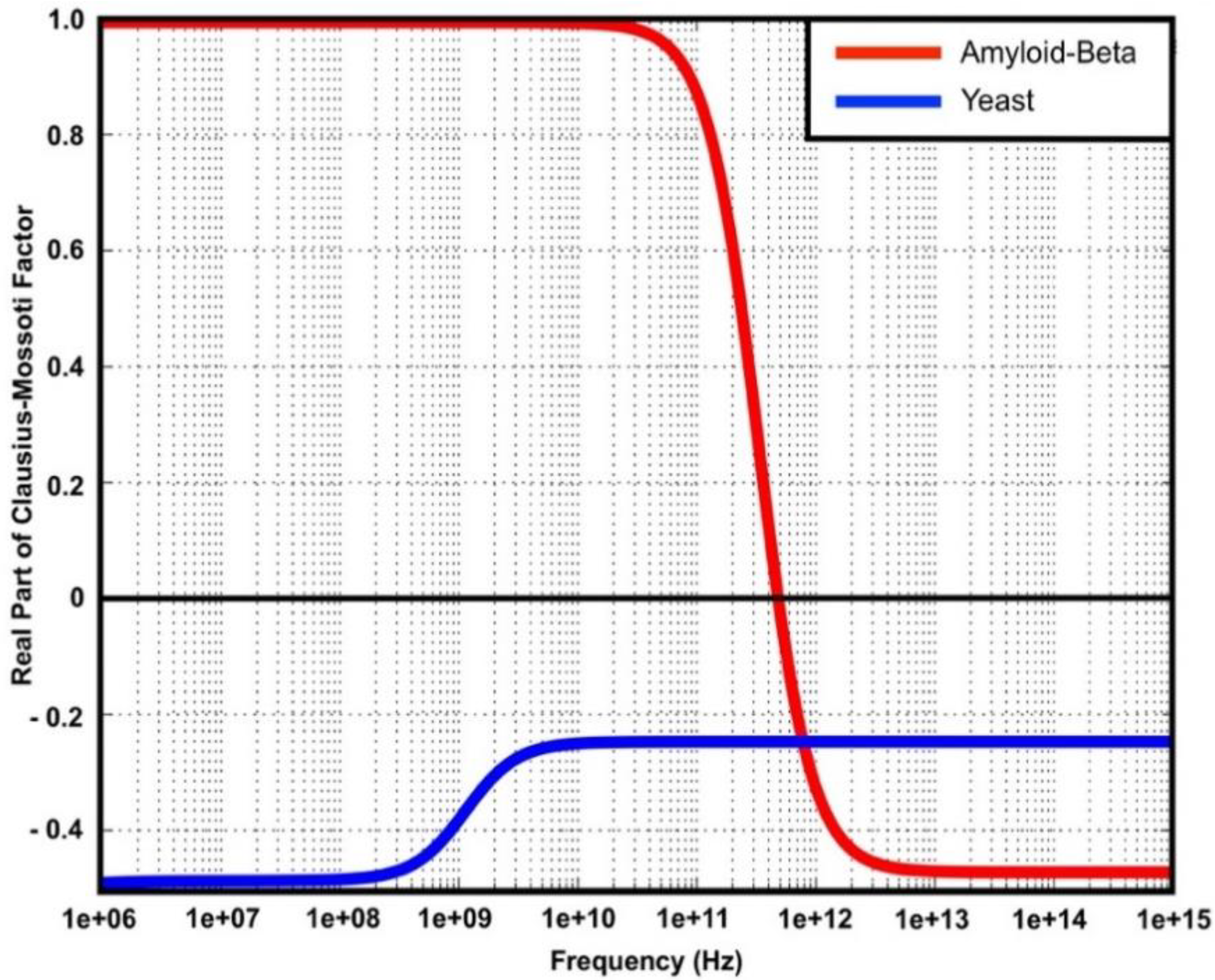
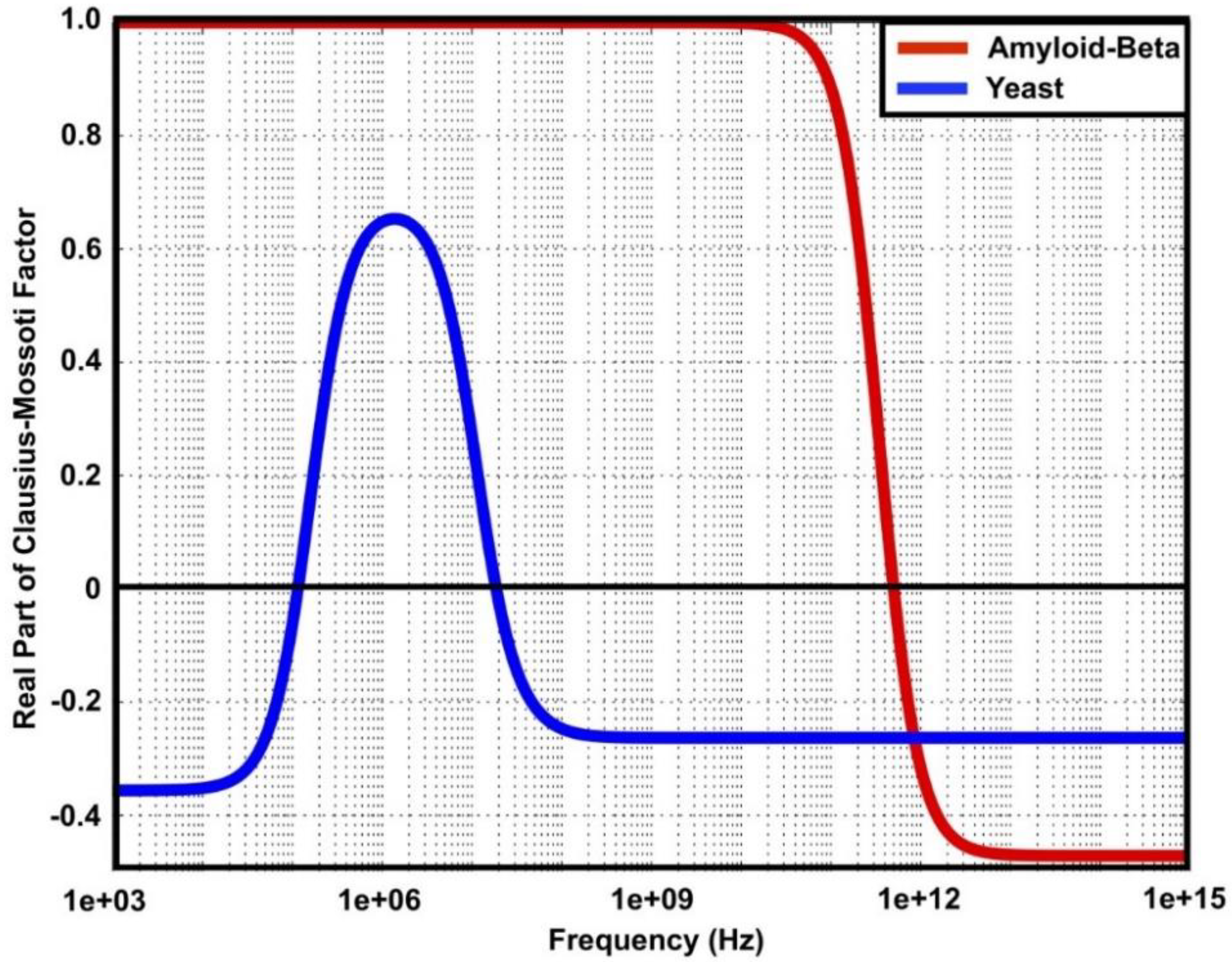
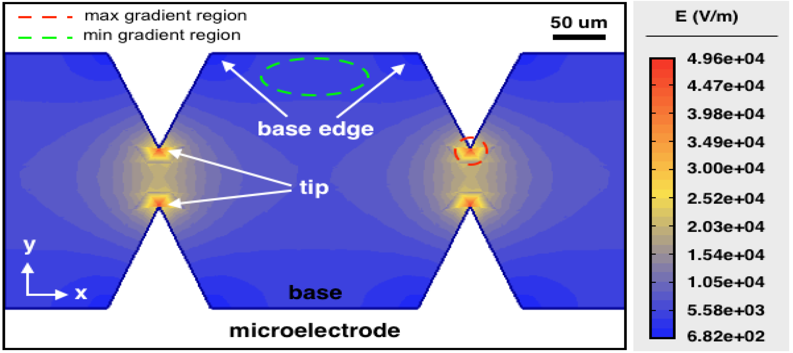
2. Results and Discussion
3. Methodology
3.1. Simulation
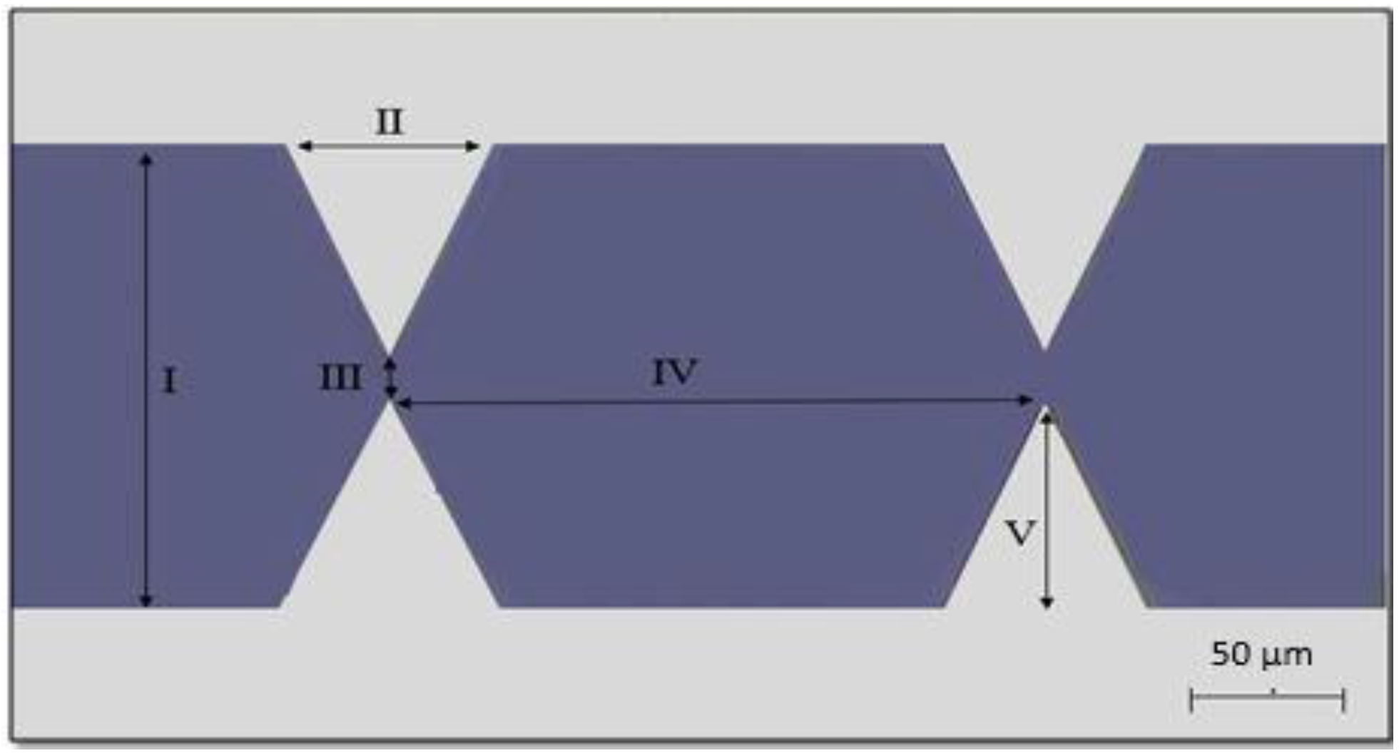
3.2. Fabrication Microelectrode Procedure
3.3. Preparing the Yeast Cells and Aβ Protein Sample
3.4. Experimental Procedure
4. Conclusions
Author Contributions
Funding
Conflicts of Interest
References
- Möller, H.J.; Graeber, M.B. The Case Described by Alois Alzheimer In 1911. Historical and Conceptual Perspectives Based on The Clinical Record And Neurohistological Sections. Eur. Arch. Psychiatry Clin. Neurosci. 1998, 248, 111–122. [Google Scholar] [PubMed]
- Aldavert-Vera, L.; Huguet, G.; Costa-Miserachs, D.; Ortiz, S.P.; Kádár, E.; Morgado-Bernal, I.; Segura-Torres, P. Intracranial Self-Stimulation Facilitates Active-Avoidance Retention and Induces Expression of C-Fos and Nurrl in Rat Brain Memory Systems. Behav. Brain Res. 2013, 250, 46–57. [Google Scholar] [CrossRef] [PubMed]
- Huguet, G.; Aldavert-Vera, L.; Kádár, E.; Peña de Ortiz, S.; Morgado-Bernal, I.; Segura-Torres, P. Intracranial Self-Stimulation to The Lateral Hypothalamus, a Memory Improving Treatment, Results in Hippocampal Changes in Gene Expression. Neuroscience 2009, 162, 359–374. [Google Scholar] [CrossRef]
- Klooster, D.C.W.; de Louw, A.J.A.; Aldenkamp, A.P.; Besseling, R.M.H. Neuroscience and Biobehavioral Reviews Technical Aspects of Neurostimulation: Focus on Equipment, Electric Field Modeling, and Stimulation Protocols. Neurosci. Biobehav. Rev. 2016, 65, 113–141. [Google Scholar] [CrossRef] [PubMed]
- Wimo, A.; Prince, M. World Alzheimer Report 2010 The Global Economic Impact of Dementia. Alzheimer’s Disease International, 2010; pp. 1–56. Available online: https://www.alz.co.uk/research/files/WorldAlzheimerReport2010ExecutiveSummary.pdf (accessed on 25 April 2019).
- Prince, M.; Knapp, M.; Guerchet, M.; McCrone, P.; Prina, M.; Comas-Herrera, A.; Wittenberg, R.; Adelaja, B.; Hu, B.; King, D.; et al. Dementia UK: Second Edition—Overview. Alzheimer’s Society, 2014; pp. 1–62. Available online: http://eprints.lse.ac.uk/59437/1/Dementia_UK_Second_edition_-_Overview.pdf (accessed on 25 April 2019).
- Leinenga, G.; Götz, J. Scanning Ultrasound Removes Amyloid-β and Restores Memory in an Alzheimer’ s Disease Mouse Model. Sci. Transl. Med. 2015, 7, 278ra33. [Google Scholar] [CrossRef] [PubMed]
- Priller, C.; Bauer, T.; Mitteregger, G.; Krebs, B.; Kretzschmar, H.A.; Herms, J. Synapse Formation and Function Is Modulated by the Amyloid Precursor Protein. J. Neurosci. 2006, 26, 7212–7221. [Google Scholar] [CrossRef]
- Querfurth, H.W.; Laferla, F.M. Alzheimer’s Disease. N. Engl. J. Med. 2010, 362, 329–344. [Google Scholar] [CrossRef]
- Gendron, T.F.; Petrucelli, L. The Role of Tau in Neurodegeneration. Mol. Neurodegener. 2009, 4, 13. [Google Scholar] [CrossRef]
- Laxton, A.W.; Tang-Wai, D.F.; McAndrews, M.P.; Zumsteg, D.; Wennberg, R.; Keren, R.; Wherrett, J.; Naglie, G.; Hamani, C.; Smith, G.S.; et al. A Phase I Trial of Deep Brain Stimulation of Memory Circuits in Alzheimer’ s Disease. Ann. Neurol. 2010, 68, 521–534. [Google Scholar] [CrossRef]
- Ren, Y. Towards Brain-on-a-Chip: Microfluidic and Microelectrode Array Platforms for Morphological and Electrophysiological Observations on the Propagation of Alzheimer’s Disease. Ph.D. Thesis, The École Polytechnique Fédérale de Lausanne, Lausanne, Switzerland, October 2015. [Google Scholar]
- Sabbagh, J.J.; Kinney, J.W.; Cummings, J.L. Animal systems in The Development of Treatments for Alzheimer’s Disease: Challenges, Methods, and Implications. Neurobiol. Aging 2013, 34, 169–183. [Google Scholar] [CrossRef]
- Exley, C.; Esiri, M.M. Severe Cerebral Congophilic Angiopathy Coincident with Increased Brain Aluminium in a Resident of Camelford, Cornwall, UK. J. Neurol. Neurosurg. Psychiatry 2006, 77, 877–879. [Google Scholar] [CrossRef] [PubMed]
- Patel, V.P.; Chu, C.T. Nuclear Transport, Oxidative Stress, and Neurodegeneration. Int. J. Clin. Exp. Pathol. 2011, 4, 215–229. [Google Scholar] [PubMed]
- Hubbard, R.E.; Haider, M.K. Hydrogen Bonds in Proteins: Role and Strength. Encycl. Life Sci. 2010. [Google Scholar] [CrossRef]
- Cooper, A. Heat Capacity of Hydrogen-Bonded Networks: An Alternative View of Protein Folding Thermodynamics. Biophys. Chem. 2000, 85, 25–39. [Google Scholar] [CrossRef]
- Khoshmanesh, K.; Nahavandi, S.; Baratchi, S.; Mitchell, A.; Kalantar-zadeh, K. Biosensors and Bioelectronics Dielectrophoretic Platforms for Bio-microfluidic Systems. Biosens. Bioelectron. 2011, 26, 1800–1814. [Google Scholar] [CrossRef] [PubMed]
- Khoshmanesh, K.; Zhang, C.; Campbell, J.L.; Kayani, A.A.; Nahavandi, S.; Mitchell, A.; Kalantar-Zadeh, K. Dielectrophoretically Assembled Particles: Feasibility for Optofluidic Systems. Microfluid. Nanofluidics 2010, 9, 755–763. [Google Scholar] [CrossRef]
- Ali, M.A.M.; Ostrikov, K.; Khalid, F.A.; Majlis, B.Y.; Kayani, A.A. Active Bioparticle Manipulation in Microfluidic Systems. RSC Adv. 2016, 6, 113066–113094. [Google Scholar] [CrossRef]
- Kung, Y.C.; Huang, K.W.; Chong, W.; Chiou, P.Y. Tunnel Dielectrophoresis for Tunable, Single-Stream Cell Focusing in Physiological Buffers in High-Speed Microfluidic Flows. Small 2016, 12, 4343–4348. [Google Scholar] [CrossRef] [PubMed]
- Kung, Y.C.; Huang, K.W.; Fan, Y.J.; Chiou, P.Y. Fabrication of 3D High Aspect Ratio PDMS Microfluidic Networks with a Hybrid stamp. Lab Chip 2015, 15, 1861–1868. [Google Scholar] [CrossRef]
- Waheed, W.; Alazzam, A.; Abu-Nada, E.; Khashan, S.; Abutayeh, M. A Microfluidics Device for 3D Switching of Microparticles Using Dielectrophoresis. J. Electrostat. 2018, 94, 1–7. [Google Scholar] [CrossRef]
- Mohamad, A.S.; Hamzah, R.; Hoettges, K.F.; Hughes, M.P. A Dielectrophoresis-Impedance Method for Protein Detection and Analysis. AIP Adv. 2017, 7, 015202. [Google Scholar] [CrossRef]
- Ali, M.A.M.; Majlis, B.Y.; Azman, Z.N.; Kayani, A.A. Cell-Cell Contact Configurations by Dielectrophoresis for Electrofusion: Study on Directions, Stability and Dielectric Heating Effect. In Proceedings of the 2016 IEEE EMBS Conference on Biomedical Engineering and Sciences (IECBES), Kuala Lumpur, Malaysia, 4–8 December 2017; pp. 539–544. [Google Scholar]
- Kayani, A.A.; Khoshmanesh, K.; Ward, S.A.; Mitchell, A.; Kalantar-zadeh, K. Optofluidics Incorporating Actively Controlled Micro- and Nano-Particles Optofluidics Incorporating Actively Controlled. Biomicrofluidics 2012, 6, 031501. [Google Scholar] [CrossRef] [PubMed]
- Kayani, A.A.; Chrimes, A.F.; Khoshmanesh, K.; Sivan, V.; Zeller, E.; Kalantar-zadeh, K.; Mitchell, A. Interaction of Guided Light in Rib Polymer Waveguides with Dielectrophoretically Controlled Nanoparticles. Microfluid. Nanofluidics 2011, 11, 93–104. [Google Scholar] [CrossRef]
- Chrimes, A.F.; Kayani, A.A.; Khoshmanesh, K.; Stoddart, P.R.; Mulvaney, P.; Mitchell, A.; Kalantar-Zadeh, K. Dielectrophoresis-Raman Spectroscopy System for Analysing Suspended Nanoparticles. Lab Chip 2010, 11, 921–928. [Google Scholar] [CrossRef] [PubMed]
- Yi, P.; Kayani, A.A.; Chrimes, A.F.; Ghorbani, K.; Nahavandi, S.; Kalantar-zadeh, K.; Khoshmanesh, K. Thermal Analysis of Nanofluids in Microfluidics Using an Infrared Camera. Lab Chip 2012, 12, 2520–2525. [Google Scholar] [CrossRef] [PubMed]
- Ramos, A.; Morgan, H.; Green, N.G.; Castellanos, A. AC Electrokinetics: A Review of Forces in Microelectrode Structures. J. Phys. D Appl. Phys. 1998, 31, 2338–2353. [Google Scholar] [CrossRef]
- Chan, J.Y.; Ahmad Kayani, A.B.; Md Ali, M.A.; Kok, C.K.; Yeop Majlis, B.; Hoe, S.L.L.; Marzuki, M.; Khoo, A.S.; Ostrikov, K.K.; Ataur Rahman, M.; et al. Dielectrophoresis-Based Microfluidic Platforms for Cancer Diagnostics. Biomicrofluidics 2018, 12, 011503. [Google Scholar] [CrossRef] [PubMed]
- Jeanmonod, D.J.; Suzuki, R.K.; Hrabovsky, M. Supramolecular Organization of Amyloid Fibrils. Intech Open 2018, 2, 64. [Google Scholar]
- Schleeger, M.; Deckert-Gaudig, T.; Deckert, V.; Velikov, K.P.; Koenderink, G.; Bonn, M. Amyloids: From Molecular Structure to Mechanical Properties. Polymer 2013, 54, 2473–2488. [Google Scholar] [CrossRef]
- Domigan, L.; Andersen, K.B.; Sasso, L.; Dimaki, M.; Svendsen, W.E.; Gerrard, J.A.; Castillo-León, J. Dielectrophoretic Manipulation and Solubility of Protein Nanofibrils Formed from Crude Crystallins. Electrophoresis 2013, 34, 1105–1112. [Google Scholar] [CrossRef]
- Fischer, H.E. Scholars’ Mine an AC Method of Measuring the Conductivity of Dielectric Liquids. Master’s Thesis, Missouri University of Science and Technology, St. Rolla, MO, USA, 1966. [Google Scholar]
- Boulanger, L. Observations on Variations in Electrical Conductivity of Pure Demineralized Water: Modification (‘Activation’) of Conductivity by Low-Frequency, Low-Level Alternativing Electric Fields. Int. J. Biometeorol. 1998, 41, 137–140. [Google Scholar] [CrossRef]
- Gitlin, I.; Carbeck, J.D.; Whitesides, G.M. Proteins Why Are Proteins Charged? Networks of Charge—Charge Interactions in Proteins Measured by Charge Ladders and Capillary Electrophoresis Angewandte. Angew. Chem. Int. Ed. 2006, 45, 3022–3060. [Google Scholar] [CrossRef] [PubMed]
- Patel, S.; Showers, D.; Vedantam, P.; Tzeng, T.R.; Qian, S.; Xuan, X. Microfluidic Separation of Live and Dead Yeast Cells Using Reservoir-Based Dielectrophoresis. Biomicrofluidics 2012, 6, 034102. [Google Scholar] [CrossRef] [PubMed]
- Zheng, Y.; Nguyen, J.; Wang, C.; Sun, Y. Electrical Measurement of Red Blood Cell Deformability on a Microfluidic Device. Lab Chip 2013, 13, 3275–3283. [Google Scholar] [CrossRef] [PubMed]
- Chaparro, C.V.; Herrera, L.V.; Meléndez, A.M.; Miranda, D.A. Considerations on electrical impedance measurements of electrolyte solutions in a four-electrode cell. J. Phys. Conf. Ser. 2016, 687, 12101. [Google Scholar] [CrossRef] [Green Version]
- Ali, M.A.M.; Kayani, A.B.A.; Yeo, L.Y.; Chrimes, A.F.; Ahmad, M.Z.; Ostrikov, K.K.; Majlis, B.Y. Microfluidic Dielectrophoretic Cell Manipulation Towards Stable Cell Contact Assemblies. Biomed. Microdevices 2018, 20, 95. [Google Scholar] [CrossRef] [PubMed]
- Yafouz, B.; Kadri, N.A.; Ibrahim, F. Microarray Dot Electrodes Utilizing Dielectrophoresis for Cell Characterization. Sensors 2013, 13, 9029–9046. [Google Scholar] [CrossRef] [PubMed] [Green Version]
- Alhammadi, F.; Waheed, W.; El-Khasawneh, B.; Alazzam, A. Continuous-Flow Cell Dipping and Medium Exchange in a Microdevice Using Dielectrophoresis. Micromachines 2018, 9, 223. [Google Scholar] [CrossRef]
- Moosavi, B.; Mousavi, B.; Macreadie, I.G. Yeast Model of Amyloid-β and Tau Aggregation in Alzheimer’s Disease. J. Alzheimer’s Dis. 2015, 47, 9–16. [Google Scholar] [CrossRef]
- Johnson, R.D.; Schauerte, J.A.; Wisser, K.C.; Gafni, A.; Steel, D.G. Direct Observation of Single Amyloid-β(1-40) Oligomers on Live Cells: Binding and Growth at Physiological Concentrations. PLoS ONE 2011, 6, e23970. [Google Scholar] [CrossRef]
- Ryan, T.M.; Caine, J.; Mertens, H.D.; Kirby, N.; Nigro, J.; Breheney, K.; Waddington, L.J.; Streltsov, V.A.; Curtain, C.; Masters, C.L.; et al. Ammonium Hydroxide Treatment of Aβ Produces an Aggregate Free Solution Suitable for Biophysical and Cell Culture Characterization. PeerJ 2013, 1, e73. [Google Scholar] [CrossRef] [PubMed]
- Hellstrand, E.; Boland, B.; Walsh, D.M.; Linse, S. Amyloid Protein Aggregation Produces Highly Reproducible Kinetic Data and Occurs by a Two-Phase Process. ACS Chem. Neurosci. 2010, 1, 13–18. [Google Scholar] [CrossRef] [PubMed]
- Kayani, A.A.; Khoshmanesh, K.; Nguyen, T.G.; Kostovski, G.; Chrimes, A.F.; Nasabi, M.; Heller, D.A.; Mitchell, A.; Kalantar-zadeh, K. Dynamic Manipulation of Modes in an Optical Waveguide Using Dielectrophoresis. Electrophoresis 2012, 13, 2075–2085. [Google Scholar] [CrossRef] [PubMed]
- Khoshmanesh, K.; Tovar-Lopez, F.J.; Baratchi, S.; Zhang, C.; Kayani, A.A.; Chrimes, A.F.; Nahavandi, S.; Wlodkowic, D.; Mitchell, A.; Kalantar-zadeh, K. Dielectrophoresis of Micro/Nano Particles Using Curved Microelectrodes. Proc. SPIE Int. Soc. Opt. Eng. 2011. [Google Scholar] [CrossRef]
- Wang, P.; Chang, H. Bacteria Capture, Concentration and Detection by Alternating Current Dielectrophoresis and Self-Assembly of Dispersed Single-Wall Carbon Nanotubes. Electrophoresis 2006, 27, 1376–1385. [Google Scholar]
- Staton, S.J.R.; Jones, P.V.; Ku, G.; Gilman, S.D.; Kheterpal, I.; Hayes, M.A. Manipulation and Capture of Aβ Amyloid Fibrils and Monomers by DC Insulator Gradient Dielectrophoresis (DC-iGDEP). Analyst 2012, 137, 3227. [Google Scholar] [CrossRef] [PubMed]


| Area | Temperature Increase, Δθ | Final Temperature (Initial = 27 °C) |
|---|---|---|
| At microtip (highest electric field region with 50 kV/m) | 5.952 °C | 32.925 °C |
| At electrode base (lowest electric field region with 680 V/m) | 0.001 °C | 27.001 °C |
| Particle | Parameter | Value | Reference |
|---|---|---|---|
| Aβ Protein | protein size, rAβ | 5 to 15 (nm) | [7,37] |
| protein conductivity, σAβ | 3 × 10³ (S/m) | ||
| core permittivity, εAβ | (2 to 4) εo (F/m) | ||
| interface permittivity, εint | (10 to 20) εo (F/m) | ||
| Yeast | membrane thickness, dmem | 8 (nm) | [38] |
| cell wall thickness, dwall | 220 (nm) | ||
| cell radius, ryeast | 3 (μm) | ||
| cytoplasm conductivity, σcyt | 0.2 (S/m) | ||
| membrane conductivity, σmem | 2.5 × 10⁻⁸ (S/m) | ||
| cell wall conductivity, σwall | 1.4 × 10⁻³ (S/m) | ||
| cytoplasm permittivity, εcyt | 50 εo (F/m) | ||
| membrane permittivity, εmem | 6 εo (F/m) | ||
| cell wall permittivity, εwall | 60 εo (F/m) | ||
| PBS (50%) | medium conductivity, σm | 6 (S/m) | [39,40] |
| medium permittivity, εm | 80 εo (F/m) | ||
| DIW | Medium conductivity, σm | 0.01(S/m) | [41] |
| Medium permittivity, εm | 78 εo (F/m) |
© 2019 by the authors. Licensee MDPI, Basel, Switzerland. This article is an open access article distributed under the terms and conditions of the Creative Commons Attribution (CC BY) license (http://creativecommons.org/licenses/by/4.0/).
Share and Cite
Al-Ahdal, S.A.; Ahmad Kayani, A.B.; Md Ali, M.A.; Chan, J.Y.; Ali, T.; Adnan, N.; Buyong, M.R.; Mhd Noor, E.E.; Majlis, B.Y.; Sriram, S. Dielectrophoresis of Amyloid-Beta Proteins as a Microfluidic Template for Alzheimer’s Research. Int. J. Mol. Sci. 2019, 20, 3595. https://0-doi-org.brum.beds.ac.uk/10.3390/ijms20143595
Al-Ahdal SA, Ahmad Kayani AB, Md Ali MA, Chan JY, Ali T, Adnan N, Buyong MR, Mhd Noor EE, Majlis BY, Sriram S. Dielectrophoresis of Amyloid-Beta Proteins as a Microfluidic Template for Alzheimer’s Research. International Journal of Molecular Sciences. 2019; 20(14):3595. https://0-doi-org.brum.beds.ac.uk/10.3390/ijms20143595
Chicago/Turabian StyleAl-Ahdal, Salman Ali, Aminuddin Bin Ahmad Kayani, Mohd Anuar Md Ali, Jun Yuan Chan, Talal Ali, Norah Adnan, Muhamad Ramdzan Buyong, Ervina Efzan Mhd Noor, Burhanuddin Yeop Majlis, and Sharath Sriram. 2019. "Dielectrophoresis of Amyloid-Beta Proteins as a Microfluidic Template for Alzheimer’s Research" International Journal of Molecular Sciences 20, no. 14: 3595. https://0-doi-org.brum.beds.ac.uk/10.3390/ijms20143595