Article

OsMSR3, a Small Heat Shock Protein, Confers Enhanced Tolerance to Copper Stress in Arabidopsis thaliana

1
Key Laboratory of Agro-ecological Processes in Subtropical Region, Institute of Subtropical Agriculture, Chinese Academy of Sciences, Changsha 410125, China
2
Zhengzhou Tobacco Research Institute of China National Tobacco Corporation, Zhengzhou 450001, China
3
State Key Laboratory of Hybrid Rice, Hunan Hybrid Rice Research Centre, Changsha 410125, China
*
Author to whom correspondence should be addressed.
Int. J. Mol. Sci. 2019, 20(23), 6096; https://0-doi-org.brum.beds.ac.uk/10.3390/ijms20236096
Submission received: 21 October 2019 / Revised: 29 November 2019 / Accepted: 30 November 2019 / Published: 3 December 2019
(This article belongs to the Special Issue Heavy Metals Accumulation, Toxicity and Detoxification in Plants)

Abstract

:
Copper is a mineral element essential for the normal growth and development of plants; however, excessive levels can severely affect plant growth and development. Oryza sativa L. multiple stress-responsive gene 3 (OsMSR3) is a small, low-molecular-weight heat shock protein (HSP) gene. A previous study has shown that OsMSR3 expression improves the tolerance of Arabidopsis to cadmium stress. However, the role of OsMSR3 in the Cu stress response of plants remains unclear, and, thus, this study aimed to elucidate this phenomenon in Arabidopsis thaliana, to further understand the role of small HSPs (sHSPs) in heavy metal resistance in plants. Under Cu stress, transgenic A. thaliana expressing OsMSR3 showed higher tolerance to Cu, longer roots, higher survival rates, biomass, and relative water content, and accumulated more Cu, abscisic acid (ABA), hydrogen peroxide, chlorophyll, carotenoid, superoxide dismutase, and peroxidase than wild-type plants did. Moreover, OsMSR3 expression in A. thaliana increased the expression of antioxidant-related and ABA-responsive genes. Collectively, our findings suggest that OsMSR3 played an important role in regulating Cu tolerance in plants and improved their tolerance to Cu stress through enhanced activation of antioxidative defense mechanisms and positive regulation of ABA-responsive gene expression.

1. Introduction

Copper is an essential mineral element for the normal growth and development of plants. In plants, Cu functions as an important cofactor for metalloproteins and participates in numerous biological processes, including photosynthesis, respiration, oxygen superoxide scavenging, cell wall metabolism and lignification, and ethylene perception [1,2,3,4,5,6]. Cu-deficient soils not only affect the quality and quantity of plant food crops but also reduce their nutritional value as the main source of essential minerals for humans [7]. In addition, exposure of plants to excess Cu interferes with normal growth, proliferation, and differentiation of most plant cells [8,9,10,11,12,13,14]. One of the earliest and most obvious symptoms of Cu stress is inhibition of primary root elongation [15,16,17], while its prominent manifestations are decreased proliferation of root meristem cells [18], impaired cell integrity [19], and cell death [20]. Excessive accumulation of Cu in plants leads to the production of reactive oxygen species (ROS), which are toxic owing to their high redox activity [21].
Plants have developed specific mechanisms to prevent Cu toxicity by tightly regulating Cu homeostasis, including Cu uptake, translocation, efflux, and sequestration [22]. Plants also activate antioxidant defense responses to mitigate oxidative damage caused by free Cu ions in the cytosol [23]. The defense system includes ROS-removing enzymes, such as superoxide dismutase (SOD), peroxidase (POD), and catalase (CAT), as well as low-molecular-weight antioxidants, such as ascorbic acid (ASC) and glutathione (GSH) [24]. These antioxidant compounds and enzymes can be used as physiological indicators for evaluating plant antioxidant defense ability [15,16,17,18,19,20,21,22,23,24,25,26].
The stress hormone abscisic acid (ABA) plays an important role in plant stress tolerance. Heavy metals such as Cd, Hg, and Cu can induce the expression of ABA synthesis genes, which in turn increase the endogenous level of ABA [27,28]. A previous study showed that cadmium treatment increases endogenous ABA levels in cattail and reed roots [29], potato tubers [30], and rice plants [31]. Exposure to high Cu concentrations also increased ABA levels in rice [31]. During the germination of wheat seeds, ABA levels increased in the presence of Hg, Cd, and Cu stress [32]. Plants exposed to heavy metal stress showed an increased concentration of ABA, which indicates that the hormone is involved in the protective mechanism against heavy metal toxicity [27,33,34].
In plants, small heat shock proteins (sHSPs) with monomer sizes ranging from 12 to 42 kDa are more diverse and abundant than those in other organisms. There are 23 sHSPs in rice, and these are proposed to be categorized into fourteen classes [35]. Classes CI-CXI (nine subfamilies) are localized in the nucleus or cytoplasm, whereas the other five are positioned in the endoplasmic reticulum, mitochondria, plastid, and peroxisome [25,26,27,28,29,30,31,32,33,34,35,36]. These sHSPs are stimulated in response to a wide range of abiotic stresses. For instance, Oshsp26, which encodes a chloroplast-localized sHSP, has been shown to enhance tolerance against oxidative and heat stress in tall fescue [37]. Overexpression of sHSP17.7 enhances drought tolerance in transgenic rice [38]. Overexpression of OsHsp18.0-CI, an sHSP-CI family gene, enhances resistance to bacterial leaf streak in rice [39]. However, there are only a few studies on the sHSPs involved in heavy metal resistance.
Our previous studies have shown that OsMSR3 belongs to the class I sHSP family [40]. The expression of the OsMSR3 gene significantly enhances tolerance of Arabidopsis thaliana (L.) Heynh (A. thaliana) to cadmium stress [41]. However, the molecular mechanism of OsMSR3-induced Cu tolerance is poorly understood. In this study, we determined that the expression of OsMSR3 was upregulated by Cu stress. Therefore, we speculated that OsMSR3 plays an important role in plant tolerance to Cu. Expression of OsMSR3 enhanced the Cu stress tolerance of A. thaliana. Our research enhances the understanding of the role of sHSPs in heavy metal resistance in plants.

2. Results

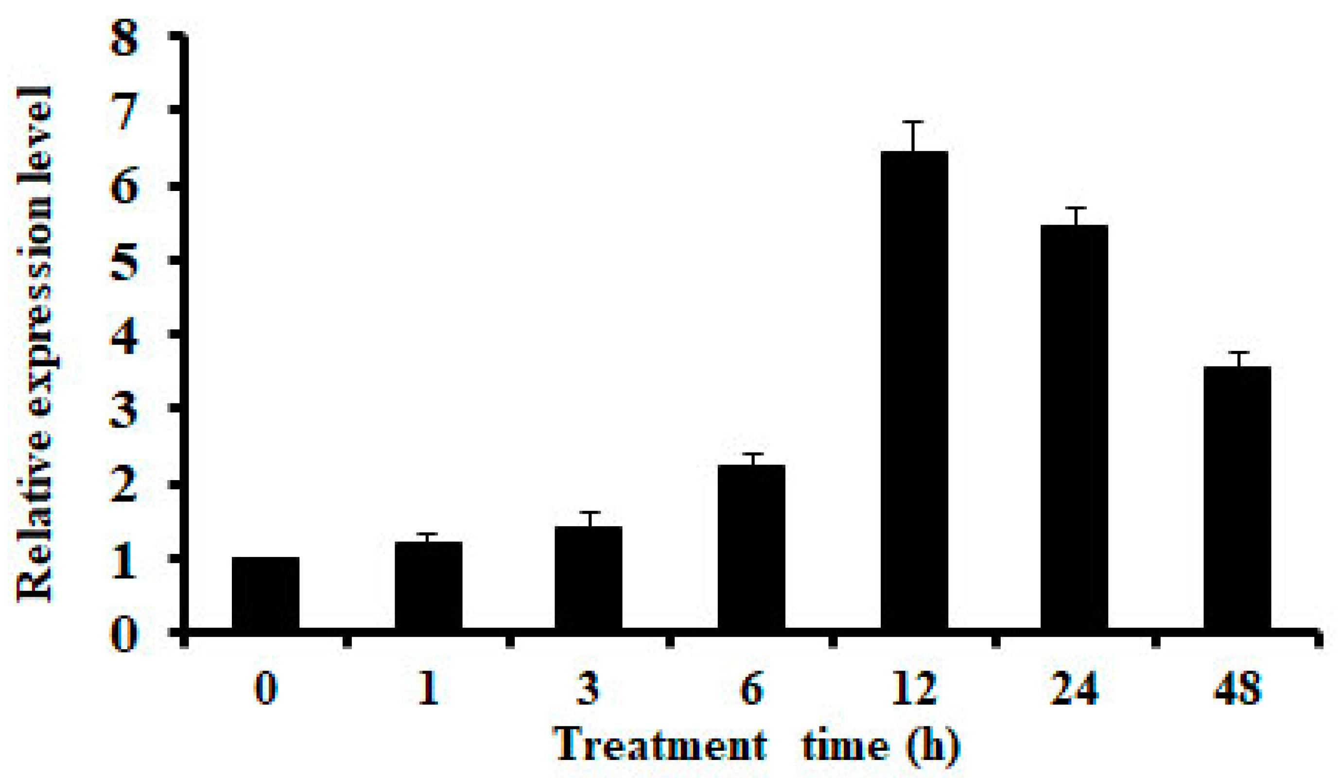
2.1. Expression of OsMSR3 is Induced by Cu Stress

The quantitative reverse transcription-polymerase chain reaction (qRT-PCR) showed that the expression of OsMSR3 increased rapidly after 6 h of Cu stress and peaked at 12 h in Pei’ai 64S rice seedlings (Figure 1). Subsequently, OsMSR3 expression decreased by nearly 3.5-fold compared to the control levels at 48 h (Figure 1).

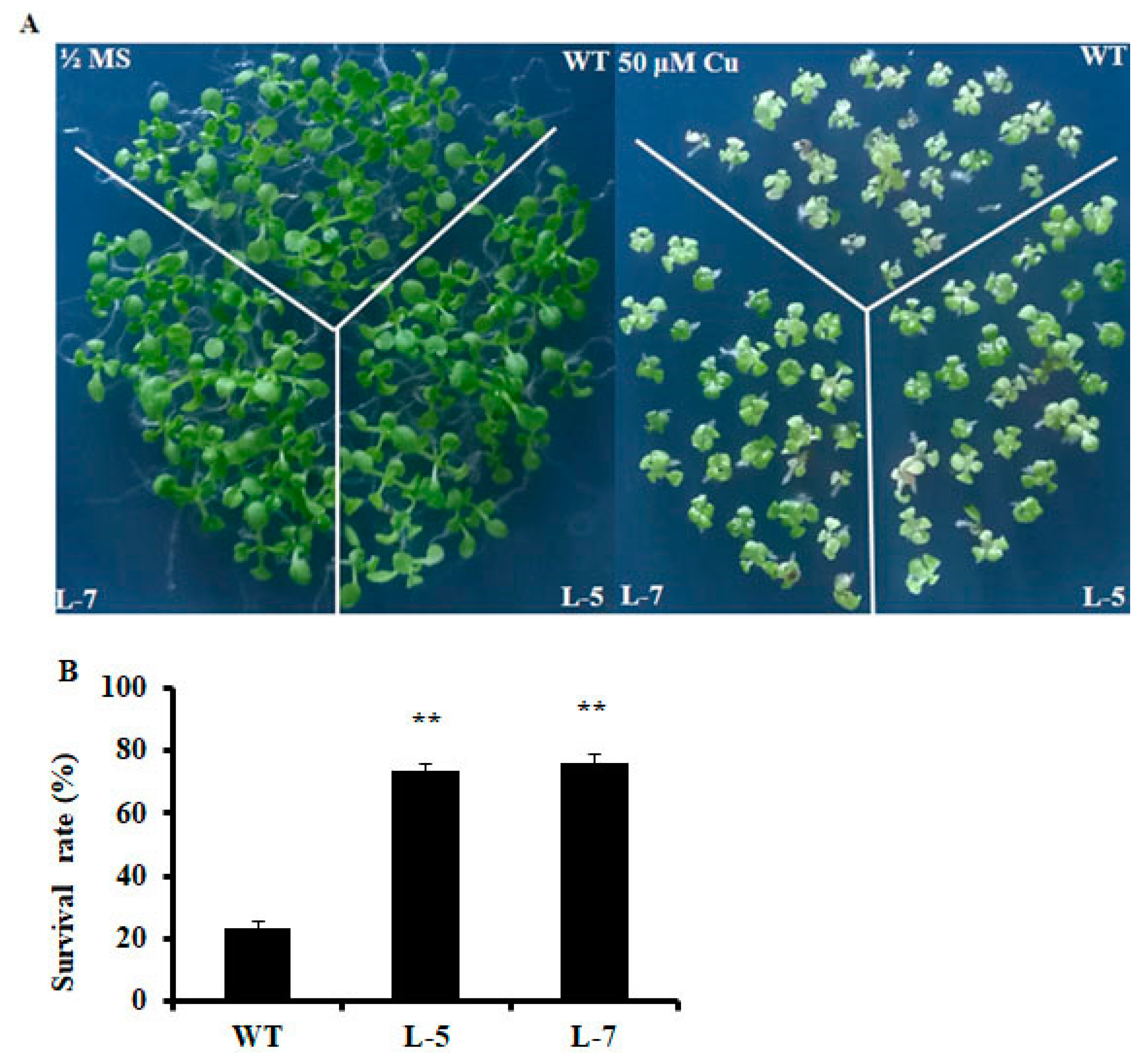
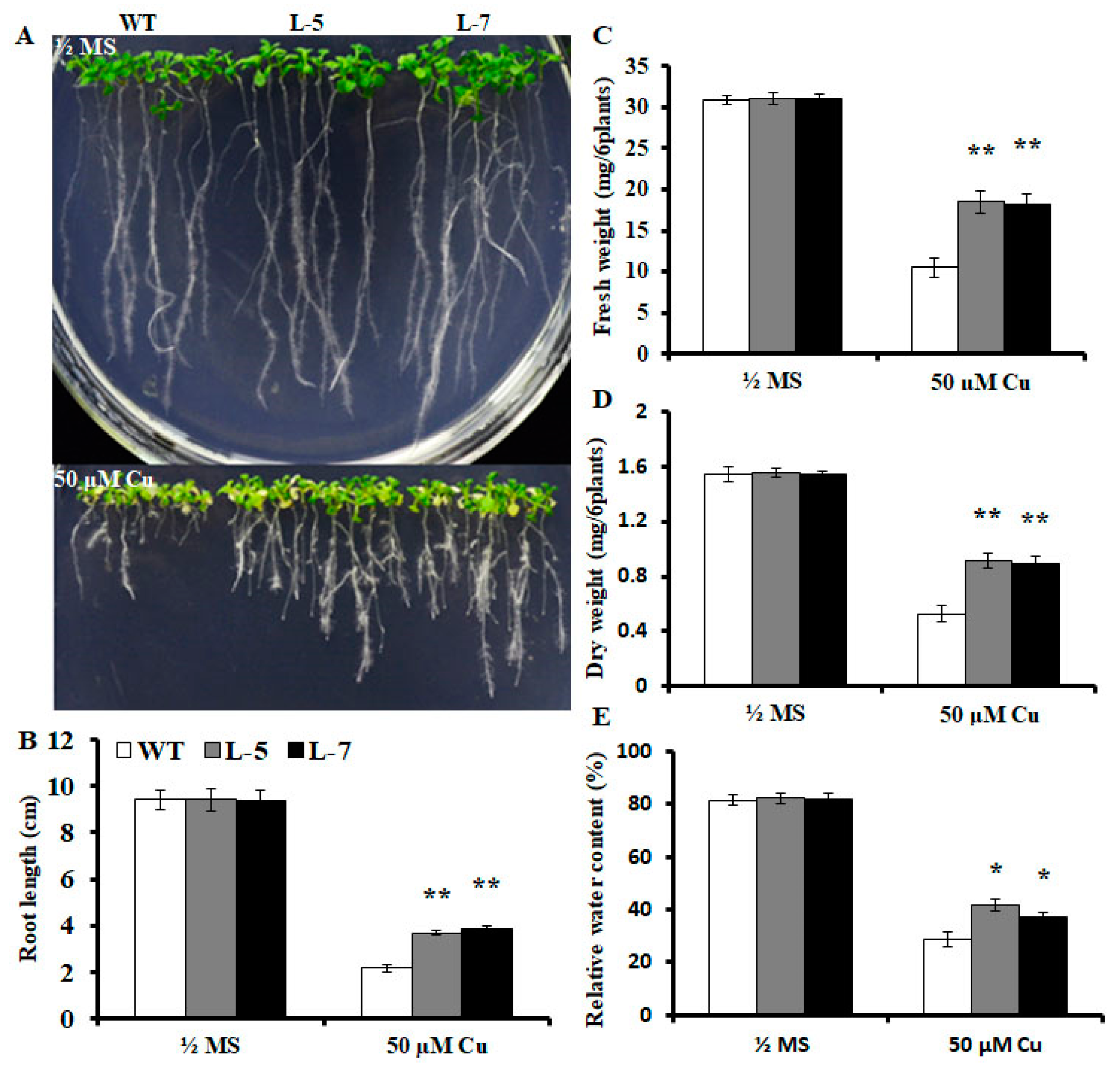
2.2. Expression of OsMSR3 Enhances Cu Tolerance of Transgenic A. thaliana

To assess whether upregulation of OsMSR3 enhances tolerance to Cu, transgenic Arabidopsis plants expressing OsMSR3 were generated and analyzed. Based on a previous study [41], two independent transgenic lines, L-5 and L-7, were chosen for further experiments. We examined survival rates and root lengths of both OsMSR3-expressing and wild-type plants treated with Cu (50 μM). As shown in Figure 2A, there was no significant difference in survival rate and root length between wild-type and transgenic seedlings in the absence of stress. However, in half-strength Murashige and Skoog (½ MS) medium supplemented with Cu, transgenic plants showed higher Cu tolerance than wild-type plants did, with a higher survival rate and longer root length (Figure 2B and Figure 3B). Furthermore, the fresh and dry weight of wild-type and transgenic plants measured under normal and Cu stress did not differ significantly in the absence of stress, but fresh and dry weight were higher in both transgenic lines than they were in wild-type plants under Cu stress (Figure 3C,D). After the application of Cu stress, the relative water content (RWC) of transgenic plants was >35%. As shown in Figure 3E, under control conditions, the RWC was almost the same for all tested lines. In the presence of 50 μM copper chloride (CuCl2), all plants showed a reduction in RWC. However, water loss was higher in the wild-type than it was in the transgenic lines.

2.3. Expression of OsMSR3 in Arabidopsis Causes Higher Accumulation of Cu

To determine whether the enhanced Cu tolerance of transgenic plants was associated with their lower Cu accumulation, Cu content was determined in the different lines at the end of Cu treatment. Cu accumulation was higher in the roots and shoots of transgenic plants than that of wild-type plants. As shown in Figure 4A,B, the Cu content of transgenic lines L-5 and L-7 was approximately 1.2 and 1.1 times higher in the roots, and 1.66 and 1.59 times higher in the shoots, respectively, than it was in the wild-type plants.

2.4. Effects of OsMSR3 Expression on ABA, Malondialdehyde (MDA), and Hydrogen Peroxide (H2O2) Content in A. thaliana

To determine whether OsMSR3 affects ABA content in A. thaliana under Cu stress, endogenous ABA content in transgenic and wild-type plants was measured. Under normal conditions, there was almost no difference in ABA content between the wild-type and transgenic lines, whereas Cu treatment increased the levels in both plant types (Figure 4C). Specifically, the mean ABA content, which was 4.12 ng g−1 fresh weight (FW) in wild-type plants, increased to 5.18 and 5.21 ng g−1 FW in the L-5 and L-7 lines, respectively (Figure 4C).
To examine the oxidative damage induced by excess Cu, we monitored the accumulation of malondialdehyde (MDA) and hydrogen peroxide (H2O2) in wild-type and transgenic plants. Under normal conditions, differences in MDA levels were not apparent between wild-type and transgenic plants, but levels were significantly increased by Cu stress (Figure 4D). Wild-type plants had a higher MDA content than transgenic plants did (Figure 4D). As shown in Figure 4D, MDA levels were approximately 1.61 and 1.59 times higher in wild-type plants than they were in transgenic lines L-5 and L-7 plants, respectively.
Cu stress can lead to H2O2 generation, which can be used to examine the oxidative damage induced by excess Cu [42]. In our study, H2O2 levels were not significantly different between transgenic and wild-type plants under controlled conditions. However, under Cu stress, H2O2 levels were lower in both transgenic lines than in wild-type plants, but no significant difference was observed between the L-5 and L-7 lines (Figure 4E).

2.5. Effects of OsMSR3 Expression in A. thaliana on Chlorophyll and Carotenoid Content

To determine whether the chlorophyll content is altered in transgenic plants under salt stress, we detected chlorophyll and carotenoid content in the leaves of wild-type and transgenic seedlings under normal conditions and Cu stress. As shown in Figure 4F,G, there was no significant difference in chlorophyll and carotenoid content between the transgenic and wild-type lines under normal growth conditions. In contrast, following Cu treatment, the chlorophyll content in the leaves of the OsMSR3 transgenic lines (L-5 and L-7) was 1.59 and 1.57 times higher than that in the wild-type plants, although the content decreased in both transgenic and wild-type plants. The carotenoid content in L-5 and L-7 plants was 1.26 and 1.23 times higher than that in the wild-type.

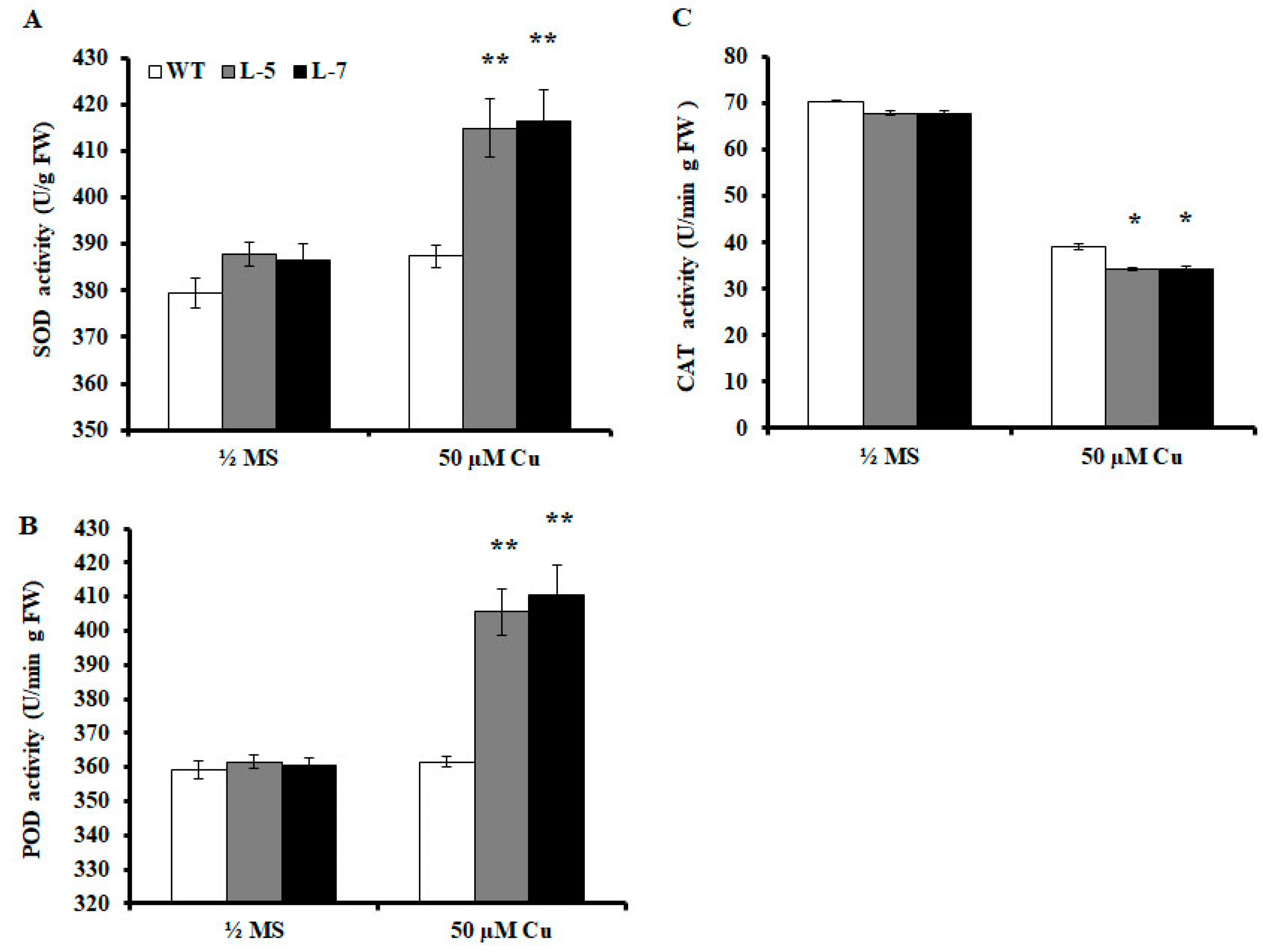
2.6. Antioxidant Enzyme Activities are Altered in Transgenic A. thaliana

To determine whether increased Cu tolerance in transgenic plants is related to changes in oxidase activity in vivo, SOD, POD, and CAT activities were measured in wild-type and transgenic plants grown in medium without (CK) or with 50 μM CuCl2. The data showed that under normal conditions, SOD and POD activities in the transgenic lines were slightly higher than those in wild-type plants (Figure 5A,B), whereas CAT activity was slightly lower in the transgenic lines than in the wild-type plants (Figure 5C). Under Cu stress, SOD and POD activities increased in both wild-type and transgenic lines. However, the SOD activity of L-5 and L-7 was 1.07 times higher (Figure 5A), and the POD activity was 1.12 and 1.14 times higher (Figure 5B) than that of wild-type plants, respectively. Cu stress decreases CAT activity in both wild-type and transgenic lines with a greater decrease in the transgenic lines than in the wild-type plants (Figure 5C). The POD activity of the two transgenic lines was only 0.87 and 0.88 times higher than that of the wild-type plants (Figure 5C).

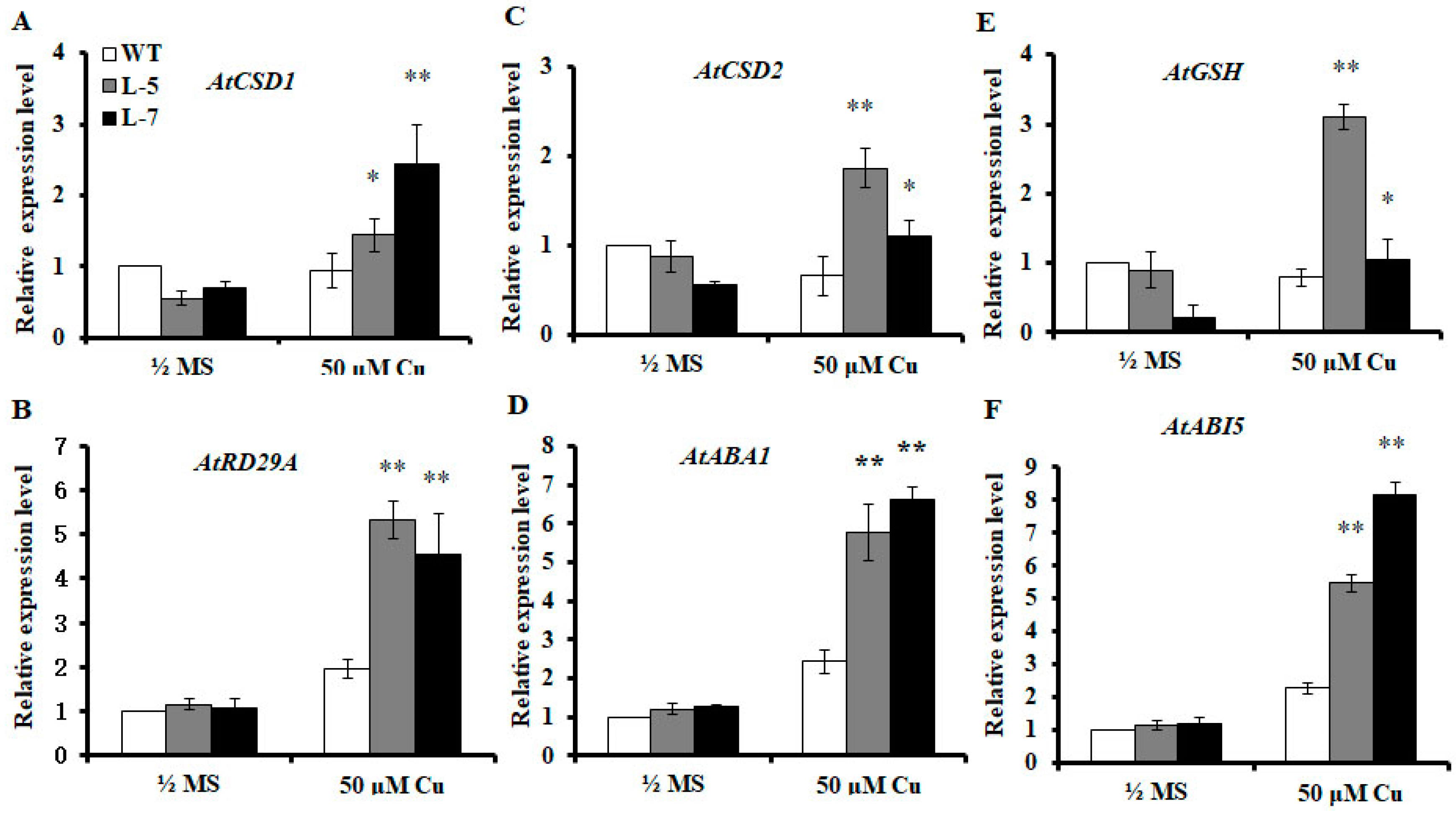
2.7. Expression of OsMSR3 Increases Expression of Antioxidant-Related and ABA-Responsive Genes

To determine the performance of transgenic plants under Cu stress and elucidate the molecular mechanism underlying the resistance of transgenic plants to Cu stress, the transcript levels of antioxidant-related (AtCSD1, AtCSD2, and AtPOD) and ABA-responsive (AtRD29A, AtABA1, and AtABI5) genes were assayed in wild-type and transgenic plants under normal and stress conditions. The expression levels of AtCSD1, AtCSD2, and AtPOD were higher in wild-type plants than they were in transgenic plants under control conditions. However, higher gene expression was observed in transgenic plants than in wild-type plants under Cu stress conditions (Figure 6A–C). Compared to the expression under normal conditions, Cu stress inhibited the expression of these genes in wild-type plants but activated their expression in transgenic lines. The ABA-responsive genes, AtRD29A, AtABA1, and AtABI5, showed no significantly different expression levels between transgenic and wild-type plants under normal conditions (Figure 6D–F). Under Cu stress conditions, the expression levels of the three genes were higher in the transgenic lines than in the wild-type plants (Figure 6D–F).

3. Discussion

Heavy metal pollution in soils is an emerging worldwide threat owing to its adverse effects on environmental safety [43]. Currently, cadmium and lead pollution in soils and their harmful effects on humans are attracting the attention of researchers globally. Cu pollution has become an important problem in the soil environment; however, studies of this phenomenon are still in infancy [44]. With the development of modern molecular biology, transgenic technology has emerged as an effective method to discover new Cu-tolerant genes in plants and cultivate plants that are highly efficient at repairing damage due to Cu contamination. In the previous study, we found that the expression of OsMSR3 in Arabidopsis significantly enhanced tolerance to cadmium stress [41]. As Cu and cadmium belong to the group of heavy metal elements, we evaluated if the transgenic lines expressing OsMSR3 in Arabidopsis could enhance the ability of copper tolerance.
The expression of OsMSR3 was enhanced by Cu stress (Figure 1), which indicated that OsMSR3 is involved in the response to Cu. Then, two transgenic lines L-5 and L-7 were used to perform the Cu tolerance experiment. The results showed that the expression of OsMSR3 enhanced the tolerance of A. thaliana to Cu stress than that of wild-type plants, manifested as higher survival rate (Figure 2), higher biomass (Figure 3), and longer root length (Figure 3B). Moreover, transgenic plants accumulated less MDA than wild-type plants under Cu stress (Figure 4D). It is well known that Cu damages cell membranes by inducing lipid peroxidation [45]. The MDA level is used to detect membrane lipid peroxidation and permeability [46]. These results suggest that the expression of OsMSR3 alleviated Cu-induced damage to the cell membrane of A. thaliana. Cell membrane stability is a major factor contributing to the maintenance of water status in plants during water deficit [47,48]. Therefore, the RWC of transgenic lines under Cu stress was higher than that of wild-type plants (Figure 3E).
Although the transgenic plants accumulated more Cu in the root and shoot (Figure 4A,B), their growth was significantly better than that of the wild-type. The accumulation of Cu in plant roots may inhibit the development of fine roots and reduce the absorption of iron and other trace elements [49]. To some extent, OsMSR3 protein may reduce the inhibition of Cu transport from the root to the shoot. We suggest that OsMSR3 may be helpful in maintaining the homeostasis of Cu metal ions at the cell and plant levels. In addition, we found that chlorophyll and carotenoid content in the leaves of OsMSR3 transgenic lines was higher than that in those of the wild-type plants under Cu stress (Figure 4F,G). Chlorophyll is an important part of the light-harvesting complex (LHCII). As an antenna for capturing light energy and transferring it to the reaction center, the chlorophyll content reflects the intensity of plant photosynthesis [50]. Carotenoids play an important role in plant growth and development. For example, they can act as a haptokine by transferring captured light to chlorophyll and can also act as a scavenger of free radicals in plant cells [51,52]. We speculated that the expression of OsMSR3 reduced the damage to chlorophyll and carotenoids under Cu stress.
SOD activity and SOD-related gene expression in OsMSR3 transgenic lines were significantly higher than those in wild-type plants (Figure 5A, Figure 6A,C). The presence of excess Cu causes the generation of ROS, such as superoxide radical (O2), H2O2, singlet oxygen (1O2), and hydroxyl radicals (OH) [53]. To scavenge ROS and alleviate their deleterious effects, plants stimulate ROS-scavenging systems such as CAT, SOD, and POD [24], to combat the oxidative injury induced by heavy metal exposure [54]. SOD is the first line of defense against ROS and catalyzes O2 to produce H2O2 and O2 [55]. An increase in SOD activity in stressed plants is an important indicator of superoxide ion production and enhancement of oxidative tolerance [55]. Therefore, the enhanced Cu tolerance of transgenic lines is related to the expression of SOD-related genes and SOD activity in A. thaliana under Cu stress induced by OsMSR3 expression.
Cu stress can lead to the generation of ROS, such as H2O2 [56]. In this study, we found that H2O2 accumulated in wild-type and transgenic seedlings significantly during Cu stress, albeit to a lower extent in the two OsMSR3 transgenic lines than in the wild-type (Figure 4E). The primary H2O2-scavenging enzymes in plant cells are CAT and POD; the former degrades H2O2 into water and oxygen. No studies, to date, have confirmed that a change in CAT activity is necessary to eliminate H2O2 in rice plants under Cu stress [55]. However, the current study revealed that Cu significantly increased POD activity in the transgenic lines but had little effect on CAT activity (Figure 5B,C). Moreover, POD gene expression was upregulated under Cu stress (Figure 6E). This is consistent with the increase in POD activity. Studies have shown that CAT has a high capacity but low affinity, whereas POD has a high affinity for H2O2 [57]. Thus, POD is the most effective H2O2-scavenging enzyme to reduce H2O2 content in plant cells under Cu stress.
Heavy metal exposure induces the expression of ABA synthesis-related genes in plants, which eventually leads to an increase in endogenous ABA levels [28]. In this study, under Cu stress conditions, ABA content and ABA-related gene expression levels in transgenic plants were significantly higher than those in wild-type plants (Figure 4C and Figure 6B,D,F). Therefore, we also propose that the expression of OsMSR3 leads to the upregulation of ABA-related genes and an increase in endogenous ABA level under Cu stress, which may partly explain the increased tolerance of transgenic plants to Cu stress.

4. Materials and Methods

4.1. Plant Material and Growth Conditions

The seeds of rice (Oryza sativa ssp. indica) cultivar Pei’ai 64S were surface-sterilized with 75% ethanol for 2 min, treated with 50% sodium hypochlorite for 20 min, and then washed with distilled water at least thrice. The sterilized seeds were germinated on half-strength Murashige and Skoog (½ MS) medium and grown in a greenhouse under conditions of a light intensity of 600 μmol/m2/s 70% relative humidity, and 28 °C temperature with a 12-h light/dark photoperiod. For the Cu stress experiment, two-week-old seedlings were exposed to a nutrient solution containing 50 μM CuCl2 for 48 h. The leaves were harvested as a pool for each sample at 0, 1, 3, 6, 12, 24, 36, and 48 h after Cu treatment.

4.2. Cu Tolerance Assay

We used 50 µM and 100 µM CuCl2 to do the pre-experiment and then selected the concentration of 50 µM CuCl2 as the most suitable. The seeds of T3 transgenic and wild-type A. thaliana (ecotype Columbia-0) were surface-sterilized and sown in Petri dishes containing ½ MS media with or without 50 μM CuCl2. The seeds were incubated in the dark at 4 °C for two days to break the dormancy and then transferred to a growth chamber. After incubation for 30 days, the survival rate of A. thaliana was determined. For measurement of root growth under Cu treatment, three-day-old A. thaliana seedlings were transferred onto ½ MS medium with or without 50 μM CuCl2, in vertically placed dishes. After incubation for 21 days, the root length (from the base of the root to the tip) and FW of six plants were measured.
Next, whole plants were rehydrated with distilled water at 4 °C for 12 h, blotted dry, and then the turgid weight (TW) was recorded. Rehydrated whole plants were oven-dried at 80 °C for 24 h, and the dry weight (DW) was recorded. RWC was calculated as follows: RWC (%) = (FW − DW)/(TW − DW) × 100. For the qRT-PCR analysis of selected genes, three-day-old A. thaliana seedlings were transferred onto ½ MS medium with or without 50 μM CuCl2. After 21 days of treatment, plant materials were harvested, and qRT-PCR was performed. The detailed procedure is provided in the next section.

4.3. RNA Extraction and qRT-PCR Analysis

Total RNAs were extracted with TRIzol reagent (Invitrogen, Burlington, ON, Canada), as described previously [58]. qPCR analysis was conducted using AceQ qPCR SYBR Green Master Mix (Vazyme Biotech, Nanjing, China), and the reactions were performed using an ABI7900HT (Applied Biosystems, Foster City, CA, USA) and run on the following schedule: 95 °C for 10 min, followed by 40 cycles at 95 °C for 15 s and 58 °C for 30 s. The internal controls were ACTIN1 and β-TUBULIN for rice and A. thaliana, respectively. The data for relative expression were analyzed using the comparative Ct method [59]. The primer pairs used in the qPCR analysis are listed in Table S1.

4.4. Measurement of Cu Content

Cu content was determined according to the method described by Li et al. [24]. Briefly, three-day-old A. thaliana seedlings were transferred to ½ MS medium with or without 50 μM CuCl2. After 21 days of treatment, the roots and shoots were harvested and dried at 80 °C for two days. Dried plant tissues (50–100 mg roots; 100–200 mg shoots) were digested with 11 N HNO3 at 200 °C for 10 h. The digested samples were then diluted with 0.1 N HNO3 and analyzed using an atomic absorption spectrometer (Solaar M6; Thermo Fisher, Boston, MA, USA). The experiments were performed in triplicate.

4.5. Measurement of MDA Content

Two-week-old transgenic and wild-type plants were cultivated on ½ MS medium with or without 50 μM CuCl2 for 24 h. Then, 0.3 g of the seedlings was harvested and ground into a powder for the determination of MDA content, which was measured according to a previously standardized method [24].

4.6. Measurement of ABA Content

Two-week-old transgenic and wild-type plants were cultivated on ½ MS media with or without 50 μM CuCl2 for 24 h. Approximately 0.2 g of the leaf tissue was harvested, ground into a powder, and then suspended in 1.8 mL 100 mM sodium phosphate buffer (PBS, pH = 7.4) for ABA leaf content detection, using a previously published method [60].

4.7. Measurement of Chlorophylls and Carotenoids

The chlorophyll and carotenoid content were determined according to a previous method [60]. Briefly, three-day-old A. thaliana seedlings were transferred onto ½ MS medium with or without 50 μM CuCl2 in vertically placed dishes. After incubation for 21 days, chlorophyll and carotenoids were extracted from the rosette leaves of the wild-type and transgenic plants with 100% alcohol. An ultraviolet-visible (UV-vis) spectrometer (UV-2600; Shimadzu Co., Kyoto, Japan) was used to measure the absorption of the extracts. The total chlorophyll and carotenoid content were calculated according to a previously published method [61].

4.8. Quantitative Analysis of H2O2

The H2O2 concentration was determined using a commercially available kit (Nanjing Jiancheng Bioengineering Institute, Nanjing, China). Briefly, two-week-old plants (wild-type and transgenic lines) cultivated in ½ MS medium were treated for 24 h with or without 50 μM CuCl2. Then, 0.5 g of the seedlings was harvested, weighed, immediately ground, and then suspended in 5 mL 0.9% sodium chloride solution. The supernatant was collected after centrifugation for 10 min at 4 °C and 3000× g, and the H2O2 content was measured according to the protocol provided by the manufacturer of the kit.

4.9. Assay of Antioxidant Enzyme Activities

To measure antioxidant enzyme activities, two-week-old plants (wild-type and transgenic lines) cultivated in ½ MS medium were treated for 24 h with or without 50 μM CuCl2. Seedling samples (0.5 g) were frozen in liquid nitrogen, rapidly ground into powder, and then homogenized in 100 mM sodium phosphate buffer (pH 7.4) on ice. After centrifugation at 3000× g for 15 min at 4 °C, the supernatant samples were immediately used for the detection of antioxidant enzymes. The activities of SOD, POD, and CAT were measured using specific assay kits (A001-1, A084-3, and A007-1, respectively) from Nanjing Jiancheng Bioengineering Institute (Nanjing, China) according to the manufacturer’s instructions.

4.10. Statistical Analysis

The experimental data were analyzed using the statistical package for the social sciences (SPSS) 17.0 statistical software (SPSS Inc., Chicago, IL, USA). At least three independent experiments were performed, and the average results are presented. Error bars represent standard deviation (SD, n > 3). Furthermore, * p < 0.05 or ** p < 0.01 indicate statistically significant means.

5. Conclusions

In conclusion, we showed the involvement of OsMSR3 in Cu tolerance in A. thaliana. OsMSR3-expressing lines exhibited enhanced Cu stress tolerance, possibly through enhanced activation of antioxidative defense mechanisms and positive regulation of ABA-responsive gene expression. In view of the good performance of the transgenic lines, OsMSR3 can be used to modify plants for remediation of Cu pollution in the soil. Therefore, this study provides an important insight into plant biology and mechanisms to overcome increasing heavy metal pollution in soils.

Supplementary Materials

Supplementary materials can be found at https://0-www-mdpi-com.brum.beds.ac.uk/1422-0067/20/23/6096/s1. Table S1. Primer sequences used for quantitative reverse transcription polymerase reaction (qRT-PCR).

Author Contributions

Y.C. designed and performed the experiments; Y.C. and X.X. analyzed the data and wrote the paper; M.W., X.Y., G.X., S.S., M.L., and K.L. contributed reagents/materials/analysis tools; X.X. supervised the work and revised the manuscript. All the authors agreed on the contents of the paper and declared no conflicting interests.

Funding

This work was financially supported by the National Natural Science Foundation of China (Grant Nos. 31301253 and 31671671) and Youth Innovation Team Project of ISA, CAS (2017QNCXTD_GTD).

Conflicts of Interest

The authors declare no conflict of interest.

Abbreviations

ABAabscisic acid
SODsuperoxide dismutase
PODperoxidase
CATcatalase
ROSreactive oxygen species
ASCascorbic acid
GSHglutathione
sHSPssmall heat shock protei
SDstandard deviation

References

  1. Yruela, I.; Pueyo, J.J.; Alonso, P.J.; Picorel, R. Photoinhibition of photosystem II from higher plants effect of copper inhibition. J. Biol. Chem. 1996, 271, 27408–27415. [Google Scholar] [CrossRef] [Green Version]
  2. Rodriguez, F.I.; Esch, J.J.; Hall, A.E.; Binder, B.M.; Schaller, G.E.; Bleecker, A.B. A copper cofactor for the ethylene receptor ETR1 from Arabidopsis. Science 1999, 283, 996–998. [Google Scholar] [CrossRef]
  3. Himelblau, E.; Amasino, R.M. Delivering copper within plant cells. Curr. Opin. Plant Biol. 2000, 3, 205–210. [Google Scholar] [CrossRef]
  4. Pilon, M.; Abdel-Ghany, S.E.; Cohu, C.M.; Gogolin, K.A.; Ye, H. Copper cofactor delivery in plant cells. Curr. Opin. Plant Biol. 2006, 9, 256–263. [Google Scholar] [CrossRef]
  5. Burkhead, J.L.; Reynolds, K.A.; Abdel-Ghany, S.E.; Cohu, C.M.; Pilon, M. Copper homeostasis. New Phytol. 2009, 182, 799–816. [Google Scholar] [CrossRef]
  6. Bernal, M.; Casero, D.; Singh, V.; Wilson, G.T.; Grande, A.; Yang, H.; Dodani, S.C.; Pellegrini, M.; Huijser, P.; Connolly, E.L.; et al. Transcriptome sequencing identifies SPL7-regulated copper acquisition genes FRO4/FRO5 and the copper dependence of iron homeostasis in Arabidopsis. Plant Cell 2012, 24, 738–761. [Google Scholar] [CrossRef] [Green Version]
  7. Song, Y.; Zhou, L.; Yang, S.; Wang, C.; Zhang, T.; Wang, J. Dose-dependent sensitivity of Arabidopsis thaliana seedling root to copper is regulated by auxin homeostasis. Environ. Exp. Bot. 2017, 139, 23–30. [Google Scholar] [CrossRef]
  8. Luna, C.M.; Gonzalez, C.A.; Trippi, V.S. Oxidative damage caused by an excess of copper in oat leaves. Plant Cell Physiol. 1994, 35, 11–15. [Google Scholar] [CrossRef]
  9. Shen, Z.G.; Zhang, F.Q.; Zhang, F.S. Toxicity of copper and zinc in seedlings of mung bean and inducing accumulation of polyamine. J. Plant Nutr. 1998, 21, 1153–1162. [Google Scholar] [CrossRef]
  10. Patsikka, E.; Kairavuo, M.; Sersen, F.; Aro, E.M.; Tyystjarvi, E. Excess copper predisposes photosystem II to photoinhibition in vivo by outcompeting iron and causing decrease in leaf chlorophyll. Plant Physiol. 2002, 129, 1359–1367. [Google Scholar] [CrossRef] [Green Version]
  11. Nielsen, H.D.; Brownlee, C.; Coelho, S.M.; Brown, M.T. Inter-population differences in inherited copper tolerance involve photosynthetic adaptation and exclusion mechanisms in Fucus serratus. New Phytol. 2003, 160, 157–165. [Google Scholar] [CrossRef]
  12. Demirevska-Kepovaa, K.; Simova-Stoilovaa, L.; Stoyanovaa, Z.; Hölzerb, R.; Feller, U. Biochemical changes in barley plants after excessive supply of copper and manganese. Environ. Exp. Bot. 2004, 52, 253–266. [Google Scholar] [CrossRef]
  13. Drazkiewicz, M.; Skorzynska-Polit, E.; Krupa, Z. Copper-induced oxidative stress and antioxidant defence in Arabidopsis thaliana. Biometals 2004, 17, 379–387. [Google Scholar] [CrossRef]
  14. Wang, P.; De Schamphelaere, K.A.; Kopittke, P.M.; Zhou, D.M.; Peijnenburg, W.J.; Lock, K. Development of an electrostatic model predicting copper toxicity to plants. J. Exp. Bot. 2012, 63, 659–668. [Google Scholar] [CrossRef] [Green Version]
  15. Navari-Izzo, F.; Cestone, B.; Cavallini, A.; Natali, L.; Giordani, T.; Quartacci, M.F. Copper excess triggers phospholipase D activity in wheat roots. Phytochemistry 2006, 67, 1232–1242. [Google Scholar] [CrossRef]
  16. Peto, A.; Lehotai, N.; Lozano-Juste, J.; Leon, J.; Tari, I.; Erdei, L.; Kolbert, Z. Involvement of nitric oxide and auxin in signal transduction of copper-induced morphological responses in Arabidopsis seedlings. Ann. Bot. 2011, 108, 449–457. [Google Scholar] [CrossRef]
  17. Marchand, L.; Nsanganwimana, F.; Lamy, J.B.; Quintela-Sabaris, C.; Gonnelli, C.; Colzi, I.; Fletcher, T.; Oustriere, N.; Kolbas, A.; Kidd, P.; et al. Root biomass production in populations of six rooted macrophytes in response to Cu exposure: Intra-specific variability versus constitutive-like tolerance. Environ. Pollut. 2014, 193, 205–215. [Google Scholar] [CrossRef] [Green Version]
  18. Liu, D.; Jiang, W.; Meng, Q.; Zou, J.; Gu, J.; Zeng, M. Cytogenetical and ultrastructural effects of copper on root meristem cells of Allium sativum L. Biocell 2009, 33, 25–32. [Google Scholar]
  19. Madejón, P.; Ramírez-Benítez, J.E.; Corrales, I.; Barceló, J.; Poschenrieder, C. Copper-induced oxidative damage and enhanced antioxidant defenses in the root apex of maize cultivars differing in Cu tolerance. Environ. Exp. Bot. 2009, 67, 415–420. [Google Scholar] [CrossRef]
  20. Yeh, C.M.; Hung, W.C.; Huang, H.J. Copper treatment activates mitogen-activated protein kinase signalling in rice. Physiol. Plant. 2003, 119, 392–399. [Google Scholar] [CrossRef]
  21. Zhan, E.; Zhou, H.; Li, S.; Liu, L.; Tan, T.; Lin, H. OTS1-dependent deSUMOylation increases tolerance to high copper levels in Arabidopsis. J. Integr. Plant Biol. 2018, 60, 310–322. [Google Scholar] [CrossRef] [PubMed]
  22. Clemens, S. Molecular mechanisms of plant metal tolerance and homeostasis. Planta 2001, 212, 475–486. [Google Scholar] [CrossRef] [PubMed]
  23. Sandalio, L.M.; Dalurzo, H.C.; Gomez, M.; Romero-Puertas, M.C.; Del Rio, L.A. Cadmium-induced changes in the growth and oxidative metabolism of pea plants. J. Exp. Bot. 2001, 52, 2115–2126. [Google Scholar] [CrossRef] [PubMed]
  24. Li, M.J.; Xu, G.Y.; Xia, X.J.; Wang, M.L.; Yin, X.M.; Zhang, B.; Zhang, X.; Cui, Y.C. Deciphering the physiological and molecular mechanisms for copper tolerance in autotetraploid Arabidopsis. Plant Cell Rep. 2017, 36, 1585–1597. [Google Scholar] [CrossRef] [PubMed]
  25. Jaleel, C.A.; Riadh, K.; Gopi, R.; Manivannan, P.; Ine’s, J.; Al-Juburi, H.J.; Zhao, C.X.; Shao, H.B.; Rajaram, P. Antioxidant defense responses: Physiological plasticity in higher plants under abiotic constraints. Acta Physiol. Plant. 2009, 31, 427–436. [Google Scholar] [CrossRef]
  26. Maksymiec, W.; Krupa, Z. The effects of short-term exposition to Cd, excess Cu ions and jasmonate on oxidative stress appearing in Arabidopsis thaliana. Environ. Exp. Bot. 2006, 57, 187–194. [Google Scholar] [CrossRef]
  27. Hollenbach, B.; Schreiber, L.; Hartung, W.; Dietz, K.J. Cadmium leads to stimulated expression of the lipid transfer protein genes in barley: Implications for the involvement of lipid transfer proteins in wax assembly. Planta 1997, 203, 9–19. [Google Scholar] [CrossRef]
  28. Bücker-Neto, L.; Paiva, A.L.S.; Machado, R.D.; Arenhart, R.; Margis-Pinheiro, M. Interactions between plant hormones and HMs responses. Genet. Mol. Biol. 2017, 40, 373–386. [Google Scholar] [CrossRef]
  29. Fediuc, E.; Lips, S.H.; Erdei, L. O-acetylserine (thiol) lyase activity in Phragmites and Typha plants under cadmium and NaCl stress conditions and the involvement of ABA in the stress response. J. Plant Physiol. 2005, 162, 865–872. [Google Scholar] [CrossRef]
  30. Stroinski, A.; Chadzinikolau, T.; Gizewska, K.; Zielezinska, M. ABA or cadmium induced phytochelatin synthesis in potato tubers. Biol. Plant. 2010, 54, 117–120. [Google Scholar] [CrossRef]
  31. Kim, Y.H.; Khan, A.L.; Kim, D.H.; Lee, S.Y.; Kim, K.M.; Waqas, M.; Jung, H.Y.; Shin, J.H.; Kim, J.G.; Lee, I.J. Silicon mitigates heavy metal stress by regulating P-type heavy metal ATPases, Oryza sativa low silicon genes, and endogenous phytohormones. BMC Plant Biol. 2014, 14, 13. [Google Scholar] [CrossRef] [PubMed] [Green Version]
  32. Munzuro, Ö.; Fikriye, K.Z.; Yahyagil, Z. The abscisic acid levels of wheat (Triticum aestivum L. cv. Çakmak 79) seeds that were germinated under heavy metal (Hg++, Cd++, Cu++) stress. Gazi Univ. J. Sci. 2008, 21, 1–7. [Google Scholar]
  33. Rauser, W.E.; Dumbroff, E.B. Effects of excess cobalt, nickel and zinc on the water relations of Phaseolus vulgaris. Environ. Exp. Bot. 1981, 21, 249–255. [Google Scholar] [CrossRef]
  34. Poschenrieder, C.; Gunsé, B.; Barceló, J. Influence of cadmium on water relations, stomatal resistance, and abscisic acid content in expanding bean leaves. Plant Physiol. 1989, 90, 1365–1371. [Google Scholar] [CrossRef]
  35. Sarkar, N.K.; Kim, Y.K.; Grover, A. Rice sHsp genes: Genomic organization and expression profiling under stress and development. BMC Genom. 2009, 10, 393. [Google Scholar] [CrossRef] [Green Version]
  36. Siddique, M.; Gernhard, S.; von Koskull-Doring, P.; Vierling, E.; Scharf, K.D. The plant sHSP superfamily: Five new members in Arabidopsis thaliana with unexpected properties. Cell Stress Chaperon. 2008, 13, 183–197. [Google Scholar] [CrossRef] [Green Version]
  37. Kim, K.H.; Alam, I.; Kim, Y.G.; Sharmin, S.A.; Lee, K.W.; Lee, S.H.; Lee, B.H. Overexpression of a chloroplast-localized small heat shock protein OsHSP26 confers enhanced tolerance against oxidative and heat stresses in tall fescue. Biotechnol. Lett. 2012, 34, 371–377. [Google Scholar] [CrossRef]
  38. Sato, Y.; Yokoya, S. Enhanced tolerance to drought stress in transgenic rice plants overexpressing a small heat-shock protein, sHSP17.7. Plant Cell Rep. 2008, 27, 329–334. [Google Scholar] [CrossRef]
  39. Ju, Y.; Tian, H.; Zhang, R.; Zuo, L.; Chu, Z. Overexpression of oshsp18.0-CI enhances resistance to bacterial leaf streak in rice. Rice 2017, 10, 12. [Google Scholar] [CrossRef] [Green Version]
  40. Cui, Y.C.; Xu, M.L.; Li, L.Y.; Wang, M.L.; Xu, G.Y.; Xia, X.J. Expression and cloning of a multiple stress responsive gene (OsMSR3) in rice. J. Wuhan. Bot. Res. 2009, 6, 574–581. [Google Scholar]
  41. Cui, Y.C.; Xu, G.Y.; Wang, M.L.; Yu, Y.; Li, M.J.; da Rocha, P.S.C.F.; Xia, X.J. Expression of OsMSR3 in Arabidopsis enhances tolerance to cadmium stress. Plant Cell Tiss. Org. Cult. 2013, 113, 331–340. [Google Scholar] [CrossRef]
  42. Kumar, G.; Kushwaha, H.R.; Panjabi-Sabharwal, V.; Kumari, S.; Joshi, R.; Karan, R.; Mittal, S.; Singla Pareek, S.L.; Pareek, A. Clustered metallothionein genes are co-regulated in rice and ectopic expression of OsMT1e-P confers multiple abiotic stress tolerance in tobacco via ROS scavenging. BMC Plant Biol. 2012, 12, 107. [Google Scholar] [CrossRef] [PubMed] [Green Version]
  43. Wang, R.; Shafi, M.; Ma, J.; Zhong, B.; Guo, J.; Hu, X.; Xu, W.; Yang, Y.; Ruan, Z.; Wang, Y.; et al. Effect of amendments on contaminated soil of multiple heavy metals and accumulation of heavy metals in plants. Environ. Sci. Pollut. Res. 2018, 25, 28695–28704. [Google Scholar] [CrossRef] [PubMed]
  44. Wang, M.; Li, S.S.; Li, X.Y.; Zhao, Z.Q.; Chen, S.B. An overview of current status of copper pollution in soil and remediation efforts in China. Earth Sci. Front. 2018, 25, 305–313. [Google Scholar]
  45. De Vos, C.H.R.; Ten Bookum, W.M.; Vooijs, R.; Schat, H.; Dekok, L.J. Effect of copper on fatty acid composition and peroxidation of lipids in the roots of copper tolerant and sensitive Silenecucubalus. Plant Physiol. Biochem. 1993, 31, 151–158. [Google Scholar]
  46. Heath, R.L.; Packer, L. Photoperoxidation in isolated chloroplasts: I. Kinetics and stoichiometry of fatty acid peroxidation. Arch. Biochem. Biophys. 1968, 125, 189–198. [Google Scholar] [CrossRef]
  47. Nishiyama, R.; Watanabe, Y.; Leyva-Gonzalez, M.A.; Van Ha, C.; Fujita, Y.; Tanaka, M.; Seki, M.; Yamaguchi-Shinozaki, K.; Shinozaki, K.; Herrera-Estrella, L.; et al. Arabidopsis AHP2, AHP3, and AHP5 histidine phosphotransfer proteins function as redundant negative regulators of drought stress response. Proc. Natl. Acad. Sci. USA 2013, 110, 4840–4845. [Google Scholar] [CrossRef] [Green Version]
  48. Demidchik, V.; Straltsova, D.; Medvedev, S.S.; Pozhvanov, G.A.; Sokolik, A.; Yurin, V. Stress-induced electrolyte leakage: The role of K+-permeable channels and involvement in programmed cell death and metabolic adjustment. J. Exp. Bot. 2014, 65, 1259–1270. [Google Scholar] [CrossRef]
  49. Rehman, M.; Liu, L.; Wang, Q.; Saleem, M.H.; Bashir, S.; Ullah, S.; Peng, D. Copper environmental toxicology, recent advances, and future outlook: A review. Environ. Sci. Pollut. Res. Int. 2019, 26, 18003–18016. [Google Scholar] [CrossRef]
  50. Sui, N.; Tian, S.; Wang, W.; Wang, M.; Fan, H. Overexpression of glycerol-3-phosphate acyltransferase from Suaeda salsa improves salt tolerance in Arabidopsis. Front. Plant Sci. 2017, 8, 1337. [Google Scholar] [CrossRef]
  51. Polívka, T.; Frank, H.A. Molecular factors controlling photosynthetic light harvesting by carotenoids. Acc. Chem. Res. 2010, 43, 1125–1134. [Google Scholar] [CrossRef] [PubMed] [Green Version]
  52. Polívka, T.; Pullerits, T.; Frank, H.A.; Cogdell, R.J.; Sundström, V. Ultrafast formation of a carotenoid radical in LH2 antenna complexes of purple bacteria. J. Phys. Chem. B 2004, 108, 15398–15407. [Google Scholar] [CrossRef]
  53. Saglam, A.; Yetiddin, F.; Demiralay, M.; Terzi, R. Copper stress and responses in plants. In Plant Metal Interaction: Emerging Remediation Techniques; Ahmad, P., Ed.; Elsevier: Amsterdam, The Netherlands, 2016; pp. 21–40. [Google Scholar] [CrossRef]
  54. Zabalza, A.; Galvez, L.; Marino, D.; Royuela, M.; Arrese-Igor, C.; Gonzalez, E.M. The application of ascorbate or its immediate precursor, galactono-1, 4-lactone, does not affect the response of nitrogen-fixing pea nodules to water stress. J. Plant Physiol. 2008, 165, 805–812. [Google Scholar] [CrossRef]
  55. Thounaojam, T.C.; Panda, P.; Mazumdar, P.; Kumar, D.; Sharma, G.; Sahoo, L.; Panda, S. Excess copper induced oxidative stress and response of antioxidants in rice. Plant Physiol. Biochem. 2012, 53, 33–39. [Google Scholar] [CrossRef] [PubMed]
  56. Gu, C.S.; Liu, L.Q.; Deng, Y.M.; Zhu, X.D.; Huang, S.Z.; Lu, X.Q. The heterologous expression of the Iris lactea var. chinensis type 2 metallothionein IlMT2b gene enhances copper tolerance in Arabidopsis thaliana. Bull. Environ. Contam. Toxicol. 2015, 94, 247–253. [Google Scholar] [CrossRef] [PubMed]
  57. Mittler, R. Oxidative stress, antioxidants and stress tolerance. Trends Plant Sci. 2002, 7, 405–410. [Google Scholar] [CrossRef]
  58. Xu, G.Y.; Rocha, P.S.C.F.; Wang, M.L.; Xu, M.L.; Cui, Y.C.; Li, L.Y.; Zhu, Y.X.; Xia, X.J. A novel rice calmodulin-like gene, OsMSR2, enhances drought and salt tolerance and increases ABA sensitivity in Arabidopsis. Planta 2011, 234, 47–59. [Google Scholar] [CrossRef]
  59. Xu, M.L.; Chen, R.J.; Rocha, P.S.C.F.; Wang, M.L.; Xu, G.Y.; Xia, X.J. Expression and cloning of a novel stress responsive gene (OsMsr1) in rice. Acta Agron. Sin. 2008, 10, 1712–1718. [Google Scholar] [CrossRef]
  60. Cui, Y.C.; Li, M.J.; Yin, X.M.; Song, S.F.; Xu, G.Y.; Wang, M.L.; Li, C.Y.; Peng, C.; Xia, X.J. OsDSSR1, a novel small peptide, enhances drought tolerance in transgenic rice. Plant Sci. 2018, 270, 85–96. [Google Scholar] [CrossRef]
  61. Lichtenthaler, H.K.; Wellburn, A.R. Determinations of total carotenoids and chlorophylls a and b of leaf extracts in different solvents. Biochem. Soc. Trans. 1983, 11, 591e592. [Google Scholar] [CrossRef] [Green Version]
Figure 1. Expression analysis of the Oryza sativa L. multiple stress-responsive gene 3 (OsMSR3) gene. Expression analysis of OsMSR3 in Pei’ai 64S rice seedlings at different time points (0, 1, 3, 6, 12, 24, and 48 h) under Cu stress using quantitative reverse transcription-polymerase reaction (qRT-PCR). The ACTIN1 gene was used as an internal control. Error bars indicate standard deviations (SD) of three independent experiments.
Ijms 20 06096 g001
Figure 2. Performance of transgenic plants and wild-types under normal and Cu stress conditions. (A) Left panel, seedlings reared under normal conditions (0 μM CuCl2) for 30 days; right panel, seedlings exposed to 50 μM CuCl2 for 30 days. WT, wild-type Arabidopsis thaliana; L-5 and L-7, transgenic lines 5 and 7. (B) Survival rates of plants after growth under Cu stress for 30 days. Each column represents an average of three replicates, and bars indicate ± standard deviation (SD); and ** p < 0.01 indicate significant differences compared to wild-type T plants under the same conditions determined using Student’s t-tests.
Ijms 20 06096 g002
Figure 3. Improved tolerance to Cu stress induced by Oryza sativa L. multiple stress-responsive gene 3 (OsMSR3) expression. (A) Images of representative plants grown on half-strength Murashige and Skoog (½ MS) medium with or without 50 μM copper chloride (CuCl2) for 21 days. (B) Effect of Cu treatment on root length of plants presented in panel A. (C) Fresh weight (FW) and (D) dry weight (DW) of wild-type and transgenic line plants treated with or without 50 μM CuCl2 for 21 days. (E) Relative water content (RWC) of wild-type and transgenic plants treated with or without 50 μM CuCl2 for 21 days. Values are means ± standard deviation (SD) of three independent biological replicates; * p < 0.05 and ** p < 0.01 indicate significant differences from the wild-type determined using Student’s t-test.
Ijms 20 06096 g003
Figure 4. Quantitative analysis of various physiological indexes in wild-type and transgenic plants. (A,B) Cu content in wild-type and transgenic plant shoots and roots treated with 50 μM copper chloride (CuCl2) for 21 days, respectively. (C) abscisic acid (ABA), (D) malondialdehyde (MDA), and (E) hydrogen peroxide (H2O2) content in wild-type and transgenic plants treated with or without 50 μM CuCl2 for 24 h. (F,G) Chlorophyll and carotenoid content in wild-type and transgenic plants treated with or without 50 μM CuCl2 for 21 days. Values are means ± standard deviation (SD) of three independent biological replicates; * p < 0.05 and ** p < 0.01 indicate significant differences from wild-type plants under the same conditions determined using Student’s t-test.
Ijms 20 06096 g004
Figure 5. Antioxidant enzyme activity determination. Quantitative analysis of (A) superoxide dismutase (SOD), (B) peroxidase (POD), (C) and catalase (CAT) activity in wild-type and transgenic plants treated with or without 50 μM copper chloride (CuCl2) for 24 h. Values are means ± standard deviation (SD) of three independent experiments; * p < 0.05 and ** p < 0.01 indicate significant differences from wild-type plants under the same conditions determined using the Student’s t-test.
Ijms 20 06096 g005
Figure 6. Quantitative reverse transcription-polymerase reaction (qRT-PCR) gene analysis. qRT-PCR analysis of relative expression of (A) AtCSD1, (B) AtRD29A, (C) AtCSD2, (D) AtABA1, (E) AtGSH, (F) and AtABI5, in two-week-old transgenic and wild-type (WT) plants treated with (Cu) or without (Control) 50 μM copper chloride (CuCl2) for 24 h. Values are means ± standard deviation (SD) of three independent biological replicates; * p < 0.05 and ** p < 0.01 indicate significant differences from wild-type plants under the same conditions, determined using the Student’s t-test.
Ijms 20 06096 g006

