Article

Downregulation of the Polycomb-Associated Methyltransferase Ezh2 during Maturation of Hippocampal Neurons Is Mediated by MicroRNAs Let-7 and miR-124

by
Laura Guajardo
1,2,
Rodrigo Aguilar
1,2,
Fernando J. Bustos
1,2,3,
Gino Nardocci
1,2,
Rodrigo A. Gutiérrez
2,4,
Brigitte van Zundert
1,3,* and
Martin Montecino
1,2,*
1
Institute of Biomedical Sciences, Faculty of Medicine and Faculty of Life Sciences, Universidad Andres Bello, Santiago 8370186, Chile
2
FONDAP Center for Genome Regulation, Santiago 8370186, Chile
3
CARE Biomedical Research Center, Santiago 83370186, Chile
4
Millennium Institute for Integrative Biology, Departamento de Genética Molecular y Microbiología, Facultad de Ciencias Biológicas, Pontificia Universidad Católica de Chile, Santiago 8331150, Chile
*
Authors to whom correspondence should be addressed.
Int. J. Mol. Sci. 2020, 21(22), 8472; https://0-doi-org.brum.beds.ac.uk/10.3390/ijms21228472
Submission received: 6 October 2020 / Revised: 2 November 2020 / Accepted: 6 November 2020 / Published: 11 November 2020
(This article belongs to the Special Issue Epigenetic Regulation in Human Brain)

Abstract

:
Ezh2 is a catalytic subunit of the polycomb repressive complex 2 (PRC2) which mediates epigenetic gene silencing through depositing the mark histone H3 lysine 27 trimethylation (H3K27me3) at target genomic sequences. Previous studies have demonstrated that Enhancer of Zeste Homolog 2 (Ezh2) was differentially expressed during maturation of hippocampal neurons; in immature neurons, Ezh2 was abundantly expressed, whereas in mature neurons the expression Ezh2 was significantly reduced. Here, we report that Ezh2 is downregulated by microRNAs (miRs) that are expressed during the hippocampal maturation process. We show that, in mature hippocampal neurons, lethal-7 (let-7) and microRNA-124 (miR-124) are robustly expressed and can target cognate motifs at the 3′-UTR of the Ezh2 gene sequence to downregulate Ezh2 expression. Together, these data demonstrate that the PRC2 repressive activity during hippocampal maturation is controlled through a post-transcriptional mechanism that mediates Ezh2 downregulation in mature neurons.

1. Introduction

Several reports have demonstrated the critical role of epigenetic mechanisms during control of gene expression associated with the physiological function of the central nervous system (CNS) in mammals [1,2]. These mechanisms include DNA methylation, histone post-translational modifications, and long and short non-coding RNA activities, among others [3]. A relevant component of epigenetic control in the CNS is mediated by the polycomb repressive complex 2 (PRC2), which catalyzes the deposit of the repressive histone mark trimethyl lysine 27 of histone H3 (H3K27me3) at target gene promoters, thereby producing their transcriptional silencing [4,5]. Our team showed that, during early stages of hippocampal neuron maturation, PRC2 activity was critical to control the expression of non-neuronal gene programs (e.g., osteogenic lineage genes), as well as the expression of key components of the dendritic arbor formation that were required at more advanced stages of maturation [6,7]. These data demonstrated that the catalytic subunit of PRC2, the methyltransferase Enhancer of Zeste Homolog 2 (Ezh2), played a key role during this gene expression silencing events in the hippocampus. Additionally, Ezh2 has been found expressed in neuronal progenitor cells and in neurons during early embryonic stages, where Ezh2 trimethylates H3K27 target regulatory sequences for controlling the balance between self-renewal and lineage commitment [8,9].
It has been determined that, in rat hippocampal neurons, the expression of Ezh2 is downregulated during development [7]. Thus, Ezh2 is significantly expressed in the hippocampus at embryonic and early postnatal stages, where, as part of PRC2, it binds to target promoters to mediate gene silencing. At later adult stages, Ezh2 mRNA and protein are largely undetectable in hippocampal neurons and Ezh2 function in the PRC2 complex can be partially replaced by Ezh1 [6,7]. Although our results indicated that an important component mediating this Ezh2 downregulation during hippocampal maturation could involve transcriptional inhibition of the Ezh2 gene, additional evidence has demonstrated the contribution of post-transcriptional and post-translational mechanisms, including the role of microRNAs [10] and ubiquitin-dependent proteasome degradation [11].
Accumulating evidence indicates that microRNAs (miRs) are key modulators of gene expression in the CNS [12,13], where approximately 50% of the total known mammalian miRs are expressed [14,15], regulating critical processes that include dendritogenesis [16] and synapse maturation [17]. Studies have shown that conditional knockout of Dicer in mice, the endoribonuclease that mediates a critical step during miR maturation, drastically affected survival of neural stem cells and decreased differentiation of newborn neurons [18,19,20]. Among the microRNAs (miRs), miR-124 has been reported to play a relevant role in neuronal function, which has been found to be upregulated during neural differentiation [14,21,22]. Forced expression of miR-124 in embryonal carcinoma P19 cells in the presence of retinoic acid promotes a neuronal-like phenotype [20,22,23]. Moreover, an increased expression of miR-124 mediates the conversion of fibroblasts to functional neurons [24,25]. Studies in vivo have further shown that miR-124 was an important regulator of adult neurogenesis in mice [26]. Conversely, knockout of the miR-124-1 gene in mice resulted in defective neuronal survival, impaired hippocampal axonal outgrowth, and reduced brain size [27]. Interestingly, it was been shown that miR-124 targeted the Ezh2 3′-UTR in early neuronal cells, thereby controlling Ezh2 expression and Ezh2-mediated gene silencing during neuron lineage commitment [28]. Additionally, miR-124 targeted and downregulated USP14, coding for a deubiquitinase that binds to Ezh2, and thus prevented ubiquitin-dependent degradation of Ezh2 via proteasome [29,30].
Another relevant microRNA molecule during brain development is let-7 (lethal-7), which has been found in elevated concentrations in this tissue during embryogenesis [31,32,33,34]. Mammals contain a let-7 family, including several members (from let-7a to let-7k), which have been shown to play a significant role in maintaining the balance between neuronal progenitor cell proliferation and commitment to engage neurogenic differentiation, as they control the expression of a number of genes that are critical during this early developmental stages [35,36,37,38]. Interestingly, it has been shown that let-7 mediated control of Ezh2 gene expression in prostate cancer cells [39], as it recognized a specific motif at the Ezh2 3′-UTR. Loss of let-7 function in these tumor cells resulted in increased Ezh2 expression that was accompanied by acquisition of a cancer stem cell signature.
Whereas these data provide support for a regulatory role of miRs over PRC2 expression and function during early neuron lineage commitment, there is a lack of studies that address the contribution of miRs during the maturation of hippocampal neurons, a process that determines the functionality of these cells during learning and memory. Here, we examine whether specific miR molecules contribute to Ezh2 downregulation during hippocampal neuronal maturation. We report that miR-124 and let-7, which are among the group of miRs that are highly expressed in mature hippocampal neurons, can target cognate motifs at the Ezh2 3′-UTR, and thereby dampen Ezh2 expression.

2. Results

2.1. MicroRNA Expression Profile in Mature Hippocampal Neurons

Previous results have indicated that expression of the methylase Ezh2, the catalytic subunit of the PRC2 complex, was downregulated during hippocampal neuronal maturation [7]. However, the mechanisms that control this reduced expression of Ezh2 (see Figure 1) have not been elucidated.
Accumulating evidence demonstrates that miRs are critical regulators of gene expression in the CNS, controlling key processes that include dendritogenesis and synapse maturation, among others [38]. Hence, we examined the miR expression profile in mature rat hippocampal neurons seeking to identify miR molecules that could mediate this Ezh2 mRNA downregulation (Figure 1). The miRs were isolated from neuron-enriched cell populations obtained from 18-day-old rat embryos (E18) and grown in culture for up to 20 days in vitro (20 DIV), under conditions that significantly reduced glia proliferation and favored neuron maturation [7,40]. The miR populations were harvested from mature (20 DIV) cell cultures, purified, properly labeled, and then analyzed using microarrays (Affimetrix Chip System). The miR expression profile data (see additional data file in Table S1) were analyzed bioinformatically to determine which miR molecules were highly expressed in these mature hippocampal neurons. Importantly, it was detected that, among the top 25 highly expressed miRs (Table 1), there were two families, i.e., miR-124 and let-7, which could potentially recognize the 3′-UTR end of the Ezh2 mRNA in these cells (see below), and therefore potentially contributed to downregulate Ezh2 expression during hippocampal maturation (Figure 1) [7].

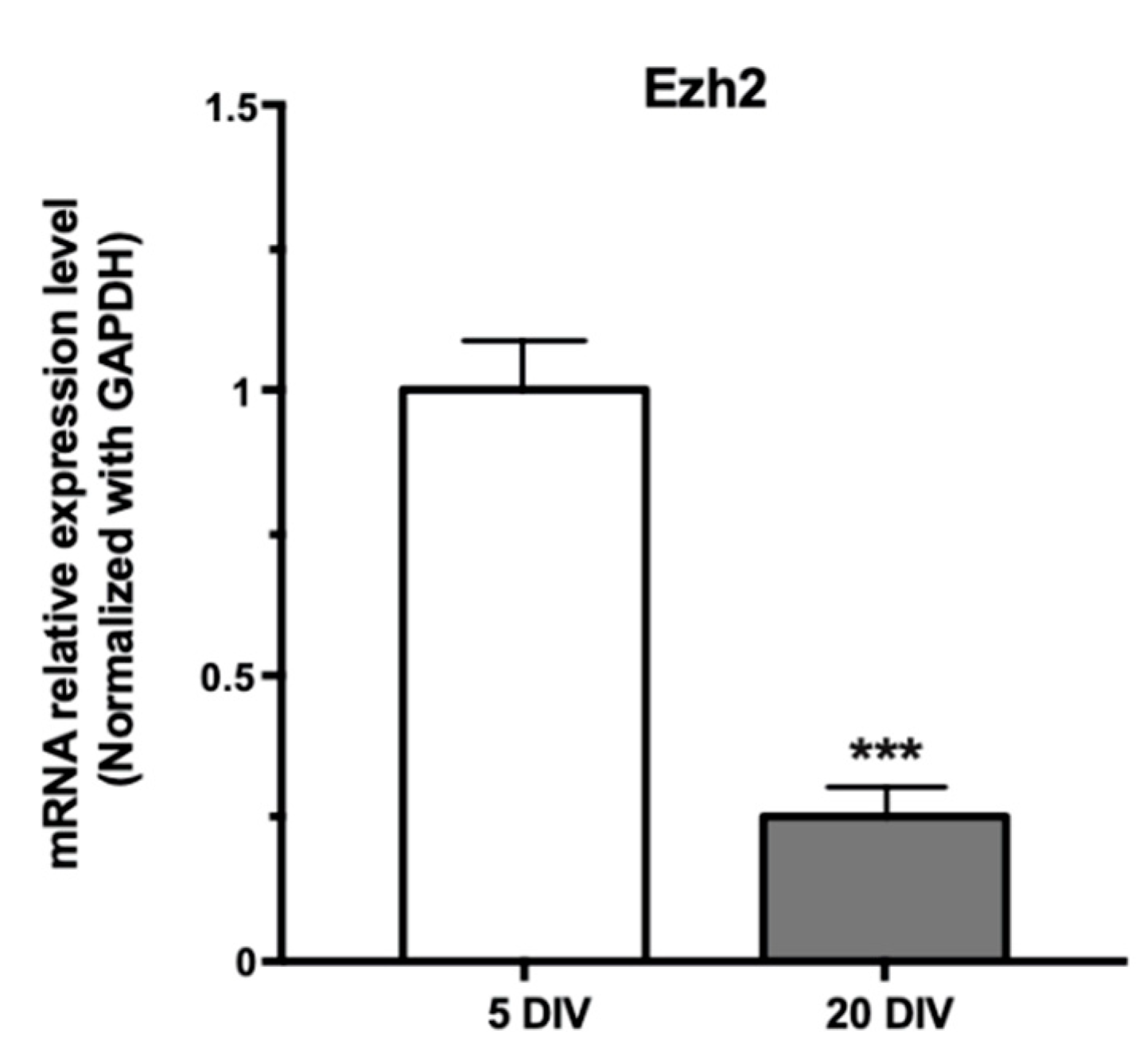
2.2. Downregulation of Ezh2 Expression during Hippocampal Maturation Requires Dicer

To begin examining the role of miRs on Ezh2 expression in hippocampal neurons, we carried out a knockdown of Dicer expression within these cells, the RNA nuclease that catalyzes the processing of immature miR precursors, thereby allowing the formation of mature and functional miR molecules [41,42]. Two shRNAs coding sequences targeting Dicer mRNA (shDicer1 and shDicer2) were produced as lentiviral vectors, and then infective viral particles were generated. These particles efficiently infected hippocampus neuronal cell types, including the mouse N2A neuroblastoma cell line and primary rat hippocampal neurons, where a significant decrease in Dicer mRNA expression was detected (Figure S1).
Next, we infected mature (from 17 DIV to 20 DIV) hippocampal neuron-enriched cultures with lentiviruses encoding for the two shRNAs against Dicer. It was found that both molecules downregulated Dicer mRNA levels as compared with an infection with lentiviruses encoding only GFP (Figure 2A, B, respectively). Importantly, we determined that this shRNA-mediated knockdown of Dicer expression (using a mix of both shDicer 1 and 2, Figure 2C) resulted in a significant increase in Ezh2 mRNA levels (Figure 2D). Together, these results indicate that the decreased Ezh2 expression during maturation of hippocampal neurons (Figure 1) can be mediated by miRs that require Dicer for adequate processing and function.

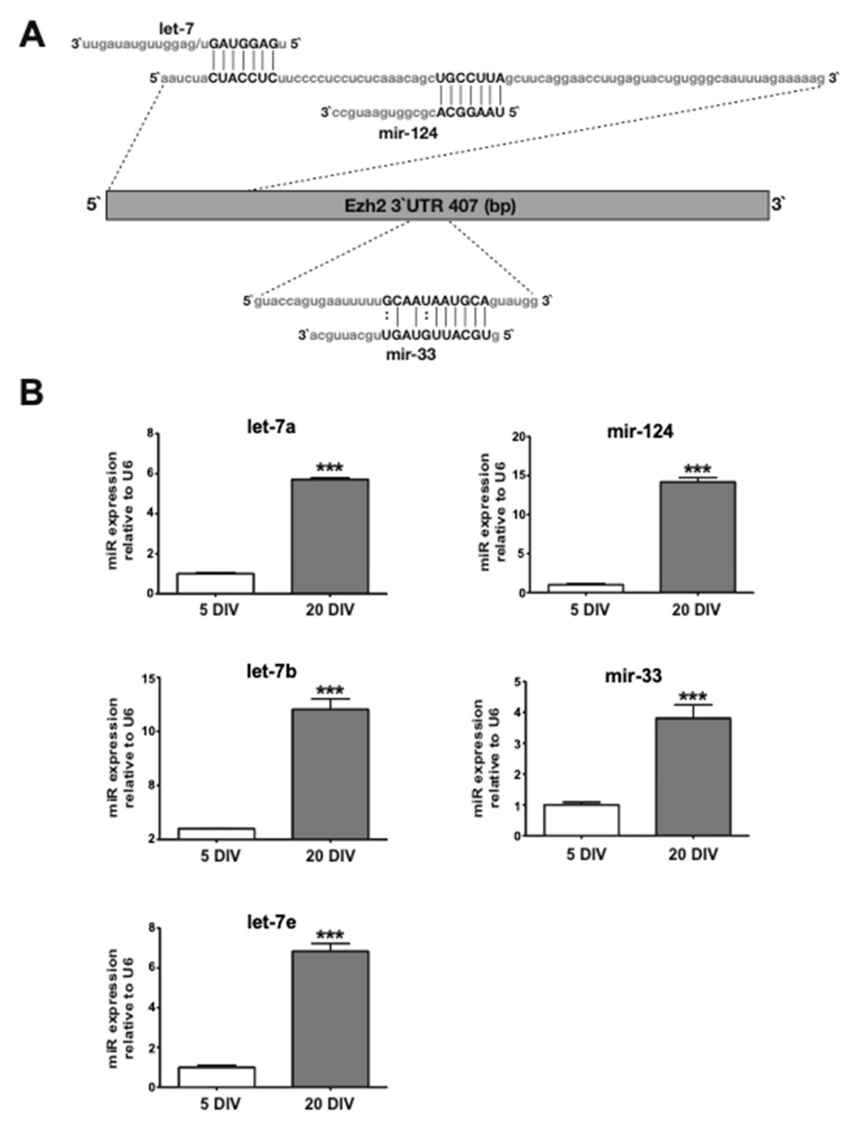
2.3. Candidate miRs for Mediating Ezh2 Downregulation Exhibit Increased Expression during Hippocampal Neuronal Maturation

Our highly expressed miR profile in mature hippocampal neurons (Table 1) revealed the presence of miRs that could potentially target the 3′-UTR of the Ezh2 mRNA (see Table 2). Among them, miR-124 has been previously shown to target and downregulate Ezh2 mRNA expression during early mouse neurogenesis [28] and let-7, which has been previously associated with Ezh2 mRNA control in prostate cancer cells [39]. Both miR families could recognize non-overlapping motifs at the Ezh2 3′-UTR (Figure 3A) and were confirmed as upregulated during hippocampal neuronal maturation by using Taqman-based quantitative PCR approaches (see Methods) (Figure 3B). We additionally identified other miRs that could potentially target the Ezh2 3′-UTR. For instance, miR-33 (Figure 3A), although it is detected at relatively lower levels than miR-124 and let-7 members in mature hippocampal neurons (see Table S1), shows an expression that increases during the transition from immature to mature neurons (Figure 3B). It has been shown that miR-33 played a principal role during control of the cholesterol metabolism as it targeted and downregulated the expression of genes associated with the biosynthesis and transport of cholesterol in the liver [43]. Importantly, miR-33 has not been found to be associated with Ezh2 expression control or to play a relevant role in hippocampal neurons.

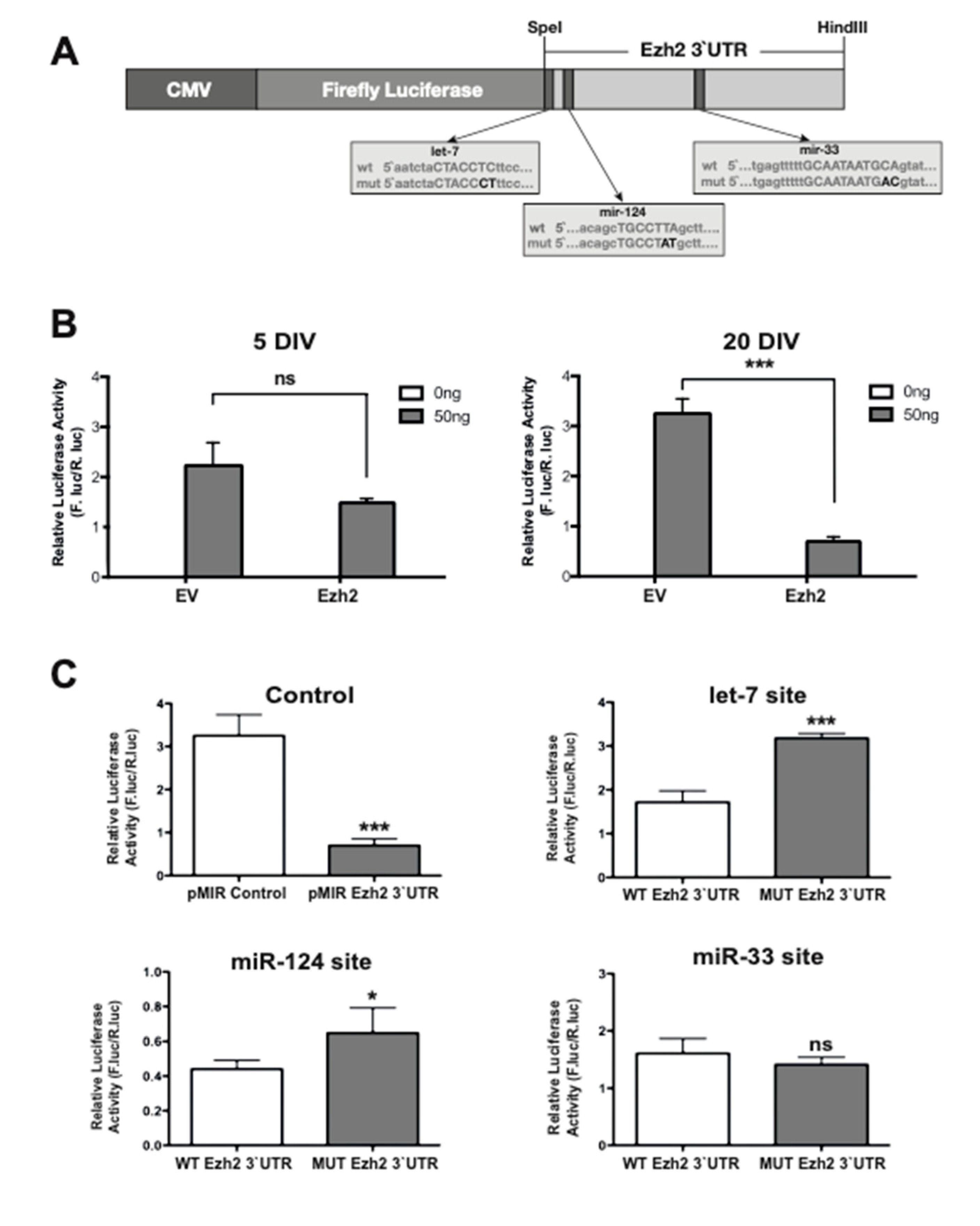
2.4. The 3′-UTR of the Ezh2 Gene Contains Sequence Elements that Mediate Decreased Expression of Immediately Upstream Protein-Coding Genes

Next, we examined the ability of the Ezh2 3′-UTR sequence to modulate the expression of an immediately upstream luciferase reporter gene in cells enriched in miRs that can potentially target motifs in this 3′-UTR. For this purpose, the entire Ezh2 3′-UTR segment (407 bp) was cloned downstream of the luciferase reporter gene within the context of Cytomegalovirus (CMV)-driven mammalian expression vector (CMV-LUC-Ezh2-3′UTR, see Figure 4A). This vector was transfected in both immature (5 DIV) and mature (20 DIV) hippocampal neuronal cultures for 24 h, after which the luciferase activity present in the transfected cells was measured. It was found that transfection of this construct in mature neurons resulted in a significantly reduced luciferase activity as compared with that obtained in immature hippocampal cells (Figure 4B). This result indicates that the 3′-UTR of the Ezh2 gene can mediate a decreased biosynthesis of the luciferase protein in mature hippocampal cells that exhibit increased expression of miRs that may potentially target this 3′-UTR sequence.
Next, we examined whether the specific motifs for let-7, miR-124, and miR-33 within the 3′-UTR of the Ezh2 gene were contributing to mediate this downregulation of luciferase expression in mature hippocampal neurons. Specific mutations at each motif were introduced within the context of the entire Ezh2 3′-UTR sequence cloned immediately downstream of the luciferase reporter gene (CMV-LUC-Ezh2-3′UTR vector) (Figure 4A). Each mutated construct was transiently transfected in mature hippocampal neurons (in parallel with the wild-type version of this expression vector) and the effect on the luciferase activity was measured 24 h later. We determined that mutation of the cognate motifs for let-7 and miR-124 prevented the reduced luciferase activity observed with the wild-type version of the construct (Figure 4C). Thus, instead of the decreased luciferase activity (five- to six-fold) produced by the WT Ezh2 3′-UTR construct (control graph), the mutation at the let-7 (let-7 site graph) and miR-124 (miR-124 site graph) elements resulted in higher reporter activity with respect to this wild-type construct (two- and 1.5-fold, respectively). These results suggest that joint or independent binding of endogenous let-7 and miR-124 family members to the Ezh2 3′-UTR sequence can mediate downregulation of the luciferase reporter activity. In addition, mutation of the miR-33 motif resulted in luciferase activity that was comparable to that produced by the wild-type version of the vector (Figure 4C). These results suggest that let-7 and miR-124 motifs, but not the miR-33 motif, can significantly contribute to the Ezh2 3′-UTR-dependent downregulation of Ezh2 expression in mature hippocampal neurons. It is necessary to consider that because mutation of the let-7 and miR-124 motifs did not completely revert the Ezh2 3′-UTR-mediated reduction in the luciferase reporter activity, there could be additional regulatory mechanisms associated with this 3′-UTR sequence that contributed to its inhibitory activity.

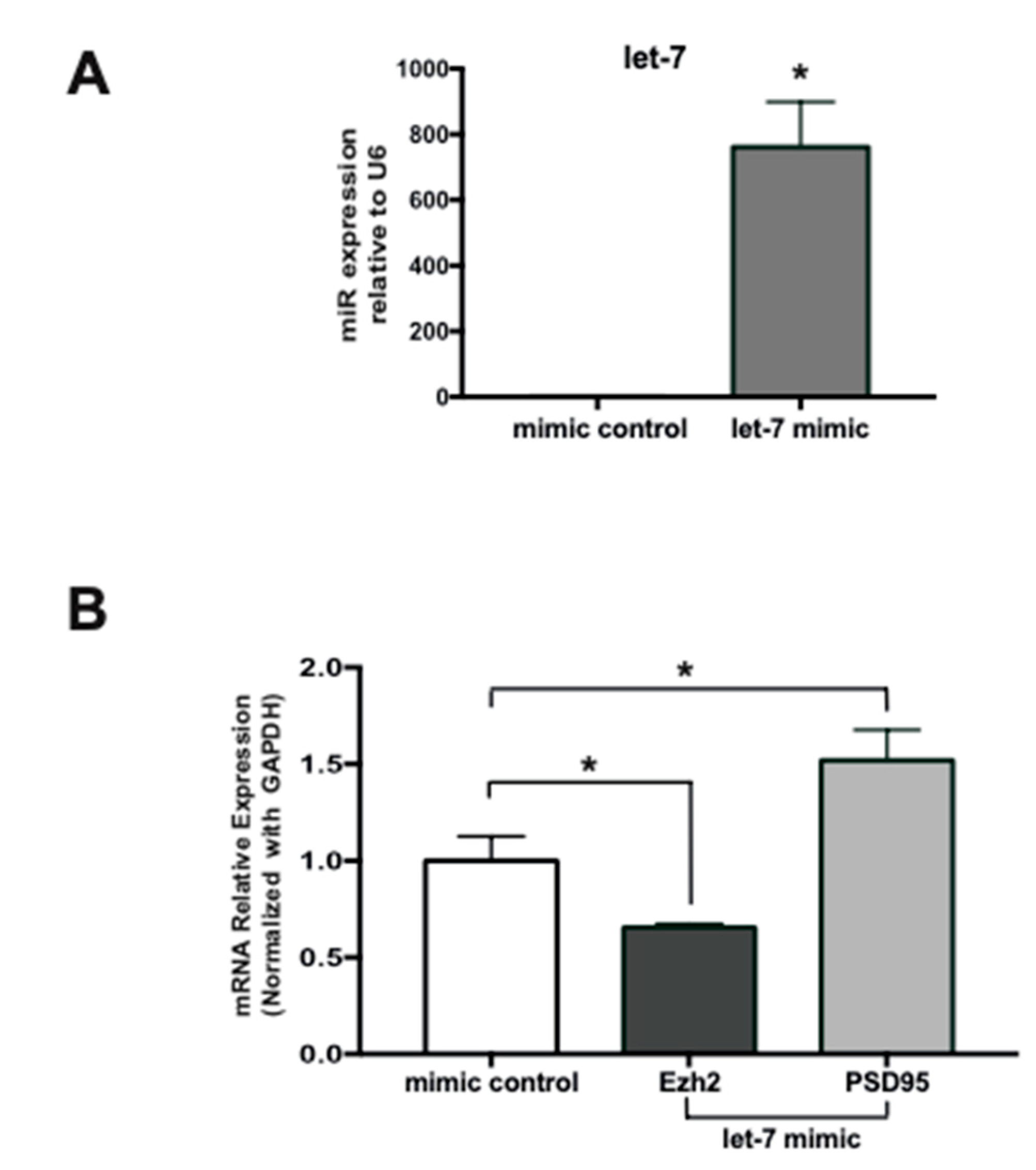
2.5. Expression of a Let-7 Mimic in Immature Hippocampal Neurons Inhibits Ezh2 Expression

To further demonstrate that miRs enriched in mature hippocampal neurons can mediate downregulation of Ezh2 expression, we performed a gain-of-function experiment using a miR mimics-based strategy [44,45]. Because the mutation of the let-7 motif produced the highest effect on the inhibitory activity of the Ezh2 3′-UTR sequence (Figure 4C), we examined the impact of an enhanced expression of let-7e mimics on the levels of the endogenous Ezh2 mRNA in immature (5 DIV) hippocampal neurons. As shown in Figure 5, transfection of these immature neurons with a let-7e mimic results in a robust increase in its expression as compared with a negative control mimic (termed mimic control) (Figure 5A). This let-7 increase is accompanied by a significant reduction in Ezh2 mRNA levels (Figure 5B), further indicating that Ezh2 expression is controlled by miRs that, like let-7, can target the Ezh2 3′-UTR in hippocampal neurons. Importantly, this reduced Ezh2 mRNA expression occurs concomitantly with an increase in PSD95/Dlg4 gene transcription (Figure 5B), a previously reported gene target of the PRC2-Ezh2 complex in immature hippocampal neurons [7].

3. Discussion

During early stages of hippocampal development, the repressive activity of the PRC2 complex is critical for both silencing the expression of non-neuronal gene programs (e.g., osteogenic lineage genes) and repressing the expression of neuronal genes coding key synaptic components (e.g., PSD95/Dlg4) that are mainly required at later stages of hippocampal maturation [6,7]. Previous work from our group demonstrated that Ezh2, the catalytic subunit of PRC2, was differentially expressed during maturation of hippocampal neurons; in immature neurons, Ezh2 was abundantly expressed, whereas in mature neurons the expression of Ezh2 was significantly reduced [6,7]. Here, we find that the microRNAs let-7 and miR-124, which are highly enriched in mature hippocampal cells (20 DIV), can mediate downregulation of Ezh2 expression in these cells. Although these data argue in favor of the hypothesis that, during hippocampal maturation, the Ezh2 repressive activity (as part of the PRC2 complex) can be controlled through a miR-mediated post-transcriptional mechanism, it is necessary to consider the contribution of additional mechanisms to this Ezh2 downregulation. These additional mechanisms include changes in histone tail modifications that can inhibit transcription at the Ezh2 gene promoter in hippocampal cells [7] and the proteasome-mediated Ezh2 protein degradation in brain cells [11]. Therefore, it is tempting to propose that Ezh2 expression is tightly controlled during hippocampal maturation by a combination of transcriptional, post-transcriptional, and post-translational mechanisms.
In agreement with the results reported here, miR-124 has been previously found upregulated during mammalian neuronal differentiation [14,21,22] where it contributed to neuronal survival [27]. Moreover, miR-124 has been shown to target the Ezh2 3′-UTR in neuronal cell lines (N2a and P19), thereby downregulating Ezh2 expression and reducing PRC2-Ezh2-mediated gene silencing required for proper progression of neuronal differentiation [28]. Interestingly, miR-124 has also been proposed to be a critical regulator of PRC2-independent functions of Ezh2 during early neural lineage commitment. In this case, miR-124 targeted and downregulated the expression of USP14 [29], a deubiquitinase enzyme that binds to the Ezh2 protein, and prevented its ubiquitin-dependent degradation via proteasome [30]. Together with these previous findings, our results provide further support for the critical role of miR-124 during neuronal function, as miR-124 may control Ezh2 expression at both early and late stages of neuronal differentiation.
The let-7 family members have previously been found to be expressed at elevated concentrations in brain tissues during embryogenesis [31,32,33,34]. However, a direct role of let-7 in hippocampal neuronal differentiation or plasticity has not been reported. Here, we find that a let-7-dependent decrease in Ezh2 mRNA is accompanied by a significant increase in PSD95/Dlg4 mRNA, further supporting the critical role of the PRC2-Ezh2 complex during inhibition of the PSD95/Dlg4 gene transcription in immature hippocampal cells [7]. Because let-7 cannot directly target the PSD95/Dlg4 gene 3′-UTR (let-7 motif is not present at this PSD95/Dlg4 sequence), our results provide a basis for proposing that, in mature hippocampal neurons, let-7 can indirectly promote the expression of relevant hippocampal plasticity components by downregulating the expression of the epigenetic repressor PRC2-Ezh2. These results extend previous findings showing that miR-125, together with fragile X mental retardation protein (FMRP), represses the translation of PSD95 mRNA in hippocampal neurons at early stages (7 DIV) of maturation [46,47].
Future studies must address the specific mechanisms mediating the increased expression of both miR-124 and let-7 during hippocampal neuron maturation. It has been reported that the role of miR-124 during neuronal differentiation was tightly associated with the activity of the Repressor Element-1 Silencing Transcription Factor (REST) [30], a key repressor of neuronal genes in neural progenitors, including sequences coding for the miR-124 RNA precursors [23]. Neuronal differentiation is accompanied with a progressive inhibition of this REST-dependent repression of miR-124 transcription, which results in miR-124 accumulation and subsequent downregulation of target transcripts, including those coding for REST and its associated protein MeCP1, as both transcripts contain miR-124 binding motifs at their 3′-UTRs [23,48]. Whether this type of negative feedback regulatory loop also operates during hippocampal neuronal maturation needs to be established. In the case of let-7, studies should clarify the potential role of the Lin28A/B proteins during control of the let-7 biogenesis in hippocampal neurons [49]. In mouse hippocampal neurons, brain-derived neurotrophic factor (BDNF) has been shown to directly stimulate Lin28 expression in a developing brain, which resulted in an inhibition of let-7 biogenesis (by preventing Dicer-mediated processing of the pre-let-7 transcript and promoting its degradation) and the concomitant increase in let-7 target transcripts, including a minor subset of mRNAs coding for proteins associated with excitatory functions [50]. As BDNF-mediated gene pathways are a relevant component in the physiological function of hippocampal neurons, the contribution of this BDNF-Lin28-miR-124 axis needs to be experimentally addressed.
The contribution of the let-7 and miR-124 motifs in the Ezh2 3′-UTR inhibitory activity appears to be selective and, likely, is related to the elevated concentration of both microRNAs in mature hippocampal cells (both among the top 10 most abundant miRs in these cells). This conclusion is supported by our results indicating that a consensus motif for miR-33, a microRNA expressed at significantly lower relative concentration than let-7 and miR-124 in these mature hippocampal neurons, does not contribute to the Ezh2 3′-UTR-dependent downregulation of the upstream luciferase reporter gene. Previous reports have extensively documented that miR-33 controlled the expression of genes associated with the metabolism of cholesterol, mainly in the liver [43]. Understanding whether this microRNA also regulates specific mRNA targets (different from Ezh2) in hippocampal neurons needs further investigation.
It also remains to be determined whether let-7 and mir-124 functionally collaborate (additively or synergistically) during downregulation of Ezh2 expression in mature hippocampal neurons. Because their specific target motifs are not overlapping at the 3′-UTR sequence of the Ezh2 gene, and because independent mutation of each of these two motifs cannot completely revert the inhibitory activity associated with of this Ezh2 3′UTR sequence, it is reasonable to hypothesize that a collaborative inhibitory mechanism involving both let-7 and miR-124 operates in physiological conditions. Moreover, the possibility that these two molecules function in concert with additional regulatory mechanisms must be addressed in future studies.

4. Materials and Methods

4.1. Primary Hippocampal Cultures

Protocols of animal management were performed according to NIH guidelines and as approved by the Ethical and Biosafety Committees of Universidad Andres Bello (014/2013; 024/2013; 001/2018). Pregnant Sprague–Dawley rats were deeply anesthetized with CO2; hippocampal cultures were prepared from E18 pups, as described previously [7,51,52].

4.2. MicroRNA Expression Analyses Using Microarrays

Microarray analyses were performed using 500 ng of miRNA, purified using a mirVanaTM miRNA isolation kit (Life Technologies Inc., Ambion, Burlington, ON, Canada). For miRNA labeling, the FlashtaqTM Biotin kit (Genisphere, Hatfield, PA, USA) was used, following the manufacturer′s recommendations. The miRNAs were processed for poly-A tail addition, and then ligated with adapters for biotin labeling and subsequent recognition. Labeled miRNAs were hybridized to GeneChipTM miRNA 2.0 Array (Affymetrix, Santa Clara, CA, USA), which contained 389 probes in triplicate to detect rat miRNAs. Results were analyzed using the software AffymetrixTM miRNA QC Tool.

4.3. MicroRNA Quantitation

The miRNAs were extracted using a mirVanaTM miRNA isolation kit (Life Technologies Inc.), according to the manufacturer’s instructions. Specific cDNA for each miRNA quantified was synthesized from 10 ng of RNA, using a TaqManTM MicroRNA Reverse Transcription Kit (Life Technologies Inc.). Then, quantitation was performed for each miRNA using TaqManTM MicroRNA Assays (Life Technologies Inc.), followed by real-time PCR with TaqManTM probes.

4.4. Plasmid Construction

Reporter plasmids were constructed by inserting the full-length 3′-UTR sequence of Ezh2 (NM_001107051.1) from Rattus norvegicus (between Spe I and Hind III sites), downstream of the firefly luciferase gene in the pMIR-reportTM vector (Promega, Madison, WI, USA). Ezh2 3′-UTR is 407 bp in length. Mutations were carried out by changing only two nucleotides of each MRE at the 3′-UTR, to prevent broad changes in the 3′-UTR secondary structure. Nucleotide changes were analyzed by using the Mfold (RNA-folding form) web server (http://mfold.rit.albany.edu/?q=mfold/) [53]. The ΔG values for the interactions between each miRNA and MREs were calculated using the RNAhybrid server [54]. Details of the mutations of the MREs generated for each miRNA, are shown in Table 3.

4.5. Transient Transfections

Functional 3′-UTR analyses were carried out in cultures of primary rat hippocampal neurons obtained from 18-day-old embryos (E18), that were maintained in NeurobasalTM growth medium (GIBCO, Gaithersburg, MD, USA) supplemented with B27 (GIBCO), 2 mM L-glutamine, 100U/mL penicillin, and 100 μg/mL streptomycin. Transfection was performed at 4 days in vitro (4 DIV) using LipofectamineTM 3000, following the manufacturer′s instructions (Invitrogen, Carlsbad, CA, USA), and then reporter activity was evaluated at 5 DIV. To evaluate cultures at day 20 DIV, transfection with NeuromagTM was performed at 19 DIV, according to the manufacturer’s instructions (OZ Biosciences, San Diego, CA, USA). Neuronal cultures were transfected with 20 ng of plasmid pMIR-reportTM containing the 3′-UTR of the rat Ezh2 gene. Cultures were co-transfected with 10 pg SV-40 pRL Renilla Luciferase control reporter vector, used as a transfection control. These plasmid quantities were selected after performing dose-response curves.

4.6. Lentivirus Production and Infection of Hippocampal Neurons

HEK293FT cells were transfected using LipofectamineTM 3000 reagent (Invitrogen), following the manufacturer’s instructions, with the pVSVg, pΔ8.9, and pLKO.1-shRNA plasmids (at a 1:2:3 ratio, respectively) and a total DNA of 10 μg. TRC1.5 pLKO.1-puro non-mammalian shRNA control plasmid DNA (SHC002, Sigma) was used as negative control. After 12 h, the medium was replaced and cells were maintained at 32 °C for 48 h. Supernatants containing lentiviral particles were collected, filtered through a PVDF filter (0.45 μm pore size), and concentrated by centrifugation at 3800× g for 30 min at 4 °C in an AmiconTM Ultra-15 centrifugal filter (100K, Merck Millipore, Burlington, MA, USA), according to the manufacturer’s instructions. Aliquots of concentrated viral particles were stored at −80 °C. Hippocampal cultures evaluated at 20 DIV were infected at 16 DIV with 1.0 × 10⁷ infectious units of virus (IFU)/cell culture plate (30 mm) containing shRNA-Dicer or shRNA-GFP control.

4.7. Reverse Transcriptase and Real-Time Quantitative PCR (RT-qPCR)

Total RNA was extracted with TRIzolTM (Invitrogen), according to the manufacturer’s protocol. Then, 2 μg of each sample were used for reverse transcription. The qPCR was performed using SYBR GreenTM PCR Master Mix (Applied Biosystems, Life Technologies Inc.). Data are presented as relative mRNA levels of the gene of interest normalized to GAPDH mRNA levels. Primers used were the following: Dicer Fw: GGCAGGTGTACTATCCGATGA, Rev: TGGTTCCATCTCAAGCAATTC; Ezh2 Fw: GCCAGACTGGGAAGAAATCTG, Rev: TCACTGGTGACTGAACACTCC; GAPDH Fw: CATGGCCTTCCGTGTTCCTA, Rev: CCTGCTTCACCACCTTCTTGAT.

4.8. Luciferase Reporter Assay

Luciferase activity was measured 24 h after transfection using a Dual-GloTM Luciferase Assay System (Promega) in a GloMaxTM 20/20 Luminometer (Promega, Turner Biosystems, Madison, WI, USA). Firefly luciferase activity was normalized to Renilla luciferase activity to minimize variations in transfection efficiency between experiments. Experiments were performed in triplicate. Quantitative data of the reporter gene assay are presented as mean ± SEM (n = 3).

4.9. MicroRNA Target Prediction

To identify miRNAs that potentially target the 3′-UTR of Ezh2 mRNAs, we used the following miRNA target prediction sites: TargetScan (http://www.targetscan.org/) [55] and miRDB (http://mirdb.org/) [56].

4.10. Expression of Let-7 Mimics

For the in vitro transfection of immature hippocampal neurons, the transfection mix was prepared using 90 pmol of let-7 mimic (mirVanaTM miRNA mimic, Life Technologies) and 15 μL of LipofectamineTM RNAiMax (Invitrogen), according to the manufacturer′s protocol, for 48 h. The transfection was confirmed by RT-qPCR. Each experiment was repeated at least 3 times.

4.11. Statistical Analysis

For the expression analysis we used Student’s t-test to determine significant differences between experiments. In all figures, error bars represent the mean ± standard error of the mean; * p < 0.05, ** p < 0.01, and *** p < 0.001.

Supplementary Materials

Author Contributions

L.G. performed most of the experimental work and data analyses; R.A. and F.J.B. carried out the transcriptomic studies and contributed to data analyses; G.N. and R.A.G. performed the transcriptomic studies, including the bioinformatics analyses; B.v.Z. and M.M. designed the experimental strategy, oversaw the experimental work, and wrote most parts of the manuscript. All authors have read and agreed to the published version of the manuscript.

Funding

This research was supported by FONDAP 15090007 (to R.G. and M.M.), FONDECYT 3140418 (to L.G.), FONDECYT 11190998 (to G.N.), FONDECYT 1170878 (to M.M.), FONDECYT 1181645 (to B.v.Z.), CONICYT PIA/BASAL AFB 170005 CARE UC (to B.v.Z.), and Nucleus UNAB DI-4-17/N (to B.v.Z.).

Conflicts of Interest

The authors declare no conflict of interests.

Abbreviations

miRsMicroRNAs
CNSCentral nervous system
IFUInfectious units of virus
FMRPFragile X mental retardation protein
BDNFBrain-derived neurotrophic factor

References

  1. Gräff, J.; Kim, D.; Dobbin, M.M.; Tsai, L.-H. Epigenetic regulation of gene expression in physiological and pathological brain processes. Physiol. Rev. 2011, 91, 603–649. [Google Scholar] [CrossRef]
  2. Sweatt, J.D. The emerging field of neuroepigenetics. Neuron 2013, 80, 624–632. [Google Scholar] [CrossRef] [Green Version]
  3. Allis, C.D.; Jenuwein, T. The molecular hallmarks of epigenetic control. Nat. Rev. Genet. 2016, 17, 487–500. [Google Scholar] [CrossRef] [PubMed]
  4. Margueron, R.; Reinberg, D. The Polycomb complex PRC2 and its mark in life. Nature 2011, 469, 343–349. [Google Scholar] [CrossRef] [PubMed] [Green Version]
  5. Yu, J.-R.; Lee, C.-H.; Oksuz, O.; Stafford, J.M.; Reinberg, D. PRC2 is high maintenance. Genes Dev. 2019, 33, 903–935. [Google Scholar] [CrossRef] [PubMed]
  6. Aguilar, R.; Bustos, F.J.; Saez, M.; Rojas, A.; Allende, M.L.; van Wijnen, A.J.; van Zundert, B.; Montecino, M. Polycomb PRC2 complex mediates epigenetic silencing of a critical osteogenic master regulator in the hippocampus. Biochim. Biophys. Acta 2016, 1859, 1043–1055. [Google Scholar] [CrossRef] [PubMed] [Green Version]
  7. Henriquez, B.; Bustos, F.J.; Aguilar, R.; Becerra, A.; Simon, F.; Montecino, M.; van Zundert, B. Ezh1 and Ezh2 differentially regulate PSD-95 gene transcription in developing hippocampal neurons. Mol. Cell. Neurosci. 2013, 57, 130–143. [Google Scholar] [CrossRef]
  8. Pereira, J.D.; Sansom, S.N.; Smith, J.; Dobenecker, M.-W.; Tarakhovsky, A.; Livesey, F.J. Ezh2, the histone methyltransferase of PRC2, regulates the balance between self-renewal and differentiation in the cerebral cortex. Proc. Natl. Acad. Sci. USA 2010, 107, 15957–15962. [Google Scholar] [CrossRef] [Green Version]
  9. Sher, F.; Boddeke, E.; Copray, S. Ezh2 expression in astrocytes induces their dedifferentiation toward neural stem cells. Cell. Reprogram. 2011, 13, 1–6. [Google Scholar] [CrossRef]
  10. Shivram, H.; Le, S.V.; Iyer, V.R. MicroRNAs reinforce repression of PRC2 transcriptional targets independently and through a feed-forward regulatory network. Genome Res. 2019, 29, 184–192. [Google Scholar] [CrossRef] [Green Version]
  11. Yu, Y.-L.; Chou, R.-H.; Shyu, W.C.; Hsieh, S.C.; Wu, C.S.; Chiang, S.Y.; Chang, W.J.; Chen, J.N.; Tseng, Y.J.; Lin, Y.H.; et al. Smurf2-mediated degradation of EZH2 enhances neuron differentiation and improves functional recovery after ischaemic stroke. EMBO Mol. Med. 2013, 5, 531–547. [Google Scholar] [CrossRef] [PubMed]
  12. Zhao, C.; Sun, G.; Ye, P.; Li, S.; Shi, Y. MicroRNA let-7d regulates the TLX/microRNA-9 cascade to control neural cell fate and neurogenesis. Sci. Rep. 2013, 3, 1–7. [Google Scholar] [CrossRef] [PubMed] [Green Version]
  13. Ji, F.; Lv, X.; Jiao, J. The role of microRNAs in neural stem cells and neurogenesis. J. Genet Genom. 2013, 40, 61–66. [Google Scholar] [CrossRef] [PubMed]
  14. Krichevsky, A.M.; King, K.S.; Donahue, C.P.; Khrapko, K.; Kosik, K.S. A microRNA array reveals extensive regulation of microRNAs during brain development. RNA 2003, 9, 1274–1281. [Google Scholar] [CrossRef] [Green Version]
  15. Somel, M.; Liu, X.; Tang, L.; Yan, Z.; Hu, H.; Guo, S.; Jiang, X.; Zhang, X.; Xu, G.; Xie, G.; et al. MicroRNA-driven developmental remodeling in the brain distinguishes humans from other primates. PLoS Biol. 2011, 9, e1001214. [Google Scholar] [CrossRef]
  16. Khudayberdiev, S.; Fiore, R.; Schratt, G. MicroRNA as modulators of neuronal responses. Commun. Integr. Biol. 2009, 2, 411–413. [Google Scholar] [CrossRef] [Green Version]
  17. Schratt, G. microRNAs at the synapse. Nat. Rev. Neurosci. 2009, 10, 842–849. [Google Scholar] [CrossRef]
  18. De Pietri Tonelli, D.; Pulvers, J.N.; Haffner, C.; Murchison, E.P.; Hannon, G.J.; Huttner, W.B. miRNAs are essential for survival and differentiation of newborn neurons but not for expansion of neural progenitors during early neurogenesis in the mouse embryonic neocortex. Development 2008, 135, 3911–3921. [Google Scholar] [CrossRef] [Green Version]
  19. Kawase-Koga, Y.; Otaegi, G.; Sun, T. Different timings of Dicer deletion affect neurogenesis and gliogenesis in the developing mouse central nervous system. Dev. Dyn. 2009, 238, 2800–2812. [Google Scholar] [CrossRef] [Green Version]
  20. Makeyev, E.V.; Zhang, J.; Carrasco, M.A.; Maniatis, T. The MicroRNA miR-124 promotes neuronal differentiation by triggering brain-specific alternative pre-mRNA splicing. Mol. Cell. 2007, 27, 435–448. [Google Scholar] [CrossRef] [Green Version]
  21. Deo, M.; Yu, J.-Y.; Chung, K.-H.; Tippens, M.; Turner, D.L. Detection of mammalian microRNA expression by in situ hybridization with RNA oligonucleotides. Dev. Dyn. 2006, 235, 2538–2548. [Google Scholar] [CrossRef] [PubMed] [Green Version]
  22. Visvanathan, J.; Lee, S.; Lee, B.; Lee, J.W.; Lee, S.-K. The microRNA miR-124 antagonizes the anti-neural REST/SCP1 pathway during embryonic CNS development. Genes Dev. 2007, 21, 744–749. [Google Scholar] [CrossRef] [PubMed] [Green Version]
  23. Conaco, C.; Otto, S.; Han, J.-J.; Mandel, G. Reciprocal actions of REST and a microRNA promote neuronal identity. Proc. Natl. Acad. Sci. USA 2006, 103, 2422–2427. [Google Scholar] [CrossRef] [PubMed] [Green Version]
  24. Yoo, A.S.; Sun, A.X.; Li, L.; Shcheglovitov, A.; Portmann, T.; Li, Y.; Lee-Messer, C.; Dolmetsch, R.E.; Tsien, R.W.; Crabtree, G.R. MicroRNA-mediated conversion of human fibroblasts to neurons. Nature 2011, 476, 228–231. [Google Scholar] [CrossRef] [PubMed]
  25. Xue, Y.; Ouyang, K.; Huang, J.; Zhou, Y.; Ouyang, H.; Li, H.; Wang, G.; Wu, Q.; Wei, C.; Bi, Y.; et al. Direct conversion of fibroblasts to neurons by reprogramming PTB-regulated microRNA circuits. Cell 2013, 152, 82–96. [Google Scholar] [CrossRef] [Green Version]
  26. Cheng, L.-C.; Pastrana, E.; Tavazoie, M.; Doetsch, F. miR-124 regulates adult neurogenesis in the subventricular zone stem cell niche. Nat. Neurosci. 2009, 12, 399–408. [Google Scholar] [CrossRef] [Green Version]
  27. Sanuki, R.; Onishi, A.; Koike, C.; Muramatsu, R.; Watanabe, S.; Muranishi, Y.; Irie, S.; Uneo, S.; Koyasu, T.; Matsui, R.; et al. miR-124a is required for hippocampal axogenesis and retinal cone survival through Lhx2 suppression. Nat. Neurosci. 2011, 14, 1125–1134. [Google Scholar] [CrossRef]
  28. Neo, W.H.; Yap, K.; Lee, S.H.; Looi, L.S.; Khandelia, P.; Neo, S.X.; Makeyev, E.V.; Su, I.-H. MicroRNA miR-124 controls the choice between neuronal and astrocyte differentiation by fine-tuning Ezh2 expression. J. Biol. Chem. 2014, 289, 20788–20801. [Google Scholar] [CrossRef] [Green Version]
  29. Doeppner, T.R.; Doehring, M.; Bretschneider, E.; Zechariah, A.; Kaltwasser, B.; Müller, B.; Koch, J.C.; Bähr, M.; Hermann, D.M.; Michel, U. MicroRNA-124 protects against focal cerebral ischemia via mechanisms involving Usp14-dependent REST degradation. Acta Neuropathol. 2013, 126, 251–265. [Google Scholar] [CrossRef]
  30. Lee, S.W.; Oh, Y.M.; Lu, Y.-L.; Kim, W.K.; Yoo, A.S. MicroRNAs Overcome Cell Fate Barrier by Reducing EZH2-Controlled REST Stability during Neuronal Conversion of Human Adult Fibroblasts. Dev. Cell 2018, 46, 73–84. [Google Scholar] [CrossRef] [Green Version]
  31. Thomson, J.M.; Parker, J.; Perou, C.M.; Hammond, S.M. A custom microarray platform for analysis of microRNA gene expression. Nat. Methods 2004, 1, 47–53. [Google Scholar] [CrossRef] [PubMed] [Green Version]
  32. Schulman, B.R.M.; Esquela-Kerscher, A.; Slack, F.J. Reciprocal expression of lin-41 and the microRNAs let-7 and mir-125 during mouse embryogenesis. Dev. Dyn. 2005, 234, 1046–1054. [Google Scholar] [CrossRef] [PubMed] [Green Version]
  33. Thomson, J.M.; Newman, M.; Parker, J.S.; Morin-Kensicki, E.M.; Wright, T.; Hammond, S.M. Extensive post-transcriptional regulation of microRNAs and its implications for cancer. Genes Dev. 2006, 20, 2202–2207. [Google Scholar] [CrossRef] [Green Version]
  34. Wulczyn, F.G.; Smirnova, L.; Rybak, A.; Brandt, C.; Kwidzinski, E.; Ninnemann, O.; Strehle, M.; Seiler, A.; Schumacher, S.; Nitsch, R. Post-transcriptional regulation of the let-7 microRNA during neural cell specification. FASEB J. 2007, 21, 415–426. [Google Scholar] [CrossRef] [PubMed] [Green Version]
  35. Schwamborn, J.C.; Berezikov, E.; Knoblich, J.A. The TRIM-NHL protein TRIM32 activates microRNAs and prevents self-renewal in mouse neural progenitors. Cell 2009, 136, 913–925. [Google Scholar] [CrossRef] [PubMed] [Green Version]
  36. Cimadamore, F.; Amador-Arjona, A.; Chen, C.; Huang, C.-T.; Terskikh, A.V. SOX2-LIN28/let-7 pathway regulates proliferation and neurogenesis in neural precursors. Proc. Natl. Acad. Sci. USA 2013, 110, E3017–E3026. [Google Scholar] [CrossRef] [PubMed] [Green Version]
  37. Zhao, C.; Sun, G.; Li, S.; Lang, M.-F.; Yang, S.; Li, W.; Shi, Y. MicroRNA let-7b regulates neural stem cell proliferation and differentiation by targeting nuclear receptor TLX signaling. Proc. Natl. Acad. Sci. USA 2010, 107, 1876–1881. [Google Scholar] [CrossRef] [Green Version]
  38. Thomas, K.T.; Gross, C.; Bassell, G.J. microRNAs Sculpt Neuronal Communication in a Tight Balance That Is Lost in Neurological Disease. Front. Mol. Neurosci. 2018, 11, 455. [Google Scholar] [CrossRef]
  39. Kong, D.; Heath, E.; Chen, W.; Cher, M.L.; Powell, I.; Heilbrun, L.; Li, Y.; Ali, S.; Sethi, S.; Hassan, O.; et al. Loss of let-7 up-regulates EZH2 in prostate cancer consistent with the acquisition of cancer stem cell signatures that are attenuated by BR-DIM. PLoS ONE 2012, 7, e33729. [Google Scholar] [CrossRef] [Green Version]
  40. Bustos, F.J.; Varela-Nallar, L.; Campos, M.; Henriquez, B.; Phillips, M.; Opazo, C.; Aguayo, L.G.; Montecino, M.; Constantine-Paton, M.; Inestrosa, N.C.; et al. PSD95 suppresses dendritic arbor development in mature hippocampal neurons by occluding the clustering of NR2B-NMDA receptors. PLoS ONE 2014, 9, e94037. [Google Scholar] [CrossRef] [Green Version]
  41. Hutvágner, G.; McLachlan, J.; Pasquinelli, A.E.; Balint, E.; Tuschl, T.; Zamore, P.D. A cellular function for the RNA-interference enzyme Dicer in the maturation of the let-7 small temporal RNA. Science 2001, 293, 834–838. [Google Scholar] [CrossRef] [PubMed] [Green Version]
  42. Martinez, J.; Patkaniowska, A.; Urlaub, H.; Lührmann, R.; Tuschl, T. Single-stranded antisense siRNAs guide target RNA cleavage in RNAi. Cell 2002, 110, 563–574. [Google Scholar] [CrossRef] [Green Version]
  43. Zhang, X.; Price, N.L.; Fernández-Hernando, C. Non-coding RNAs in lipid metabolism. Vascul. Pharmacol. 2019, 114, 93–102. [Google Scholar] [CrossRef] [PubMed]
  44. Kuhn, D.E.; Martin, M.M.; Feldman, D.S.; Terry, A.V.; Nuovo, G.J.; Elton, T.S. Experimental validation of miRNA targets. Methods 2008, 44, 47–54. [Google Scholar] [CrossRef] [PubMed] [Green Version]
  45. Rebustini, I.T. A Functional MicroRNA Screening Method for Organ Morphogenesis. Curr. Protoc. Cell Biol. 2017, 74, 19.19.1–19.19.17. [Google Scholar]
  46. Muddashetty, R.S.; Nalavadi, V.C.; Gross, C.; Yao, X.; Xing, L.; Laur, O.; Warren, S.T.; Bassell, G.J. Reversible inhibition of PSD-95 mRNA translation by miR-125a, FMRP phosphorylation, and mGluR signaling. Mol. Cell. 2011, 42, 673–688. [Google Scholar] [CrossRef] [Green Version]
  47. Ifrim, M.F.; Williams, K.R.; Bassell, G.J. Single-Molecule Imaging of PSD-95 mRNA Translation in Dendrites and Its Dysregulation in a Mouse Model of Fragile X Syndrome. J. Neurosci. 2015, 35, 7116–7130. [Google Scholar] [CrossRef] [Green Version]
  48. Wu, J.; Xie, X. Comparative sequence analysis reveals an intricate network among REST, CREB and miRNA in mediating neuronal gene expression. Genome Biol. 2006, 7, R85. [Google Scholar] [CrossRef] [Green Version]
  49. Lee, H.; Han, S.; Kwon, C.S.; Lee, D. Biogenesis and regulation of the let-7 miRNAs and their functional implications. Protein Cell 2016, 7, 100–113. [Google Scholar] [CrossRef] [Green Version]
  50. Huang, Y.-W.A.; Ruiz, C.R.; Eyler, E.C.H.; Lin, K.; Meffert, M.K. Dual regulation of miRNA biogenesis generates target specificity in neurotrophin-induced protein synthesis. Cell 2012, 148, 933–946. [Google Scholar] [CrossRef] [Green Version]
  51. Sepulveda, F.J.; Bustos, F.J.; Inostroza, E.; Zúñiga, F.A.; Neve, R.L.; Montecino, M.; van Zundert, B. Differential roles of NMDA Receptor Subtypes NR2A and NR2B in dendritic branch development and requirement of RasGRF1. J. Neurophysiol. 2010, 103, 1758–1770. [Google Scholar] [CrossRef] [PubMed] [Green Version]
  52. Varela-Nallar, L.; Grabowski, C.P.; Alfaro, I.E.; Alvarez, A.R.; Inestrosa, N.C. Role of the Wnt receptor Frizzled-1 in presynaptic differentiation and function. Neural Dev. 2009, 4, 41. [Google Scholar] [CrossRef] [PubMed] [Green Version]
  53. Zuker, M. Mfold web server for nucleic acid folding and hybridization prediction. Nucleic Acids Res. 2003, 31, 3406–3415. [Google Scholar] [CrossRef] [PubMed]
  54. Rehmsmeier, M.; Steffen, P.; Hochsmann, M.; Giegerich, R. Fast and effective prediction of microRNA/target duplexes. RNA 2004, 10, 1507–1517. [Google Scholar] [CrossRef] [PubMed] [Green Version]
  55. Grimson, A.; Farh, K.K.-H.; Johnston, W.K.; Garrett-Engele, P.; Lim, L.P.; Bartel, D.P. MicroRNA targeting specificity in mammals: Determinants beyond seed pairing. Mol. Cell. 2007, 27, 91–105. [Google Scholar] [CrossRef] [PubMed] [Green Version]
  56. Wong, N.; Wang, X. miRDB: An online resource for microRNA target prediction and functional annotations. Nucleic Acids Res. 2015, 43, D146–D152. [Google Scholar] [CrossRef]
Figure 1. Expression of Enhancer of Zeste Homolog 2 (Ezh2) mRNA is downregulated during hippocampal neuronal maturation. mRNA samples were collected from hippocampal neuron-enriched cultures at 5 and 20 days in vitro (DIV). Then, real-time quantitative PCR (RT-qPCR) analysis was performed to determine the Ezh2 mRNA levels. All values are presented as relative expression normalized to Glyceraldehyde-3-Phosphate Dehydrogenase (GAPDH). (n = 3, *** p < 0.001).
Ijms 21 08472 g001
Figure 2. Dicer knockdown in primary mature hippocampal neurons results in enhanced Ezh2 mRNA expression. Reduced Dicer mRNA expression is found after lentiviral transduction of shDicer 1 (A) and 2 (B) molecules; (C) Hippocampal neuron-enriched cultures at 17 DIV were transduced for 72 h with a mix of lentivirus coding shRNA 1 and shRNA 2 against Dicer, and Dicer mRNA expression then analyzed; (D) Ezh2 mRNA expression in mature hippocampal neurons (20 DIV) infected with shRNA against Dicer. Results are shown as mean ± SEM (n = 3, * p < 0.05 and ** p < 0.01).
Ijms 21 08472 g002
Figure 3. Expression of miRs that potentially bind at the 3′-UTR of Ezh2 mRNAs is upregulated during hippocampal neuronal maturation. (A) Schematic representation of Ezh2 3’-UTR and the predicted miR-124, let-7, and miR-33 target sites; (B) Quantification of miR-124, let-7 (let-7a, -b, and -e family members) and miR-33 increased expression during hippocampal neuronal maturation (5 to 20 DIV). Each miRNA was quantified using specific TaqMan® probes. Expression of the different miRNAs was normalized against snU6 RNA. Results are shown as mean ± SEM. (n = 3, *** p < 0.001).
Ijms 21 08472 g003
Figure 4. 3′-UTR sequence of the Ezh2 gene contains miR motifs that can downregulate the expression of an immediately up-stream luciferase reporter gene in rat hippocampal neurons. (A) Schematic representation of the CMV(Cytomegalovirus)-driven luciferase Ezh2 3`-UTR reporter gene (pMIR-report-Ezh2-3′UTR) construct expressed in hippocampal neurons following transient transfection. Wild-type (wt) and mutated (mut) motifs for miR-124, let-7, and miR-33 are shown below the construct. The specific mutations introduced are indicated in black-bold; (B) Immature (5 DIV) and mature (20 DIV) hippocampal neurons were transfected with 50 ng of empty vector (EV) or pMIR-report-Ezh2-3′UTR vector and luciferase activity measured 24 h later; (C) Relative luciferase activity after expression of wild-type (control) and mutant (indicated at the top of each graph) versions of let-7, miR-124, and miR-33 motifs in mature (20 DIV) hippocampal neurons. Results are shown as mean ± SEM (n = 3, * p < 0.05 and *** p < 0.001).
Ijms 21 08472 g004
Figure 5. Forced expression of a let-7 mimic downregulates Ezh2 mRNA expression in immature hippocampal neurons. (A) Expression of let-7 was significantly higher in immature hippocampal neurons (5 DIV) following a let-7 mimic transfection for 48 h. Quantification was assessed by TaqMan® probes as shown in Figure 3 legend; (B) Significantly decreased expression of Ezh2 mRNA follows let-7 mimic transfection in immature hippocampal neurons. In contrast, expression of PSD95/Dlg4 gene mRNA, an Ezh2 target gene in these cells, was found to be significantly enhanced. (n = 3, * p < 0.05).
Ijms 21 08472 g005
Table 1. List of top 25 highly expressed microRNAs in mature hippocampal neurons. The data was originated by microarray analyses from three independent experiments (n = 1, 2, and 3).
ProbeSetID20 DIV n120 DIV n220 DIV n3Mean 20 DIV
rno-miR-125b-5p_st14,1958673514,1152463414,0220194514,1110444
rno-let-7c_st14,0565437113,781126114,3390840414,058918
rno-miR-124_st13,1452883114,6763791713,7468701313,8561792
rno-let-7b_st13,7721592513,4853391314,0216206713,7597064
hp_rno-mir-124-3_s_st12,6646819814,1740883313,411135313,4166352
hp_rno-mir-124-1_s_st12,5526354514,1253350213,4048624413,3609443
hp_rno-mir-124-2_s_st12,5096909913,998420813,4029301913,3036807
rno-miR-103_st12,8602098913,5864468613,0242865213,1569811
rno-miR-99a_st13,426562313,0379083612,682461613,0489774
rno-miR-107_st12,5482707713,3345996112,9207448812,9345384
rno-miR-16_st13,0670410913,0851143512,4582926412,8701494
rno-miR-138_st11,9740753413,4638400513,0213129212,8197428
rno-miR-24_st13,2250758712,540345612,4565239312,7406485
rno-miR-22_st12,8071227512,9243975212,487362212,7396275
rno-let-7a_st12,744834412,6542931312,6639577912,6876951
rno-miR-181a_st12,1808116213,0570869812,7154261412,6511082
rno-miR-191_st12,9715573912,5497254812,4277597912,6496809
rno-miR-23a_st13,461149512,4186415912,0628872612,6475594
rno-let-7d_st12,3306972512,73661212,8671422812,6448172
rno-miR-26a_st12,5020737412,9482149412,2146120812,5549669
rno-let-7e_st12,1386431112,4711762612,4888492612,3662229
rno-miR-125a-5p_st12,6796712,3032767611,8651182212,2826883
rno-miR-130a_st12,3827945212,6225652511,7559660312,2537753
rno-miR-127_st11,3813731712,7965064212,2806567912,1528455
rno-miR-99b_st12,3170208811,7008487211,972361211,9967436
Table 2. The microRNA (miR) families that can potentially target the 3′-UTR region of Ezh2 mRNAs in mature rat hippocampal neuron-enriched cultures (20 DIV). Information about the specific chromosome location of each miR family member is provided. The position of miRs within a sequence that clusters additional microRNA- and other-coding genes is also indicated. IR, intergenic region.
TargetmicroRNAChromosomeCoding Details
let-7a-117Clustered miR-3596b, miR-3596d, let-7d; let-7f-1
let-7a-28Clustered miR-100, miR-3695a, in lnc215 gene
let-7b7Clustered let-7c-2
let-7c-111Clustered miR-99a
let-7c-27Clustered let-7b
let-7d17Clustered miR-3596b, miR-3596d, let-7a-1, let-7f-1, in Spaca6 gene
let-7e1Clustered miR-99b, miR-3596c, miR-125a
Ezh2let-7f-117Clustered 3596b, miR-3596d, let-7a-1, let-7d
let-7f-2XClustered miR-98, in Huwe1 gene
let-7g8IR
let-7i7IR
miR-124-115IR
miR-124-22IR
miR-124-33IR
miR-337In Srebf2 gene
Table 3. Mutations performed at miR-124, let-7, and miR-33 motifs within the Ezh2 3′-UTR region. Specific mutated nucleotides for each miRNA recognition element (MRE) are indicated in bold black. Changes in the predicted ΔG values for both wild-type (wt) and mutated miR motifs are also included.
miR AnalyzedEzh2 3′UTRMRE—miR Alignments PredictedΔG MRE WTΔG MRE MUT
WT 5′AAUCUACUACCUCU 3
let-77–133′UGAUAUGUUGGAGGAUGGAGU 5−20.1−15.4
MUT 5′AAUCUACUACCCUU 3
WT 5′CUCCUCUCAAACAGCUGCCUUAG 3
miR-12433–393′CCGUAAGUGGCGC-ACGGAAU 5−19.9−15.8
MUT 5′CUCCUCUCAAACAGCUGCCUAUG 3
WT 5′UGAAUUUUUGCAAUAAUGCAG 3
miR-33195–2163′ACGUUACGUUGAUGUUACGUG 5−18.2−14.2
MUT 5′UGAAUUUUUGCAAUAAUGACG 3
Publisher’s Note: MDPI stays neutral with regard to jurisdictional claims in published maps and institutional affiliations.

