Article

Genetic Deletion of NOD1 Prevents Cardiac Ca2+ Mishandling Induced by Experimental Chronic Kidney Disease

by
Marta Gil-Fernández
1,†,
José Alberto Navarro-García
2,†,
Almudena Val-Blasco
1,†,
Laura González-Lafuente
2,†,
José Carlos Martínez
2,
Angélica Rueda
3,
Maria Tamayo
4,
José Luis Morgado
4,
Carlos Zaragoza
5,6,
Luis Miguel Ruilope
2,7,8,
Carmen Delgado
4,6,†,
Gema Ruiz-Hurtado
2,8,* and
María Fernández-Velasco
1,6,*
1
IdiPAZ: Hospital La Paz Institute for Health Research, 28046 Madrid, Spain
2
Cardiorenal Translational Laboratory, Institute of Research i+12, Hospital Universitario 12 de Octubre, 28041 Madrid, Spain
3
Departamento de Bioquímica, Centro de Investigación y de Estudios Avanzados del IPN, México City 07360, Mexico
4
Biomedical Research Institute “Alberto Sols” CSIC-UAM, 28046 Madrid, Spain
5
Departamento de Cardiología, Unidad de Investigación Mixta Universidad Francisco de Vitoria, 28223 Madrid, Spain
6
Centro de Investigación Biomédica en Red en Enfermedades Cardiovasculares (CIBERCV), 28029 Madrid, Spain
7
School of Doctoral Studies and Research, European University of Madrid, 28224 Madrid, Spain
8
CIBER-CV, Hospital Universitario 12 de Octubre, 28029 Madrid, Spain
*
Authors to whom correspondence should be addressed.
Contributed equally to this work.
Int. J. Mol. Sci. 2020, 21(22), 8868; https://0-doi-org.brum.beds.ac.uk/10.3390/ijms21228868
Submission received: 5 October 2020 / Revised: 16 November 2020 / Accepted: 19 November 2020 / Published: 23 November 2020

Abstract

:
Risk of cardiovascular disease (CVD) increases considerably as renal function declines in chronic kidney disease (CKD). Nucleotide-binding oligomerization domain-containing protein 1 (NOD1) has emerged as a novel innate immune receptor involved in both CVD and CKD. Following activation, NOD1 undergoes a conformational change that allows the activation of the receptor-interacting serine/threonine protein kinase 2 (RIP2), promoting an inflammatory response. We evaluated whether the genetic deficiency of Nod1 or Rip2 in mice could prevent cardiac Ca2+ mishandling induced by sixth nephrectomy (Nx), a model of CKD. We examined intracellular Ca2+ dynamics in cardiomyocytes from Wild-type (Wt), Nod1−/− and Rip2−/− sham-operated or nephrectomized mice. Compared with Wt cardiomyocytes, Wt-Nx cells showed an impairment in the properties and kinetics of the intracellular Ca2+ transients, a reduction in both cell shortening and sarcoplasmic reticulum Ca2+ load, together with an increase in diastolic Ca2+ leak. Cardiomyocytes from Nod1−/−-Nx and Rip2−/−-Nx mice showed a significant amelioration in Ca2+ mishandling without modifying the kidney impairment induced by Nx. In conclusion, Nod1 and Rip2 deficiency prevents the intracellular Ca2+ mishandling induced by experimental CKD, unveiling new innate immune targets for the development of innovative therapeutic strategies to reduce cardiac complications in patients with CKD.

1. Introduction

Chronic kidney disease (CKD) is a complex pathology characterized by a reduced glomerular filtration rate, increased urinary albumin excretion and kidney damage [1]. Recent studies have found that cardiac complications are frequent in patients with CKD [2,3]. The United States Renal Data System (USRDS) 2014 annual report stated that the prevalence of any cardiovascular disease (CVD) is about 2-fold higher in patients with CKD than in patients without CKD. Heart failure (HF) is the main cardiovascular risk in patients with CKD from the outset, and increases gradually with the progression of renal dysfunction [4]. Patients with CKD also have a higher prevalence of cardiac systolic and diastolic dysfunction [5]. These data underscore the sharp increase in mortality in advanced CKD, due mainly to the occurrence of cardiovascular events, such as arrhythmias [6,7]. The complex association between CKD and cardiac dysfunction can likely be explained by the clustering of several risk factors, including uremia and inflammatory mediators [8,9]. Yet, little is known about the underlying mechanisms of cardiac dysfunction in CKD.
Cardiac muscle cell contraction is tightly regulated by the change in intracellular Ca2+ levels, acting as a key mediator of excitation-contraction (EC)-coupling. The initial depolarization event of the action potential activates L-type Ca2+ channels (LTCCs) of the sarcolemma, firing an inward voltage-dependent Ca2+ current type l (ICaL) from the extracellular medium. Ca2+ entry triggers a large release of Ca2+ from the sarcoplasmic reticulum (SR) by ryanodine receptor (RyR2) channels, resulting in an increased intracellular Ca2+ concentration ([Ca2+]i) that prompts cell contraction. For relaxation to occur, [Ca2+]i must return to diastolic levels and this occurs mainly by two mechanisms: (i) Ca2+ re-uptake by the Sarco/Endoplasmic Ca2+ pump (SERCA) 2a and (ii) Ca2+ extrusion by the Na+/Ca2+ exchanger (NCX). During diastole, RyR2 channels are mostly closed; however, there is always a low but finite probability that a RyR2 channel will spontaneously open, mediating Ca2+ flux into the cytosol—known as Ca2+ sparks. Ca2+ spark frequency is normally low during diastole but, in some pathological conditions, abnormally large or frequent sparks can activate the RyR2 channels at neighboring release sites, generating SR Ca2+ waves that favor Ca2+ extrusion by NCX, and providing a substrate to initiate a life-threatening arrhythmia. Dysregulation of any of these Ca2+ handling mechanisms is commonly associated with the development of cardiac dysfunction [10], but whether this is coming from an indirect kidney damage is less known.
There is a growing body of evidence suggesting that inflammation induced by innate immune system activation can contribute to cardiac dysfunction [11,12]. Indeed, some receptors of the innate immune system are known to play a significant role in the host response after cardiac and renal damage [13,14]. The nucleotide-like receptors (NLRs) are a family of receptors of the innate immune system with a relevant role in several CVDs. Indeed, specific activators of NLRs have a role in the progression of some CVDs [15]. Nucleotide-binding oligomerization domain (NOD) 1 (NOD1) and 2 (NOD2) are members of the NLR family that present a few differences in their stimulatory molecules and tissue location. Regarding their cellular location, NOD1 is broadly expressed in many cell types and organs, such as heart, lung, skeletal muscle and kidney [14,16,17,18], whereas NOD2 expression is more restricted to immune cells and endothelial cells [14,19,20,21,22]. Importantly, different groups including ours have reported an association between NOD1 and CVDs [11,23,24,25,26,27,28,29]. NOD proteins are involved in host defense that respond rapidly to certain pathogens or endogenous molecules released during cell injury by triggering an inflammatory response [17]. NOD1 is a cytosolic protein that contains a caspase activation domain, a recruitment domain (CARD), a nucleotide-binding oligomerization domain (NOD) and a leucine-rich repeat domain. Upon activation, NOD1 undergoes a conformational change, leading to self-oligomerization that allows the recruitment and activation of the receptor-interacting serine/threonine protein kinase 2 (RIP2) through CARD-CARD interactions [30]. Activated RIP2 in turn mediates the recruitment and activation of several mediators that allow the translocation of nuclear factor κB (NF-κB) into the nucleus to initiate the inflammatory response [18].
Several groups, including ours, have analyzed the role of NOD1 in CVD [11,23,31,32,33,34,35]. We previously showed that NOD1 is up-regulated in both mouse and human failing myocardium and its genetic deletion or pharmacological blockade in mice with experimental HF impedes the development of cardiac dysfunction, mainly by preventing cardiac Ca2+ mishandling [11]. NOD1 expression has been reported in human and mouse renal tubular epithelial cells [14,36]. Supporting the involvement of NOD proteins in renal disease, Shigeoka et al. demonstrated that the deficiency of Nod1/2 or Rip2 deletion, was protective against acute kidney ischemia/reperfusion injury, suggesting that NODs respond to endogenous ligands after injury [14]. By contrast, Stroo et al. reported that the double Nod1/2 deletion had no impact on the chronic renal damage induced by ureteral obstruction [37].
The potential role of NOD1 in the cardiovascular complications caused by specific renal disease is unknown. Accordingly, the main goal of the present study was to investigate whether the NOD1-dependent pathway was implicated in cardiac dysfunction and Ca2+ mishandling induced by experimental CKD.

2. Results

2.1. Macroscopic and Microscopic Cardiac Features and Biochemical Parameters of Renal Function in Wild-Type and Nod1−/− Mice at Baseline and after Experimental CKD

Cardiac macroscopic and microscopic parameters of the mice are summarized in Table 1. Macroscopic analysis revealed that the Nx surgery induced an overall loss in BW (body weight) in wild-type (Wt) mice, accompanied by a reduction in kidney weight (KW). Notably, the KW in Nx mice considers the remaining one-third of the left kidney after the surgery, which is hypertrophied in both Wt and Nod1−/− mice (Table 1). The weight of the partial kidney resulting from the surgery (1/6 of the kidney) was similar to the weight of the complete kidney in sham-operated mice, demonstrating that the Nx remaining kidney is hypertrophied in both Wt and Nod1−/− mice. KW from Wt and Nod1−/−-sham-operated mice was similar. All these results were supported by KW/BW data (Table 1). Heart weight (HW) and HW/BW ratio were similar between the different groups (Table 1). Additionally, no differences in cardiomyocyte area were observed between Wt-sham, Wt-Nx, Nod1−/−-sham and Nod1−/−-Nx hearts, indicating that Nx surgery does not induce cardiac hypertrophy in Wt or Nod1−/− mice. Examination of biochemical parameters related to renal function showed comparable kidney impairment in both groups of mice subjected to Nx, as demonstrated by the significantly higher levels of plasma urea and BUN, compared with sham-operated mice (Table 2). FGF-23 levels were higher in Wt-Nx and Nod1−/−-Nx mice compared to sham animals, although they were only significantly increased in Wt-Nx, compared to Wt-sham. Phosphate levels were not different between the groups. Biochemical kidney parameters were similar between Wt-Nx and Nod1−/−-Nx mice, indicating that the deficiency of NOD1 does not prevent the renal impairment induced by Nx.

2.2. Deficiency of NOD1 Prevents both Systolic Ca2+ Release Impairment and the Decrease in SR Ca2+ Load Triggered by Experimental CKD

Given the link between CKD and the prevalence of cardiac and systolic dysfunction [2,3] and the involvement of NOD1 in preventing cardiac Ca2+ mishandling [11], we evaluated systolic Ca2+ release after Nx and the possible participation of NOD1. To do this, we analyzed cardiomyocyte Ca2+ transients electrically-evoked by field stimulation at 2 Hz using confocal microscopy. Representative line-scan images from field-stimulated cardiomyocytes from Wt-sham, Wt-Nx, Nod1−/−-sham and Nod1−/−-Nx mice are shown in Figure 1A. Wt-Nx cells clearly displayed a lower amplitude of the intracellular Ca2+ transients, slower kinetics and lower cell shortening values than Wt-sham cardiomyocytes (Figure 1A–D). By contrast, the amplitude of Ca2+ transients in Nod1−/−-sham and Nod1−/−-Nx myocytes was very similar, and close to the values in Wt-sham cells (Figure 1B). These data indicate that deficiency of NOD1 prevents the decrease in the Ca2+ transient’s amplitude, and the impairment in their decay time and cell shortening induced by Nx. Since changes in systolic Ca2+ release are closely related to an alteration in the amount of Ca2+ that entries though L-type Ca2+ channels (LTCCs), we measured the density of ICaL using patch-clamp technique in the whole cell configuration in cardiomyocytes isolated from Wt-sham and Wt-Nx mice. Supplementary Figure S1 shows a similar ICaL density in both experimental groups, indicating that the Nx did no induce any change in the density of Ca2+ entering through LTCCs.
We next examined whether the observed differences in Ca2+ transients between the different groups were related to changes in the cardiomyocyte SR Ca2+ load by measuring caffeine-evoked Ca2+ transients. Figure 1E shows representative line-scan images of caffeine-evoked Ca2+ transients in each group. The amplitude of caffeine-evoked Ca2+ transients was significantly lower in Wt-Nx cells than in Wt-sham cardiomyocytes (Figure 1E,F). By contrast, this parameter was similar between Nod1−/−-Nx and Nod1−/−-sham cardiomyocytes (Figure 1E,F), indicating that NOD1 deficiency prevents the impairment of systolic Ca2+ release induced by Nx and contributes to the maintenance of the physiological levels of the SR Ca2+ load, allowing adequate systolic Ca2+ release and regular cell shortening after experimental CKD.

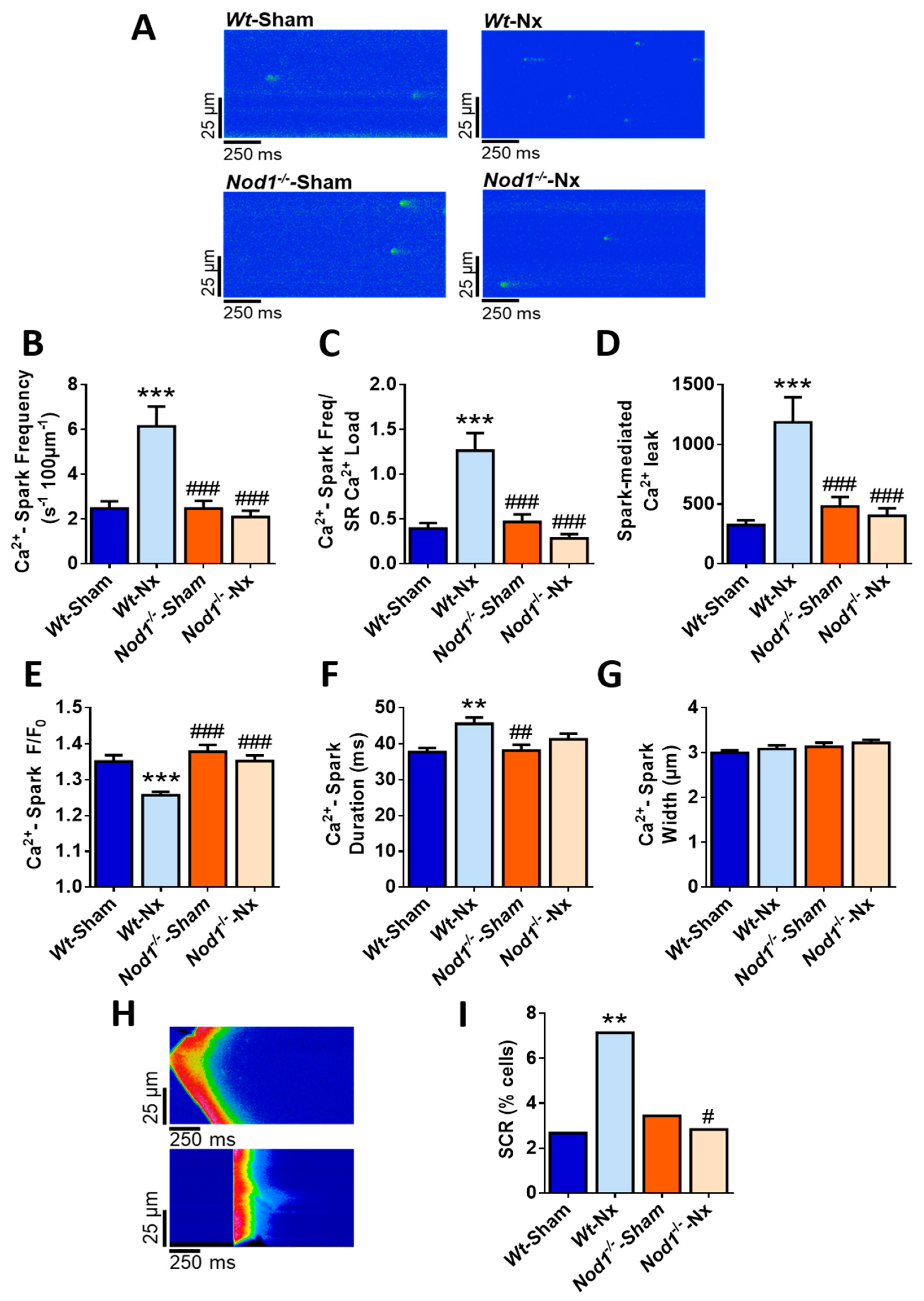
2.3. Deficiency of NOD1 Blunts the Increase in Diastolic Ca2+ Release Induced by Nx

Since impairment of SR Ca2+ load is frequently associated with alterations in diastolic Ca2+ release, we analyzed the frequency and properties of Ca2+ sparks to measure the spark-mediated Ca2+ leak from RyR2 channels. Representative line-scan confocal images of quiescent cardiomyocytes from Wt-sham, Wt-Nx, Nod1−/−-sham and Nod1−/−-Nx mice are shown in Figure 2A. Results showed a significantly higher frequency of Ca2+ sparks in Wt-Nx cells than in Wt-sham counterparts, whereas Ca2+ spark frequency in Nod1−/−-Nx cardiomyocytes was similar to those of in Nod1−/−-sham cells, and both were comparable with those of Wt-sham cells (Figure 2B). Estimation of Ca2+ spark frequency normalized to the SR Ca2+ load showed that this was significantly higher in Wt-Nx cardiomyocytes (Figure 2C). Confirming these data, the overall spark-mediated Ca2+ leak was substantially increased in Wt-Nx cells (Figure 2D). By contrast, both the Ca2+ spark frequency/SR Ca2+ load (Figure 2C) and the overall spark-mediated Ca2+ leak (Figure 2D) were similar in Nod1−/−-Nx and Nod1−/−-sham cardiomyocytes.
Examination of the biophysical characteristics of Ca2+ sparks revealed that their amplitude was significantly lower in Wt-Nx cardiomyocytes than in Wt-sham cells (Figure 2E), whereas the opposite was observed for the average duration of Ca2+ sparks (Figure 2F). Ca2+ spark amplitude and duration in Nod1−/−-Nx cells was similar to those of Nod1−/−-sham cells, and both were comparable with those of the Wt-sham group (Figure 2E,F). Conversely, the average width of Ca2+ sparks was unchanged between groups (Figure 2G).
We next analyzed other forms of spontaneous Ca2+ release (SCR), such as Ca2+ waves and spontaneous Ca2+ transients, in ventricular quiescent cardiomyocytes. Figure 2H illustrates an example of a Ca2+ wave (upper panel) and spontaneous Ca2+ transient release (lower panel) from cells isolated from Wt-Nx mice. Results showed that the occurrence of SCR was almost 3-fold higher in Wt-Nx cardiomyocytes than in Wt-sham cells (Figure 2I). By contrast, the occurrence of SCR in Nod1−/−-Nx cells was significantly lower than in Wt-Nx cells, and similar to that obtained in Nod1−/−-sham and Wt-sham myocytes (Figure 2I).
Taken together, these results confirm that the loss of NOD1 prevents the increase in diastolic Ca2+ leak induced by Nx, a beneficial effect that can also be related to the maintenance of the SR Ca2+ load, as observed in Nod1−/−-Nx cardiomyocytes. This provides an explanation not only for the improvement in the SR Ca2+ load, but also for the better systolic Ca2+ release observed in Nod1−/−-Nx cells relative to the Wt-Nx group.

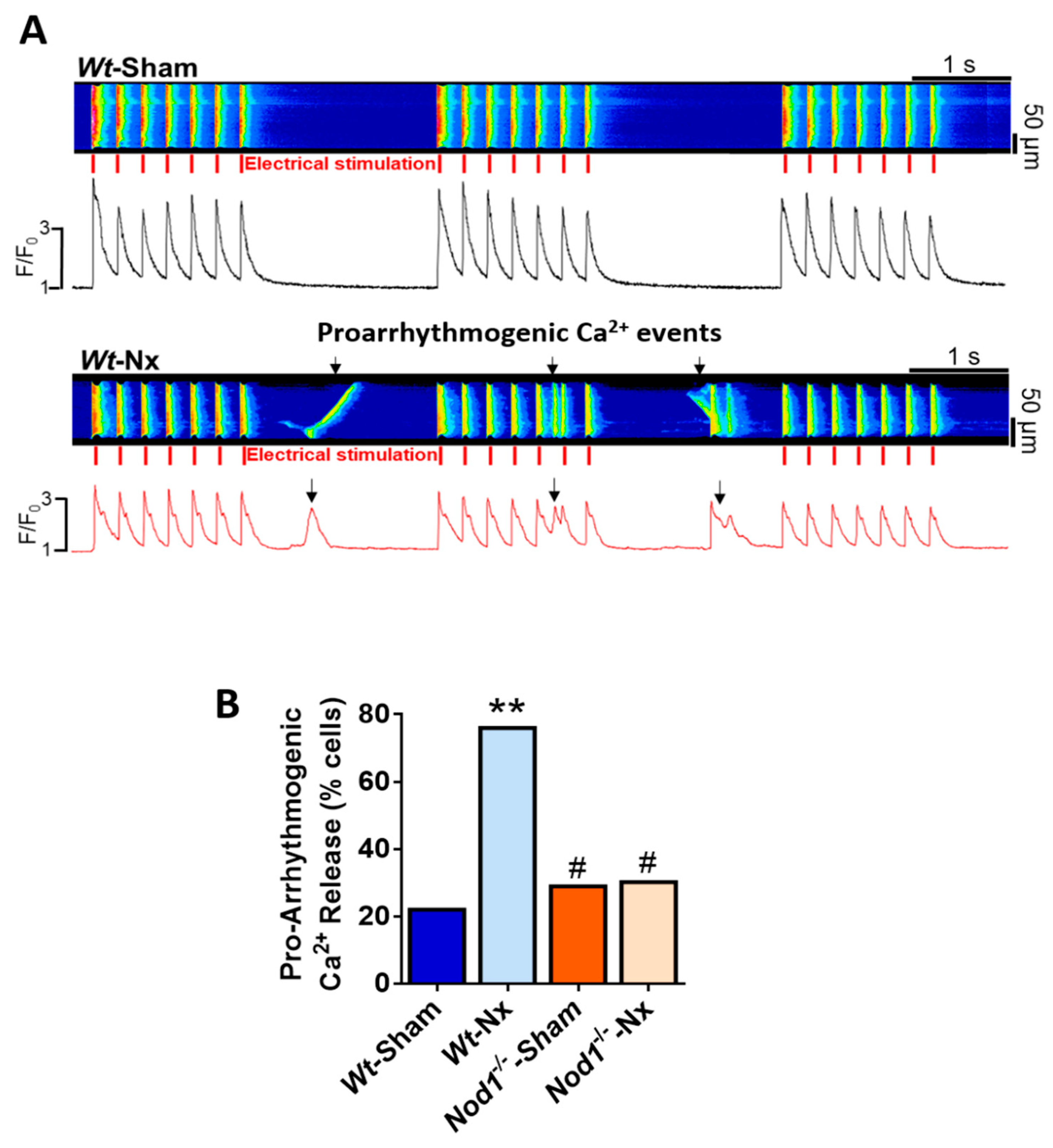
2.4. Deficiency of NOD1 Prevents the Increase in the Rate of Pro-Arrhythmogenic Ca2+ Events Induced by Nx

A close relationship exists between altered intracellular Ca2+ dynamics and ventricular arrhythmias, which are the most common causes of sudden death in advanced stages of renal disease. We analyzed the occurrence of pro-arrhythmic behavior as spontaneous Ca2+ waves or Ca2+ transients in ventricular cardiomyocytes field stimulated at 2 Hz for three cycles. Representative line-scan images of a regular Ca2+ transient (upper panel) in a Wt-sham cell and pro-arrhythmogenic Ca2+ transients and waves (lower panel) in a Wt-Nx cell are shown in Figure 3A. Results indicated that the occurrence of abnormal Ca2+ events was significantly higher in Wt-Nx cardiomyocytes than in Wt-sham cells, being this pro-arrhythmogenic Ca2+ release more than 2-fold higher in Wt-Nx (Figure 3B). By contrast, a lower percentage of Nod1−/−-sham and Nod1−/−-Nx cells showed this aberrant behavior (Figure 3B). These results indicate that the genetic deletion of NOD1 significantly prevents the increased pro-arrhythmogenic Ca2+ release induced by Nx.

2.5. Macroscopic and Microscopic Cardiac Features and Biochemistry Parameters of Renal Function of Rip2−/− Mice at Baseline and after Experimental CKD

As the majority of NOD1-derived effects are mediated via RIP2 activation [38] we next analyzed whether the lack of RIP2 also ameliorates Ca2+ mishandling linked to CKD.
Similar to the studies in Nod1−/− mice, we characterized the model by analyzing the structural properties of both the kidney and heart. Analysis revealed no differences in HW, HW/BW ratio and cardiomyocyte area between Rip2−/−-sham and Rip2−/−-Nx mice, indicating that Nx did not induce cardiac hypertrophy (Supplementary Table S1). By contrast, the surgery induced overall BW loss in the Wt and Rip2−/−-Nx mice, along with a reduction in KW in both groups, although the remnant KW after the Nx was higher than one third of the sham-operated mice (Supplementary Table S1). These results were also supported by KW/BW data. Analysis of biochemical indicators of renal function revealed that the levels of plasma urea, BUN and FGF-23 were significantly higher in Rip2−/−-Nx mice than in Rip2−/−-sham mice, whereas no differences were observed in phosphate levels between different groups (Supplementary Table S2). The results are similar to those observed in Wt-Nx mice, indicating that the loss of RIP2 does not prevent renal impairment induced by Nx.

2.6. Deficiency of RIP2 Prevents Ca2+ Mishandling Induced by Experimental CKD

Analysis of systolic Ca2+ release and cell shortening showed that deficiency of RIP2 prevented the lower amplitude of intracellular Ca2+ transients (Figure 4A,B), the slower kinetics (Figure 4C) and the decreased cell shortening (Figure 4D) induced by Nx. The changes were also associated with a recovery in the depleted SR Ca2+ load levels in Rip2−/−-Nx cardiomyocytes compared with Wt-Nx cells (Figure 4E). Accordingly, cardiomyocytes from Rip2−/−-Nx mice showed similar systolic Ca2+ release and SR Ca2+ load, as observed in cells from sham-operated Rip2−/− and Wt mice.
Deficiency of RIP2 also prevented the increased diastolic Ca2+ leak induced by the Nx surgery. Cells from Rip2−/−-Nx mice showed similar values of Ca2+ sparks frequency, Ca2+ sparks frequency normalized by SR Ca2+ load and spark-mediated leak to sham-operated Rip2−/− and Wt cardiomyocytes (Figure 5A–D). These data indicate that deficiency of RIP2 also prevents the increased Ca2+ leak during diastole and this effect can explain the maintenance of the SR Ca2+ load and the physiological systolic Ca2+ release observed in the Rip2−/− model of experimental CKD.
Finally, we determined whether the absence of RIP2 could also modulate the incidence of pro-arrhythmogenic Ca2+ release in isolated cardiomyocytes. Results established that only a small number of cells from Rip2−/−-Nx mice showed pro-arrhythmogenic Ca2+ events in paced cells, with the percentage of these events significantly lower than that in the Wt-Nx group, and similar to that in sham-operated Rip2−/− and Wt cells (Figure 5E). Overall, these results support the data in Nod1−/− mice, and point to a key role for the NOD1 adapter RIP2 in the prevention of Ca2+ mishandling induced by experimental CKD.

3. Discussion

Our study demonstrates that genetic deletion of either Nod1 or Rip2 prevents Ca2+ mishandling associated with experimental CKD. Much research has focused on determining the interplay between CVD and CKD [39]; however, many questions remain unanswered, especially in relation to the mechanisms involved in the development of cardiac events after renal damage. Among the multiple risk factors that can explain the high prevalence of CVD in CKD are mineral and bone disorders, oxidative stress, accumulation of uremic toxins and an increased inflammatory response. Regarding CKD, serum Pi levels have been considered a classical biomarker of renal severity and dysfunction together with others such as BUN or urea. However, several authors have already demonstrated that an increase in circulating FGF-23 is the earliest alteration observed in CKD patients even before the increase in serum Pi [40]. In fact, our results point out a similar condition in the experimental CKD developed in mice by the 5/6Nx. Our results demonstrated that the genetic deletion of Nod1 did not affect the increase in FGF-23 plasma levels. Moreover, the acute incubation of Wt and Nod1−/− cardiomyocytes with FGF-23 induced a similar systolic and diastolic Ca2+ mishandling (Supplementary Figure S2), suggesting that probably NOD-1 and the FGF-23 axis are involved in independent pathways that contribute to the regulation of Ca2+ handling, at least in our experimental model of CKD. Importantly, patients with advanced CKD with secondary hyperparathyroidism and hypocalcemia harbor arrhythmias and changes in cardiac electrical conduction [41]. However, an unresolved issue is whether the presence of inflammation is linked to a worse prognosis because of the cardiac events in patients with CKD. Interestingly, the use of specific inhibitors targeting proinflammatory mediators contributes to the prevention of some CKD comorbidities, including cardiovascular complications [42,43]. Sustained activation of the innate immune response leads to increased inflammation and frequently results in maladaptive responses that can promote deleterious cardiac remodeling [13]. NOD1 is an innate immune mediator known to be involved in both CKD and CVD [11,14,23,33,34,35,37,44,45]. With respect to renal diseases, Shigeoka et al. demonstrated that the double Nod1/2 deletion, as well as Rip2 deletion, is protective against acute kidney damage induced by ischemia/reperfusion in mice [14]. By contrast, in experimental CKD induced by unilateral ureteral obstruction, Stroo et al. found similar renal damage in Wt and double Nod1−/−/Nod2−/− mice [37]. These conflicting results might be related to the different experimental procedures used to induce either acute or chronic kidney damage.
We show that deficiency of NOD1 prevents cardiac Ca2+ mishandling in a mouse model of CKD induced by 5/6 nephrectomy, suggesting a specific protective cardiac role of this receptor independent of renal damage. The classical 5/6 nephrectomy model of CKD reproduces many of the main features found in human CKD [46,47] and we recently showed that this model presents with elevated cardiac Ca2+ mishandling, which can explain the cardiac dysfunction that accompanies CKD [48]. Interestingly, the altered pattern of Ca2+ cycling in cardiomyocytes in nephrectomised Wt mice has important similarities to that found in HF [11,49], where cardiomyocyte contraction is also strongly compromised. The majority of studies provide evidence that failing hearts show a depressed systolic Ca2+ release. As expected, and similar to what occurs in HF, our results show that Wt-Nx cardiomyocytes present with a significant decrease in the Ca2+ transient amplitude together with a significant slower decay time constant, and having a decreased systolic Ca2+ release and depressed cell contraction. All these alterations were not associated with changes in mRNA levels of Nod1 or Rip2 (Supplementary Figure S3), suggesting that posttranslational modifications or downstream factors derived from the NOD1-pathway activation can be involved in the observed effects.
In contrast to what occurs in Wt-Nx cell, Ca2+ mishandling is blunted in Nod1−/−-Nx mice, chiefly by the prevention of three effects: (i) the decrease in the Ca2+ transient amplitude; (ii) the increase in their decay time constant; and (iii) the depressed cell contraction. Thus, the loss of NOD1 prevents the decline in systolic Ca2+ release induced by the Nx surgery. This improvement in systolic Ca2+ release can be related to the levels of SR Ca2+ load. Indeed, the reduction in the SR Ca2+ load observed in Wt-Nx cardiomyocytes was also prevented by the loss of NOD1. Thus, both the maintenance of the SR Ca2+ load and the improvement in the systolic Ca2+ release can explain the better cardiac parameters exhibited by Nod1−/−-Nx cells, compared with Wt-Nx counterparts.
Depressed SR Ca2+ load can result from an increase in the Ca2+ leak during diastole. In this regard, Wt-Nx cardiomyocytes showed an increase in diastolic Ca2+ leak represented by a higher frequency of Ca2+ sparks, Ca2+ waves and spontaneous Ca2+ transients, as compared with Wt-sham cells. Supporting these results, we previously showed that RyR2 channel activity is increased in hearts from Wt-Nx mice [48]. The increased diastolic Ca2+ leak observed in Wt-Nx mice is potentially a good substrate for the induction of cardiac arrhythmias since the released Ca2+ diffuses to neighboring RyR2 clusters inducing SCR and triggering cardiac arrhythmias. The genetic deletion of NOD1 reduces the occurrence of Ca2+ sparks, Ca2+ waves and spontaneous Ca2+ transients after Nx surgery, similar to those found in sham-operated Wt and Nod1−/− mice. Thus, NOD1 deficiency prevents the abnormal diastolic Ca2+ leak induced by the Nx, along with a reduction of pro-arrhythmogenic Ca2+ events. A likely explanation for this is that NOD1 deficiency rescues the SR Ca2+ content and improves cell contractility, recovering the impaired cardiac outcome observed in Wt-Nx mice. These features also bear a resemblance to those found in HF, since SCR is ameliorated in Nod1−/−-sham mice with experimental HF and the deletion of NOD1 prevents RyR2 hyperactivity [11].
To comprehensively study the pathway involved in Ca2+ cycling-dysregulation evident in our mouse model of CKD, we also determined whether the deficiency of RIP2 plays a role in the regulation of Ca2+ dynamics. RIP2 is the adapter kinase that mediates the majority of NOD1 actions. Accordingly, RIP2-deficient cells are hyporesponsive to signaling through NOD proteins and show severely reduced NFκB activation [50]. Although the role of RIP2 in renal diseases remains enigmatic, its expression has been shown to be strongly induced in failing murine and human myocardium [11]. We demonstrate here that loss of RIP2 prevents the CKD-induced Ca2+ mishandling, as cardiomyocytes from Rip2−/−-Nx mice exhibit improved Ca2+ transients amplitude, kinetic rates and cell contractility compared with their Wt-Nx counterparts. This improvement in systolic Ca2+ release can be due to the rescue of the SR Ca2+ content found in the absence of RIP2. Moreover, we also demonstrate that RIP2 deficiency prevents the increased diastolic Ca2+ release observed in Wt-Nx mice, reducing the Ca2+ spark frequency and the occurrence of pro-arrhythmogenic events. These results are in line with those from the analysis of Nod1−/− mice. It would be of great interest to determine whether NOD1/RIP2 antagonists can also prevent the Ca2+ mishandling linked to CKD in future studies.
In conclusion, we establish, for the first time, to our knowledge, that the genetic deletion of two different components of the NOD1 signaling pathway prevents Ca2+ mishandling induced by experimental CKD. Our findings suggest that the NOD1 proinflammatory pathway could be targeted for the development of new therapies to reduce the risk of cardiovascular complications in patients with CKD.

4. Methods

4.1. Animal Care

The study was conducted following recommendations of the Spanish Animal Care and Use Committee, according to the guidelines for ethical care of experimental animals of the European Union (2010/63/EU), and was approved by the General Direction of Agriculture and the Environment at the Environment Council of Madrid (PROEX: 053/16 and 272.5/20). Male Nod1−/− and Rip2−/− mice on a C57BL/6J (6B; 129P2-NOD1tm1Nnz/J: 6B; 129P2-RIP2tm1Nnz/J) background were used. Mice were bred and housed under specific pathogen-free conditions in the Experimental Animal Centre of Instituto de Investigación Hospital Universitario la PAZ, IdiPAZ. Mice were maintained at controlled temperature (23–25 °C) on a 12-hr light/dark cycle with ad libitum access to water and a standard diet. Wild-type (Wt) C57BL/6J mice (The Jackson Laboratory, Bar Harbor, ME, USA) were employed as controls. Nod1−/− and Rip2−/− were kindly provided by Dr. Gabriel Nuñez (Ann Arbor, MI, USA). Gene expression analysis of Nod1 and Rip2 confirmed the absence of signal in Nod1- and Rip2-deficient mice (not shown). Macroscopic cardiac and kidney parameters were analyzed in all experimental groups as before [35,49].

4.2. Serology

Blood plasma samples were used to analyze the levels of phosphate (Abcam, Cambridge, UK), blood urea nitrogen (BUN) and urea (BioAssays System, Hayword, CA, USA) and fibroblast growth factor-23 (FGF-23) (Immunotopics, Inc., San Clemente, CA, USA) following the manufacturers’ instructions.

4.3. Experimental CKD

Six-week-old male were randomly assigned to either five-sixth nephrectomy (Nx) or sham surgery under isoflurane (1.5% v/v, isoflurane/oxygen) anesthesia and preoperative analgesia (Metacam, 0.05 mg/kg intramuscular) in a two-stage approach, as described [48]. Briefly, in the first stage an abdominal midline incision was made and the left kidney was exposed. Both the upper and lower poles were tied with a polyglycolic acid suture line (Dexon®, 4-0), which was subsequently removed. After a recovery period of one week, the entire right kidney was removed, following ligation of the renal blood vessels and cauterization of the ureter. The peritoneum and skin were then sutured, and the animals were returned to their cages. In control mice, sham surgeries involved midline incision, exposure of both kidneys, but no removal of tissue. The same timings were used as for Nx surgery. Blood plasma was employed for biochemical assays and isolated ventricular cardiomyocytes for Ca2+ recordings.

4.4. Cardiomyocyte Isolation

Six weeks after the second surgery, ventricular cardiomyocytes were isolated using standard enzymatic digestion [51]. Briefly, mice were anesthetized with sodium pentobarbital (100 mg/kg intraperitoneal) and heparinized (4 U/g intraperitoneal). The heart was rapidly excised and cannulated via the ascending aorta on a Langendorff perfusion apparatus. Retrograde perfusion was initiated with a standard Ca2+-free Tyrode’s solution containing 0.2 mmol/L EGTA over 2–3 min at room temperature, and continued for ~3–5 min with the same solution containing collagenase type II (1 mg/mL) (Worthington Biochemical, Lakewood, NY, USA) and CaCl2 (0.1 mmol/L). The heart was then removed from the Langendorff apparatus and the ventricles were cut out, finely minced into small pieces and mechanically dissociated in the enzymatic solution (standard Tyrode’s solution containing 0.1 mmol/L CaCl2). The cardiomyocyte cell suspension was filtered through a nylon mesh (250 µm), pelleted by centrifugation for 3 min at 300 rpm and suspended in Tyrode’s solution containing 0.5 mmol/L CaCl2. Cells were centrifuged as before and suspended in a storage solution containing 1 mmol/L CaCl2. Tyrode’s solution comprised (in mmol/L): 130 NaCl, 5.4 KCl, 0.5 MgCl2, 25 HEPES, 0.4 NaH2PO4, 22 glucose; pH = 7.4 adjusted with NaOH. Cardiomyocytes were immediately used for calcium imaging analyses and patch-clamp experiments.

4.5. Intracellular Calcium Imaging

Experiments were performed at room temperature (20–23 °C). Images were obtained with a Zeiss LSM 710 Meta confocal microscope (Carl Zeiss, Germany; 40× oil immersion objective with a 1.2 NA), by scanning the cardiomyocytes with an Argon laser every 1 s. Experiments were performed at room temperature (20–23 °C). To record intracellular Ca2+ transients, cells were first loaded with the Ca2+-sensitive probe Fluo-3 (5 µmol/L; Invitrogen Life Technologies, Carslbad, CA, USA), and were then electrically excited at 2 Hz by field stimulation using two parallel platinum electrodes. Fluo-3 was excited at 488 nm and emitted fluorescence was collected at >505 nm. The fluorescence values (F) were normalized by the basal fluorescence (F0) to obtain the fluorescence ratio (F/F0). All Ca2+ images were corrected for the background fluorescence. The decay time constant of Ca2+ transients (Tau) was obtained by fitting the decay trace. Cell contraction was calculated as the difference of cardiomyocyte length between rest and contraction (during electrical stimulation) and expressed as a percentage of shortening of cell length. Spontaneous Ca2+ sparks, and spontaneous Ca2+ transients and waves were acquired once stimulation was stopped. Ca2+ sparks were considered as located and fast increments in Ca2+ fluorescence. Total spark-mediated Ca2+ leak was calculated by multiplying spark frequency × peak × duration × width. SR Ca2+ load was assessed by rapid caffeine (10 mmol/L) application to deplete the SR of Ca2+ stores, after field-stimulation to reach the steady-state. Arrhythmic activity was analyzed as abnormal spontaneous Ca2+ release (SCR) by applying 3 cycles of field electrical stimulation at 2 Hz paced, consisting of 7 electric pulses. Data analysis was performed with homemade routines using IDL 8 software (Research System Inc. Boulder, CO, USA) and Image J 1.50i software (NIH). Images were corrected for background fluorescence. Cardiomyocyte surface area was quantified with the LSM Zeiss Image Browser 4.2 software (Carl Zeiss).

4.6. Statistical Analysis

Results are reported as mean ± SEM. Statistical analysis was performed using one-way analysis of variance (ANOVA) or the chi-square test, as appropriate. If a significant level of p was reached (p < 0.05) and there was no significant variance in homogeneity, Tukey’s post hoc multicomparison analysis was applied. All statistical analyses were performed with the SPSS 15.0 software (SPSS Inc., Chicago, IL, USA) and significance was assumed when p < 0.05.

Supplementary Materials

Author Contributions

M.F.-V. and G.R.-H. designed the experiments. M.G.-F., J.A.N.-G., A.V.-B., L.G.-L. and J.C.M. performed experiments, analysed the data and performed the figures. M.T., A.R. and J.L.M. contributed to some calcium recordings, C.D. performed ICaL recordings and provided reagents and materials and helped with data interpretation. C.Z. and L.M.R. contributed with data interpretation. M.F.-V., M.G.-F. and G.R.-H. wrote the manuscript. All authors have read and agreed to the published version of the manuscript.

Funding

This work was supported by Spanish Ministry of Economy and Competitiveness and European Regional Development Fund (SAF-2017-84777R), Institute of Health Carlos III (PI17/01093 and PI17/01344), Sociedad Española de Cardiología, Proyecto Traslacional 2019, Fundación Renal Íñigo Álvarez de Toledo (FRIAT), Fondo Europeo de Desarrollo Regional (FEDER), FSE, and CIBER-CV, a network funded by ISCIII. M.F.-V. is Miguel Servet II researcher of ISCIII (MSII16/00047 Carlos III Health Institute). G.R.-H. is Miguel Servet I researcher of ISCIII (CP15/00129 Carlos III Health Institute). M.T. is a PhD student funded by the FPU program of the Spanish Ministry of Science, Innovation and Universities (FPU17/06135). A.R. was supported by Fondo SEP-Cinvestav project #601410 FIDSC 2018/2; and Fondo SEP-Conacyt Ciencia Básica A1-S-9082.

Acknowledgments

The technical assistance of Jennifer Aceves-Ripoll, Laura Martin-Nunes, Monica Martin-Belinchón and Lucía Guerrero-Lopez are gratefully acknowledged.

Conflicts of Interest

The authors declare no conflict of interest.

Abbreviations

BWbody weight
CARDcaspase activation and recruitment domain
CKDchronic kidney disease
CVDcardiovascular disease
ECexcitation-contraction
FGF23fibroblast growth factor-23
HFheart failure
HWheart weight
KWkidney weight
LTCCssarcolemma L-type Ca2+ channels
NCXNa+/Ca2+ exchanger
NFκBnuclear factor kappa B
NOD1nucleotide-binding oligomerization domain-containing protein 1
NOD2nucleotide-binding oligomerization domain-containing protein 2
Nxfive-sixth nephrectomy
RIP2receptor-interacting-serine/threonine-protein kinase 2
RyR2ryanodine receptor type 2
SCRspontaneous Ca2+ release
SERCA2asarco/endoplasmic reticulum Ca2+ pump subtype
SRsarcoplasmic reticulum
TLtibia length
Wtwild-type

References

  1. Plantinga, L.C.; Boulware, L.E.; Coresh, J.; Stevens, L.A.; Miller, E.R.; Saran, R.; Messer, K.L.; Levey, A.S.; Powe, N.R. Patient awareness of chronic kidney disease: Trends and predictors. Arch. Intern. Med. 2008, 168, 2268–2275. [Google Scholar] [CrossRef] [PubMed]
  2. Gansevoort, R.T.; Correa-Rotter, R.; Hemmelgarn, B.R.; Jafar, T.H.; Heerspink, H.J.L.; Mann, J.F.; Matsushita, K.; Wen, C.P. Chronic kidney disease and cardiovascular risk: Epidemiology, mechanisms, and prevention. Lancet 2013, 382, 339–352. [Google Scholar] [CrossRef]
  3. Schefold, J.C.; Filippatos, G.; Hasenfuss, G.; Anker, S.D.; von Haehling, S. Heart failure and kidney dysfunction: Epidemiology, mechanisms and management. Nat. Rev. Nephrol. 2016, 12, 610–623. [Google Scholar] [CrossRef] [PubMed]
  4. House, A.A.; Wanner, C.; Sarnak, M.J.; Piña, I.L.; McIntyre, C.W.; Komenda, P.; Kasiske, B.L.; Deswal, A.; deFilippi, C.R.; Cleland, J.G.F.; et al. Heart failure in chronic kidney disease: Conclusions from a Kidney Disease: Improving Global Outcomes (KDIGO) Controversies Conference. Kidney Int. 2019, 95, 1304–1317. [Google Scholar] [CrossRef] [PubMed] [Green Version]
  5. Sarnak, M.J.; Levey, A.S.; Schoolwerth, A.C.; Coresh, J.; Culleton, B.; Hamm, L.L.; McCullough, P.A.; Kasiske, B.L.; Kelepouris, E.; Klag, M.J.; et al. Kidney disease as a risk factor for development of cardiovascular disease: A statement from the American Heart Association Councils on Kidney in Cardiovascular Disease, High Blood Pressure Research, Clinical Cardiology, and Epidemiology and Prevention. Circulation 2003, 108, 2154–2169. [Google Scholar] [CrossRef]
  6. Rantanen, J.M.; Riahi, S.; Schmidt, E.B.; Johansen, M.B.; Søgaard, P.; Christensen, J.H. Arrhythmias in Patients on Maintenance Dialysis: A Cross-sectional Study. Am. J. Kidney Dis. 2020, 75, 214–224. [Google Scholar] [CrossRef]
  7. Di Lullo, L.; Rivera, R.; Barbera, V.; Bellasi, A.; Cozzolino, M.; Russo, D.; De Pascalis, A.; Banerjee, D.; Floccari, F.; Ronco, C. Sudden cardiac death and chronic kidney disease: From pathophysiology to treatment strategies. Int. J. Cardiol. 2016, 217, 16–27. [Google Scholar] [CrossRef]
  8. Navarro-García, J.A.; Fernández-Velasco, M.; Delgado, C.; Delgado, J.F.; Kuro-O, M.; Ruilope, L.M.; Ruiz-Hurtado, G. PTH, vitamin D, and the FGF-23-klotho axis and heart: Going beyond the confines of nephrology. Eur. J. Clin. Investig. 2018, 48, e12902. [Google Scholar] [CrossRef] [Green Version]
  9. Ruiz-Hurtado, G.; Ruilope, L.M. Hypertension and obesity: Correlates with renin-angiotensin-aldosterone system and uric acid. J. Clin. Hypertens. 2014, 16, 559–560. [Google Scholar] [CrossRef] [Green Version]
  10. Bers, D.M. Cardiac excitation-contraction coupling. Nature 2002, 415, 198–205. [Google Scholar] [CrossRef]
  11. Val-Blasco, A.; Piedras, M.J.G.M.; Ruiz-Hurtado, G.; Suarez, N.; Prieto, P.; Gonzalez-Ramos, S.; Gómez-Hurtado, N.; Delgado, C.; Pereira, L.; Benito, G.; et al. Role of NOD1 in Heart Failure Progression via Regulation of Ca 2+ Handling. J. Am. Coll. Cardiol. 2017, 69, 423–433. [Google Scholar] [CrossRef] [PubMed]
  12. Yao, C.; Veleva, T.; Scott, L.; Cao, S.; Li, L.; Chen, G.; Jeyabal, P.; Pan, X.; Alsina, K.M.; Abu-Taha, I.; et al. Enhanced Cardiomyocyte NLRP3 Inflammasome Signaling Promotes Atrial Fibrillation. Circulation 2018, 138, 2227–2242. [Google Scholar] [CrossRef] [PubMed]
  13. Mann, D.L. Innate immunity and the failing heart: The cytokine hypothesis revisited. Circ. Res. 2015, 116, 1254–1268. [Google Scholar] [CrossRef] [PubMed] [Green Version]
  14. Shigeoka, A.A.; Kambo, A.; Mathison, J.C.; King, A.J.; Hall, W.F.; da Silva Correia, J.; Ulevitch, R.J.; McKay, D.B. Nod1 and nod2 are expressed in human and murine renal tubular epithelial cells and participate in renal ischemia reperfusion injury. J. Immunol. 2010, 184, 2297–2304. [Google Scholar] [CrossRef] [PubMed]
  15. Laman, J.D.; Schoneveld, A.H.; Moll, F.L.; Van Meurs, M.; Pasterkamp, G. Significance of peptidoglycan, a proinflammatory bacterial antigen in atherosclerotic arteries and its association with vulnerable plaques. Am. J. Cardiol. 2002, 90, 119–123. [Google Scholar] [CrossRef]
  16. Inohara, N.; Koseki, T.; del Peso, L.; Hu, Y.; Yee, C. Nod1, an Apaf-1-like activator of caspase-9 and nuclear factor-κB. J. Biol. Chem. 1999, 274, 14560–14567. [Google Scholar] [CrossRef] [Green Version]
  17. Moreno, L.; Gatheral, T. Therapeutic targeting of NOD1 receptors. Br. J. Pharmacol. 2013, 170, 475–485. [Google Scholar] [CrossRef] [Green Version]
  18. Park, J.; Kim, Y.; Shaw, M.; Kanneganti, T.; Fujimoto, Y. Nod1/RICK and TLR signaling regulate chemokine and antimicrobial innate immune responses in mesothelial cells. J. Immunol. 2007, 179, 514–521. [Google Scholar] [CrossRef] [Green Version]
  19. Ogura, Y.; Inohara, N.; Benito, A.; Chen, F.F.; Yamaoka, S.; Nunez, G. Nod2, a Nod1/Apaf-1 family member that is restricted to monocytes and activates NF-kappaB. J. Biol. Chem. 2001, 276, 4812–4818. [Google Scholar] [CrossRef] [Green Version]
  20. Ogura, Y.; Lala, S.; Xin, W.; Smith, E.; Dowds, T.A.; Chen, F.F.; Zimmermann, E.; Tretiakova, M.; Cho, J.H.; Hart, J.; et al. Expression of NOD2 in Paneth cells: A possible link to Crohn’s ileitis. Gut 2003, 52, 1591–1597. [Google Scholar] [CrossRef]
  21. Davey, M.P.; Martin, T.M.; Planck, S.R.; Lee, J.; Zamora, D.; Rosenbaum, J.T. Human endothelial cells express NOD2/CARD15 and increase IL-6 secretion in response to muramyl dipeptide. Microvasc. Res. 2006, 71. [Google Scholar] [CrossRef] [PubMed]
  22. Liu, H.Q.; Zhang, X.Y.; Edfeldt, K.; Nijhuis, M.O.; Idborg, H.; Bäck, M.; Roy, J.; Hedin, U.; Jakobsson, P.J.; Laman, J.D.; et al. NOD2-mediated innate immune signaling regulates the eicosanoids in atherosclerosis. Arterioscler. Thromb. Vasc. Biol. 2013, 33, 2193–2201. [Google Scholar] [CrossRef] [PubMed] [Green Version]
  23. Prieto, P.; Vallejo-Cremades, M.T.; Benito, G.; González-Peramato, P.; Francés, D.; Agra, N.; Terrón, V.; Gónzalez-Ramos, S.; Delgado, C.; Ruiz-Gayo, M.; et al. NOD1 receptor is up-regulated in diabetic human and murine myocardium. Clin. Sci. 2014, 127, 665–677. [Google Scholar] [CrossRef] [PubMed] [Green Version]
  24. Loyer, X.; Gómez, A.M.; Milliez, P.; Fernandez-Velasco, M.; Vangheluwe, P.; Vinet, L.; Charue, D.; Vaudin, E.; Zhang, W.; Sainte-Marie, Y.; et al. Cardiomyocyte overexpression of neuronal nitric oxide synthase delays transition toward heart failure in response to pressure overload by preserving calcium cycling. Circulation 2008, 117, 3187–3198. [Google Scholar] [CrossRef] [PubMed] [Green Version]
  25. Ferrand, A.; Al Nabhani, Z.; Tapias, N.S.; Mas, E.; Hugot, J.P.; Barreau, F. NOD2 Expression in Intestinal Epithelial Cells Protects Toward the Development of Inflammation and Associated Carcinogenesis. CMGH 2019, 7, 357–369. [Google Scholar] [CrossRef] [PubMed] [Green Version]
  26. Al Nabhani, Z.; Dietrich, G.; Hugot, J.P.; Barreau, F. Nod2: The intestinal gate keeper. PLoS Pathog. 2017, 13, e1006177. [Google Scholar] [CrossRef] [PubMed] [Green Version]
  27. Mirkov, M.U.; Verstockt, B.; Cleynen, I. Genetics of inflammatory bowel disease: Beyond NOD2. Lancet Gastroenterol. Hepatol. 2017, 2, 224–234. [Google Scholar] [CrossRef]
  28. Berlin, P.; Reiner, J.; Witte, M.; Wobar, J.; Lindemann, S.; Barrantes, I.; Kreikemeyer, B.; Bastian, M.; Schäffler, H.; Bannert, K.; et al. Nod2 deficiency functionally impairs adaptation to short bowel syndrome via alterations of the epithelial barrier function. Am. J. Physiol. Gastrointest. Liver Physiol. 2019, 317, G727–G738. [Google Scholar] [CrossRef]
  29. Kobayashi, K.S.; Chamaillard, M.; Ogura, Y.; Henegariu, O.; Inohara, N.; Nuñez, G.; Flavell, R.A. Nod2-dependent regulation of innate and adaptive immunity in the intestinal tract. Science 2005, 307, 731–734. [Google Scholar] [CrossRef]
  30. Caruso, R.; Warner, N.; Inohara, N.; Núñez, G. NOD1 and NOD2: Signaling, Host Defense, and Inflammatory Disease. Immunity 2014, 41, 898–908. [Google Scholar] [CrossRef] [Green Version]
  31. Marchitti, S.A.; Brocker, C.; Stagos, D.; Vasiliou, V. Non-P450 aldehyde oxidizing enzymes: The aldehyde dehydrogenase superfamily. Expert Opin. Drug Metab. Toxicol. 2008, 4, 697–720. [Google Scholar] [CrossRef] [PubMed] [Green Version]
  32. Motomura, Y.; Kanno, S.; Asano, K.; Tanaka, M.; Hasegawa, Y.; Katagiri, H.; Saito, T.; Hara, H.; Nishio, H.; Hara, T.; et al. Identification of Pathogenic Cardiac CD11c+ Macrophages in Nod1-Mediated Acute Coronary Arteritis. Arterioscler. Thromb. Vasc. Biol. 2015, 35, 1423–1433. [Google Scholar] [CrossRef] [PubMed] [Green Version]
  33. Delgado, C.; Ruiz-Hurtado, G.; Gómez-Hurtado, N.; González-Ramos, S.; Rueda, A.; Benito, G.; Prieto, P.; Zaragoza, C.; Delicado, E.G.; Pérez-Sen, R.; et al. NOD1, a new player in cardiac function and calcium handling. Cardiovasc. Res. 2015, 106, 375–386. [Google Scholar] [CrossRef] [PubMed] [Green Version]
  34. Val-Blasco, A.; Navarro-García, J.A.; Tamayo, M.; Piedras, M.J.; Prieto, P.; Delgado, C.; Ruiz-Hurtado, G.; Rozas-Romero, L.; Gil-Fernández, M.; Zaragoza, C.; et al. Deficiency of NOD1 improves the β-adrenergic modulation of Ca2+ handling in a mouse model of heart failure. Front. Physiol. 2018, 9. [Google Scholar] [CrossRef] [Green Version]
  35. Val-Blasco, A.; Prieto, P.; Gonzalez-Ramos, S.; Benito, G.; Vallejo-Cremades, M.T.; Pacheco, I.; González-Peramato, P.; Agra, N.; Terrón, V.; Delgado, C.; et al. NOD1 activation in cardiac fibroblasts induces myocardial fibrosis in a murine model of type 2 diabetes. Biochem. J. 2017, 474, 399–410. [Google Scholar] [CrossRef]
  36. Uehara, A.; Fujimoto, Y.; Fukase, K.; Takada, H. Various human epithelial cells express functional Toll-like receptors, NOD1 and NOD2 to produce anti-microbial peptides, but not proinflammatory cytokines. Mol. Immunol. 2007, 44, 3100–3111. [Google Scholar] [CrossRef]
  37. Stroo, I.; Emal, D.; Butter, L.M.; Teske, G.J.; Claessen, N.; Dessing, M.C.; Girardin, S.E.; Florquin, S.; Leemans, J.C. No difference in renal injury and fibrosis between wild-type and NOD1/NOD2 double knockout mice with chronic kidney disease induced by ureteral obstruction. BMC Nephrol. 2018, 19, 78. [Google Scholar] [CrossRef] [Green Version]
  38. Park, J.-H.; Kim, Y.-G.; McDonald, C.; Kanneganti, T.-D.; Hasegawa, M.; Body-Malapel, M.; Inohara, N.; Núñez, G. RICK/RIP2 Mediates Innate Immune Responses Induced through Nod1 and Nod2 but Not TLRs. J. Immunol. 2007, 178, 2380–2386. [Google Scholar] [CrossRef]
  39. Rafiq, K.; Noma, T.; Fujisawa, Y.; Ishihara, Y.; Arai, Y.; Nabi, A.H.M.N.; Suzuki, F.; Nagai, Y.; Nakano, D.; Hitomi, H.; et al. Renal sympathetic denervation suppresses de novo podocyte injury and albuminuria in rats with aortic regurgitation. Circulation 2012, 125, 1402–1413. [Google Scholar] [CrossRef]
  40. Wolf, M. Forging forward with 10 burning questions on FGF23 in kidney disease. J. Am. Soc. Nephrol. 2010, 21, 1427–1435. [Google Scholar] [CrossRef] [Green Version]
  41. Brown, S.J.; Ruppe, M.D.; Tabatabai, L.S. The Parathyroid Gland and Heart Disease. Methodist DeBakey Cardiovasc. J. 2017, 13, 49–54. [Google Scholar] [CrossRef] [Green Version]
  42. Ridker, P.M.; MacFadyen, J.G.; Glynn, R.J.; Koenig, W.; Libby, P.; Everett, B.M.; Lefkowitz, M.; Thuren, T.; Cornel, J.H. Inhibition of Interleukin-1β by Canakinumab and Cardiovascular Outcomes in Patients With Chronic Kidney Disease. J. Am. Coll. Cardiol. 2018, 71, 2405–2414. [Google Scholar] [CrossRef] [PubMed]
  43. Ridker, P.M. Clinician’s Guide to Reducing Inflammation to Reduce Atherothrombotic Risk: JACC Review Topic of the Week. J. Am. Coll. Cardiol. 2018, 72, 3320–3331. [Google Scholar] [CrossRef] [PubMed]
  44. Tourneur, E.; Ben Mkaddem, S.; Chassin, C.; Bens, M.; Goujon, J.-M.; Charles, N.; Pellefigues, C.; Aloulou, M.; Hertig, A.; Monteiro, R.C.; et al. Cyclosporine A Impairs Nucleotide Binding Oligomerization Domain (Nod1)-Mediated Innate Antibacterial Renal Defenses in Mice and Human Transplant Recipients. PLoS Pathog. 2013, 9, e1003152. [Google Scholar] [CrossRef] [PubMed] [Green Version]
  45. Stroo, I.; Butter, L.M.; Claessen, N.; Teske, G.J.; Rubino, S.J.; Girardin, S.E.; Florquin, S.; Leemans, J.C. Phenotyping of Nod1/2 double deficient mice and characterization of Nod1/2 in systemic inflammation and associated renal disease. Biol. Open 2012, 1, 1239–1247. [Google Scholar] [CrossRef] [PubMed] [Green Version]
  46. Gava, A.L.; Freitas, F.P.; Balarini, C.M.; Vasquez, E.C.; Meyrelles, S.S. Effects of 5/6 nephrectomy on renal function and blood pressure in mice. Int. J. Physiol. Pathophysiol. Pharmacol. 2012, 4, 167–173. [Google Scholar] [PubMed]
  47. Kren, S.; Hostetter, T.H. The course of the remnant kidney model in mice. Kidney Int. 1999, 56, 333–337. [Google Scholar] [CrossRef] [PubMed] [Green Version]
  48. Navarro-García, J.A.; Rueda, A.; Romero-García, T.; Aceves-Ripoll, J.; Rodríguez-Sánchez, E.; González-Lafuente, L.; Zaragoza, C.; Fernández-Velasco, M.; Kuro-o, M.; Ruilope, L.M.; et al. Enhanced Klotho availability protects against cardiac dysfunction induced by uraemic cardiomyopathy by regulating Ca2+ handling. Br. J. Pharmacol. 2020. [Google Scholar] [CrossRef]
  49. Ruiz-Hurtado, G.; Li, L.; Fernández-Velasco, M.; Rueda, A.; Lefebvre, F.; Wang, Y.; Mateo, P.; Cassan, C.; Gellen, B.; Benitah, J.P.; et al. Reconciling depressed Ca2+ sparks occurrence with enhanced RyR2 activity in failing mice cardiomyocytes. J. Gen. Physiol. 2015, 146, 295–306. [Google Scholar] [CrossRef] [Green Version]
  50. Kobayashi, K.; Inohara, N.; Hernandez, L.D.; Galán, J.E.; Núñez, G.; Janeway, C.A.; Medzhitov, R.; Flavell, R.A. RICK/Rip2/CARDIAK mediates signalling for receptors of the innate and adaptive immune systems. Nature 2002, 416, 194–199. [Google Scholar] [CrossRef] [Green Version]
  51. Shioya, T. A Simple Technique for Isolating Healthy Heart Cells from Mouse Models. J. Physiol. Sci. 2007, 57, 327–335. [Google Scholar] [CrossRef] [PubMed] [Green Version]
Figure 1. Deficiency of nucleotide-binding oligomerization domain-containing protein 1 (NOD1) prevents the dysregulation of systolic Ca2+ release, cell contraction impairment and the reduction in sarcoplasmic reticulum (SR) Ca2+-load triggered by 5/6 nephrectomy. (A) Representative line-scan confocal images of Ca2+ transients in cardiomyocytes from Wt-sham, Wt-Nx, Nod1−/−-sham and Nod1−/−-Nx mice electrically evoked by field stimulation at 2 Hz. Mean values of (B) peak fluorescence of Ca2+ transients, (C) decay time constant and (D) cell shortening obtained in cells from Wt-sham (n = 45 cells/five mice), Wt-Nx (n = 43 cells/five mice), Nod1−/−-sham (n = 39 cells/five mice) and Nod1−/−-Nx (n = 50 cells/five mice) mice. (E) Representative line-scan confocal images of caffeine-evoked Ca2+ transients in cardiomyocytes from all groups. (F) Mean values of caffeine-evoked Ca2+ transients amplitude obtained in cells from Wt-sham (n = 33 cells/five mice), Wt-Nx (n = 37 cells/five mice), Nod1−/−-sham (n = 34 cells/5 mice) and Nod1−/−-Nx (n = 35 cells/5 mice) mice. Results show mean ± SEM. *** p < 0.001 vs. Wt-sham; # p < 0.05, ### p < 0.001 vs. Wt-Nx.
Ijms 21 08868 g001
Figure 2. Prevents the increased frequency of Ca2+-sparks and spontaneous Ca2+ release (SCR) induced by 5/6 nephrectomy. (A) Representative line-scan confocal images of Ca2+ sparks recordings obtained in a quiescent cardiomyocyte isolated from Wt-sham, Wt-Nx, Nod1−/−-sham and Nod1−/−-Nx mice. Average data of (B) Ca2+ spark frequency, (C) normalization of Ca2+ spark frequency by SR-Ca2+ load, (D) spark-mediated Ca2+ leak and Ca2+ sparks properties: (E) peak, (F) duration and (G) width obtained in cells isolated from Wt-sham (n = 45 cells/five mice), Wt-Nx (n = 47 cells/five mice), Nod1−/−-sham (n = 40 cells/five mice) and Nod1−/−-Nx (n = 46 cells/five mice) mice. (H) Representative line-scan confocal images of SCR recordings (Ca2+ wave [upper panel]; spontaneous Ca2+ transients release (lower panel)) from cardiomyocytes isolated from Wt-Nx mice. (I) Average data of SCR occurrence obtained in cells isolated from all groups. Histograms show mean ± SEM. ** p < 0.01; *** p < 0.001 vs. Wt-sham; # p < 0.05; ## p < 0.01; ### p < 0.001 vs. Wt-Nx.
Ijms 21 08868 g002
Figure 3. Deficiency of NOD1 prevents the increase of aberrant pro-arrhythmogenic Ca2+ events provoked by 5/6 nephrectomy. (A) Representative line-scan images of a cardiomyocyte paced at 2 Hz for three cycles isolated from Wt-sham (upper panel) and Wt-Nx mice (lower panel). The corresponding fluorescence [Ca2+]i profiles appear below line-scan images. Red marks indicate electrical stimulation. (B) Percentage of cells with pro-arrhythmogenic Ca2+ release in cells isolated from Wt-sham (n = 45 cells/5 mice), Wt-Nx (n = 47 cells/five mice), Nod1−/−-sham (n = 40 cells/five mice) and Nod1−/−-Nx (n = 46 cells/five mice) mice. Histograms show the mean values. ** p < 0.01 vs. Wt-sham; # p < 0.05 vs. Wt-Nx.
Ijms 21 08868 g003
Figure 4. Deficiency of receptor-interacting serine/threonine protein kinase 2 (RIP2) prevents the systolic Ca2+ mishandling, contractile dysfunction and depressed SR Ca2+-load induced by 5/6 nephrectomy. (A) Representative line-scan confocal images of Ca2+ transients obtained from Wt-sham, Wt-Nx, Rip2−/−-sham and Rip2−/−-Nx cardiomyocytes electrically evoked under field stimulation at 2 Hz. Mean values of (B) peak fluorescence of Ca2+ transients; (C) decay time constant; and (D) cell shortening obtained in cells from Wt-sham (n = 37 cells/four mice), Wt-Nx (n = 37 cells/four mice), Rip2−/−-sham (n = 35 cells/four mice) and Rip2−/−-Nx (n = 56 cells/four mice) mice. (E) Mean values of caffeine-evoked Ca2+ transients amplitude obtained in cardiomyocytes from Wt-sham (n = 29 cells/four mice), Wt-Nx (n = 32 cells/four mice), Rip2−/−-sham (n = 24 cells/four mice) and Rip2−/−-Nx (n = 36 cells/four mice) mice. Results show mean ± SEM. ** p < 0.01; *** p < 0.001 vs. Wt-sham; ### p < 0.001 vs. Wt-Nx.
Ijms 21 08868 g004
Figure 5. Deficiency of RIP2 prevents the increased frequency of Ca2+ sparks, diastolic Ca2+ leak and pro-arrhythmogenic Ca2+ events provoked by 5/6 nephrectomy. (A) Representative line-scan confocal images of Ca2+ sparks recordings obtained in a quiescent cardiomyocyte obtained from Wt-sham, Wt-Nx, Rip2−/−-sham and Rip2−/−-Nx. Average data of (B) Ca2+ spark frequency; (C) normalization of Ca2+ spark frequency by SR-Ca2+ load; (D) spark-mediated Ca2+ leak in cells isolated from Wt-sham (n = 35 cells/four mice), Wt-Nx (n = 38 cells/four mice), Rip2−/−-sham (n = 27 cells/four mice) and Rip2−/−-Nx (n = 47 cells/four mice) mice. (E) Percentage of cells with pro-arrhythmogenic Ca2+ release in cardiomyocytes isolated all groups. Histograms show mean ± SEM. *** p < 0.001 vs. Wt-sham; # p < 0.05; ## p < 0.01; ### p < 0.001 vs. Wt-Nx.
Ijms 21 08868 g005
Table 1. Macroscopic parameters in Wild-type (Wt) and Nod1−/− mice subjected or not to experimental chronic kidney disease (CKD).
Wt-shamWt-NxNod1−/−-shamNod1−/−-Nx
HW
(mg)
186.89 ± 6.08 (10)169.63 ± 4.65 (8)198.27 ± 6.66 # (10)185.03 ± 12.05 (9)
BW
(g)
26.24 ± 0.25 (10)23.11 ± 0.83 (8)26.99 ± 0.76 ## (10)25.26 ± 0.76 (9)
HW/BW
(mg/g)
7.11 ± 0.19 (10)7.43 ± 0.42 (8)7.38 ± 0.28 (10)7.31 ± 0.37 (9)
KW
(mg)
183.80 ± 4.78 (10)155.83 ± 8.62 * (8)190.96 ± 7.02 ## (10)165.90 ± 7.39 (9)
KW/BW
(mg/g)
7.01 ± 0.18 (10)6.76 ± 0.33 (8)7.09 ± 0.24 (10)6.58 ± 0.26 (9)
Cell area
(μm2)
3482.21 ± 109.25
(72 cells/10)
3215.90 ± 119.73
(54 cells/8)
3396.71 ± 94.36
(67 cells/10)
3271.32 ± 116.62
(59 cells/9)
Data from 8–10 animals for macroscopic parameters per experimental group are reported as mean ± SEM. HW: heart weight, BW: body weight, KW: kidney weight. * p < 0.05 vs. Wt-sham; # p < 0.05, ## p < 0.01 vs. Wt-Nx. Statistical significance was determined by one-way analysis of variance (ANOVA).
Table 2. Biochemical plasma parameters in Wild-type and Nod1−/− mice subjected or not to experimental CKD.
Wt-shamWt-NxNod1−/−-shamNod1−/−-Nx
Urea
(mg/dL)
37.24 ± 3.55 (5) 83.30 ± 5.45 *** (8)39.80 ± 4.78 ### (6)83.68 ± 8.04 ***,&&& (8)
BUN
(mg/dL)
17.40 ± 1.66 (5)38.93 ± 2.55 *** (8)18.60 ± 2.23 ### (6)39.10 ± 3.76 ***,&&& (8)
Pi
(mg/dL)
6.19 ± 0.65 (7)6.91 ± 0.55 (8)6.21 ± 0.90 (6)6.92 ± 1.01 (8)
FGF-23 (pg/mL)140.3 ± 22.72 (7)294.00 ± 47.47 * (8)137.30 ± 24.66 (5)256.90 ± 38.51 (8)
Data from 5–8 animals for biochemical parameters per experimental group are reported as mean ± SEM. BUN: blood urea nitrogen; FGF-23: fibroblast growth factor 23; Pi: phosphates. * p < 0.05, *** p < 0.001 vs. Wt-sham; ### p < 0.001 vs. Wt-Nx; &&& p < 0.001 vs. Nod1−/−-sham. Statistical significance was determined by one-way ANOVA.
Publisher’s Note: MDPI stays neutral with regard to jurisdictional claims in published maps and institutional affiliations.

