Sequence of Polyurethane Ionomers Determinative for Core Structure of Surfactant–Copolymer Complexes


 ,
, 
Abstract
:1. Introduction
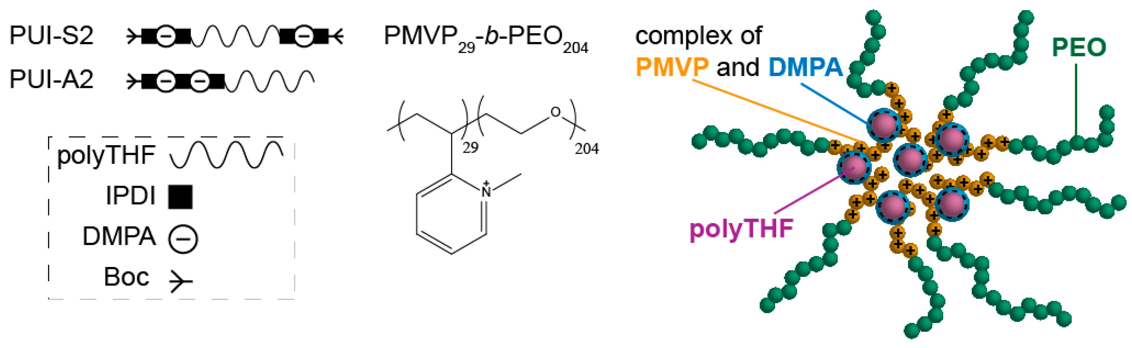
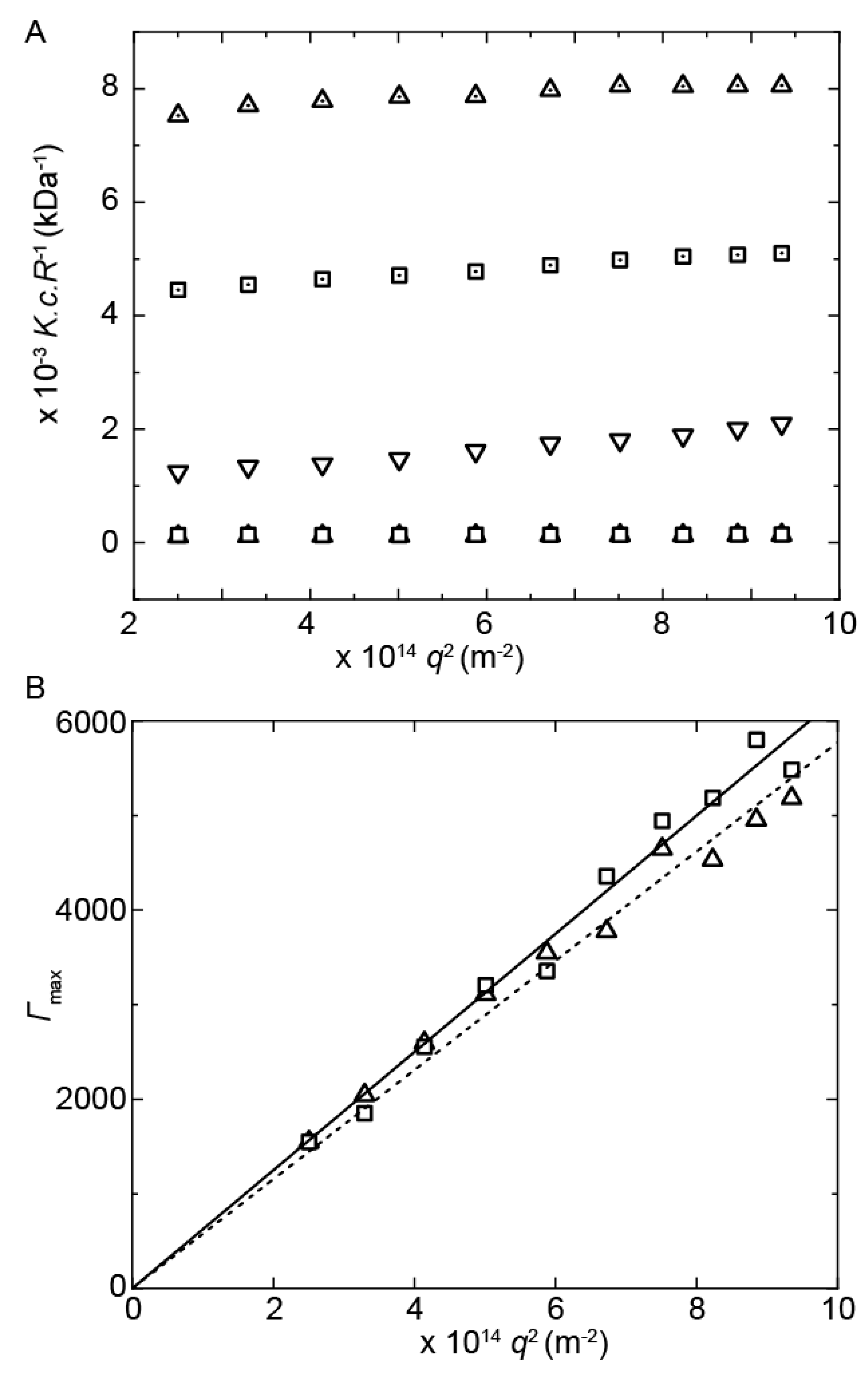
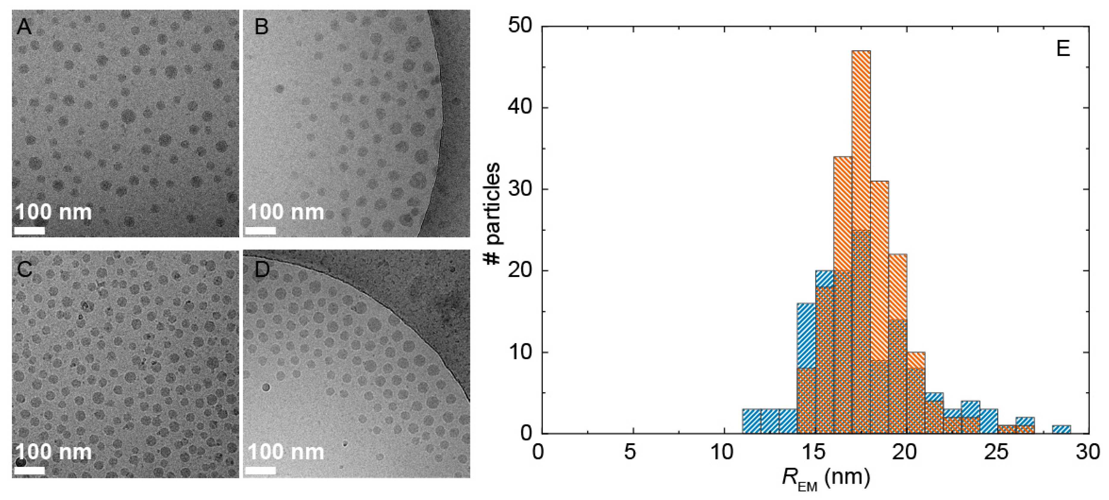
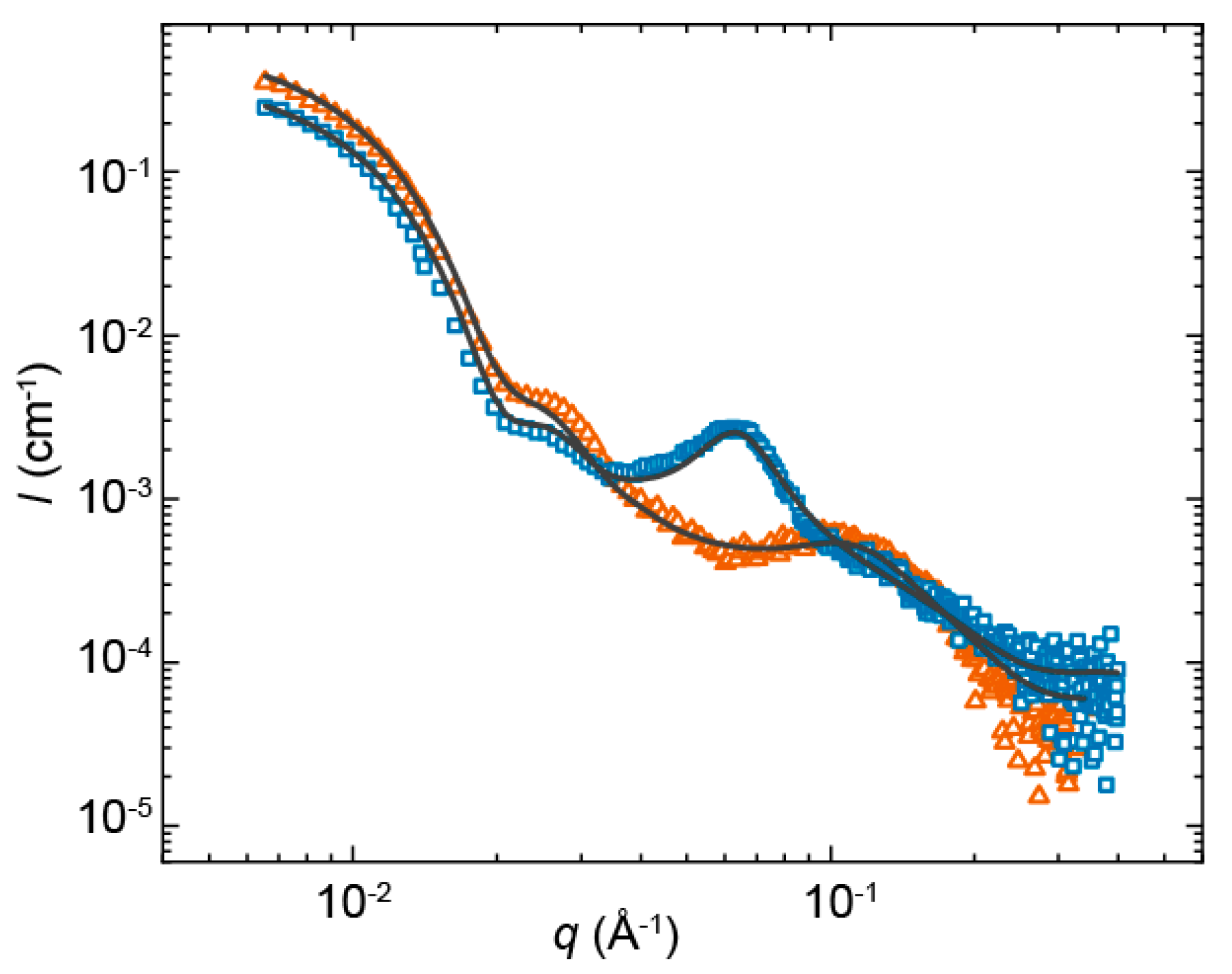
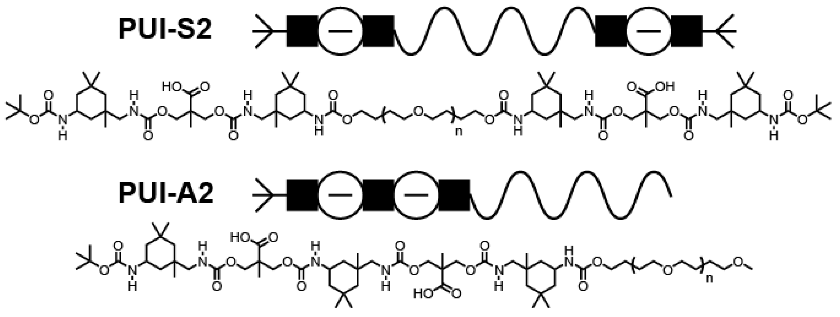
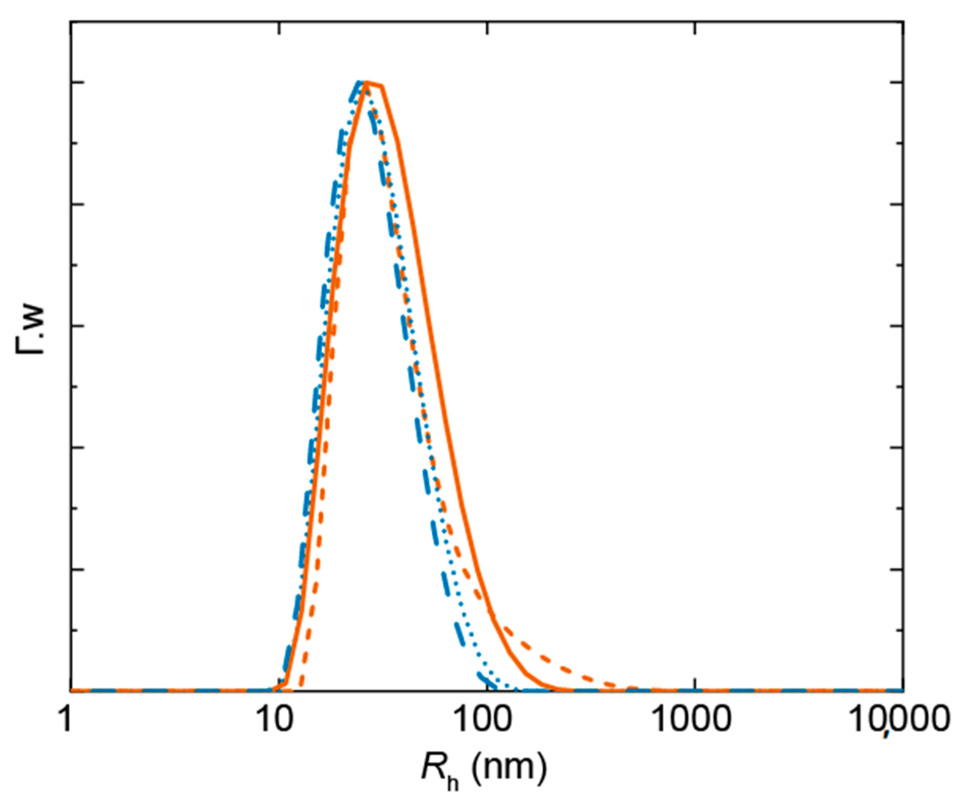
2. Results
3. Materials and Methods
3.1. Materials
3.2. Sample Preparation
3.3. Static Light Scattering (SLS) and Dynamic Light Scattering (DLS)
3.4. Small-Angle X-ray Scattering (SAXS)
3.5. Cryogenic Transmission Electron Microscopy (cryoTEM)
4. Conclusions
Author Contributions
Funding
Data Availability Statement
Acknowledgments
Conflicts of Interest
Abbreviations
| PUI | Polyurethane ionomer |
| PMVP29-b-PEO204 | Poly(N-methyl-2-vinylpyridinium iodide)29-b-poly(ethylene oxide)204 |
| THF | Tetrahydrofuran |
| DLS | Dynamic light scattering |
| SLS | Static light scattering |
| SAXS | Small-angle X-ray scattering |
| cryoTEM | Cryogenic transmission electron microscopy |
Appendix A


| System | PUI-S2:PMVP29-b-PEO204 | PUI-A2:PMVP29-b-PEO204 |
|---|---|---|
| sc1 | 1 × 10⁻³ | 1 × 10⁻³ |
| bkg (cm−1) | 1 × 10⁻⁴ | 1 × 10⁻⁴ |
| SLD r1 (Å−2) | 330.4 | 330.3 |
| SLDsolvent r1 (Å−2) | 330 | 330 |
| r1 (nm) | 20 | 20 |
| ÐSAXS r1 | 0.17 | 0.17 |
| sc2 | 4 × 10⁻³ | 4 × 10⁻³ |
| SLD r2 (Å−2) | 330.4 | 330.3 |
| SLDsolvent r2 (Å−2) | 330.6 | 330.6 |
| r2 (nm) | 1.5 | 1.3 |
| Imax | 3.0 × 10⁻⁴ | 21 × 10−4 |
| qmax (Å−1) | 0.108 | 0.061 |
| σ | 0.04 | 0.015 |
References
- Kralova, I.; Sjöblom, J. Surfactants used in food industry: A review. J. Dispers. Sci. Technol. 2009, 30, 1363–1383. [Google Scholar] [CrossRef]
- Kim, S.; Shi, Y.; Kim, J.Y.; Park, K.; Cheng, J.X. Overcoming the barriers in micellar drug delivery: Loading efficiency, in vivo stability, and micelle–cell interaction. Expert Opin. Drug Deliv. 2010, 7, 49–62. [Google Scholar] [CrossRef] [PubMed]
- Yokoyama, M. Clinical applications of polymeric micelle carrier systems in chemotherapy and image diagnosis of solid tumors. J. Exp. Clin. Med. 2011, 3, 151–158. [Google Scholar] [CrossRef]
- Rosen, M.J.; Kunjappu, J.T. Surfactants and Interfacial Phenomena; John Wiley & Sons: Hoboken, NJ, USA, 2012. [Google Scholar]
- Schmolka, I.R. A review of block polymer surfactants. J. Am. Oil Chem. Soc. 1977, 54, 110–116. [Google Scholar] [CrossRef]
- Nicolai, T.; Colombani, O.; Chassenieux, C. Dynamic polymeric micelles versus frozen nanoparticles formed by block copolymers. Soft Matter 2010, 6, 3111–3118. [Google Scholar] [CrossRef]
- Borisov, O.V.; Zhulina, E.B. Effect of salt on self-assembly in charged block copolymer micelles. Macromolecules 2002, 35, 4472–4480. [Google Scholar] [CrossRef]
- Harada, A.; Kataoka, K. Formation of polyion complex micelles in an aqueous milieu from a pair of oppositely-charged block copolymers with poly (ethylene glycol) segments. Macromolecules 1995, 28, 5294–5299. [Google Scholar] [CrossRef]
- Wu, H.; Ting, J.M.; Werba, O.; Meng, S.; Tirrell, M.V. Non-equilibrium phenomena and kinetic pathways in self-assembled polyelectrolyte complexes. J. Chem. Phys. 2018, 149, 163330. [Google Scholar] [CrossRef]
- Kabanov, A.V.; Bronich, T.K.; Kabanov, V.A.; Yu, K.; Eisenberg, A. Spontaneous formation of vesicles from complexes of block ionomers and surfactants. J. Am. Chem. Soc. 1998, 120, 9941–9942. [Google Scholar] [CrossRef]
- Berret, J.-F.; Cristobal, G.; Hervé, P.; Oberdisse, J.; Grillo, I. Structure of colloidal complexes obtained from neutral/poly-electrolyte copolymers and oppositely charged surfactants. Eur. Phys. J. E 2002, 9, 301–311. [Google Scholar] [CrossRef]
- Courtois, J.; Berret, J.-F. Probing oppositely charged surfactant and copolymer interactions by isothermal titration microcalorimetry. Langmuir 2010, 26, 11750–11758. [Google Scholar] [CrossRef] [PubMed] [Green Version]
- Berret, J.-F.; Vigolo, B.; Eng, R.; Hervé, P.; Grillo, I.; Yang, L. Electrostatic self-assembly of oppositely charged copolymers and surfactants: A light, neutron, and X-ray scattering study. Macromolecules 2004, 37, 4922–4930. [Google Scholar] [CrossRef]
- Voets, I.K.; Moll, P.M.; Aqil, A.; Jérôme, C.; Detrembleur, C.; De Waard, P.; De Keizer, A.; Cohen Stuart, M. A Temperature responsive complex coacervate core micelles with a PEO and PNIPAAm corona. J. Phys. Chem. B 2008, 112, 10833–10840. [Google Scholar] [CrossRef] [PubMed]
- Solomatin, S.V.; Bronich, T.K.; Bargar, T.W.; Eisenberg, A.; Kabanov, V.A.; Kabanov, A.V. Environmentally responsive nanoparticles from block ionomer complexes: Effects of pH and ionic strength. Langmuir 2003, 19, 8069–8076. [Google Scholar] [CrossRef]
- Solomatin, S.V.; Bronich, T.K.; Eisenberg, A.; Kabanov, V.A.; Kabanov, A.V. Colloidal stability of aqueous dispersions of block ionomer complexes: Effects of temperature and salt. Langmuir 2004, 20, 2066–2068. [Google Scholar] [CrossRef] [PubMed]
- Solomatin, S.V.; Bronich, T.K.; Eisenberg, A.; Kabanov, V.A.; Kabanov, A.V. Fluorescence anisotropy study of aqueous dispersions of block ionomer complexes. J. Phys. Chem. B 2005, 109, 4303–4308. [Google Scholar] [CrossRef] [PubMed]
- Balomenou, I.; Bokias, G. Water-soluble complexes between cationic surfactants and comb-type copolymers consisting of an anionic backbone and hydrophilic nonionic poly(N,N-dimethylacrylamide) side chains. Langmuir 2005, 21, 9038–9043. [Google Scholar] [CrossRef]
- Chen, W.; Chen, H.; Hu, J.; Yang, W.; Wang, C. Synthesis and characterization of polyion complex micelles between poly(ethylene glycol)-grafted poly(aspartic acid) and cetyltrimethyl ammonium bromide. Colloids Surf. A Physicochem. Eng. Asp. 2006, 278, 60–66. [Google Scholar] [CrossRef]
- Vitorazi, L.; Berret, J.-F.; Loh, W. Self-assembly of complex salts of cationic surfactants and anionic–neutral block copolymers. Dispersions with liquid-crystalline internal structure. Langmuir 2013, 29, 14024–14033. [Google Scholar] [CrossRef] [Green Version]
- Masahiro, F.; Pan, P.; Shan, G.; Bao, Y.; Fujita, M.; Maeda, M. Core–Shell structure, biodegradation, and drug release behavior of poly(lactic acid)/poly(ethylene glycol) block copolymer micelles tuned by macromolecular stereostructure. Langmuir 2015, 31, 1527–1536. [Google Scholar] [CrossRef]
- Yang, L.; Wu, X.-H.; Liu, F.; Duan, Y.; Li, S. Novel biodegradable polylactide/poly(ethylene glycol) micelles prepared by direct dissolution method for controlled delivery of anticancer drugs. Pharm. Res. 2009, 26, 2332–2342. [Google Scholar] [CrossRef] [PubMed]
- Li, W.; Li, J.; Gao, J.; Li, B.; Xia, Y.; Meng, Y.; Yu, Y.; Chen, H.; Dai, J.; Wang, H.; et al. The fine-tuning of thermosensitive and degradable polymer micelles for enhancing intracellular uptake and drug release in tumors. Biomaterials 2011, 32, 3832–3844. [Google Scholar] [CrossRef] [PubMed]
- Adams, M.L.; Kwon, G.S. Relative aggregation state and hemolytic activity of amphotericin B encapsulated by poly(ethylene oxide)-block–poly(N-hexyl-l-aspartamide)-acyl conjugate micelles: Effects of acyl chain length. J. Control. Release 2003, 87, 23–32. [Google Scholar] [CrossRef]
- Zhuo, X.; Lei, T.; Miao, L.; Chu, W.; Li, X.; Luo, L.; Gou, J.; Zhang, Y.; Yin, T.; He, H.; et al. Disulfiram-loaded mixed nanoparticles with high drug-loading and plasma stability by reducing the core crystallinity for intravenous delivery. J. Colloid Interface Sci. 2018, 529, 34–43. [Google Scholar] [CrossRef] [PubMed]
- Unsal, H.; Onbulak, S.; Calik, F.; Er-Rafik, M.; Schmutz, M.; Sanyal, A.; Rzayev, J. Interplay between molecular packing, drug loading, and core cross-linking in bottlebrush copolymer micelles. Macromolecules 2017, 50, 1342–1352. [Google Scholar] [CrossRef]
- Kang, N.; Perron, M.-È.; Prud’Homme, R.E.; Zhang, Y.; Gaucher, G.; Leroux, J.-C. Stereocomplex block copolymer micelles: Core−shell nanostructures with enhanced stability. Nano Lett. 2005, 5, 315–319. [Google Scholar] [CrossRef]
- Monaghan, O.R.; Bomans, P.H.H.; Sommerdijk, N.A.J.M.; Holder, S.J. Controlling the melting transition of semi-crystalline self-assembled block copolymer aggregates: Controlling release rates of ibuprofen. Polym. Chem. 2017, 8, 5303–5316. [Google Scholar] [CrossRef]
- Jeong, Y.-H.; Shin, H.-W.; Kwon, J.-Y.; Lee, S.-M. Cisplatin-encapsulated polymeric nanoparticles with molecular geometry-regulated colloidal properties and controlled drug release. ACS Appl. Mater. Interfaces 2018, 10, 23617–23629. [Google Scholar] [CrossRef]
- Timmers, E.M.; Fransen, M.; Magana, J.R.; Janssen, H.M.; Voets, I.K. Micellization of sequence-controlled polyurethane ionomers in mixed solvents. Macromolecules. under review.
- Voets, I.K.; De Vries, R.; Fokkink, R.; Sprakel, J.; May, R.P.; De Keizer, A.; Cohen Stuart, M.A. Towards a structural characterization of charge-driven polymer micelles. Eur. Phys. J. E 2009, 30, 351–359. [Google Scholar] [CrossRef] [Green Version]
- Van Der Kooij, H.M.; Spruijt, E.; Voets, I.K.; Fokkink, R.; Cohen Stuart, M.A.; Van Der Gucht, J. On the stability and morphology of complex coacervate core micelles: From spherical to wormlike micelles. Langmuir 2012, 28, 14180–14191. [Google Scholar] [CrossRef] [PubMed]
- Liu, S.; Hu, C.; Wei, Y.; Duan, M.; Chen, X.; Hu, Y. Transformation of H-aggregates and J-dimers of water-soluble tetrakis (4-carboxyphenyl) porphyrin in polyion complex micelles. Polymers 2018, 10, 494. [Google Scholar] [CrossRef] [PubMed] [Green Version]
- Pergushov, D.V.; Remizova, E.V.; Gradzielski, M.; Lindner, P.; Feldthusen, J.; Zezin, A.B.; Müller, A.H.; Kabanov, V.A. Micelles of polyisobutylene-block-poly(methacrylic acid) diblock copolymers and their water-soluble interpolyelectrolyte complexes formed with quaternized poly(4-vinylpyridine). Polymers 2004, 45, 367–378. [Google Scholar] [CrossRef]
- Berret, J.-F.; Hervé, P.; Aguerre-Chariol, O.; Oberdisse, J. Colloidal complexes obtained from charged block copolymers and surfactants: A comparison between small-angle neutron scattering, Cryo-TEM, and simulations. J. Phys. Chem. B 2003, 107, 8111–8118. [Google Scholar] [CrossRef]
- Berret, J.-F. Evidence of overcharging in the complexation between oppositely charged polymers and surfactants. J. Chem. Phys. 2005, 123, 164703. [Google Scholar] [CrossRef] [PubMed] [Green Version]
- Fanova, A.; Janata, M.; Filippov, S.K.; Slouf, M.; Netopilík, M.; Mariani, A.; Štěpánek, M. Evolution of structure in a comb copolymer–surfactant coacervate. Macromolecules 2019, 52, 6303–6310. [Google Scholar] [CrossRef]
- Cingil, H.E.; Meertens, N.C.; Voets, I.K. Temporally programmed disassembly and reassembly of C3Ms. Small 2018, 14, 1802089. [Google Scholar] [CrossRef]
- Wu, H. Correlations between the Rayleigh ratio and the wavelength for toluene and benzene. Chem. Phys. 2010, 367, 44–47. [Google Scholar] [CrossRef]
- Rasolonjatovo, B.; Gomez, J.-P.; Même, W.; Gonçalves, C.; Huin, C.; Bennevault-Celton, V.; Le Gall, T.; Montier, T.; Lehn, P.; Cheradame, H.; et al. Poly(2-methyl-2-oxazoline)-b-poly(tetrahydrofuran)-b-poly(2-methyl-2-oxazoline) amphiphilic triblock copolymers: Synthesis, physicochemical characterizations, and hydrosolubilizing properties. Biomacromolecules 2015, 16, 748–756. [Google Scholar] [CrossRef]
- Huglin, M.B. Specific refractive index increments of polymer solutions. Part I. Literature values. J. Appl. Polym. Sci. 2003, 9, 3963–4001. [Google Scholar] [CrossRef]
- Brzozowska, A.M.; De Keizer, A.; Detrembleur, C.; Cohen Stuart, M.A.; Norde, W. Grafted ionomer complexes and their effect on protein adsorption on silica and polysulfone surfaces. Colloid Polym. Sci. 2010, 288, 1621–1632. [Google Scholar] [CrossRef] [PubMed] [Green Version]
- Guinier, A.; Fournet, G.; Yudowitch, K.L. Small-Angle Scattering of X-rays; John Wiley & Sons: New York, NY, USA, 1955. [Google Scholar]


Publisher’s Note: MDPI stays neutral with regard to jurisdictional claims in published maps and institutional affiliations. |
© 2020 by the authors. Licensee MDPI, Basel, Switzerland. This article is an open access article distributed under the terms and conditions of the Creative Commons Attribution (CC BY) license (http://creativecommons.org/licenses/by/4.0/).
Share and Cite
Timmers, E.M.; Magana, J.R.; Schoenmakers, S.M.C.; Fransen, P.M.; Janssen, H.M.; Voets, I.K. Sequence of Polyurethane Ionomers Determinative for Core Structure of Surfactant–Copolymer Complexes. Int. J. Mol. Sci. 2021, 22, 337. https://0-doi-org.brum.beds.ac.uk/10.3390/ijms22010337
Timmers EM, Magana JR, Schoenmakers SMC, Fransen PM, Janssen HM, Voets IK. Sequence of Polyurethane Ionomers Determinative for Core Structure of Surfactant–Copolymer Complexes. International Journal of Molecular Sciences. 2021; 22(1):337. https://0-doi-org.brum.beds.ac.uk/10.3390/ijms22010337
Chicago/Turabian StyleTimmers, Elizabeth M., Jose Rodrigo Magana, Sandra M. C. Schoenmakers, P. Michel Fransen, Henk M. Janssen, and Ilja K. Voets. 2021. "Sequence of Polyurethane Ionomers Determinative for Core Structure of Surfactant–Copolymer Complexes" International Journal of Molecular Sciences 22, no. 1: 337. https://0-doi-org.brum.beds.ac.uk/10.3390/ijms22010337