Review

Viral Infection Modulates Mitochondrial Function

1
College of Veterinary Medicine, South China Agricultural University, No. 483 Wushan Road, Tianhe District, Guangzhou 510642, China
2
Guangdong Laboratory for Lingnan Modern Agriculture, College of Veterinary Medicine, South China Agricultural University, Guangzhou 510642, China
3
Key Laboratory of Zoonosis Prevention and Control of Guangdong Province, Guangzhou 510642, China
*
Authors to whom correspondence should be addressed.
Int. J. Mol. Sci. 2021, 22(8), 4260; https://0-doi-org.brum.beds.ac.uk/10.3390/ijms22084260
Submission received: 30 March 2021 / Revised: 14 April 2021 / Accepted: 15 April 2021 / Published: 20 April 2021
(This article belongs to the Section Molecular Pathology, Diagnostics, and Therapeutics)

Abstract

:
Mitochondria are important organelles involved in metabolism and programmed cell death in eukaryotic cells. In addition, mitochondria are also closely related to the innate immunity of host cells against viruses. The abnormality of mitochondrial morphology and function might lead to a variety of diseases. A large number of studies have found that a variety of viral infections could change mitochondrial dynamics, mediate mitochondria-induced cell death, and alter the mitochondrial metabolic status and cellular innate immune response to maintain intracellular survival. Meanwhile, mitochondria can also play an antiviral role during viral infection, thereby protecting the host. Therefore, mitochondria play an important role in the interaction between the host and the virus. Herein, we summarize how viral infections affect microbial pathogenesis by altering mitochondrial morphology and function and how viruses escape the host immune response.

1. Physiological Morphology of Mitochondria

Mitochondria originated from an ancient bacterial endosymbiont, are important organelles found in almost all cells. In the nearly 130 years since mitochondria were first reported, new functions have been discovered. Mitochondria maintain the dynamic balance of the mitochondrial network through the fission and fusion mediated by a dedicated set of dynamin-related GTPases, provide energy for cells, and regulate processes such as autophagy, calcium homeostasis, innate immunity, signal transduction, and apoptosis [1].
Mitochondria are in a highly dynamic process within the cell, undergoing fission and fusion cycles to control the mitochondria’s morphology. The Fuzzy onion (Fzo) is the first protein discovered to mediate mitochondrial fusion during Drosophila spermatogenesis, and mutations in the Fzo gene can cause mitochondrial fusion disorders and abnormal accumulation in Drosophila sperm cells [2]. In mammals, the proteins that mediate mitochondrial fusion mainly include Mfn1 (Mitofusin1), Mfn2 (Mitofusin2), and OPA1 (Optic atrophy 1) [3,4,5]. Mfns contain heptad repeat regions (HR2), and Mfn1 and Mfn2, located on the outer mitochondrial membrane (OMM), interact to form Mfn1/Mfn2 homodimers or Mfn1/Mfn2 heterodimers through oligomerization of the HR2 structures, thereby promoting phase trans-plugging of the adjacent OMM [6,7], and involving GTP hydrolysis, which eventually leads to the fusion of the OMM [8,9]. OPA1, a dynamically related GTPase and localized to IMM, participate in IMM fusion. OPA1 protein was hydrolyzed into different fragments in the intermembrane space: one is the long subtype L-OPA1 associated with mitochondrial fusion, the other is the short subtype S-OPA1 [10,11]. L-OPA1 achieves selective mitochondrial fusion through the heteromorphic interaction between its GTPase domain and its adjacent mitochondrial membrane cardiolipid (CL). Loss of fusion-mediating proteins (MFN1, MFN2, and OPA1) can cause changes in mitochondrial morphology, leading to mitochondrial fragmentation. Mitochondrial fusion is a necessary cellular process that facilitates the merging of mitochondrial fragments and mediates the exchange of mitochondrial DNA, proteins, and metabolites.
Mitochondrial consonance proteins were deleted by gene knockout and RNA interference techniques, leading to mitochondrial fragmentation [3,5]. Mitochondria can also break down damaged mitochondria through “mitochondrial fission”, breaking them down into smaller fragments. In mammalian cells, Drp1 is a significant protein that mediates mitochondrial fission. After Drp1 is activated, it is recruited from the cytosol to the OMM, where oligomerization occurs. Drp1 forms rings and spirals within the diameter of the OMM and hydrolyzes GTP depending on its GTP-enzyme activity, resulting in membrane constriction and scission [12,13]. The transport and function of Drp1 are rapidly regulated by the opposing effects of phosphorylation at two key serines. In general, phosphorylation of serine 616 enhances Drp1 activity and promotes its targeting of mitochondrial aggregation, while phosphorylation at serine 637 reduces the activity of Drp1, keeping it in the cytoplasm [14]. For example, RIP1 phosphorylates Ser616 residue of Drp1, thereby inducing mitochondrial fission and eliminating damaged mitochondria through mitophagy, when cells are in a state of energy stress [15]. Phosphorylation at Ser637 of Drp1 inhibits the interaction of GTP-binding/middle domains with the GED domain, thereby reducing GTPase activity and altering Drp1 function and mitochondrial morphology [16]. Drp1 requires proteins of various accessories to perform its function. At present, mitochondrial fission factor (mitochondrial fission factor, Mff), mitochondrial fission protein 1 (mitochondrial fission protein 1, Fis1), mitochondrial dynamin 49 (mitochondrial dynamics proteins of 49 kDa, Mi D49), and mitochondrial dynamin 51 (mitochondrial dynamics proteins of 51 kDa, MiD51) located on the mitochondria have been found to act as ligands for Drp1, which recruit Drp1 to the mitochondria and regulate mitochondrial fission [17]. Fis1, the only Dnm1 receptor in yeast cells, is controversial for recruiting Drp1 to mitochondria in mammalian cells. For example, Fis1 and Drp1 interact in mammalian cells, and increasing Fis1 levels will promote mitochondrial fission [18]. However, Fis1 deletion in colon cancer cells suggests that it is not necessary for mitochondrial division [19]. A recent study found that human Fis1 blocks mitochondrial fusion mechanisms by binding to Mfn1, Mfn2, and OPA1, suggesting that Drp1 is dispensable for human Fis1 function [20]. Mff protein is also a receptor molecule of Drp1, it interacts with Drp1 through the amino-terminal cytoplasmic region, and is distributed homogeneously on the OMM, mainly in the same places as Drp1 [19]. Overexpression of Mff can promote the recruitment of Drp1 to mitochondria, while silencing of Mff expression can promote mitochondrial fusion. In addition, mitochondrial dynamics proteins (MiDs) are involved in mitochondrial fission in fis1 and Drp1 deficient cells. When MiDs are overexpressed, they recruit a large number of inactive S637 phosphorylated Drp1 into mitochondria to mediate mitochondrial elongation [21,22].
Mitochondria are involved in a series of cellular activities such as cell metabolism, programmed cell death and innate immunity, and the host response to viral infection. In addition, in the long-term evolutionary process, viruses have evolved a pathway to affect their intracellular survival by targeting mitochondria, and by mediating mitochondria-induced cell death, they can spread or evade host immunity. In this review, we explore how viruses manipulate mitochondria and how this manipulation affects microbial pathogenesis.

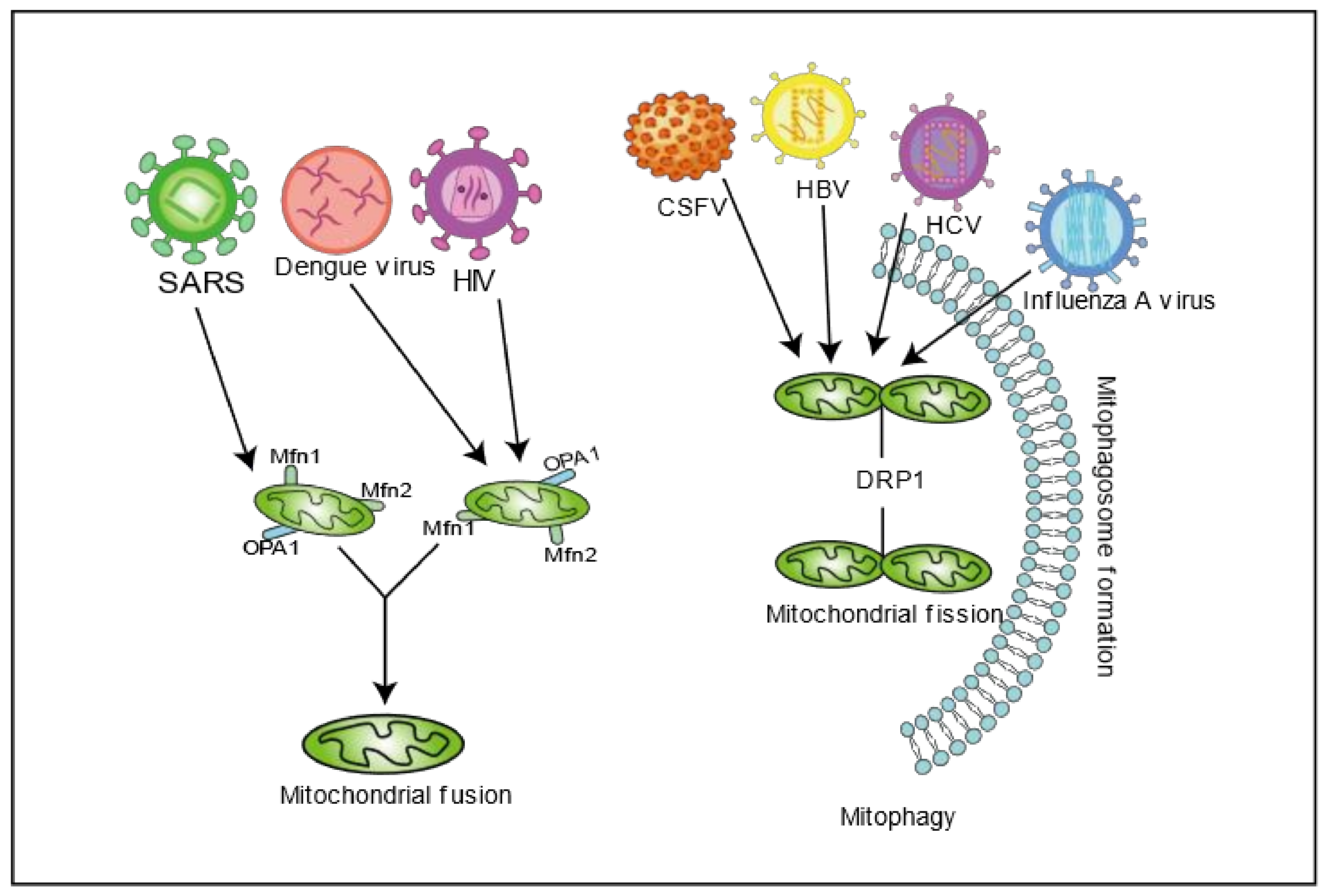
2. Viral Infection Disrupts Mitochondrial Dynamics

A variety of viral infections can induce mitochondrial autophagy by destroying the dynamic balance of mitochondria, which is conducive to viral self-infection. Since the early discovery of mitochondrial morphological changes in patients with Hepatitis C virus (HCV), more and more studies have focused on the changes in mitochondrial function caused by HCV infection, which is a positive-strand RNA virus [23]. The HCV core protein can be targeted and located on the OMM, resulting in a decrease in electron transport complex I, inhibition of mitochondrial electron transport, and an increase in the production of reactive oxygen species (ROS) [24,25]. HCV also induces ROS production through Core, E1, and NS3 proteins, which triggers mitochondrial permeability transition, leading to DNA damage and STAT3 activation [26]. Reducing the mitochondrial permeability threshold induced by Ca2+ and ROS is a feature of hepatitis C virus infection. It is a direct result of the interaction of HCV core proteins with mitochondria [27]. HCV infection also disrupts mitochondrial dynamics by promoting mitochondrial fission and mitophagy to promote viral persistence. HCV induces the phosphorylation of Drp1 (Ser616) and transports it to the mitochondria to mediate mitochondrial fission, thereby causing mitophagy [28]. Interference of HCV-induced mitochondrial fission and mitophagy can reduce glycolysis and ATP production as well as increase interferon synthesis, thereby inhibiting viral secretion [28]. Another study showed that HCV-induced mitochondrial fission is not only dependent on DRP1 protein, but HCV NS5A protein can also interact with phosphatidylinositol 4-kinase IIIα, which induces mitochondrial fragmentation [29]. HCV induces the expression of Parkin and PINK1, and triggers the translocation of Parkin into mitochondria to mediate mitophagy. Inhibition of mitophagy by silencing Parkin and PINK1 can partially rescue mitochondrial complex I enzyme activity and inhibit HCV replication [28]. Interestingly, the HCV core protein interacts with Parkin, inhibiting Parkin translocation to mitochondria, leading to the formation of mitochondrial autophagosomes and the failure of autophagy degradation [30]. Classical swine fever virus (CSFV) and Dengue virus (DENV) belong to the same family of flaviviruses as HCV does, and infection can also facilitate self-replication by affecting mitochondria’ function [31,32,33,34,35]. CSFV infection causes MNF2 to be ubiquitinated and degraded and stimulates the expression of Parkin and PINK1 and mitochondrial translocation, leading to mitochondrial fission and increased mitophagy. Silencing DRP1 and Parkin resulted in a decline in CSFV replication [31]. DENV proteins NS4B and NS3 mediate an imbalance in mitochondrial dynamics by inhibiting Drp1-triggered mitochondrial fission, which is conducive to the replication of DENV. In addition, NS4B protein of DENV can inactivate DRP1 and mediate mitochondrial elongation [34]. Mitochondrial extension brings mitochondria into contact with convoluted membranes (CMs) and destroys the integrity of the mitochondria-endoplasmic reticulum binding site on the mitochondrial-associated membrane (MAM), resulting in RLR signal transduction failure and reduced interferon production. However, another study found that DENV can also inhibit mitochondrial fusion through NS2B3 protein cleavage of MFN1 and MFN2, blocking RLR signal transduction and destroying mitochondrial membrane potential, thereby enhancing DENV infection [35].
It has also been reported that hepatitis B virus (HBV), a partially double-stranded DNA virus belonging to the Hepatoviridae family, may mediate mitochondrial damage in liver cells by changing mitochondrial dynamics, thus causing liver diseases. Many studies have reported that HBV HBx protein can target mitochondria and be situated in the OMM, IMM, or matrix. Studies have shown that MARCH5, a mitochondrial E3 ubiquitin ligase, can degrade HBx accumulated on mitochondria through polyubiquitination and regulate mitochondrial dynamics through ubiquitination of Drp1, Fis1, and Mfn1, thereby negatively regulating HBV [36]. HBx recruits Parkin to destroy depolarized/dysfunctional mitochondria by up-regulating PINK1 expression [37]. Other studies have shown that HBV and HBx protein promoted mitochondrial fission by promoting the expression of DRP1. HBV and HBx protein also promote cell survival and persistent viral infection via stimulating Parkin-mediated mitophagy [37]. PB1-F2 is a crucial virulence factor for the influenza virus’ pathogenicity, which is an enveloped RNA virus of the orthomyxoviridae family. PB1-F2 targets mitochondria and is transported to the IMM through the TOMM40 channel, causing loss of mitochondrial membrane potential and disrupting mitochondrial function [38,39,40]. In contrast, the low pathogenic subtype influenza A PB1-F2, lacking the c-terminal region, does not cause mitochondrial dysfunction [41]. PB1-F2 interacts with TUFM (Tu translation elongation factor, mitochondrial) on mitochondria and induces mitophagy and inhibits type I interferon expression [42]. However, a recent study showed that H1N1 infection can promote mitochondrial elongation and alter the host cell’s endoplasmic reticulum–mitochondrial contact sites by increasing OPA1 expression and decreasing DRP1 expression, thereby altering mitochondrial morphology dynamics. In addition, the treatment of cells with Mito-C (a novel pro-fission compound) significantly reduced viral replication by restoring part of the mitochondrial function [43]. Severe Acute Respiratory Syndrome Coronavirus (SARS-CoV) is a single-stranded positive-stranded RNA virus belonging to the genus Coronavirus. Its NSP2 interacts with PHB1 and PHB2, implicated in several cellular functions, thereby impairing intracellular signaling and affecting mitochondrial biogenesis [44,45]. SARS-CoV virulence factor ORF-9B also degrades DRP1 through the proteasome, leading to mitochondrial fusion that evades the host innate immune response [46]. Severe Acute Respiratory Syndrome Coronavirus 2 (SARS-CoV-2), a member of the same family as SARS-CoV, has caused global social and economic disruption. Recent studies have shown that SARS-CoV-2 could manipulate immune response and cell metabolism to promote cell replication by regulating autophagy, increasing ROS processes, and decreasing mitochondrial function [47]. In SARS-CoV-2, ORF9b interacts with the TOM70 subunit of the OMM protein import mechanism [48], which has a potential regulatory effect on MAVS. SARS-CoV-2 Nsp4, required for CM formation in SARS-CoV, potentially interacts with the mitochondrial import machinery (TIM) complexes [48]. SARS-CoV-2 Nsp8 also interacts with mitochondrial ribosomes [48]. More and more studies have shown that viruses maintain the viral replication’s ecological sites by manipulating mitochondrial dynamics (Figure 1). Therefore, the study of virus and mitochondrial dynamics may become the critical drug targets for viral infection treatment.

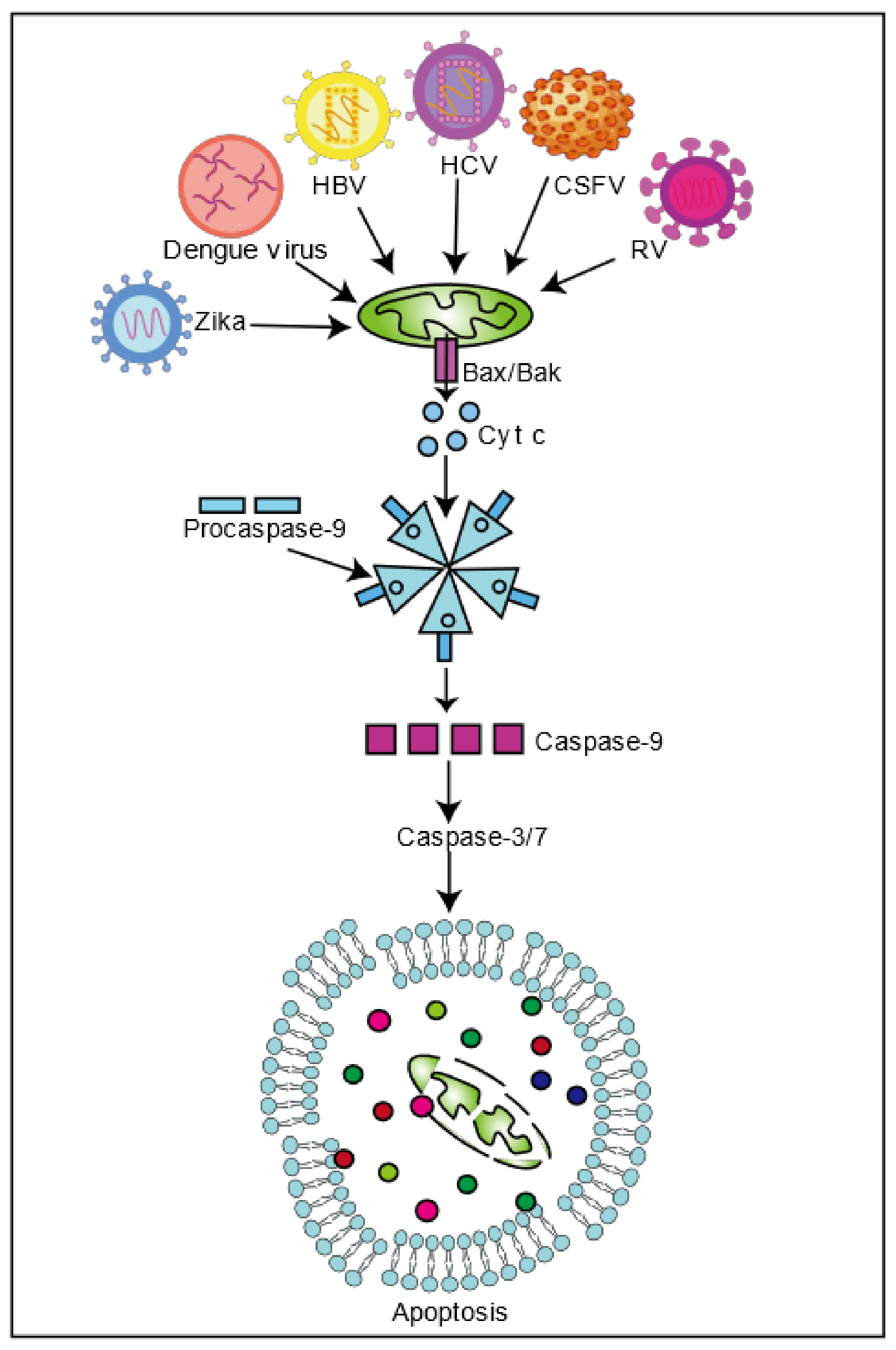
3. Viral Infection Regulates Mitochondria-Induced Cell Death

Apoptosis is the process of cell autonomy and programmed death, controlled by genes, to maintain the stability of the internal environment. At present, cell apoptosis can be divided into three pathways. Mitochondria influence cell death through the intrinsic apoptotic pathway. When apoptosis is induced, mitochondrial membrane protein activation using the Bcl-2 family protein channels triggers mitochondrial outer membrane permeability and releases apoptosis proteins (such as Cyt c, Smac, etc.) into the cytoplasm. Cyt c and apoptotic protease activating factor 1 (APAF1) interact, forming apoptosomes and activating procaspase-9, which cracks caspase-3 and caspase-7, thus inducing cell apoptosis [49]. Many viruses promote viral spread by inducing cell death or maintaining persistent infection via inhibiting cell death. HCV inhibits cell apoptosis via disrupting mitochondrial dynamics. HCV infection induces the phosphorylation of DRP1Ser616, which triggers mitochondrial fission and mitophagy, thereby inhibiting cell apoptosis, which eventually promotes viral persistence [28]. CSFV infection is similar to HCV infection. CSFV and HCV infection trigger the occurrence of mitophagy via activating the PINK1 and Parkin pathways to clear impaired mitochondria and prevent the release of pro-apoptotic proteins, thereby inhibiting cell apoptosis and maintaining viral infection [28,31]. Drp1 silencing blocks mitochondrial fission, mitophagy, and up-regulated apoptosis signals induced by HCV and CSFV, reducing virion secretion [28,31]. Interestingly, HCV viral proteins play a different role in inducing apoptosis. For example, NS4A protein changes the intracellular distribution of mitochondria, causing mitochondrial damage and the release of Cyt c into the cytoplasm, thereby activating Caspase-3-mediated apoptosis [50]. E2 protein, transfected in Huh-7 cells, down-regulates Bcl-2 and up-regulates Bax, which may induce apoptosis through a mitochondrial-dependent caspase pathway [51]. Interaction of the core protein with 14-3-3ε protein releases Bax to activate apoptosis [52]. NS4B causes a decrease in mitochondrial membrane potential, activates caspase 9, and releases Cyt c, inducing apoptosis through the mitochondrial death pathway [53]. NS4A and NS3-4A proteins up-regulate Bax and translocate to the mitochondria, down-regulating the expression of anti-apoptotic protein Bcl-xL and activating caspase-9, thereby inducing mitochondrial-mediated death through the Bax and caspase cascade reaction, which eventually induces cell death [54]. Further research on the function and mechanism of viral proteins and the substances that inhibit the activity of viral proteins may provide new ideas for the treatment and drug development of chronic hepatitis.
HBV virus also induces apoptosis. HBx protein can strongly interact with p53 in the aggregated mitochondrial structure, leading to cell death [55]. Similarly, DENV induces p53-dependent mitochondria-mediated apoptosis [56]. By binding with Bax, HBx interferes with the interaction between Bax and 14-3-3epsilon, enhancing the transmigration of Bax to mitochondria, regulating the opening of mitochondrial permeability transition pores and releasing Caspase-3 and cytochrome C, and then mediating endogenous mitochondrial apoptosis [57,58]. HBV also inhibits apoptosis and maintains viral infection by changing mitochondrial dynamics. HBx can induce the ubiquitination of Mfn2, promote the expression of DRP1, lead to mitochondrial fission, and induce mitophagy through the PINK1-Parkin pathway to inhibit cell apoptosis and maintain cell survival and persistent infection of the virus [37]. In addition, SARS-CoV can also induce cell apoptosis. SARS-CoV 3a protein can activate caspase-9 and cytochrome c protein release from the mitochondria or activate caspase-8 through extrinsic signal(s) and cause Bid activation to modulate the mitochondrial death pathway [59]. SARS-CoV N protein induces the decrease of mitochondrial membrane potential and the increase of ROS and cytochrome C release, which mediate apoptosis [59,60]. In addition, SARS-CoV M protein induces mitochondrial cytochrome c protein release, which mediates cell apoptosis [61]. Similarly, SARS-CoV-2 3a protein may induce apoptosis [48].
Moreover, viruses can promote replication and spread by regulating cell death. For example, Rotavirus, a double-stranded RNA virus belonging to the Reoviridae family, can induce apoptosis. Recent studies have shown that NSP4 changes the mitochondrial membrane potential and mitochondrial permeability through interaction with the mitochondrial membrane protein adenine nucleotide translocator and voltage-dependent anion channel (VDAC), releasing cytochrome C, activating caspase, and up-regulating the apoptosis signal to mediate cell apoptosis [62]. In addition, Rotavirus infection can up-regulate the concentration of Bax and mediate apoptosis through the mitochondrial pathway [63]. On the other hand, in the early stage of Rotavirus infection, NSP1 inhibits cell apoptosis by activating the PI3K/Akt signaling pathway or inhibiting p53 regulation and ensuring early replication of the virus in the cell [64]. Rotavirus infection also mediates apoptosis by regulating mitochondrial dynamics. In the late stage of Rotavirus infection, NSP4 induces the Ser616 phosphorylation of Drp1 through CDK1 and participates in the recruitment of DRP1 to mitochondria, mediating mitochondrial fragmentation, releasing Cyt c, and activating caspase-9 and caspase-3 to induce apoptosis and facilitate the spread of the virus [65]. Similarly, influenza A virulence factor PB1-F2 targets the IMM and causes mitochondrial dysfunction and induces cell death through the endogenous mitochondrial pathway [38,66]. Zika virus is a single-stranded positive-stranded RNA virus belonging to the genus Flavivirus, and Zika virus infection can also reduce mitochondrial transmembrane potential, reduce the expression of Mfn2, and promote mitochondrial fragmentation, inducing cell apoptosis. Mitochondrial division inhibitor 1 (Mdivi-1), a small molecule inhibiting mitochondria fission, blocks mitochondrial fission and improves mitochondrial dynamics after Zika virus infection, thereby increasing cell survival [67].
Interestingly, various strategies to evade cellular immunity have evolved in viruses. For example, virus infection can induce cell apoptosis to facilitate shedding and hence dissemination. In addition, viruses can inhibit cell apoptosis through mitophagy, thereby ensuring their replication. At present, the mechanism between apoptosis and autophagy is not fully understood, but regulation of each process keeps cells in a balanced state [68,69]. Several studies have shown that many viruses can maintain viral infection by triggering mitophagy to prevent apoptosis. HCV clears fission mitochondria through mitophagy, thereby inhibiting cell apoptosis. Silencing DRP61 or Parkin can increase the secretion of cytochrome C, significantly increasing the apoptosis signaling and enhancing the activity of caspase3. These results suggest that HCV promotes viral persistence by attenuating apoptosis through mitophagy [28]. Porcine reproductive and respiratory syndrome (PRRSV), a single positive-strand RNA virus of the Arteriviridae family, can promote self-replication by disrupting mitochondrial dynamics, inducing mitophagy, and inhibiting cell apoptosis [70]. HBV induces mitochondrial fission and mitophagy molecules, which mediate mitochondrial fission and mitophagy, and reduces virus-induced cell apoptosis. Interfering with the production of mitophagy enhances the apoptosis signal and reduces virus replication [37]. Similarly, Newcastle disease virus (NDV), a single-stranded negative RNA virus that belongs to the Paramyxoviridae family, Porcine reproductive virus, and CSFV can inhibit cell apoptosis by inducing mitophagy, thereby promoting virus infection [31,71]. In the context of viral infection, how apoptosis regulates mitophagy and the molecular mechanism of the mutual regulation between apoptosis and mitophagy need further study.
In conclusion, viruses inducing apoptosis through the mitochondrial pathway maintains the niche of self-replication (Figure 2). Therefore, further research into the specific mechanism of virus-induced cell apoptosis will facilitate new antiviral drugs for different viruses.

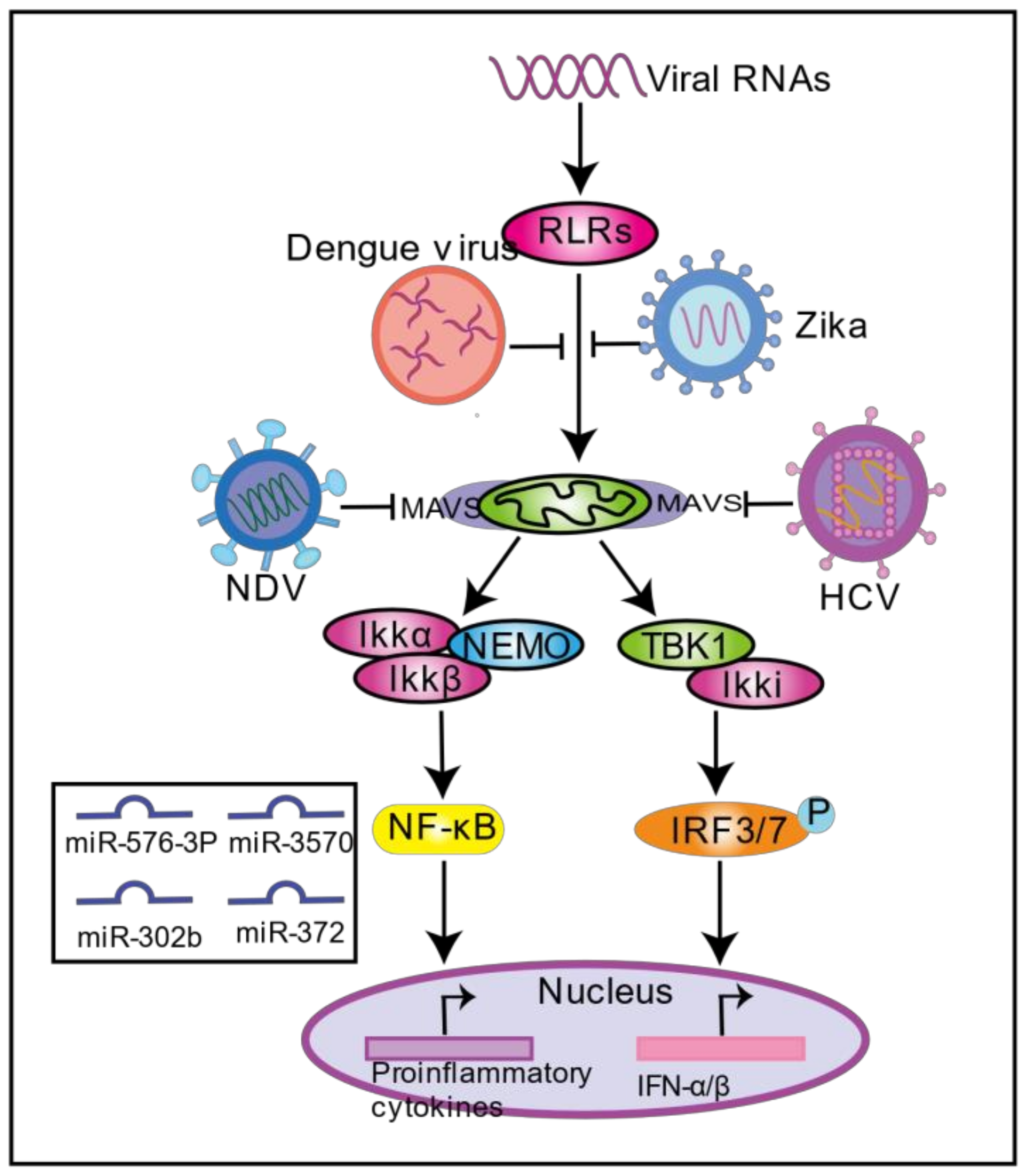
4. Viral Infection Regulates Mitochondria-Induced Innate Immunity

When a virus infects cells, the host activates the innate immune system to recognize the virus through pathogen recognition receptors (PRRs), such as TLRs, RLRs, and NLRs. Here, we focus on viral infection regulating the mitochondria-mediated RLR signaling pathway. Many viral pathogen-associated molecular patterns (PAMPs) are recognized by the retinoic acid-inducible gene I (RIG-I) and melanoma differentiation-associated gene 5 (MDA5); RIG-1 and MDA5 undergo conformational changes that result in the exposure of the CARD domain to form a homologous oligomer. RIG-1 and MDA5 recognize and bind to each other through the N-terminal CARD domain and the N-terminal CARD domain of MAVS, forming MAVS prion-like polymers and activating downstream signaling pathways such as NF-κB and IRF3/7, thereby inducing the expression of inflammatory cytokines and interferons involved in the innate antiviral response. MAVS is located in the OMM as the pivotal adaptor protein of the RLR pathway. MAVS’ functions depend on its mitochondrial localization, confirming that mitochondria play an important role in the innate immune signaling pathway.
More and more studies have shown that viruses developed a series of strategies to antagonize the RLR signaling pathway in mitochondria during the their evolution to escape from the host immune system (Figure 3). SARS-CoV 3b protein inhibits type I IFN production by blocking MAVS activity [72]. In addition, the Nsp13 protein and 9C protein of SARS-CoV-2 may be involved in the regulation of MAVS signal transduction, thereby mediating the innate immune response [48]. A recent study showed that SARS-CoV-2 infection in human colon epithelial cancer cells Caco-2 resulted in decreased expression of MAVS [73]. HCV can evade host immunity, causing chronic infection. NS3/4A can be localized in mitochondria and combined with MAVS. NS3/4A cleaves MAVS at Cys-508, causing the N-terminal fragment of MAVS to dislocate from the mitochondria and become an inactive fragment, which prevents the production of IFN [74,75]. Similarly, Bat Hepatovirus and Seneca Valley virus are RNA viruses, both belonging to the Picornaviridae family, that can also interfere with innate immune signal transduction by interacting with MAVs protein, thus maintaining viral infection [76,77]. Bat Hepatovirus 3ABC proteases interact with human MAVS and cleave MAVS at Glu463/Gly464 to inhibit the activation of IRF3 and NF-κB, thereby blocking the production of type I interferon in human cells [76]. Seneca Valley virus 3C protease depends on its protease activity to cleave MAVS at Q148, inhibiting the type I interferon [77]. In addition, the virus can also degrade MAVS through the proteasome pathway and block the RLR signaling pathway. For example, HBV HBx protein can interact with MAVS, promote the ubiquitination and degradation of MAVS, and inhibit the RIG-I-MDA5 pathway, which together reduce the production of IFN-β [78]. NDV V protein recruits E3 ubiquitin ligase RNF5 to mediate MAVS degradation through the proteasomal pathway to prevent IFN production [79]. Rotavirus VP3 protein targets mitochondria and mediates the phosphorylation of the SPLTSS motif in the proline-rich region of MAVS, causing MAVS to be degraded through the proteasome pathway, blocking the production of IFN-γ during Rotavirus infection of intestinal epithelial cells [80]. Viruses also inhibit the RLR signaling pathway by blocking the binding of MAVS to RIG-1 and MDA5. By binding to the 14-3-3-binding motif, Zika virus NS3 prevents RIG-1 and MDA5 from being transported to mitochondria, thereby blocking interferon production mediated by the RLR signaling pathway [81]. DENV NS4A binds to the N-terminal CARD-like (CL) domain and the C-terminal transmembrane (TM) domain of MAVS, which prevents MAVS from binding to RIG-I and inhibits the production of interferon [82]. Viruses can also evade host innate immunity by manipulating microRNAs, regulating a range of host immune systems through post-transcriptional regulation to block RLR signaling pathways. Vesicular Stomatitis Virus (VSV), a single-stranded negative RNA (ssRNA) virus of the family Rhabviridae, infection induces miR-576-3p through IRF3 and regulates MAVS and TRAF3 mRNAs to reduce type I expression interferon and avoid excessive inflammation [83]. Rhabdovirus infects miiuy croaker macrophages, inducing miR-3570 expression and targeting and suppressing MAVS expression, thus promoting the virus [84]. Studies have shown that miR-302b and miR-372 induced by viral infection may manipulate cell function and mitochondrial metabolism through aspartate glutamate transporter SLC25A12, thereby impairing MAVS-mediated innate immunity to antiviral viruses [85]. Interestingly, the introduction of mimics of miR-302b and miR-372 into cells can reduce NADH levels, resulting in an increase in the NAD/NADH ratio by up to 50%, a decrease in mitochondrial oxygen consumption, and ultimately a change in cellular metabolic pathways from the citric acid cycle to sugar digestion, while increasing the content of lactate [85]. The latest research shows that hepatitis B virus infection directly binds to MAVS through lactate dehydrogenase-dependent lactic acid to prevent MAVS from mitochondrial aggregation and localization, thereby blocking the RLR signaling pathway [86]. Since lactic acid plays a negative regulatory role in calf-mediated innate immune response [87], these two miRNAs may affect innate immunity through lactic acid regulation.

5. Viral Infection Regulates Mitochondrial Metabolism

Mitochondria are the energy metabolism centers of cells; they produce ATP by regulating macromolecular metabolism of carbohydrates, amino acids, and fatty acids. The main energy source of the cell is dephosphorylated by the ATP molecule into an adenosine diphosphate (ADP) molecule. For this process to continue, cells need to break down some macromolecular metabolites through pathways such as glycolysis, the tricarboxylic acid cycle, and oxidative phosphorylation. Glucose is the primary source of energy for cells. In the cytoplasm, two molecules of ATP are produced by glycolysis from one molecule of glucose, generating two pyruvate molecules. To optimize ATP production, cells undergo oxidative phosphorylation (OXPHOS), which oxidizes pyruvate into the mitochondrial matrix through the mitochondrial pyruvate carrier (MPC) with the tricarboxylic acid cycle. Finally, complete oxidation of one glucose molecule through the mitochondrial electron transport chain generates 36 ATP molecules. Although oxidative phosphorylation produces high energy efficiency, it is a slow process and cannot meet the energy requirements of rapidly dividing cells, such as activated immune cells or cancer cells. Therefore, these cells need to initiate aerobic glycolysis (also called the Warburg effect) to produce energy quickly to maintain their activity. In addition, during starvation and emergency, lipase degrades lipids into free fatty acids, which enter mitochondria for fatty acid β oxidation, thus maintaining the balance of cellular energy metabolism.
Many viruses can actively reshape host cell metabolism to enhance intracellular survival. HCV infection causes changes in cell metabolism, which increases the carbohydrate flux during glycolysis and decreases the activities of aerobic oxidative phosphorylation and the citric acid cycle, which may direct the cell towards the Warburg Effect quite quickly, within a few days or weeks after infection of a cell [88,89,90]. In a recent study, some critical components of the mitochondrial respiratory chain complex were found to be down-regulated six days after HCV infection, including MT-ND1, MT-ND3, MT-ND4, MT-ND4L, and MT-CO2 [91]. In addition, the down-regulation of MTND, COX, and F0/F1ATP synthase was found in the HCV-infected CD8+T cell cycle [92,93]. It has been shown that HCV systematically limits the activity of oxidative phosphorylation by altering the expression of the mitochondrial respiratory chain complex [94]. HIF-1α and the proto-oncogene c-myc are significantly expressed in HCV-infected cells, inducing the expression of several glycolytic key enzymes, including glucokinase (G.K.), phosphoglucose-1 (PFK-1), and pyruvate kinase (P.K.) [95,96,97]. In addition, HCV infection induces upregulation of Hexokinase 2 expression and enhances Hexokinase activity through interaction with HCV protein NS5a [98]. DENV infection also induces the upregulation of glucose transporter 1 and hexokinase 2 [99]. Inhibition of the glycolytic pathway significantly reduced the RNA synthesis of DENV and the production of infectious virions, revealing that DENV can remodel cellular glycolysis to maintain its replication [99]. Interestingly, DENV proteins have different effects on host metabolism. DENV NS1 protein interacts with GAPDH to enhance the glycolytic activity of GADPH [100]. However, the interaction of DENV NS3 protein with GAPDH resulted in reduced GAPDH glycolysis activity [101]. HCV and DENV infection can reshape cell metabolism, enhance mitochondrial fatty acid oxidation, and provide energy [102,103,104]. Meanwhile, inhibition of fatty acid transport to mitochondria and regulation of β-oxidation can affect viral replication [103]. The Zika virus can utilize host resources and reprogram cell metabolism in different cells to regulate the cell’s state in different metabolic pathways, thus facilitating its self-replication [105,106,107,108]. HIV replicates in CD4+ T cells and leads to metabolic reprogramming from oxidative phosphorylation to aerobic glycolysis [109]. HIV infection induces the increase of glucose transporter-1, the uptake of more glucose, and the up-regulation of glycolytic enzymes lactate dehydrogenase A (LDHA) hexokinase-1, thus activating aerobic glycolysis, which is conducive to HIV reverse transcription, integration, and virion production [110,111,112,113]. In addition to increasing aerobic glycolysis, HIV-infected CD4+ T cells can cause glutamine metabolism and reuse glutamine during productive HIV infection [114,115]. In addition to glucose and glutamine as the primary energy sources, HIV also uses fatty acid oxidation as an energy source to infect CD4+ T cells [115]. A recent study showed that HIV infection induces aerobic glycolysis, which helps control the quality of the virus by controlling the factors packaged into the particles to maintain infectivity [116]. Although mitochondrial metabolism is closely related to viral infection, the mechanism by which viruses target mitochondrial metabolism and how viruses utilize the energy produced by cell metabolism is still unclear.

6. Concluding Remarks

Over the past few decades, mitochondria have been shown to play an important role in viral infection and host innate immunity; however, the role of mitochondria in the host–virus interaction needs to be further studied. Viral infections can create a viable ecological niche for themselves by manipulating mitochondrial function. The virus induces mitochondria-induced cell death and the mitochondria-mediated innate immune system to facilitate its replication and transmission by regulating mitochondrial dynamics. In recent years, the role of mitochondria as the regulatory center of cell metabolism has attracted more attention. Viruses can manipulate cell metabolism, reprogram metabolic pathways, and reuse metabolites to maintain viral niches in cells. However, research on the mitochondria and its metabolism is still in its infancy. Delving into the mechanisms by which viruses use mitochondria-mediated cell metabolism to maintain infection is an exciting area for future research.

Author Contributions

Conceptualization, J.C., S.F. and X.L.; methodology, K.W.; software, S.Z. and F.Z.; writing—original draft preparation, X.L., K.W., Z.L., J.F.; writing—review and editing, J.C., S.F. and X.L.; visualization, L.Y., H.D., M.Z.; funding acquisition, J.C. All authors have read and agreed to the published version of the manuscript.

Funding

This work was supported by grants from the Science and Technology Program of Guangzhou, China (201803020005), the Science and Technology Program of Guangdong, China (2019B020211003), the Major Program of Guangdong Basic and Applied Research (202019071850100001), the Key Research Projects of Universities in Guangdong Province (2019KZDXM026), and 111 Project [D20008].

Institutional Review Board Statement

Not applicable.

Informed Consent Statement

Not applicable.

Data Availability Statement

Not applicable.

Acknowledgments

We thanks for the authors of references for providing the data of the final manuscript.

Conflicts of Interest

The authors declare no conflict of interest.

References

  1. Kiriyama, Y.; Nochi, H. Intra- and Intercellular Quality Control Mechanisms of Mitochondria. Cells 2017, 7, 1. [Google Scholar] [CrossRef] [PubMed] [Green Version]
  2. Hales, K.G.; Fuller, M.T. Developmentally regulated mitochondrial fusion mediated by a conserved, novel, predicted GTPase. Cell 1997, 90, 121–129. [Google Scholar] [CrossRef] [Green Version]
  3. Chen, H.; Detmer, S.A.; Ewald, A.J.; Griffin, E.E.; Fraser, S.E.; Chan, D.C. Mitofusins Mfn1 and Mfn2 coordinately regulate mitochondrial fusion and are essential for embryonic development. J. Cell Biol. 2003, 160, 189–200. [Google Scholar] [CrossRef]
  4. Chen, H.; Chomyn, A.; Chan, D.C. Disruption of fusion results in mitochondrial heterogeneity and dysfunction. J. Biol. Chem. 2005, 280, 26185–26192. [Google Scholar] [CrossRef] [Green Version]
  5. Cipolat, S.; Martins de Brito, O.; Dal Zilio, B.; Scorrano, L. OPA1 requires mitofusin 1 to promote mitochondrial fusion. Proc. Natl. Acad. Sci. USA 2004, 101, 15927–15932. [Google Scholar] [CrossRef] [PubMed] [Green Version]
  6. Koshiba, T.; Detmer, S.A.; Kaiser, J.T.; Chen, H.; McCaffery, J.M.; Chan, D.C. Structural basis of mitochondrial tethering by mitofusin complexes. Science 2004, 305, 858–862. [Google Scholar] [CrossRef] [Green Version]
  7. Cao, Y.L.; Meng, S.; Chen, Y.; Feng, J.X.; Gu, D.D.; Yu, B.; Li, Y.J.; Yang, J.Y.; Liao, S.; Chan, D.C.; et al. Mfn1 structures reveal nucleotide-triggered dimerization critical for mitochondrial fusion. Nature 2017, 542, 372–376. [Google Scholar] [CrossRef]
  8. Otera, H.; Mihara, K. Molecular mechanisms and physiologic functions of mitochondrial dynamics. J. Biochem. 2011, 149, 241–251. [Google Scholar] [CrossRef] [Green Version]
  9. Rojo, M.; Legros, F.; Chateau, D.; Lombès, A. Membrane topology and mitochondrial targeting of mitofusins, ubiquitous mammalian homologs of the transmembrane. J. Cell Sci. GTPase Fzo. 2002, 115, 1663–1674. [Google Scholar]
  10. Anand, R.; Wai, T.; Baker, M.J.; Kladt, N.; Schauss, A.C.; Rugarli, E.; Langer, T. The i-AAA protease YME1L and OMA1 cleave OPA1 to balance mitochondrial fusion and fission. J. Cell Biol. 2014, 204, 919–929. [Google Scholar] [CrossRef] [PubMed]
  11. Tondera, D.; Grandemange, S.; Jourdain, A.; Karbowski, M.; Mattenberger, Y.; Herzig, S.; Da Cruz, S.; Clerc, P.; Raschke, I.; Merkwirth, C.; et al. SLP-2 is required for stress-induced mitochondrial hyperfusion. EMBO J. 2009, 28, 1589–1600. [Google Scholar] [CrossRef] [PubMed] [Green Version]
  12. Ingerman, E.; Perkins, E.M.; Marino, M.; Mears, J.A.; McCaffery, J.M.; Hinshaw, J.E.; Nunnari, J. Dnm1 forms spirals that are structurally tailored to fit mitochondria. J. Cell Biol. 2005, 170, 1021–1027. [Google Scholar] [CrossRef] [Green Version]
  13. Ji, W.K.; Hatch, A.L.; Merrill, R.A.; Strack, S.; Higgs, H.N. Actin filaments target the oligomeric maturation of the dynamin GTPase Drp1 to mitochondrial fission sites. Elife 2015, 4, e11553. [Google Scholar] [CrossRef] [PubMed]
  14. Taguchi, N.; Ishihara, N.; Jofuku, A.; Oka, T.; Mihara, K. Mitotic phosphorylation of dynamin-related GTPase Drp1 participates in mitochondrial fission. J. Biol. Chem. 2007, 282, 11521–11529. [Google Scholar] [CrossRef] [PubMed] [Green Version]
  15. Saito, T.; Nah, J.; Oka, S.I.; Mukai, R.; Monden, Y.; Maejima, Y.; Ikeda, Y.; Sciarretta, S.; Liu, T.; Li, H.; et al. An alternative mitophagy pathway mediated by Rab9 protects the heart against ischemia. J. Clin. Investig. 2019, 129, 802–819. [Google Scholar] [CrossRef] [Green Version]
  16. Chang, C.R.; Blackstone, C. Cyclic AMP-dependent protein kinase phosphorylation of Drp1 regulates its GTPase activity and mitochondrial morphology. J. Biol. Chem. 2007, 282, 21583–21587. [Google Scholar] [CrossRef] [PubMed] [Green Version]
  17. Richter, V.; Singh, A.P.; Kvansakul, M.; Ryan, M.T.; Osellame, L.D. Splitting up the powerhouse: Structural insights into the mechanism of mitochondrial fission. Cell Mol. Life Sci. 2015, 72, 3695–3707. [Google Scholar] [CrossRef]
  18. Yoon, Y.; Krueger, E.W.; Oswald, B.J.; McNiven, M.A. The mitochondrial protein hFis1 regulates mitochondrial fission in mammalian cells through an interaction with the dynamin-like protein DLP1. Mol. Cell Biol. 2003, 23, 5409–5420. [Google Scholar] [CrossRef] [Green Version]
  19. Otera, H.; Wang, C.; Cleland, M.M.; Setoguchi, K.; Yokota, S.; Youle, R.J.; Mihara, K. Mff is an essential factor for mitochondrial recruitment of Drp1 during mitochondrial fission in mammalian cells. J. Cell Biol. 2010, 191, 1141–1158. [Google Scholar] [CrossRef] [Green Version]
  20. Yu, R.; Jin, S.B.; Lendahl, U.; Nistér, M.; Zhao, J. Human Fis1 regulates mitochondrial dynamics through inhibition of the fusion machinery. EMBO J. 2019, 38, e99748. [Google Scholar] [CrossRef]
  21. Palmer, C.S.; Osellame, L.D.; Laine, D.; Koutsopoulos, O.S.; Frazier, A.E.; Ryan, M.T. MiD49 and MiD51, new components of the mitochondrial fission machinery. EMBO Rep. 2011, 12, 565–573. [Google Scholar] [CrossRef]
  22. Zhao, J.; Liu, T.; Jin, S.; Wang, X.; Qu, M.; Uhlén, P.; Tomilin, N.; Shupliakov, O.; Lendahl, U.; Nistér, M. Human MIEF1 recruits Drp1 to mitochondrial outer membranes and promotes mitochondrial fusion rather than fission. EMBO J. 2011, 30, 2762–2778. [Google Scholar] [CrossRef]
  23. Barbaro, G.; Di Lorenzo, G.; Asti, A.; Ribersani, M.; Belloni, G.; Grisorio, B.; Filice, G.; Barbarini, G. Hepatocellular mitochondrial alterations in patients with chronic hepatitis C: Ultrastructural and biochemical findings. Am. J. Gastroenterol. 1999, 94, 2198–2205. [Google Scholar] [CrossRef]
  24. Schwer, B.; Ren, S.; Pietschmann, T.; Kartenbeck, J.; Kaehlcke, K.; Bartenschlager, R.; Yen, T.S.; Ott, M. Targeting of hepatitis C virus core protein to mitochondria through a novel C-terminal localization motif. J. Virol. 2004, 78, 7958–7968. [Google Scholar] [CrossRef] [PubMed] [Green Version]
  25. Korenaga, M.; Wang, T.; Li, Y.; Showalter, L.A.; Chan, T.; Sun, J.; Weinman, S.A. Hepatitis C virus core protein inhibits mitochondrial electron transport and increases reactive oxygen species (ROS) production. J. Biol. Chem. 2005, 280, 37481–37488. [Google Scholar] [CrossRef] [PubMed] [Green Version]
  26. Machida, K.; Cheng, K.T.; Lai, C.K.; Jeng, K.S.; Sung, V.M.; Lai, M.M. Hepatitis C virus triggers mitochondrial permeability transition with production of reactive oxygen species, leading to DNA damage and STAT3 activation. J. Virol. 2006, 80, 7199–7207. [Google Scholar] [CrossRef] [PubMed] [Green Version]
  27. Wang, T.; Campbell, R.V.; Yi, M.K.; Lemon, S.M.; Weinman, S.A. Role of Hepatitis C virus core protein in viral-induced mitochondrial dysfunction. J. Viral. Hepat. 2010, 17, 784–793. [Google Scholar] [CrossRef] [Green Version]
  28. Kim, S.J.; Syed, G.H.; Khan, M.; Chiu, W.W.; Sohail, M.A.; Gish, R.G.; Siddiqui, A. Hepatitis C virus triggers mitochondrial fission and attenuates apoptosis to promote viral persistence. Proc. Natl. Acad. Sci. USA 2014, 111, 6413–6418. [Google Scholar] [CrossRef] [Green Version]
  29. Siu, G.K.; Zhou, F.; Yu, M.K.; Zhang, L.; Wang, T.; Liang, Y.; Chen, Y.; Chan, H.C.; Yu, S. Hepatitis C virus NS5A protein cooperates with phosphatidylinositol 4-kinase IIIα to induce mitochondrial fragmentation. Sci. Rep. 2016, 6, 23464. [Google Scholar] [CrossRef] [Green Version]
  30. Hara, Y.; Yanatori, I.; Ikeda, M.; Kiyokage, E.; Nishina, S.; Tomiyama, Y.; Toida, K.; Kishi, F.; Kato, N.; Imamura, M.; et al. Hepatitis C virus core protein suppresses mitophagy by interacting with Parkin in the context of mitochondrial depolarization. Am. J. Pathol. 2014, 184, 3026–3039. [Google Scholar] [CrossRef]
  31. Gou, H.; Zhao, M.; Xu, H.; Yuan, J.; He, W.; Zhu, M.; Ding, H.; Yi, L.; Chen, J. CSFV induced mitochondrial fission and mitophagy to inhibit apoptosis. Oncotarget 2017, 8, 39382–39400. [Google Scholar] [CrossRef] [Green Version]
  32. Fan, S.; Wu, K.; Zhao, M.; Yuan, J.; Ma, S.; Zhu, E.; Chen, Y.; Ding, H.; Yi, L.; Chen, J. LDHB inhibition induces mitophagy and facilitates the progression of CSFV infection. Autophagy 2020, 16, 1–20. [Google Scholar] [CrossRef] [PubMed]
  33. Xie, B.; Zhao, M.; Song, D.; Wu, K.; Yi, L.; Li, W.; Li, X.; Wang, K.; Chen, J. Induction of autophagy and suppression of type I IFN secretion by CSFV. Autophagy 2020, 16, 1–23. [Google Scholar] [CrossRef]
  34. Barbier, V.; Lang, D.; Valois, S.; Rothman, A.L.; Medin, C.L. Dengue virus induces mitochondrial elongation through impairment of Drp1-triggered mitochondrial fission. Virology 2017, 500, 149–160. [Google Scholar] [CrossRef]
  35. Yu, C.Y.; Liang, J.J.; Li, J.K.; Lee, Y.L.; Chang, B.L.; Su, C.I.; Huang, W.J.; Lai, M.M.; Lin, Y.L. Dengue Virus Impairs Mitochondrial Fusion by Cleaving Mitofusins. PLoS Pathog. 2015, 11, e1005350. [Google Scholar] [CrossRef] [Green Version]
  36. Yoo, Y.S.; Park, Y.J.; Lee, H.S.; Oanh, N.T.K.; Cho, M.Y.; Heo, J.; Lee, E.S.; Cho, H.; Park, Y.Y.; Cho, H. Mitochondria ubiquitin ligase, MARCH5 resolves hepatitis B virus X protein aggregates in the liver pathogenesis. Cell Death Dis. 2019, 10, 938. [Google Scholar] [CrossRef] [PubMed]
  37. Kim, S.J.; Khan, M.; Quan, J.; Till, A.; Subramani, S.; Siddiqui, A. Hepatitis B virus disrupts mitochondrial dynamics: Induces fission and mitophagy to attenuate apoptosis. PLoS Pathog. 2013, 9, e1003722. [Google Scholar] [CrossRef] [PubMed] [Green Version]
  38. Chen, W.; Calvo, P.A.; Malide, D.; Gibbs, J.; Schubert, U.; Bacik, I.; Basta, S.; O’Neill, R.; Schickli, J.; Palese, P.; et al. A novel influenza A virus mitochondrial protein that induces cell death. Nat. Med. 2001, 7, 1306–1312. [Google Scholar] [CrossRef]
  39. Gibbs, J.S.; Malide, D.; Hornung, F.; Bennink, J.R.; Yewdell, J.W. The influenza A virus PB1-F2 protein targets the inner mitochondrial membrane via a predicted basic amphipathic helix that disrupts mitochondrial function. J. Virol. 2003, 77, 7214–7224. [Google Scholar] [CrossRef] [Green Version]
  40. Yamada, H.; Chounan, R.; Higashi, Y.; Kurihara, N.; Kido, H. Mitochondrial targeting sequence of the influenza A virus PB1-F2 protein and its function in mitochondria. FEBS Lett. 2004, 578, 331–336. [Google Scholar] [CrossRef] [PubMed] [Green Version]
  41. Yoshizumi, T.; Ichinohe, T.; Sasaki, O.; Otera, H.; Kawabata, S.; Mihara, K.; Koshiba, T. Influenza A virus protein PB1-F2 translocates into mitochondria via Tom40 channels and impairs innate immunity. Nat. Commun. 2014, 5, 4713. [Google Scholar] [CrossRef]
  42. Wang, R.; Zhu, Y.; Ren, C.; Yang, S.; Tian, S.; Chen, H.; Jin, M.; Zhou, H. Influenza A virus protein PB1-F2 impairs innate immunity by inducing mitophagy. Autophagy 2021, 17, 496–511. [Google Scholar] [CrossRef]
  43. Pila-Castellanos, I.; Molino, D.; McKellar, J.; Lines, L.; Da Graca, J.; Tauziet, M.; Chanteloup, L.; Mikaelian, I.; Meyniel-Schicklin, L.; Codogno, P.; et al. Mitochondrial morphodynamics alteration induced by influenza virus infection as a new antiviral strategy. PLoS Pathog. 2021, 17, e1009340. [Google Scholar]
  44. Cornillez-Ty, C.T.; Liao, L.; Yates, J.R., 3rd; Kuhn, P.; Buchmeier, M.J. Severe acute respiratory syndrome coronavirus nonstructural protein 2 interacts with a host protein complex involved in mitochondrial biogenesis and intracellular signaling. J. Virol. 2009, 83, 10314–10318. [Google Scholar] [CrossRef] [Green Version]
  45. Merkwirth, C.; Langer, T. Prohibitin function within mitochondria: Essential roles for cell proliferation and cristae morphogenesis. Biochim. Biophys. Acta 2009, 1793, 27–32. [Google Scholar] [CrossRef] [Green Version]
  46. Shi, C.S.; Qi, H.Y.; Boularan, C.; Huang, N.N.; Abu-Asab, M.; Shelhamer, J.H.; Kehrl, J.H. SARS-coronavirus open reading frame-9b suppresses innate immunity by targeting mitochondria and the MAVS/TRAF3/TRAF6 signalosome. J. Immunol. 2014, 193, 3080–3089. [Google Scholar] [CrossRef] [PubMed] [Green Version]
  47. Singh, M.; Bansal, V.; Feschotte, C. A Single-Cell RNA Expression Map of Human Coronavirus Entry Factors. Cell Rep. 2020, 32, 108175. [Google Scholar] [CrossRef] [PubMed]
  48. Gordon, D.E.; Jang, G.M.; Bouhaddou, M.; Xu, J.; Obernier, K.; White, K.M.; O’Meara, M.J.; Rezelj, V.V.; Guo, J.Z.; Swaney, D.L.; et al. A SARS-CoV-2 protein interaction map reveals targets for drug repurposing. Nature 2020, 583, 459–468. [Google Scholar] [CrossRef] [PubMed]
  49. Acehan, D.; Jiang, X.; Morgan, D.G.; Heuser, J.E.; Wang, X.; Akey, C.W. Three-dimensional structure of the apoptosome: Implications for assembly, procaspase-9 binding, and activation. Mol. Cell 2002, 9, 423–432. [Google Scholar] [CrossRef]
  50. Nomura-Takigawa, Y.; Nagano-Fujii, M.; Deng, L.; Kitazawa, S.; Ishido, S.; Sada, K.; Hotta, H. Non-structural protein 4A of Hepatitis C virus accumulates on mitochondria and renders the cells prone to undergoing mitochondria-mediated apoptosis. J. Gen. Virol. 2006, 87, 1935–1945. [Google Scholar] [CrossRef] [PubMed]
  51. Chiou, H.L.; Hsieh, Y.S.; Hsieh, M.R.; Chen, T.Y. HCV E2 may induce apoptosis of Huh-7 cells via a mitochondrial-related caspase pathway. Biochem. Biophys. Res. Commun. 2006, 345, 453–458. [Google Scholar] [CrossRef]
  52. Lee, S.K.; Park, S.O.; Joe, C.O.; Kim, Y.S. Interaction of HCV core protein with 14-3-3epsilon protein releases Bax to activate apoptosis. Biochem. Biophys. Res. Commun. 2007, 352, 756–762. [Google Scholar] [CrossRef] [PubMed]
  53. Zhao, P.; Han, T.; Guo, J.J.; Zhu, S.L.; Wang, J.; Ao, F.; Jing, M.Z.; She, Y.L.; Wu, Z.H.; Ye, L.B. HCV NS4B induces apoptosis through the mitochondrial death pathway. Virus Res. 2012, 169, 1–7. [Google Scholar] [CrossRef] [PubMed]
  54. Javed, F.; Manzoor, S. HCV non-structural NS4A protein of genotype 3a induces mitochondria mediated death by activating Bax and the caspase cascade. Microb. Pathog. 2018, 124, 346–355. [Google Scholar] [CrossRef] [PubMed]
  55. Takada, S.; Shirakata, Y.; Kaneniwa, N.; Koike, K. Association of hepatitis B virus X protein with mitochondria causes mitochondrial aggregation at the nuclear periphery, leading to cell death. Oncogene 1999, 18, 6965–6973. [Google Scholar] [CrossRef] [PubMed] [Green Version]
  56. Nasirudeen, A.M.; Wang, L.; Liu, D.X. Induction of p53-dependent and mitochondria-mediated cell death pathway by dengue virus infection of human and animal cells. Microbes Infect. 2008, 10, 1124–1132. [Google Scholar] [CrossRef]
  57. Gao, W.Y.; Li, D.; Cai, D.E.; Huang, X.Y.; Zheng, B.Y.; Huang, Y.H.; Chen, Z.X.; Wang, X.Z. Hepatitis B virus X protein sensitizes HL-7702 cells to oxidative stress-induced apoptosis through modulation of the mitochondrial permeability transition pore. Oncol. Rep. 2017, 37, 48–56. [Google Scholar] [CrossRef] [PubMed] [Green Version]
  58. Kim, H.J.; Kim, S.Y.; Kim, J.; Lee, H.; Choi, M.; Kim, J.K.; Ahn, J.K. Hepatitis B virus X protein induces apoptosis by enhancing translocation of Bax to mitochondria. IUBMB Life 2008, 60, 473–480. [Google Scholar] [CrossRef]
  59. Padhan, K.; Minakshi, R.; Towheed, M.A.B.; Jameel, S. Severe acute respiratory syndrome coronavirus 3a protein activates the mitochondrial death pathway through p38 MAP kinase activation. J. Gen. Virol. 2008, 89, 1960–1969. [Google Scholar] [CrossRef] [PubMed]
  60. Zhang, L.; Wei, L.; Jiang, D.; Wang, J.; Cong, X.; Fei, R. SARS-CoV nucleocapsid protein induced apoptosis of COS-1 mediated by the mitochondrial pathway. Artif. Cells Blood Substit. Immobil. Biotechnol. 2007, 35, 237–253. [Google Scholar] [CrossRef] [Green Version]
  61. Chan, C.M.; Ma, C.W.; Chan, W.Y.; Chan, H.Y. The SARS-Coronavirus Membrane protein induces apoptosis through modulating the Akt survival pathway. Arch Biochem. Biophys. 2007, 459, 197–207. [Google Scholar]
  62. Guerrero, R.; Guerrero, C.; Acosta, O. Induction of Cell Death in the Human Acute Lymphoblastic Leukemia Cell Line Reh by Infection with Rotavirus Isolate Wt1-5. Biomedicines 2020, 8, 242. [Google Scholar] [CrossRef] [PubMed]
  63. Martin-Latil, S.; Mousson, L.; Autret, A.; Colbère-Garapin, F.; Blondel, B. Bax is activated during rotavirus-induced apoptosis through the mitochondrial pathway. J. Virol. 2007, 81, 4457–4464. [Google Scholar] [CrossRef] [PubMed] [Green Version]
  64. Bhowmick, R.; Halder, U.C.; Chattopadhyay, S.; Nayak, M.K.; Chawla-Sarkar, M. Rotavirus-encoded nonstructural protein 1 modulates cellular apoptotic machinery by targeting tumor suppressor protein p53. J. Virol. 2013, 87, 6840–6850. [Google Scholar] [CrossRef] [Green Version]
  65. Mukherjee, A.; Patra, U.; Bhowmick, R.; Chawla-Sarkar, M. Rotaviral nonstructural protein 4 triggers dynamin-related protein 1-dependent mitochondrial fragmentation during infection. Cell Microbiol. 2018, 20, e12831. [Google Scholar] [CrossRef] [PubMed]
  66. Zamarin, D.; García-Sastre, A.; Xiao, X.; Wang, R.; Palese, P. Influenza virus PB1-F2 protein induces cell death through mitochondrial ANT3 and VDAC1. PLoS Pathog. 2005, 1, e4. [Google Scholar] [CrossRef]
  67. Yang, S.; Gorshkov, K.; Lee, E.M.; Xu, M.; Cheng, Y.S.; Sun, N.; Soheilian, F.; de Val, N.; Ming, G.; Song, H.; et al. Zika Virus-Induced Neuronal Apoptosis via Increased Mitochondrial Fragmentation. Front. Microbiol. 2020, 11, 598203. [Google Scholar] [CrossRef]
  68. Neumann, S.; El Maadidi, S.; Faletti, L.; Haun, F.; Labib, S.; Schejtman, A.; Maurer, U.; Borner, C. How do viruses control mitochondria-mediated apoptosis? Virus Res. 2015, 209, 45–55. [Google Scholar] [CrossRef] [PubMed]
  69. Maiuri, M.C.; Zalckvar, E.; Kimchi, A.; Kroemer, G. Self-eating and self-killing: Crosstalk between autophagy and apoptosis. Nat. Rev. Mol. Cell Biol. 2007, 8, 741–752. [Google Scholar] [CrossRef]
  70. Li, S.; Wang, J.; Zhou, A.; Khan, F.A.; Hu, L.; Zhang, S. Porcine reproductive and respiratory syndrome virus triggers mitochondrial fission and mitophagy to attenuate apoptosis. Oncotarget 2016, 7, 56002–56012. [Google Scholar]
  71. Meng, G.; Xia, M.; Wang, D.; Chen, A.; Wang, Y.; Wang, H.; Yu, D.; Wei, J. Mitophagy promotes replication of oncolytic Newcastle disease virus by blocking intrinsic apoptosis in lung cancer cells. Oncotarget 2014, 5, 6365–6374. [Google Scholar]
  72. Freundt, E.C.; Yu, L.; Park, E.; Lenardo, M.J.; Xu, X.N. Molecular determinants for subcellular localization of the severe acute respiratory syndrome coronavirus open reading frame 3b protein. J. Virol. 2009, 83, 6631–6640. [Google Scholar] [CrossRef] [Green Version]
  73. Bojkova, D.; Klann, K.; Koch, B.; Krause, D.; Ciesek, S.; Cinatl, J.; Münch, C. Proteomics of SARS-CoV-2-infected host cells reveals therapy targets. Nature 2020, 583, 469–472. [Google Scholar] [CrossRef] [PubMed]
  74. Meylan, E.; Curran, J.; Hofmann, K.; Moradpour, D.; Binder, M.; Bartenschlager, R.; Tschopp, J. Cardif is an adaptor protein in the RIG-I antiviral pathway and is targeted by hepatitis C virus. Nature 2005, 437, 1167–1172. [Google Scholar] [CrossRef] [PubMed]
  75. Li, X.D.; Sun, L.; Seth, R.B.; Pineda, G.; Chen, Z.J. Hepatitis C virus protease NS3/4A cleaves mitochondrial antiviral signaling protein off the mitochondria to evade innate immunity. Proc. Natl. Acad. Sci. USA 2005, 102, 17717–17722. [Google Scholar] [CrossRef] [PubMed] [Green Version]
  76. Feng, H.; Sander, A.L.; Moreira-Soto, A.; Yamane, D.; Drexler, J.F.; Lemon, S.M. Hepatovirus 3ABC proteases and evolution of mitochondrial antiviral signaling protein (MAVS). J. Hepatol. 2019, 71, 25–34. [Google Scholar] [CrossRef] [PubMed]
  77. Qian, S.; Fan, W.; Liu, T.; Wu, M.; Zhang, H.; Cui, X.; Zhou, Y.; Hu, J.; Wei, S.; Chen, H.; et al. Seneca Valley Virus Suppresses Host Type I Interferon Production by Targeting Adaptor Proteins MAVS, TRIF, and TANK for Cleavage. J. Virol. 2017, 91, e00823-17. [Google Scholar] [CrossRef] [PubMed] [Green Version]
  78. Wei, C.; Ni, C.; Song, T.; Liu, Y.; Yang, X.; Zheng, Z.; Jia, Y.; Yuan, Y.; Guan, K.; Xu, Y.; et al. The hepatitis B virus X protein disrupts innate immunity by downregulating mitochondrial antiviral signaling protein. J. Immunol. 2010, 185, 1158–1168. [Google Scholar] [CrossRef]
  79. Sun, Y.; Zheng, H.; Yu, S.; Ding, Y.; Wu, W.; Mao, X.; Liao, Y.; Meng, C.; Ur Rehman, Z.; Tan, L.; et al. Newcastle Disease Virus V Protein Degrades Mitochondrial Antiviral Signaling Protein to Inhibit Host Type I Interferon Production via E3 Ubiquitin Ligase RNF5. J. Virol. 2019, 93, e00322-19. [Google Scholar] [CrossRef] [Green Version]
  80. Ding, S.; Zhu, S.; Ren, L.; Feng, N.; Song, Y.; Ge, X.; Li, B.; Flavell, R.A.; Greenberg, H.B. Rotavirus VP3 targets MAVS for degradation to inhibit type III interferon expression in intestinal epithelial cells. Elife 2018, 7, e39494. [Google Scholar] [CrossRef] [PubMed]
  81. Riedl, W.; Acharya, D.; Lee, J.H.; Liu, G.; Serman, T.; Chiang, C.; Chan, Y.K.; Diamond, M.S.; Gack, M.U. Zika Virus NS3 Mimics a Cellular 14-3-3-Binding Motif to Antagonize RIG-I- and MDA5-Mediated Innate Immunity. Cell Host Microbe. 2019, 26, 493–503.e6. [Google Scholar] [CrossRef] [PubMed]
  82. He, Z.; Zhu, X.; Wen, W.; Yuan, J.; Hu, Y.; Chen, J.; An, S.; Dong, X.; Lin, C.; Yu, J.; et al. Dengue Virus Subverts Host Innate Immunity by Targeting Adaptor Protein MAVS. J. Virol. 2016, 90, 7219–7230. [Google Scholar] [CrossRef] [Green Version]
  83. Yarbrough, M.L.; Zhang, K.; Sakthivel, R.; Forst, C.V.; Posner, B.A.; Barber, G.N.; White, M.A.; Fontoura, B.M. Primate-specific miR-576-3p sets host defense signalling threshold. Nat. Commun. 2014, 5, 4963. [Google Scholar] [CrossRef]
  84. Xu, T.; Chu, Q.; Cui, J.; Bi, D. Inducible MicroRNA-3570 Feedback Inhibits the RIG-I-Dependent Innate Immune Response to Rhabdovirus in Teleost Fish by Targeting MAVS/IPS-1. J. Virol. 2018, 92, e01594-17. [Google Scholar] [CrossRef] [PubMed] [Green Version]
  85. Yasukawa, K.; Kinoshita, D.; Yaku, K.; Nakagawa, T.; Koshiba, T. The microRNAs miR-302b and miR-372 regulate mitochondrial metabolism via the SLC25A12 transporter, which controls MAVS-mediated antiviral innate immunity. J. Biol. Chem. 2020, 295, 444–457. [Google Scholar] [CrossRef] [PubMed]
  86. Zhou, L.; He, R.; Fang, P.; Li, M.; Yu, H.; Wang, Q.; Yu, Y.; Wang, F.; Zhang, Y.; Chen, A.; et al. Hepatitis B virus rigs the cellular metabolome to avoid innate immune recognition. Nat. Commun. 2021, 12, 98. [Google Scholar] [CrossRef]
  87. Zhang, W.; Wang, G.; Xu, Z.G.; Tu, H.; Hu, F.; Dai, J.; Chang, Y.; Chen, Y.; Lu, Y.; Zeng, H.; et al. Lactate Is a Natural Suppressor of RLR Signaling by Targeting MAVS. Cell 2019, 178, 176–189.e15. [Google Scholar] [CrossRef] [PubMed]
  88. Bandiera, S.; Billie Bian, C.; Hoshida, Y.; Baumert, T.F.; Zeisel, M.B. Chronic hepatitis C virus infection and pathogenesis of hepatocellular carcinoma. Curr. Opin. Virol. 2016, 20, 99–105. [Google Scholar] [CrossRef] [Green Version]
  89. Irshad, M.; Gupta, P.; Irshad, K. Molecular basis of hepatocellular carcinoma induced by hepatitis C virus infection. World J. Hepatol. 2017, 9, 1305–1314. [Google Scholar] [CrossRef] [PubMed] [Green Version]
  90. Gerresheim, G.K.; Roeb, E.; Michel, A.M.; Niepmann, M. Hepatitis C Virus Downregulates Core Subunits of Oxidative Phosphorylation, Reminiscent of the Warburg Effect in Cancer Cells. Cells 2019, 8, 1410. [Google Scholar] [CrossRef] [Green Version]
  91. Gerresheim, G.K.; Bathke, J.; Michel, A.M.; Andreev, D.E.; Shalamova, L.A.; Rossbach, O.; Hu, P.; Glebe, D.; Fricke, M.; Marz, M.; et al. Cellular Gene Expression during Hepatitis C Virus Replication as Revealed by Ribosome Profiling. Int. J. Mol. Sci. 2019, 20, 1321. [Google Scholar] [CrossRef] [PubMed] [Green Version]
  92. Wolski, D.; Foote, P.K.; Chen, D.Y.; Lewis-Ximenez, L.L.; Fauvelle, C.; Aneja, J.; Walker, A.; Tonnerre, P.; Torres-Cornejo, A.; Kvistad, D.; et al. Early Transcriptional Divergence Marks Virus-Specific Primary Human CD8+ T Cells in Chronic versus Acute Infection. Immunity 2017, 47, 648–663.e8. [Google Scholar] [CrossRef] [Green Version]
  93. Wolski, D.; Lauer, G.M. Hepatitis C Virus as a Unique Human Model Disease to Define Differences in the Transcriptional Landscape of T Cells in Acute versus Chronic Infection. Viruses 2019, 11, 683. [Google Scholar] [CrossRef] [PubMed] [Green Version]
  94. Piccoli, C.; Scrima, R.; Quarato, G.; D’Aprile, A.; Ripoli, M.; Lecce, L.; Boffoli, D.; Moradpour, D.; Capitanio, N. Hepatitis C virus protein expression causes calcium-mediated mitochondrial bioenergetic dysfunction and nitro-oxidative stress. Hepatology 2007, 46, 58–65. [Google Scholar] [CrossRef]
  95. Ripoli, M.; D’Aprile, A.; Quarato, G.; Sarasin-Filipowicz, M.; Gouttenoire, J.; Scrima, R.; Cela, O.; Boffoli, D.; Heim, M.H.; Moradpour, D.; et al. Hepatitis C virus-linked mitochondrial dysfunction promotes hypoxia-inducible factor 1 alpha-mediated glycolytic adaptation. J. Virol. 2010, 84, 647–660. [Google Scholar] [CrossRef] [PubMed] [Green Version]
  96. Jung, G.S.; Jeon, J.H.; Choi, Y.K.; Jang, S.Y.; Park, S.Y.; Kim, S.W.; Byun, J.K.; Kim, M.K.; Lee, S.; Shin, E.C.; et al. Pyruvate dehydrogenase kinase regulates hepatitis C virus replication. Sci. Rep. 2016, 6, 30846. [Google Scholar] [CrossRef] [PubMed] [Green Version]
  97. Diamond, D.L.; Syder, A.J.; Jacobs, J.M.; Sorensen, C.M.; Walters, K.A.; Proll, S.C.; McDermott, J.E.; Gritsenko, M.A.; Zhang, Q.; Zhao, R.; et al. Temporal proteome and lipidome profiles reveal hepatitis C virus-associated reprogramming of hepatocellular metabolism and bioenergetics. PLoS Pathog. 2010, 6, e1000719. [Google Scholar] [CrossRef] [Green Version]
  98. Ramière, C.; Rodriguez, J.; Enache, L.S.; Lotteau, V.; André, P.; Diaz, O. Activity of hexokinase is increased by its interaction with hepatitis C virus protein NS5A. J. Virol. 2014, 88, 3246–3254. [Google Scholar] [CrossRef] [PubMed] [Green Version]
  99. Fontaine, K.A.; Sanchez, E.L.; Camarda, R.; Lagunoff, M. Dengue virus induces and requires glycolysis for optimal replication. J. Virol. 2015, 89, 2358–2366. [Google Scholar] [CrossRef] [Green Version]
  100. Allonso, D.; Andrade, I.S.; Conde, J.N.; Coelho, D.R.; Rocha, D.C.; da Silva, M.L.; Ventura, G.T.; Silva, E.M.; Mohana-Borges, R. Dengue Virus NS1 Protein Modulates Cellular Energy Metabolism by Increasing Glyceraldehyde-3-Phosphate Dehydrogenase Activity. J. Virol. 2015, 89, 11871–11883. [Google Scholar] [CrossRef] [Green Version]
  101. Silva, E.M.; Conde, J.N.; Allonso, D.; Ventura, G.T.; Coelho, D.R.; Carneiro, P.H.; Silva, M.L.; Paes, M.V.; Rabelo, K.; Weissmuller, G.; et al. Dengue virus nonstructural 3 protein interacts directly with human glyceraldehyde-3-phosphate dehydrogenase (GAPDH) and reduces its glycolytic activity. Sci. Rep. 2019, 9, 2651. [Google Scholar] [CrossRef]
  102. Fernandes-Siqueira, L.O.; Zeidler, J.D.; Sousa, B.G.; Ferreira, T.; Da Poian, A.T. Anaplerotic Role of Glucose in the Oxidation of Endogenous Fatty Acids during Dengue Virus Infection. MSphere 2018, 3, e00458-17. [Google Scholar] [CrossRef] [Green Version]
  103. Rasmussen, A.L.; Diamond, D.L.; McDermott, J.E.; Gao, X.; Metz, T.O.; Matzke, M.M.; Carter, V.S.; Belisle, S.E.; Korth, M.J.; Waters, K.M.; et al. Systems virology identifies a mitochondrial fatty acid oxidation enzyme, dodecenoyl coenzyme A delta isomerase, required for hepatitis C virus replication and likely pathogenesis. J. Virol. 2011, 85, 11646–11654. [Google Scholar] [CrossRef] [PubMed] [Green Version]
  104. Douglas, D.N.; Pu, C.H.; Lewis, J.T.; Bhat, R.; Anwar-Mohamed, A.; Logan, M.; Lund, G.; Addison, W.R.; Lehner, R.; Kneteman, N.M. Oxidative Stress Attenuates Lipid Synthesis and Increases Mitochondrial Fatty Acid Oxidation in Hepatoma Cells Infected with Hepatitis C Virus. J. Biol. Chem. 2016, 291, 1974–1990. [Google Scholar] [CrossRef] [PubMed] [Green Version]
  105. Thaker, S.K.; Chapa, T.; Garcia, G., Jr.; Gong, D.; Schmid, E.W.; Arumugaswami, V.; Sun, R.; Christofk, H.R. Differential Metabolic Reprogramming by Zika Virus Promotes Cell Death in Human versus Mosquito Cells. Cell Metab. 2019, 29, 1206–1216.e4. [Google Scholar] [CrossRef]
  106. Beys-da-Silva, W.O.; Rosa, R.L.; Santi, L.; Berger, M.; Park, S.K.; Campos, A.R.; Terraciano, P.; Varela, A.P.M.; Teixeira, T.F.; Roehe, P.M.; et al. Zika Virus Infection of Human Mesenchymal Stem Cells Promotes Differential Expression of Proteins Linked to Several Neurological Diseases. Mol. Neurobiol. 2019, 56, 4708–4717. [Google Scholar] [CrossRef] [PubMed]
  107. Tiwari, S.K.; Dang, J.; Qin, Y.; Lichinchi, G.; Bansal, V.; Rana, T.M. Zika virus infection reprograms global transcription of host cells to allow sustained infection. Emerg. Microbes Infect. 2017, 6, e24. [Google Scholar] [CrossRef] [Green Version]
  108. Rothan, H.A.; Fang, S.; Mahesh, M.; Byrareddy, S.N. Zika Virus and the Metabolism of Neuronal Cells. Mol. Neurobiol. 2019, 56, 2551–2557. [Google Scholar] [CrossRef]
  109. Frauwirth, K.A.; Thompson, C.B. Regulation of T lymphocyte metabolism. J. Immunol. 2004, 172, 4661–4665. [Google Scholar] [CrossRef] [PubMed] [Green Version]
  110. Hollenbaugh, J.A.; Munger, J.; Kim, B. Metabolite profiles of human immunodeficiency virus infected CD4+ T cells and macrophages using LC-MS/MS analysis. Virology 2011, 415, 153–159. [Google Scholar] [CrossRef] [PubMed] [Green Version]
  111. Palmer, C.S.; Ostrowski, M.; Gouillou, M.; Tsai, L.; Yu, D.; Zhou, J.; Henstridge, D.C.; Maisa, A.; Hearps, A.C.; Lewin, S.R.; et al. Increased glucose metabolic activity is associated with CD4+ T-cell activation and depletion during chronic HIV infection. AIDS 2014, 28, 297–309. [Google Scholar] [CrossRef] [PubMed]
  112. Kavanagh Williamson, M.; Coombes, N.; Juszczak, F.; Athanasopoulos, M.; Khan, M.B.; Eykyn, T.R.; Srenathan, U.; Taams, L.S.; Dias Zeidler, J.; Da Poian, A.T.; et al. Upregulation of Glucose Uptake and Hexokinase Activity of Primary Human CD4+ T Cells in Response to Infection with HIV-1. Viruses 2018, 10, 114. [Google Scholar] [CrossRef] [PubMed] [Green Version]
  113. Hegedus, A.; Kavanagh Williamson, M.; Huthoff, H. HIV-1 pathogenicity and virion production are dependent on the metabolic phenotype of activated CD4+ T cells. Retrovirology 2014, 11, 98. [Google Scholar] [CrossRef] [PubMed] [Green Version]
  114. Hegedus, A.; Kavanagh Williamson, M.; Khan, M.B.; Dias Zeidler, J.; Da Poian, A.T.; El-Bacha, T.; Struys, E.A.; Huthoff, H. Evidence for Altered Glutamine Metabolism in Human Immunodeficiency Virus Type 1 Infected Primary Human CD4+ T Cells. AIDS Res. Hum. Retrovir. 2017, 33, 1236–1247. [Google Scholar] [CrossRef] [Green Version]
  115. Valle-Casuso, J.C.; Angin, M.; Volant, S.; Passaes, C.; Monceaux, V.; Mikhailova, A.; Bourdic, K.; Avettand-Fenoel, V.; Boufassa, F.; Sitbon, M.; et al. Cellular Metabolism Is a Major Determinant of HIV-1 Reservoir Seeding in CD4+ T Cells and Offers an Opportunity to Tackle Infection. Cell Metab. 2019, 29, 611–626.e5. [Google Scholar] [CrossRef] [Green Version]
  116. Kishimoto, N.; Yamamoto, K.; Abe, T.; Yasuoka, N.; Takamune, N.; Misumi, S. Glucose-dependent aerobic glycolysis contributes to recruiting viral components into HIV-1 particles to maintain infectivity. Biochem. Biophys. Res. Commun. 2021, 549, 187–193. [Google Scholar] [CrossRef] [PubMed]
Figure 1. Viral infection disrupts mitochondrial dynamics. Different viruses affect mitochondrial dynamics through mitochondrial fusion proteins (MFNs, OPA1) or fission proteins (DRP1) and induce mitophagy to clear damaged mitochondria to enhance cell survival and viral persistence.
Ijms 22 04260 g001
Figure 2. Viral infection regulates mitochondria-induced cell death. Different viruses mediate B-cell lymphoma 2 (Bcl-2) family proteins, release Cyt c, activate procaspase-9, and form apoptosomes, thereby inducing cell apoptosis.
Ijms 22 04260 g002
Figure 3. Viral infection regulates mitochondria-induced innate immunity. After the virus invades the cell, RLRs recognize the viral RNA and interact with the mitochondrial antiviral signal (MAVS) to activate the antiviral signal pathway. Different viruses evade host innate immunity by blocking the RLR signaling pathway.
Ijms 22 04260 g003
Publisher’s Note: MDPI stays neutral with regard to jurisdictional claims in published maps and institutional affiliations.

