Review

Saccharomyces cerevisiae, a Powerful Model for Studying rRNA Modifications and Their Effects on Translation Fidelity

by
Agnès Baudin-Baillieu
and
Olivier Namy
*
Institute for Integrative Biology of the Cell (I2BC), Université Paris-Saclay, CEA, CNRS, 91198 Gif-sur-Yvette, France
*
Author to whom correspondence should be addressed.
Int. J. Mol. Sci. 2021, 22(14), 7419; https://0-doi-org.brum.beds.ac.uk/10.3390/ijms22147419
Submission received: 11 June 2021 / Revised: 3 July 2021 / Accepted: 6 July 2021 / Published: 10 July 2021
(This article belongs to the Special Issue Yeast Research in the Post-genomic Era)

Abstract

:
Ribosomal RNA is a major component of the ribosome. This RNA plays a crucial role in ribosome functioning by ensuring the formation of the peptide bond between amino acids and the accurate decoding of the genetic code. The rRNA carries many chemical modifications that participate in its maturation, the formation of the ribosome and its functioning. In this review, we present the different modifications and how they are deposited on the rRNA. We also describe the most recent results showing that the modified positions are not 100% modified, which creates a heterogeneous population of ribosomes. This gave rise to the concept of specialized ribosomes that we discuss. The knowledge accumulated in the yeast Saccharomyces cerevisiae is very helpful to better understand the role of rRNA modifications in humans, especially in ribosomopathies.

1. Introduction

The ribosome is a complex system that translates the nucleotide code of messenger RNA into protein in cells. Eukaryotic ribosomes consist of four RNA species and more than 80 proteins, forming two independent subunits—the large subunit (LSU) and the small subunit (SSU)—which are connected when mRNA is recruited, adopting a conformation that delimits regions in which base pairing can occur between tRNA and mRNA, leading to the synthesis of a nascent polypeptide chain. In eukaryotes, the RNA moiety—consisting of the 25S, 5.8S and 5S rRNAs for the LSU and the 18S rRNA for the SSU, in yeast—has long been known to contain two regions essential for key catalytic activities for translation: the decoding center (DC) and the peptidyl transferase center (PTC). These rRNAs are transcribed as a long 35S primary transcript encompassing the 18S, 5.8S and 25S mature rRNAs, the 5S rRNA being transcribed independently. Maturation steps occur in parallel with rRNA folding and ribosomal protein assembly and are driven by more than 200 trans-acting partners (for review see [1]). During these complex processing events, rRNA undergoes many posttranscriptional modifications. In total, 112 positions have been shown to be modified in the yeast Saccharomyces cerevisiae, with 12 different classes of modified nucleotides based on simple chemical transformations and a single, complex, multistep modification. Uridine-to-pseudouridine isomerization and methylation at the ribose 2′OH predominate. Both these modifications are mediated by RNP complexes consisting of an enzyme catalyzing the modification itself and a guide RNA, which identifies the nucleotide targeted. In this review, we will focus on rRNA modifications, explaining how they occur, their mode of action and their functional consequences for ribosome function. The ribosome was long thought to have a very stable structure, but it has emerged in recent years that, on the contrary, it is highly flexible in terms of its composition and modifications. Our vision of the ribosome has, thus, evolved considerably, from a ribosome with a fixed composition reading the genetic code linearly, to a ribosome with a dynamic composition playing an integral role in the regulation of gene expression. This is how the notion of specialized ribosomes emerged, according to which some mRNAs are translated by ribosomes with a specific composition. The rRNA biogenesis and modification pathways in the yeast Saccharomyces cerevisiae are a fantastic tool, providing us with essential knowledge to underpin future studies in more complex organisms.

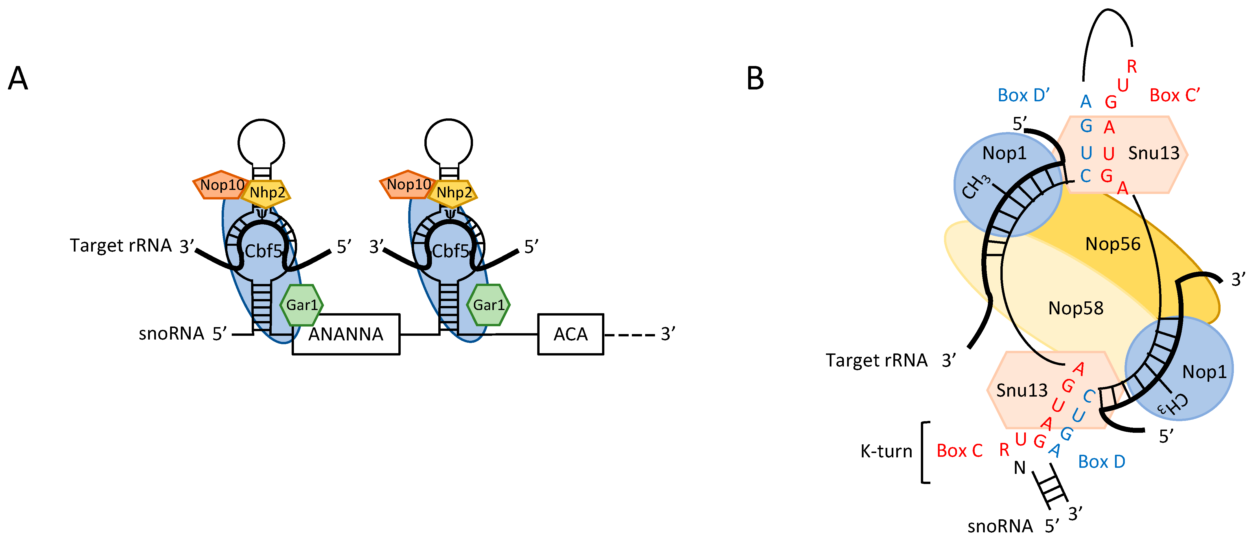
2. RNA-Guided Modifications

The vast majority of rRNA modifications are guided by two classes of small nucleolar RNAs (snoRNAs)—box H/ACA and C/D snoRNAs—in a process based on mechanisms that have been extensively studied. In the late 1980s and early 1990s, a large number of snoRNAs were identified on the basis of three criteria: (i) nucleolar localization, (ii) association with rRNA or nucleolar protein (Nop1) and (iii) conservation of sequences present in already identified snoRNAs. Their importance in rRNA processing was demonstrated early on (for review see [2,3]), but their role in rRNA modification was not formally demonstrated until 1996, when box C/D snoRNAs were shown to be involved in 2′-O methylation ([4,5]), with box H/ACA snoRNAs subsequently implicated in pseudouridine formation [6,7,8].
Box H/ACA snoRNAs are involved in the base isomerization of uridine to generate pseudouridine (Figure 1A). They fold into two hairpin structures connected by a hinge containing the H motif (5′-ANANNA-3′), followed by a tail harboring the second ACA motif. The functional complex consists of a box H/ACA snoRNA specifying the site of modification on the rRNA, the core proteins Nhp2, Nop10, Gar1 and the Cbf5 pseudouridine synthase. Recognition of the substrate uridine is directed by two short sequences complementary to the rRNA and surrounding the target. These sequences are located on the opposite strands of the internal loops of the hairpins [7].
Ribose 2′-O methylations in rRNA (Nm) are driven by the box C/D snoRNA (Figure 1B). They possess highly conserved structural elements—the C/D and C′/D′ boxes—and sequences complementary to mature rRNA. The C (5′-RUGAUGA-3′)/D (5′-CUGA-3′) boxes establish partial base pairing and fold into a kink-turn (K-turn), facilitating interactions with proteins crucial for the modification process. The C′/D′ boxes are degenerate, but they also participate, through base pairing, to the overall single-hairpin structure of the box C/D snoRNA. The snoRNA is part of a small nucleolar ribonucleoprotein (snoRNP), which forms when Snu13 is recruited through specific binding with the K-turn [9], followed by the recruitment of Nop56, Nop58 and the methyltransferase Nop1. The nucleotide to be modified is determined by two elements: the interaction between the snoRNA and the rRNA through the formation of a Watson-Crick helix of 10 to 21 nucleotides (this length heterogeneity being due to heteroduplex thermodynamic stability or local pre-rRNA conformation) and the exact positioning of the modified nucleotide five nucleotides upstream from the D/D′ box, regardless of the nature of the base (A, G, U, C).
Interestingly, two orphan C/D snoRNAs—snR4 and snR45—were recently shown to be responsible for the acetylation of C1773 and C1280 in the 18S rRNA. The mechanism of action resembles that of the box H/ACA snoRNA, with the two guide sequences base pairing on either side of the target rRNA, generating a protruding bulge exposing the C substrate. This bulge is recognized by the Kre33 acetyltransferase, the only protein identified to date as associated with this snoRNP. These two snoRNAs have retained the ability to form a snoRNP with Nop1 and the other core proteins, but are no longer able to attach a methyl group to ribose, presumably because the base-pairing/distance rules are no longer respected [10].
In yeast, 30 box H/ACA snoRNAs are responsible for the pseudouridylation of 46 of the 47 sites in the four rRNA species. Some snoRNAs can direct the modification of up to four different positions within a single or different mature rRNA molecules. Similarly, 54 of the 55 2′-O methylations observed can be attributed to 43 box C/D snoRNAs. Most snoRNA genes exist as individual transcription units (TU), but a few are present in polycistronic TUs or are encoded by introns [11]. They undergo 3′ and 5′ processing and assemble with core proteins with the assistance of many other factors. These complex events are not dealt with here but have been reviewed elsewhere [12,13,14].
In addition to snoRNA-guided processes, several modifications are catalyzed by stand-alone enzymes. Most of these modifications are base modifications (mN), with the exception of the conversion of 5S-U50 into pseudouridine by Psu7 through recognition of the canonical UGΨAR sequence and 25S-G2922 2′-O methylation, which is directed by Spb1. Surprisingly, Spb1 can also support 25S-Um2921 modification in the absence of snR52, making 25S-Um2921 the only site targeted by two independent modification processes [15].
S. cerevisiae displays six different types of base modification in the 18S and 25S rRNAs, at 10 sites: one N7-methylguanosine, two N6-dimethyladenosines, two N1-methyladenosines, two N3-methyluridines, two C5-methylcytosines and one complex N1-methyl-N3-aminocarboxypropylpseudouridine. All the enzymes responsible for these base modifications have been identified and their mode of action is known (Table 1).

3. SnoRNA Engineering

The mode of action of snoRNPs provides flexibility for the development of biotechnology tools. This has made it possible to modify rRNAs artificially for studies of the importance of individual nucleotides and the influence of the presence of a methyl group at positions that are not methylated in natural conditions.
Due to the architecture of the rDNA locus, with more than 200 copies of the rRNA genes, it is difficult to generate cellular ribosomes with modified rRNA. The very specific nature of the mechanism of snoRNA-guided modification (see above) has provided a tool for overcoming this obstacle. A study on U24, which methylates C1436 and A1448 in the 25S rRNA, showed that the deletion of a single nucleotide within the sequence complementary to the rRNA (the antisense element) shifted the D box one nucleotide downstream, resulting in the modification of U1437 instead of C1436 [16]. Modification of the antisense element from the mouse U20 similarly resulted in a new site-directed nucleotide modification [4], indicating that the modified nucleotide is specified exclusively by the D and D′ boxes and the antisense element. It has therefore been suggested that snoRNA engineering could be used to facilitate studies of the ribose methylation machinery, snoRNA function and the effect of point mutations within rRNA in vivo [17]. A plasmid based on the snR38 gene expressed under the control of the GAL1 promoter has been designed to target modification by simply replacing the antisense element with a specific sequence or with random sequences covering a large region of the rRNA (the 810 nt domain V, encompassing PTC). Studies with this plasmid have identified nucleotides essential for ribosome structure or function [18]. These random and specific approaches have been used for more detailed explorations of the interference effect on important nucleotides located within the PTC. The principal conclusion of these studies was that the observed defect was correlated with methylation itself, rather than the antisense effect. Most artificial modifications at positions unmodified in nature have no physiological consequences. Those leading to growth defects are also associated with slow protein synthesis, consistent with the interference effect observed for nucleotides already identified as involved in tRNA binding, subunit association or decoding [19]. Interestingly, the expression of a few engineered snoRNAs did not alter modification status. This lack of alteration was subsequently shown to be due to rRNA degradation, caused either by the modification itself, or by some unexplained snoRNP effect [20]. This finding raised questions about the need for snoRNA-rRNA co-evolution. A similar proof-of-concept was also established for the SnR36 H/ACA snoRNA, for which a substitution in the guide sequence led to an ectopic Ψ modification [6], but no tool has yet been developed for studying the ectopic targeting of Ψ within rRNA. This snoRNA-based tool was developed principally for rRNA modification, but it has also been successfully used to modify mRNA [21].

4. Detection of rRNA Modifications

A global view of rRNA modifications is required, to determine their roles. However, until recently and the development of NGS approaches, it was very difficult to develop such a view. Classical approaches for detecting modified ribonucleosides are based on thin-layer chromatography, capillary electrophoresis, or related techniques. In all cases, the sample is reduced to nucleotides, which are then separated on the basis of their chemical properties [22]. Unfortunately, these methods are labor-intensive, require the use of radioactive labeling and do not allow precise mapping of the modified residue. RP-HPLC, coupled or not with Mung bean protection assays, associated with tandem mass spectrometry methodologies can also be used [23,24] to quantify multiple RNA modifications accurately across conditions and cell types. However, although they are quantitative and reliable they require that the RNA of interest is isolated free from contaminants and partially digested, such methods cannot provide information about the molecule itself or the position of the modification. This technique is therefore suitable for rRNAs and tRNAs, which are abundant. Until the development of techniques based on deep sequencing (next-generation sequencing or NGS), it remained difficult to obtain a global view of the modifications carried by an RNA molecule at single-nucleotide resolution. NGS approaches have provided access to genome-wide quantitative data, shedding light on the positions only partially modified and those with a modification efficiency that varies as a function of the conditions tested. Various techniques are available for mapping RNA modifications [25,26,27] and chemical detection methods have been particularly successful in studies of rRNA modifications. One of these methods, RibomethSeq, is based on the protection, by ribose 2′-O-methylation, of the phosphodiester bond between nucleotides N and N + 1 generated by alkaline fragmentation. This protection is demonstrated by the absence of reads starting at the methylated position. This approach can be used to detect 2′-O-methylation in various types of RNA, including rRNA and tRNA [28,29,30,31]. Efficient quantitative mapping of pseudouridines by the HydraPsiSeq approach also recently became possible [26]. This method is based on RNA cleavage at random uridine residues by hydrazine, followed by aniline treatment for RNA chain scission at abasic sites. The protected residues (positions not cleaved by hydrazine) reveal the presence of pseudouridine residues. Conversely, m7G and m3C modifications can be detected on the basis of abasic site formation under alkaline conditions. These sites are then cleaved by aniline treatment and the 5′-phosphate generated is used for selective ligation for sequencing (Alkaniline-Seq). A frequency of m7G modifications as low as 2% can be detected with this method, which has been used to confirm the presence of a single m7G site, at position 1575, in the S. cerevisiae 18S rRNA (Table 1) [32]. These approaches are highly efficient, but modification-specific, because specific treatment of the RNA is required for detection of the modified positions.
Direct single-RNA molecule sequencing (without the need to generate cDNA) using the nanopore technology has the potential for the direct detection of all RNA modifications, provided that they disturb the resulting sequencing signal sufficiently, which is the case for the most common modifications: m6A, m5C, m7G and pseudouridines [33,34,35]. Direct RNA sequencing could also potentially be used to decipher the structure of single RNA molecules [36]. This approach is very promising but requires further improvement. In terms of bioinformatics pipelines, there is a real need to develop efficient scripts [37]. One major limitation is the need to sequence an artificial modification-free mirror RNA for comparison to the natural RNA [37]. The modified position is identified as a “sequencing error” at specific positions. As this approach is not hypothesis-dependent, the nature of the modification is completely unknown and further experiments are therefore required to determine the chemical nature of the modification.

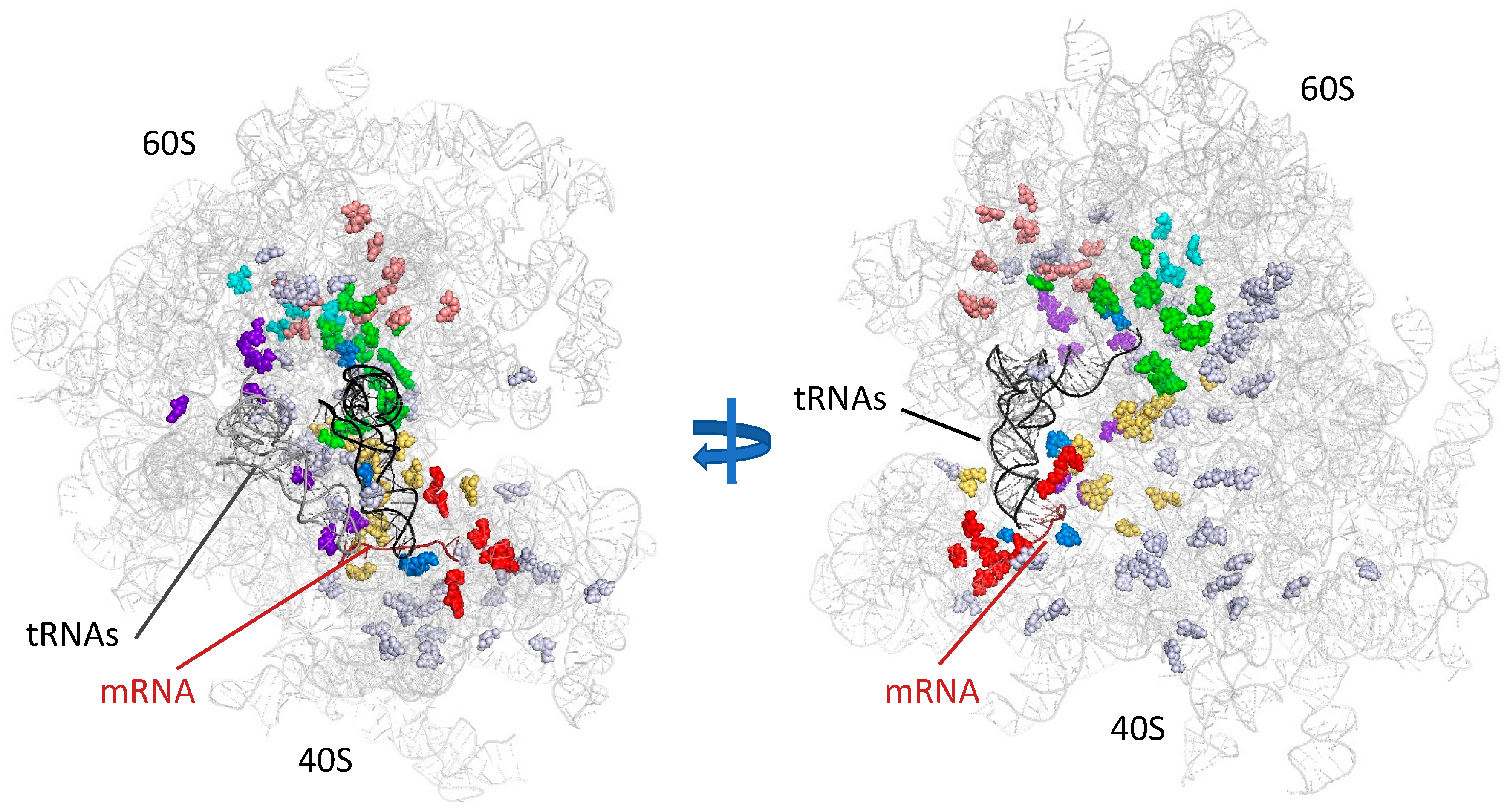
5. Function in Translation Fidelity

The two most frequent rRNA modifications have a clear and significant effect on RNA structure. Indeed, the presence of multiple modified nucleotides influences the secondary and tertiary structure of the ribosome and its interactions with its partners: tRNA, mRNA and proteins. Pseudouridine provides an extra hydrogen bond that can take part in additional pairing interactions with RNA, thereby increasing backbone rigidity and the stability of the structure. It also enhances base stacking [38]. Hydrophobicity is increased by 2′-O-methylation, protecting against nucleolytic attack, stabilizing helices [39] and increasing steric hindrance. Based on recent analyses of the 2′-O-methylation of mRNA, it appears likely that Nm modifications also disrupt diverse other RNA interactions dependent on 2′-OH groups [40].
The mapping of modifications onto the three-dimensional (3D) structure of the ribosome highlights the non-random distribution of these modifications within the rRNA. The location of modifications depends on four criteria: (1) most modifications occur in conserved regions; (2) they are concentrated in the interior of the subunits and not exposed at the periphery of the ribosome, where protein concentration is high; (3) at least 60% of the modified nucleotides occur in functionally important regions; (4) the modifications are conserved throughout evolution (Figure 2) (for a review, see [41]). All of these observations suggest a possible effect of these modifications on translation. However, it is difficult to determine the importance of each single modification for translation fidelity, for many reasons.
First, almost all modifications occur very early on the 35S pre-rRNA transcript, through the association of the snoRNP or modification enzyme with the rRNA and are therefore involved in processing itself, ribosome assembly, export to the cytoplasm and quality control for the processed subunits. This is illustrated by Nop1 mutants, in which the methylation and assembly processes are uncoupled [42] (for a review on snoRNP function in ribosome biogenesis see [43]). Studies of the dual-function snR10, which is involved in rRNA processing and modification, have shown that the two functional domains (one for each function) act cooperatively [44].
Second, some snoRNAs target multiple positions (up to four, in both the 18S and 25S rRNAs, for snR49). Furthermore, some enzymes target other RNA species in addition to rRNA. This is the case for Kree33, which acetylates the leucine and serine tRNAs with the help of the Tan1 adaptor protein. Pus7 also has the ability to use multiple substrates: it catalyzes Ψ35 formation in the spliceosomal snRNA U2 [45] and pseudouridine conversion at position 13 of the cytosolic tRNA and position 5 of the pre-tRNAtyr [46]. This site-directed pseudouridine synthase also mediates the modification of more than 200 uridine residues in mRNA upon heat-shock treatment [47], or about 260 mRNA sites during post-diauxic growth [48]. As mRNA and tRNA are the principal actors in translation, it remains difficult to determine the effect of rRNA modification in isolation.
The disruption of most individual snoRNAs, impairing single or multiple modifications, has been shown to have little or no impact on cell growth or translation, although some exceptions exist, such as 25S-Um2921 [49,50]. This particular modification site is highly conserved and under the control of two independent mechanisms dependent on a box C/D snoRNP and the stand-alone methyltransferase Spb. The abolition of this modification through coupled mutations of Spb1 and snR52 greatly decreases growth rates, alters polyribosome profiles and LSU structure and confers paromomycin sensitivity, whilst decreasing translational accuracy [15,51]. The combined loss of 25S-Gm2288 and 25S-m5C2278 also has a dramatic effect on 25S rRNA structure, impairing the recruitment of several ribosomal proteins.
Despite the rarity of examples of losses of individual modifications having a major impact on ribosome integrity and translation efficiency, a growing number of studies have concluded that the modification state of the ribosome has an impact on the decoding capacity of the ribosome. The first evidence in support of this conclusion was provided by treatment with ethionine, which inhibits methylation and by the nop1.3 mutant allele of the methyltransferase Nop1, which abolishes methylation without disturbing pre-rRNA processing other than by slightly delaying late 60S rRNA processing. Both these conditions result in a severe impairment of growth, indicating that methylation is important for ribosome structure, or directly required for translational accuracy [42]. This hypothesis was confirmed by the description of the D95A mutant allele of Cbf5A, which abolishes Ψ formation and results in a low growth rate [52] and poor translational accuracy [53] with no major defect of rRNA processing. Conversely, single box C/D snoRNA deletions individually subjected to functional profiling for growth or antibiotic susceptibility revealed subtle effects under nonoptimal growth conditions [54]. The analysis of combinatorial snRNA deletions targeting modifications of the same functional region of the ribosome has proved a major advance. Dissections of the peptidyl transferase center, the inter-subunit bridges and the decoding center suggested that modifications acted together to fine-tune translation and adapt the ribosomes to functional requirements. The influence of modifications to peptide bond formation and decoding was investigated in strains lacking up to six pseudouridine residues, including the aforementioned snR10-guided Ψ2919. Synergic effects on growth rate, protein synthesis and ribosome structure were observed [55], together with effects on translational accuracy [51]. This detrimental effect was attributed to structural changes affecting tRNA accommodation at the A site. Inter-subunit bridges have a key function, as they promote associations between subunits and movements from one subunit relative to the other, thereby controlling the various steps of translation. Two such bridges, the B2a (helix 69 of the LSU) and B1a (helix H38 of the SSU, also known as the A-site finger) bridges, have been studied, as they contain numerous modifications. H69 in the 25S rRNA and the SSU H44 together form the B2a bridge, establishing close contact with the A-site and P-site tRNAs. A role for this bridge in translation fidelity has already been demonstrated [56]. Five modifications, four Ψ and one 2′-O methylation, conserved in humans, are present in this small domain. The Fournier laboratory showed, in standard combinatory studies, that it was the loss of modifications, rather than the impairment of snoRNP-rRNA association, that enhanced rRNA turnover [57]. As expected, the loss of one or two modifications led to changes in elongation and termination accuracy, highlighting the key role of this region. Surprisingly, increasing the number of snoRNA deletions had the opposite effect, with antibiotic resistance observed [58]. Similarly, the LSU H38, known as ASF, constitutes the B1a inter-subunit bridge, contacting the A-site tRNA via the tip of the stem-loop, which is devoid of modifications. It is very rich in Ψ, with six positions in H38 and another four positions in the two adjacent helices. Deletion analysis showed that yeast cells are less sensitive to the loss of modifications in H38, the three central Ψ in the loop (Ψs 1004, 1042 and 1052) being more important for subunit association [59]. Translational accuracy is barely affected, with a very specific effect on UGA readthrough in the absence of the six modifications [58]. The decoding center has also been studied. This center has the particular feature of consisting of rRNA segments separated in the primary sequence, but coming together in the tertiary structure to surround the E, A and P-sites of the ribosome. Eight modifications are present (5 Nm and 3 Ψ, including the highly modified m1acp3Ψ1191), seven of which are conserved in humans. Remarkably, m1acp3Ψ is not conserved in E. coli but corresponds to the modified m2G966 which contributes to the initiation step of translation (PMID: 23530111, 22649054). These modifications may come into contact with either the tRNA o0r the mRNA, thereby influencing the decoding capacity of the ribosome. Indeed, it has been concluded that modifications located above the A-site tRNA (Cm1428 and Ψ1187) and in the P-site tRNA (Cm1639 and m1acp3Ψ1191) influence the rates of growth and translation, antibiotic resistance [60], the termination process and reading frame maintenance [58]. Finally, a methylation cluster is present in helices H70 and H71 of domain IV in the 25S rRNA. This region harbors 3 2′O-methylation and one 5-methyl cytosine, which is of particular interest (see below). Competition assays showed that the absence of Cm2288 and/or m5C2278 affected the fitness of yeast cells and ribosome stability [61]. These experiments, all performed before 2014, suggest that rRNA nucleotide modifications are important to ensure a stable structural conformation of the ribosome, but may, to a lesser extent, modulate the decoding capacity of the ribosome, depending on the particular combination of modifications present. Unfortunately, at the time at which these studies were performed, technology had not yet advanced sufficiently to obtain a global view of the modification status of the ribosomes within the cell population, to confirm the possibility of a diverse composition capable of adapting gene expression to environmental changes.

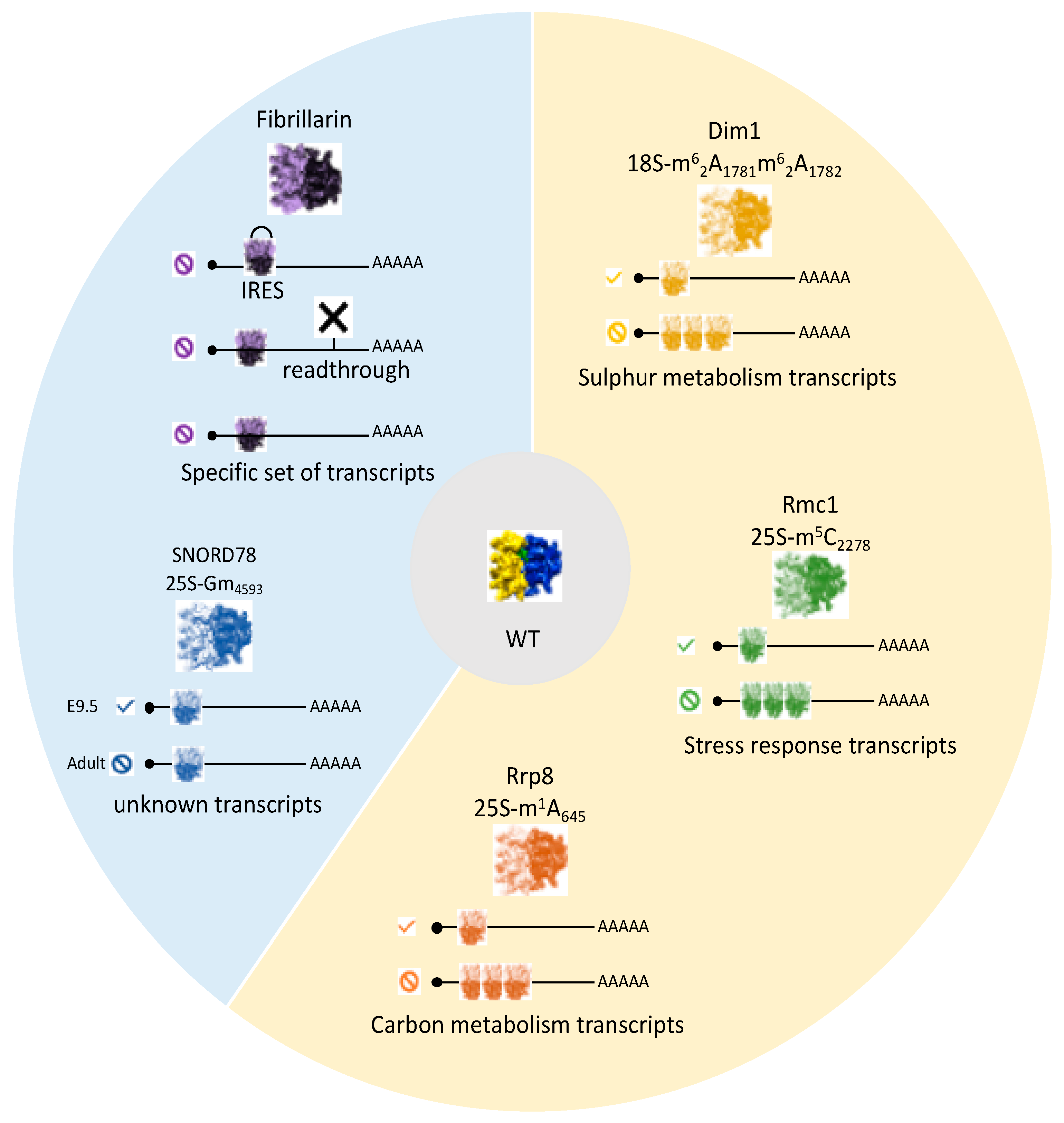
6. Specialized Ribosomes

Previous studies have demonstrated a link between the particular effects of certain modifications on precise aspects of translational accuracy and the heterogeneous nature of ribosome modification patterns. It is, therefore, of particular interest to determine whether this could influence translation of specific mRNAs. This issue was first addressed in yeast, in studies of lifespan and stress resistance. In a screen aiming to identify lifespan mediators, the methyltransferase Rcm1 was shown to be downregulated in chronologically aged cells. This enzyme drives 25S-m5C2278 cytosine methylation. These cellular processes are accompanied by a decrease in general protein synthesis, raising questions about the involvement of rRNA modification in aging. Indeed, RCM1 deletion completely abolish C2278 methylation, resulting in a longer lifespan and greater stress resistance after exposure to H2O2. An investigation of the pattern of mRNA recruitment to polysomes showed that the absence of m5C2278 results in a completely different set of mRNAs being translated relative to that translated by methylated ribosomes; more surprisingly, this set of mRNAs resembled the mRNAs of wild-type cells exposed to oxidative stress (Figure 3). The authors of the study concerned concluded that cells depleted of m5C2278 were in a pre-active state of responsiveness to translational stress that might account for their longer lifespan and greater stress resistance [62]. It remains unclear whether RCM1 activity is actually modulated in response to cellular signals. Epigenetic inactivation of the human counterpart of RCM1, NSUN5, has been observed in 38% of glioma-derived cell lines displaying the expected decreases in protein synthesis and, more surprisingly, a reprogramming towards stress proteins, enabling the tumor cells to cope with hostile environments [63]. The impact of a second stand-alone methyltransferase has also been investigated. Rrp8 directs the m1A645 modification in domain I of the 25S rRNA [64]. As expected, the loss of this modification affects the local structure of the 60S subunit, disturbing interactions with ribosomal proteins, especially eL32. -accordingly, polysome profiling shows halfmers typical of 43S initiation complexes awaiting the addition of the 60S subunit. The authors concluded that this observation reflected alterations to the recruitment of the 60S subunit, rather than an initiation defect per se. They explored the consequences of this defect for cellular mRNA translation, by performing two-dimensional gel analysis (DIGE) and mRNA determinations for proteins displaying changes in expression. Overall, 18 of the 1900 proteins detected displayed >1.5-fold differences in expression and clustered either with enzymes involved in carbon metabolism (with Sol3 protein regulated exclusively at the protein level) or with proteins related to translation itself. The authors concluded that this modification affected the translation of a subset of mRNAs with metabolic roles, either directly, or indirectly, due to the impact on components of the translational machinery [65]. In wild-type cells cultured in standard conditions, the nucleotide displays almost 100% modification. No conditions altering this modification have ever been found, raising questions about the potential regulation of this modification. However, certain diseases, such as ribosomopathies, may be partly explained by the impact of the lost modifications on the production on particular proteins. The concept of specialized ribosomes, differing in terms of modification status, was recently illustrated by a study of the rare dimethyl adenosine modification [66]. This modification, driven by the essential dimethylase Dim1 [67], concerns exclusively the two adjacent adenosines, A1781 and A1782, within the 18S rRNA. These residues are located in the decoding center and are universally conserved. The authors demonstrated the coexistence of a monomethylated m6A form within translating ribosomes, also under the control of Dim1, albeit present at only very low levels (4%). Interestingly, the monomethylation-to-dimethylation ratio changes specifically in conditions of sulfur deprivation (10% m6A in the polysomal fraction). This sensing of sulfur availability is mediated by Dim1 itself. Similar observations have been reported for cultured mammalian cells, indicating that this adaptation to sulfur deprivation is conserved among eukaryotes. The impact of the switch from m62A to m6A on mRNA translation has been investigated by ribosome profiling, a powerful method providing quantitative and qualitative information on translation at whole-cell level (for a review, see [68,69]). Using the D87E Dim1 mutant, which can tolerate 80% m6A modification with no effect on rRNA processing or subunit formation, the authors showed a significant and specific effect on the translation efficiency (TE) of 16 genes relative to WT (4% m6A); 12 of these genes are involved in sulfur metabolism. As expected, this differential behavior disappeared in conditions of sulfur deprivation. Based on these findings, the authors suggested that the modification status of the ribosome conferred specificity for decoding certain mRNAs closely linked to environmental signals. However, the molecular mechanism underlying this specificity remains unknown and ribosomal heterogeneity may also reflect the presence of different ribosomal proteins (Figure 3).
The notion of specialized ribosomes is not limited to yeast or unicellular organisms and seems to be generalizable to all organisms. In multicellular organisms, cells are generally less subject to environmental stresses. Conversely, in these organisms, protein synthesis must be adapted and synchronized to different tissues and developmental phases. For these reasons, it may be beneficial to have different populations of ribosomes, for the fine regulation and synchronization of translation according to the needs of the body.
In human cancer cells, which have high proliferation rates, a hyperactivation of ribosome biogenesis occurs, leading to high levels of protein synthesis. This is made possible, in part, by the deregulation of RNA polymerase I-mediated rRNA transcription due to a loss of p53 function [70]. Fibrillarin (FBL), the mammalian homolog of Nop1 and snoRNAs are also overexpressed in breast cancer [71]. FBL expression is also downregulated by p53 and p53 inactivation therefore leads to increases in fibrillarin activity and, thus, rRNA hypermethylation [72]. This modification pattern, in turn, alters the fidelity of translation, with more efficient IRES-dependent translation initiation for the growth factor IGF-1R. An FBL knockdown experiment was conducted to provide an additional demonstration of the link between rRNA methylation and the specificity of translation for particular mRNAs [73]. As expected, ribosome biogenesis was affected, but only mildly, as inactivation was achieved with an siRNA under controlled transfection conditions. Methylation status was investigated by ribomethseq, which revealed site-specific variations rather than a global effect, with the identification of partially methylated sites potentially subject to regulation. It was, therefore, possible to explore the specificity of such “variant” ribosomes by ribosome profiling. Translation was found to be up- or downregulated for a small set of mRNAs, 8% of which contained IRES. This category of mRNAs is enriched in genes implicated in tumorigenesis: oncogenes (p53, c-myc), growth factor receptors (IGF-1R, VGEF, FGF) or apoptosis modulators. Thus, in the context of cancer cells, fibrillarin levels and specific patterns of rRNA modification can modulate translation, thereby contributing to tumorigenesis (Figure 3).
Progress has been made with studies of methylation status based on ribomethseq on adult and embryonic tissues from mice. It was first established that the sites targeted for methylation in mouse rRNA have an equivalent in HeLa and HTC116 human cancer cell lines [31]. Conversely, in searches for sites modified in humans but not in the tissues of adult mice, the authors investigated the methylation status of A1310 and the presence in the genome of SNORD126, the corresponding guide snoRNA, in both mice and rats. They confirmed the absence of Am 1310 in mice, but found that this modification was tissue-specific in rats, due to the differential expression of SNORD126. The physiological consequences of this unexpected observation were not explored further. One third of human sites are partially methylated, but the vast majority of sites from the adult mouse tissues studied have methylation rates above 90%. Using this important observation as a starting point, the authors were able to detect decreases in methylation level. Indeed, embryonic tissues (E9.5 and E16.5) are characterized by a set of 59 sites with lower levels of methylation in at least one tissue. This elegant work then focused on Gm4593, which displays a change in methylation rate from 63% in E9.5 to 0% in adult brain tissue. The corresponding snoRNA, SNORD78, is hosted by intron 6 of the GAS5 gene, which contains eight other snoRNAs distributed between the other 10 introns. The authors showed that SNORD78 was no longer present in adult brain cells, a situation resembling that for rat SNORD126, except that the other eight SNORDs present within the RNA molecule were not affected. This discrepancy can be attributed to the alternative splicing of exon 7 in adult cells, resulting in defective SNOD78 processing. The biological purpose of this ribosome heterogeneity remains unclear, but this work highlights the modulation of rRNA methylation during developmental process (Figure 3).

7. Perspectives

During the last decade, the progress in our knowledge of rRNA modifications has been overwhelming. Despite the lack of ribosome structures at a sufficiently high resolution for the direct visualization of modifications in many cases, advances in our ability to map and quantify precisely major rRNA modifications have proved a real breakthrough, leading to the emergence of the concept of “specialized ribosomes”. This notion of “specialized ribosomes” is sometimes difficult to impose on the human context, due to difficulties demonstrating the specificity of ribosome action, but also because, in most cases, studies are performed in the context of disease. Thanks to the co-evolution of guide snoRNAs and ribosomes, S. cerevisiae constitutes an ideal model for addressing these questions more efficiently, in a much simpler system than human cells. Indeed, it remains difficult to perform systematic position-by-position studies of each modification in humans, but approaches of this kind are entirely feasible in yeast. Moreover, as yeasts are subject to highly fluctuating environments, it would be interesting to study the relationship between the pattern of modification and the response to environmental stress, which would undoubtedly provide important clues to the role of ribosome modifications in humans.

Author Contributions

Writing original draft, A.B.-B.; funding acquisition, O.N. All authors have read and agreed to the published version of the manuscript.

Funding

ANR Rescue ribosome program (17-CE12-0024-01) and ActiMeth (ANR-19-CE12-0004).

Acknowledgments

The English of this manuscript was corrected by Alex Edelman & Associates. We acknowledge support from the ANR Rescue_ribosome program (17-CE12-0024-01) and ActiMeth (ANR-19-CE12-0004).

Conflicts of Interest

The authors declare no conflict of interest.

References

  1. Woolford, J.L.; Baserga, S.J. Ribosome Biogenesis in the Yeast Saccharomyces cerevisiae. Genetics 2013, 195, 643–681. [Google Scholar] [CrossRef] [Green Version]
  2. Bachellerie, J.P.; Michot, B.; Nicoloso, M.; Balakin, A.; Ni, J.; Fournier, M.J. Antisense snoRNAs: A family of nucleolar RNAs with long complementarities to rRNA. Trends Biochem. Sci. 1995, 20, 261–264. [Google Scholar] [CrossRef]
  3. Maxwell, E.S.; Fournier, M.J. The Small Nucleolar RNAs. Annu. Rev. Biochem. 1995, 64, 897–934. [Google Scholar] [CrossRef]
  4. Cavaille, J.; Nicoloso, M.; Bachellerie, J.P. Targeted ribose methylation of RNA in vivo directed by tailored an-tisense RNA guides. Nature 1996, 383, 732–735. [Google Scholar] [CrossRef] [PubMed]
  5. Kiss-László, Z.; Henry, Y.; Bachellerie, J.-P.; Caizergues-Ferrer, M.; Kiss, T. Site-Specific Ribose Methylation of Preribosomal RNA: A Novel Function for Small Nucleolar RNAs. Cell 1996, 85, 1077–1088. [Google Scholar] [CrossRef] [Green Version]
  6. Bortolin, M.; Ganot, P.; Kiss, T. Elements essential for accumulation and function of small nucleolar RNAs directing site-specific pseudouridylation of ribosomal RNAs. EMBO J. 1999, 18, 457–469. [Google Scholar] [CrossRef] [PubMed] [Green Version]
  7. Ganot, P.; Bortolin, M.-L.; Kiss, T. Site-Specific Pseudouridine Formation in Preribosomal RNA Is Guided by Small Nucleolar RNAs. Cell 1997, 89, 799–809. [Google Scholar] [CrossRef] [Green Version]
  8. Balakin, A.G.; Smith, L.; Fournier, M.J. The RNA world of the nucleolus: Two major families of small RNAs defined by different box elements with related functions. Cell 1996, 86, 823–834. [Google Scholar] [CrossRef] [Green Version]
  9. Chagot, M.-E.; Quinternet, M.; Rothé, B.; Charpentier, B.; Coutant, J.; Manival, X.; Lebars, I. The yeast C/D box snoRNA U14 adopts a “weak” K-turn like conformation recognized by the Snu13 core protein in solution. Biochimie 2019, 164, 70–82. [Google Scholar] [CrossRef]
  10. Sharma, S.; Yang, J.; Van Nues, R.; Watzinger, P.; Kötter, P.; Lafontaine, D.; Granneman, S.; Entian, K.-D. Specialized box C/D snoRNPs act as antisense guides to target RNA base acetylation. PLoS Genet. 2017, 13, e1006804. [Google Scholar] [CrossRef]
  11. Piekna-Przybylska, D.; Decatur, W.A.; Fournier, M.J. New bioinformatic tools for analysis of nucleotide modifications in eukaryotic rRNA. RNA 2007, 13, 305–312. [Google Scholar] [CrossRef] [Green Version]
  12. Paul, A.; Tiotiu, D.; Bragantini, B.; Marty, H.; Charpentier, B.; Massenet, S.; Labialle, S. Bcd1p controls RNA loading of the core protein Nop58 during C/D box snoRNP biogenesis. RNA 2019, 25, 496–506. [Google Scholar] [CrossRef]
  13. Grzechnik, P.; Szczepaniak, S.A.; Dhir, S.; Pastucha, A.; Parslow, H.; Matuszek, Z.; Mischo, H.E.; Kufel, J.; Proudfoot, N.J. Nuclear fate of yeast snoRNA is determined by co-transcriptional Rnt1 cleavage. Nat. Commun. 2018, 9, 1–14. [Google Scholar] [CrossRef] [Green Version]
  14. Massenet, S.; Bertrand, E.; Verheggen, C. Assembly and trafficking of box C/D and H/ACA snoRNPs. RNA Biol. 2017, 14, 680–692. [Google Scholar] [CrossRef] [Green Version]
  15. Bonnerot, C.; Pintard, L.; Lutfalla, G. Functional Redundancy of Spb1p and a snR52-Dependent Mechanism for the 2′-O-Ribose Methylation of a Conserved rRNA Position in Yeast. Mol. Cell 2003, 12, 1309–1315. [Google Scholar] [CrossRef]
  16. Schmitt, H.G. Granulation tumor in the horse following barbed wire injury: X-ray therapy. Tierarztl. Prax. 1977, 5, 219–221. [Google Scholar]
  17. Ni, J.; Samarsky, D.; Liu, B.; Ferbeyre, G.; Cedergren, R.; Fournier, M.J. SnoRNAs as tools for RNA cleavage and modification. Nucleic Acids Symp. Ser. 1997, 36, 61–63. [Google Scholar]
  18. Liu, B.; Ni, J.; Fournier, M.J. Probing RNA in vivo with methylation guide small nucleolar RNAs. Methods 2001, 23, 276–286. [Google Scholar] [CrossRef]
  19. Liu, B.; Fournier, M.J. Interference probing of rRNA with snoRNPs: A novel approach for functional mapping of RNA in vivo. RNA 2004, 10, 1130–1141. [Google Scholar] [CrossRef] [PubMed] [Green Version]
  20. Liu, B.; Liang, X.-H.; Piekna-Przybylska, R.; Liu, Q.; Fournier, M.J. Mis-targeted methylation in rRNA can severely impair ribosome synthesis and activity. RNA Biol. 2008, 5, 249–254. [Google Scholar] [CrossRef] [PubMed] [Green Version]
  21. Karijolich, J.; Yu, Y.-T. Converting nonsense codons into sense codons by targeted pseudouridylation. Nature 2011, 474, 395–398. [Google Scholar] [CrossRef] [Green Version]
  22. Keith, G. Mobilities of modified ribonucleotides on two-dimensional cellulose thin-layer chromatography. Biochimie 1995, 77, 142–144. [Google Scholar] [CrossRef]
  23. Sloan, K.E.; Warda, A.S.; Sharma, S.; Entian, K.-D.; Lafontaine, D.; Bohnsack, M.T. Tuning the ribosome: The influence of rRNA modification on eukaryotic ribosome biogenesis and function. RNA Biol. 2017, 14, 1138–1152. [Google Scholar] [CrossRef]
  24. Su, D.; Chan, C.T.Y.; Gu, C.; Lim, K.S.; Chionh, Y.H.; McBee, M.E.; Russell, B.S.; Babu, I.R.; Begley, T.J.; Dedon, P.C. Quantitative analysis of ribonucleoside modifications in tRNA by HPLC-coupled mass spectrometry. Nat. Protoc. 2014, 9, 828–841. [Google Scholar] [CrossRef] [PubMed]
  25. Wang, Y.; Jia, G. Detection methods of epitranscriptomic mark N6-methyladenosine. Essays Biochem. 2020, 64, 967–979. [Google Scholar] [PubMed]
  26. Marchand, V.; Pichot, F.; Neybecker, P.; Ayadi, L.; Bourguignon-Igel, V.; Wacheul, L.; Lafontaine, D.L.J.; Pinzano, A.; Helm, M.; Motorin, Y. HydraPsiSeq: A method for systematic and quantitative mapping of pseudouridines in RNA. Nucleic Acids Res. 2020, 48, e110. [Google Scholar] [CrossRef]
  27. Krogh, N.; Nielsen, H. Sequencing-based methods for detection and quantitation of ribose methylations in RNA. Methods 2019, 156, 5–15. [Google Scholar] [CrossRef] [PubMed]
  28. Angelova, M.T.; Dimitrova, D.G.; Da Silva, B.; Marchand, V.; Jacquier, C.; Achour, C.; Brazane, M.; Goyenvalle, C.; Bourguignon-Igel, V.; Shehzada, S.; et al. tRNA 2′-O-methylation by a duo of TRM7/FTSJ1 proteins modulates small RNA silencing in Drosophila. Nucleic Acids Res. 2020, 48, 2050–2072. [Google Scholar] [CrossRef] [Green Version]
  29. Marchand, V.; Pichot, F.; Thuring, K.; Ayadi, L.; Freund, I.; Dalpke, A.; Helm, M.; Motorin, Y. Next-Generation Sequencing-Based RiboMethSeq Protocol for Analysis of tRNA 2′-O-Methylation. Biomolecules 2017, 7, 13. [Google Scholar] [CrossRef] [PubMed] [Green Version]
  30. Marchand, V.; Blanloeil-Oillo, F.; Helm, M.; Motorin, Y. Illumina-based RiboMethSeq approach for mapping of 2′-O-Me residues in RNA. Nucleic Acids Res. 2016, 44, e135. [Google Scholar] [CrossRef] [Green Version]
  31. Krogh, N.; Jansson, M.; Häfner, S.; Tehler, D.E.; Birkedal, U.; Christensen-Dalsgaard, M.; Lund, A.H.; Nielsen, H. Profiling of 2′-O-Me in human rRNA reveals a subset of fractionally modified positions and provides evidence for ribosome heterogeneity. Nucleic Acids Res. 2016, 44, 7884–7895. [Google Scholar] [CrossRef] [Green Version]
  32. Marchand, V.; Ayadi, L.; Ernst, F.G.M.; Hertler, J.; Bourguignon-Igel, V.; Galvanin, A.; Kotter, A.; Helm, M.; Lafontaine, D.L.J.; Motorin, Y. AlkAniline-Seq: Profiling of m(7) G and m(3) C RNA Modifications at Single Nucleotide Resolution. Angew. Chem. 2018, 57, 16785–16790. [Google Scholar] [CrossRef]
  33. Zheng, H.-X.; Zhang, X.-S.; Sui, N. Advances in the profiling of N6-methyladenosine (m6A) modifications. Biotechnol. Adv. 2020, 45, 107656. [Google Scholar] [CrossRef]
  34. Liu, H.; Begik, O.; Lucas, M.C.; Ramírez, J.M.; Mason, C.E.; Wiener, D.; Schwartz, S.; Mattick, J.; Smith, M.A.; Novoa, E.M. Accurate detection of m6A RNA modifications in native RNA sequences. Nat. Commun. 2019, 10, 1–9. [Google Scholar] [CrossRef] [Green Version]
  35. Smith, A.M.; Jain, M.; Mulroney, L.; Garalde, D.R.; Akeson, M. Reading canonical and modified nucleobases in 16S ribosomal RNA using nanopore native RNA sequencing. PLoS ONE 2019, 14, e0216709. [Google Scholar] [CrossRef] [Green Version]
  36. Jain, M.; Olsen, H.; Akeson, M.; Abu-Shumays, R. Adaptation of Human Ribosomal RNA for Nanopore Sequencing of Canonical and Modified Nucleotides. In RNA Modifications; Humana: New York, NY, USA, 2021; pp. 53–74. [Google Scholar]
  37. Jenjaroenpun, P.; Wongsurawat, T.; Wadley, T.D.; Wassenaar, T.M.; Liu, J.; Dai, Q.; Wanchai, V.; Akel, N.S.; Jamshidi-Parsian, A.; Franco, A.T.; et al. Decoding the epitranscriptional landscape from native RNA sequences. Nucleic Acids Res. 2021, 49, e7. [Google Scholar] [CrossRef] [PubMed]
  38. Charette, M.; Gray, M.W. Pseudouridine in RNA: What, where, how, and why. IUBMB Life 2000, 49, 341–351. [Google Scholar]
  39. Assi, H.A.; Rangadurai, A.K.; Shi, H.; Liu, B.; Clay, M.C.; Erharter, K.; Kreutz, C.; Holley, C.L.; Al-Hashimi, H.M. 2′-O-Methylation can increase the abundance and lifetime of alternative RNA conformational states. Nucleic Acids Res. 2020, 48, 12365–12379. [Google Scholar] [PubMed]
  40. Elliott, B.A.; Ho, H.-T.; Ranganathan, S.V.; Vangaveti, S.; Ilkayeva, O.; Assi, H.A.; Choi, A.K.; Agris, P.F.; Holley, C.L. Modification of messenger RNA by 2′-O-methylation regulates gene expression in vivo. Nat. Commun. 2019, 10, 1–9. [Google Scholar]
  41. Decatur, W.; Fournier, M.J. rRNA modifications and ribosome function. Trends Biochem. Sci. 2002, 27, 344–351. [Google Scholar] [CrossRef]
  42. Tollervey, D.; Lehtonen, H.; Jansen, R.; Kern, H.; Hurt, E.C. Temperature-sensitive mutations demonstrate roles for yeast fibrillarin in pre-rRNA processing, pre-rRNA methylation, and ribosome assembly. Cell 1993, 72, 443–457. [Google Scholar] [CrossRef]
  43. Ojha, S.; Malla, S.; Lyons, S.M. snoRNPs: Functions in Ribosome Biogenesis. Biomolecules 2020, 10, 783. [Google Scholar] [CrossRef]
  44. Griffiths, C.J.; Cooper, B.; Gibson, G.J. A video system for investigating breathing disorders during sleep. Thorax 1991, 46, 136–140. [Google Scholar] [CrossRef] [Green Version]
  45. Ma, X.; Zhao, X.; Yu, Y. Pseudouridylation (Ψ) of U2 snRNA in S.cerevisiae is catalyzed by an RNA-independent mechanism. EMBO J. 2003, 22, 1889–1897. [Google Scholar] [CrossRef] [Green Version]
  46. Behm-Ansmant, I.; Urban, A.; Ma, X.; Yu, Y.T.; Motorin, Y.; Branlant, C. The Saccharomyces cerevisiae U2 snRNA: Pseudouridine-synthase Pus7p is a novel multisite-multisubstrate RNA:Psi-synthase also acting on tRNAs. RNA 2003, 9, 1371–1382. [Google Scholar] [CrossRef] [Green Version]
  47. Schwartz, S.; Bernstein, D.A.; Mumbach, M.R.; Jovanovic, M.; Herbst, R.H.; Leon-Ricardo, B.X.; Engreitz, J.M.; Guttman, M.; Satija, R.; Lander, E.S.; et al. Transcriptome-wide mapping reveals widespread dynamic-regulated pseudouri-dylation of ncRNA and mRNA. Cell 2014, 159, 148–162. [Google Scholar] [CrossRef] [Green Version]
  48. Carlile, T.; Rojas-Duran, M.F.; Zinshteyn, B.; Shin, H.; Bartoli, K.M.; Gilbert, W.V. Pseudouridine profiling reveals regulated mRNA pseudouridylation in yeast and human cells. Nature 2014, 515, 143–146. [Google Scholar] [CrossRef] [PubMed] [Green Version]
  49. Lowe, T.M.; Eddy, S.R. A computational screen for methylation guide snoRNAs in yeast. Science 1999, 283, 1168–1171. [Google Scholar] [CrossRef] [PubMed] [Green Version]
  50. Qu, L.-H.; Henras, A.; Lu, Y.-J.; Zhou, H.; Zhou, W.-X.; Zhu, Y.-Q.; Zhao, J.; Henry, Y.; Caizergues-Ferrer, M.; Bachellerie, J.-P. Seven Novel Methylation Guide Small Nucleolar RNAs Are Processed from a Common Polycistronic Transcript by Rat1p and RNase III in Yeast. Mol. Cell. Biol. 1999, 19, 1144–1158. [Google Scholar] [CrossRef] [PubMed] [Green Version]
  51. Baxter-Roshek, J.L.; Petrov, A.N.; Dinman, J.D. Optimization of Ribosome Structure and Function by rRNA Base Modification. PLoS ONE 2007, 2, e174. [Google Scholar] [CrossRef] [PubMed]
  52. Zebarjadian, Y.; King, T.; Fournier, M.J.; Clarke, L.; Carbon, J. Point Mutations in Yeast CBF5 Can Abolish In Vivo Pseudouridylation of rRNA. Mol. Cell. Biol. 1999, 19, 7461–7472. [Google Scholar] [CrossRef] [Green Version]
  53. Jack, K.; Bellodi, C.; Landry, D.M.; Niederer, R.; Meskauskas, A.; Musalgaonkar, S.; Kopmar, N.; Krasnykh, O.; Dean, A.M.; Thompson, S.; et al. rRNA Pseudouridylation Defects Affect Ribosomal Ligand Binding and Translational Fidelity from Yeast to Human Cells. Mol. Cell 2011, 44, 660–666. [Google Scholar] [CrossRef] [Green Version]
  54. Esguerra, J.; Warringer, J.; Blomberg, A. Functional importance of individual rRNA 2′-O-ribose methylations revealed by high-resolution phenotyping. RNA 2008, 14, 649–656. [Google Scholar] [CrossRef] [Green Version]
  55. King, T.H.; Liu, B.; McCully, R.R.; Fournier, M.J. Ribosome Structure and Activity Are Altered in Cells Lacking snoRNPs that Form Pseudouridines in the Peptidyl Transferase Center. Mol. Cell 2003, 11, 425–435. [Google Scholar] [CrossRef]
  56. Hirabayashi, N.; Sato, N.S.; Suzuki, T. Conserved Loop Sequence of Helix 69 in Escherichia coli 23 S rRNA Is Involved in A-site tRNA Binding and Translational Fidelity. J. Biol. Chem. 2006, 281, 17203–17211. [Google Scholar] [CrossRef] [Green Version]
  57. Liang, X.-H.; Liu, Q.; Fournier, M.J. rRNA Modifications in an Intersubunit Bridge of the Ribosome Strongly Affect Both Ribosome Biogenesis and Activity. Mol. Cell 2007, 28, 965–977. [Google Scholar] [CrossRef]
  58. Baudin-Baillieu, A.; Fabret, C.; Liang, X.-H.; Piekna-Przybylska, D.; Fournier, M.J.; Rousset, J.-P. Nucleotide modifications in three functionally important regions of the Saccharomyces cerevisiae ribosome affect translation accuracy. Nucleic Acids Res. 2009, 37, 7665–7677. [Google Scholar] [CrossRef] [PubMed] [Green Version]
  59. Piekna-Przybylska, D.; Przybylski, P.; Baudin-Baillieu, A.; Rousset, J.-P.; Fournier, M.J. Ribosome Performance Is Enhanced by a Rich Cluster of Pseudouridines in the A-site Finger Region of the Large Subunit. J. Biol. Chem. 2008, 283, 26026–26036. [Google Scholar] [CrossRef] [PubMed] [Green Version]
  60. Liang, X.H.; Liu, Q.; Fournier, M.J. Loss of rRNA modifications in the decoding center of the ribosome impairs translation and strongly delays pre-rRNA processing. RNA 2009, 15, 1716–1728. [Google Scholar] [CrossRef] [PubMed] [Green Version]
  61. Gigova, A.; Duggimpudi, S.; Pollex, T.; Schaefer, M.; Kos, M. A cluster of methylations in the domain IV of 25S rRNA is required for ribosome stability. RNA 2014, 20, 1632–1644. [Google Scholar] [CrossRef] [Green Version]
  62. Schosserer, M.; Minois, N.; Angerer, T.B.; Amring, M.; Dellago, H.; Harreither, E.; Calle-Perez, A.; Pircher, A.; Gerstl, M.P.; Pfeifenberger, S.; et al. Methylation of ribosomal RNA by NSUN5 is a conserved mechanism modulating organismal lifespan. Nat. Commun. 2015, 6, 6158. [Google Scholar] [CrossRef] [PubMed] [Green Version]
  63. Janin, M.; Ortiz-Barahona, V.; de Moura, M.C.; Martinez-Cardus, A.; Llinas-Arias, P.; Soler, M.; Nachmani, D.; Pelletier, J.; Schumann, U.; Calleja-Cervantes, M.E.; et al. Epigenetic loss of RNA-methyltransferase NSUN5 in glioma targets ribosomes to drive a stress adaptive translational program. Acta Neuropathol. 2019, 138, 1053–1074. [Google Scholar] [CrossRef] [PubMed] [Green Version]
  64. Peifer, C.; Sharma, S.; Watzinger, P.; Lamberth, S.; Kotter, P.; Entian, K.D. Yeast Rrp8p, a novel méthyltransférase responsible for m1A 645 base modification of 25S rRNA. Nucleic Acids Res. 2013, 41, 1151–1163. [Google Scholar] [CrossRef] [PubMed] [Green Version]
  65. Sharma, S.; Hartmann, J.D.; Watzinger, P.; Klepper, A.; Peifer, C.; Kötter, P.; Lafontaine, D.L.J.; Entian, K.-D. A single N1-methyladenosine on the large ribosomal subunit rRNA impacts locally its structure and the translation of key metabolic enzymes. Sci. Rep. 2018, 8, 11904. [Google Scholar] [CrossRef] [PubMed]
  66. Liu, K.; Santos, D.A.; Hussmann, J.A.; Wang, Y.; Sutter, B.M.; Weissman, J.S.; Tu, B.P. Regulation of translation by methylation multiplicity of 18S rRNA. Cell Rep. 2021, 34, 108825. [Google Scholar] [CrossRef] [PubMed]
  67. Lafontaine, D.; Delcour, J.; Glasser, A.L.; Desgres, J.; Vandenhaute, J. The DIM1 gene responsible for the conserved m6(2)Am6(2)A dimethylation in the 3′-terminal loop of 18 S rRNA is essential in yeast. J. Mol. Biol. 1994, 241, 492–497. [Google Scholar] [CrossRef]
  68. Ingolia, N.T.; Hussmann, J.A.; Weissman, J.S. Ribosome Profiling: Global Views of Translation. Cold Spring Harb. Perspect. Biol. 2019, 11, a032698. [Google Scholar] [CrossRef]
  69. Baudin-Baillieu, A.; Hatin, I.; Legendre, R.; Namy, O. Translation Analysis at the Genome Scale by Ribosome Profiling. Methods Mol. Biol. 2016, 1361, 105–124. [Google Scholar]
  70. Bywater, M.J.; Poortinga, G.; Sanij, E.; Hein, N.; Peck, A.; Cullinane, C.; Wall, M.; Cluse, L.; Drygin, D.; Anderes, K.; et al. Inhibition of RNA Polymerase I as a Therapeutic Strategy to Promote Cancer-Specific Activation of p53. Cancer Cell 2012, 22, 51–65. [Google Scholar] [CrossRef] [Green Version]
  71. Su, H.; Xu, T.; Ganapathy, S.; Shadfan, M.; Long, M.; Huang, T.H.-M.; Thompson, I.; Yuan, Z.-M. Elevated snoRNA biogenesis is essential in breast cancer. Oncogene 2013, 33, 1348–1358. [Google Scholar] [CrossRef]
  72. Marcel, V.; Ghayad, S.E.; Belin, S.; Therizols, G.; Morel, A.-P.; Solano-Gonzàlez, E.; Vendrell, J.A.; Hacot, S.; Mertani, H.C.; Albaret, M.A.; et al. p53 Acts as a Safeguard of Translational Control by Regulating Fibrillarin and rRNA Methylation in Cancer. Cancer Cell 2013, 24, 318–330. [Google Scholar] [CrossRef] [PubMed] [Green Version]
  73. Erales, J.; Marchand, V.; Panthu, B.; Gillot, S.; Belin, S.; Ghayad, S.E.; Garcia, M.; Laforets, F.; Marcel, V.; Baudin-Baillieu, A.; et al. Evidence for rRNA 2′-O-methylation plasticity: Control of intrinsic translational capabilities of human ribosomes. Proc. Natl. Acad. Sci. USA 2017, 114, 12934–12939. [Google Scholar] [CrossRef] [Green Version]
Figure 1. Structures of box H/ACA and box C/D snoRNP. (A) Diagram of a typical box H/ACA snoRNP. The target rRNA is in bold and is bound to complementary regions of the H/ACA guide snoRNA. The uridine residues converted into pseudouridine lie within a bulge known as the pseudouridine pocket and are indicated (Ψ). The two H and ACA sequences are boxed. Associated proteins are shown in color. (B) Diagram of a typical box C/D snoRNP. Two target rRNA regions are represented in bold and are bound to complementary regions of the C/D guide snoRNA. The nucleosides methylated lie 5 nucleotides upstream from a D or D′ box and are indicated (-CH3). The C and C′ boxes are highlighted in red and the D and D′ boxes are highlighted in blue. The associated proteins are shown in color.
Ijms 22 07419 g001
Figure 2. Localization of rRNA modifications on the ribosome. The modified positions are indicated by spheres on the structure of the yeast ribosome (PDB: 1VXX). The P-site tRNA is indicated in black, the E-site tRNA is shown in dark gray and the mRNA is indicated in dark red. Each modified nucleotide is shown in color according to its position with respect to the functional regions of the ribosome (green: PTC; red: A-site; cyan: peptide-exit channel; blue: P-site; purple: E-site, pink: A-site finger; yellow: inter-subunit bridges). Positions lying outside the functional regions are shown in gray. Blue arrow represents a rotation by 90°.
Ijms 22 07419 g002
Figure 3. Models of specialized ribosomes in yeast and mammalian cells. The yellow part of the diagram illustrates examples related to yeast and blue part illustrates those related to human or mouse cells. (🛇) absence or (✓) presence of the modification concerned.
Ijms 22 07419 g003
Table 1. Listing of the rRNA modifications present in Saccharomyces cerevisiae. The modification type, position and the snoRNA or enzyme responsible are given, together with the modification rate, conservation between species (H for Human and Ec for Echerichia coli) and location within functional or structural domains of the ribosome.
ModificationPositionEnzyme/snoRNAModification RateConservationFunctional Domain
Ψ5S-50Pus7>85%H
Ψ5,8S-73snR43<85%
Am18S-28snR74>85%H
Am18S-100snR51<85%H
Ψ18S-106snR44>85%H
Ψ18S-120snR49>85%H
Ψ18S-211snR49<85%
Ψ18S-302snR49>85%
Cm18S-414U14>85%H
Am18S-420snR52>85%H
Am18S-436snR87>85% dH
Ψ18S-466snR189<85%
Am18S-541snR41>85%H
Gm18S-562snR40>85% d
Um18S-578snR77>85%HDC-A site
Am18S-619snR47>85%H
Ψ18S-632snR161<85%H
Ψ18S-759snR80>85%H
Ψ18S-766snR161>85%H
Am18S-796snR53>85%
Am18S-974snR54>85%H
Ψ18S-999snR31<85%HIS bridge DC-E site
Cm18S-1007snR79>85% IS bridge DC-E site
Gm18S-1126snR41>85% IS bridge
Ψ18S-1181snR85>85%HIS bridge
Ψ18S-1187snR36>85%HDC-A site
m1acp3Ψ18S-1191snR35,Emg1, Tsr3>85%HDC-P site
Um18S-1269snR55>85%HDC-A site
Gm18S-1271snR40>85%HDC-A site
ac4C18S-1280snR4, Kre33>85%HDC-A site
Ψ18S-1290snR83>85%H
Ψ18S-1415snR83<85%
Gm18S-1428snR56>85%HDC-A site
Gm18S-1572snR57>85% E-site
m7G18S-1575Bud23>85% dH
Cm18S-1639snR70>85% dEc, HDC-P site
ac4C18S-1773snR45, Kre33>85%HIS bridge
m26A18S-1781Dim1>85%Ec, HIS bridge
m26A18S-1782Dim1>85%Ec, HIS bridge
m1A25S-645Bmt1 (Rrp8)>85%HPTC
Am25S-649U18>85%HPeptide exit tunnel
Cm25S-650U18>85% Peptide exit tunnel
Cm25S-663snR58<85% dHPeptide exit tunnel
Ψ25S-776snR80>85%
Gm25S-805snR39b>85%HPeptide exit tunnel
Am25S-807snR39, snR59>85%H
Am25S-817snR60>85%H
Gm25S-867snR50>85% d
Am25S-876snR72>85% d
Um25S-898snR40>85% d
Gm25S-908snR60>85%H
Ψ25S-960snR8>85%HA site finger helix 37
Ψ25S-966snR43>85%HA site finger helix 37
Ψ25S-986snR8>85% A site finger helix 38
Ψ25S-990snR49>85% A site finger helix 38
Ψ25S-1004snR5<85%HA site finger helix 38
Ψ25S-1042snR33>85%HA site finger helix 38
Ψ25S-1052snR81>85%HA site finger helix 38
Ψ25S-1056snR44>85% A site finger helix 38
Ψ25S-1110snR82>85%
Ψ25S-1124snR5>85%HA site finger helix 39
Am25S-1133snR61>85%HA site finger helix 39
Cm25S-1437U24>85%HPeptide exit tunnel
Am25S-1449U24>85% dH
Gm25S-1450U24>85%H
Um25S-1888snR62>85%H
Ψ25S-2129snR5>85%
Ψ25S-2133snR3>85%H
m1A25S-2142Bmt2>85% Subunit surface
Ψ25S-2191snR32>85%H
Cm25S-2197snR76>85% dHE-site
Am25S-2220snR47>85%HE-site
Am25S-2256snR63>85%HIS bridge helix 69 A site tRNA
Ψ25S-2258snR191>85%Ec, HIS bridge helix 69 A site tRNA
Ψ25S-2260snR191>85%Ec, HIS bridge helix 69
Ψ25S-2264snR3>85%HIS bridge helix 69
Ψ25S-2266snR84>85%HIS bridge helix 69 P-site tRNA
m5C25S-2278Bmt3 (Rcm1)>85%HIS bridge
Am25S-2280snR13>85% IS bridge
Am25S-2281snR13>85%HIS bridge
Gm25S-2288snR75>85%HIS bridge
Ψ25S-2314snR86>85%H
Cm25S-2337snR64>85%H
Ψ25S-2340snR9>85%H
Um, Ψ, Ψm25S-2347snR65, snR9 H
Ψ25S-2349snR82>85%H
Ψ25S-2351snR82>85%
Ψ25S-2416snR11>85%H
Um25S-2417snR66>85%
Um25S-2421snR78>85%HE-site
Gm25S-2619snR67>85%Ec, HP-site
m3U25S-2634Bmt5>85% PTC
Am25S-2640snR68>85%
Um25S-2724snR67>85%
Um25S-2729snR51>85% dH
Ψ25S-2735snR189>85%H
Gm25S-2791snR48>85% E-site
Gm25S-2793snR48>85%HE-site
Gm25S-2815snR38>85%HPTC
Ψ25S-2826snR34>85%HPTC
m3U25S-2843Bmt6>85%H
Ψ25S-2865snR46>85%HPTC
m5C25S-2870Bmt4 (Nop2)>85%Ec, HPTC
Ψ25S-2880snR34>85%HPTC
Um25S-2921snR52, Spb1>85%Ec, HPTC
Gm25S-2922Spb1>85%HPTC
Ψ25S-2923snR10>85%HPTC
Ψ25S-2944snR37<85%HPTC
Am25S-2946snR71>85%HPTC
Cm25S-2948snR69>85% d PTC
Cm25S-2959snR73>85%HPTC
Ψ25S-2975snR42<85%HPTC
d: discrepancy, with different values in different publications.
Publisher’s Note: MDPI stays neutral with regard to jurisdictional claims in published maps and institutional affiliations.

