Review

Elucidating the Neuroprotective Role of PPARs in Parkinson’s Disease: A Neoteric and Prospective Target

1
Chitkara College of Pharmacy, Chitkara University, Punjab 140401, India
2
Natural & Medical Sciences Research Centre, University of Nizwa, Birkat Al Mauz 616, Nizwa P.O. Box 33, Oman
3
School of Health Science, University of Petroleum and Energy Studies, Dehradun 248007, India
4
Department of Medicinal Chemistry and Pharmacognosy, College of Pharmacy, Qassim University, Buraydah 52571, Saudi Arabia
5
Translational and Clinical Research Institute, Newcastle University, Newcastle-upon-Tyne NE1 7RU, UK
6
Department of Pharmacy, Faculty of Medicine and Pharmacy, University of Oradea, 410028 Oradea, Romania
7
Doctoral School of Biological and Biomedical Sciences, University of Oradea, 410073 Oradea, Romania
*
Authors to whom correspondence should be addressed.
Int. J. Mol. Sci. 2021, 22(18), 10161; https://0-doi-org.brum.beds.ac.uk/10.3390/ijms221810161
Submission received: 29 August 2021 / Revised: 17 September 2021 / Accepted: 19 September 2021 / Published: 21 September 2021
(This article belongs to the Special Issue Neuroprotection: Rescue from Neuronal Death in the Brain 2.0)

Abstract

:
One of the utmost frequently emerging neurodegenerative diseases, Parkinson’s disease (PD) must be comprehended through the forfeit of dopamine (DA)-generating nerve cells in the substantia nigra pars compacta (SN-PC). The etiology and pathogenesis underlying the emergence of PD is still obscure. However, expanding corroboration encourages the involvement of genetic and environmental factors in the etiology of PD. The destruction of numerous cellular components, namely oxidative stress, ubiquitin-proteasome system (UPS) dysfunction, autophagy-lysosome system dysfunction, neuroinflammation and programmed cell death, and mitochondrial dysfunction partake in the pathogenesis of PD. Present-day pharmacotherapy can alleviate the manifestations, but no therapy has been demonstrated to cease disease progression. Peroxisome proliferator-activated receptors (PPARs) are ligand-directed transcription factors pertaining to the class of nuclear hormone receptors (NHR), and are implicated in the modulation of mitochondrial operation, inflammation, wound healing, redox equilibrium, and metabolism of blood sugar and lipids. Numerous PPAR agonists have been recognized to safeguard nerve cells from oxidative destruction, inflammation, and programmed cell death in PD and other neurodegenerative diseases. Additionally, various investigations suggest that regular administration of PPAR-activating non-steroidal anti-inflammatory drugs (NSAIDs) (ibuprofen, indomethacin), and leukotriene receptor antagonists (montelukast) were related to the de-escalated evolution of neurodegenerative diseases. The present review elucidates the emerging evidence enlightening the neuroprotective outcomes of PPAR agonists in in vivo and in vitro models experiencing PD. Existing articles up to the present were procured through PubMed, MEDLINE, etc., utilizing specific keywords spotlighted in this review. Furthermore, the authors aim to provide insight into the neuroprotective actions of PPAR agonists by outlining the pharmacological mechanism. As a conclusion, PPAR agonists exhibit neuroprotection through modulating the expression of a group of genes implicated in cellular survival pathways, and may be a propitious target in the therapy of incapacitating neurodegenerative diseases like PD.

1. Introduction

Parkinson’s disease (PD) is a common, intricate, progressive, multifaceted, and debilitating neurodegenerative disease, which is portrayed by the forfeiture of dopamine (DA) generating nerve cells in the substantia nigra pars compacta (SN-PC). Moreover, a pathogenic feature of PD is the accumulation of protein named α-synuclein in Lewy bodies (LBs) and Lewy neurites pinpointed within the nerve cells [1]. Tremor, bradykinesia, rigor, and postural abnormalities emerge as an integral manifestation associated with PD [2]. In those under the age of 40, PD is exceedingly rare, but it affects nearly 1–2% of people over 60–65 years of age and presents a comparative higher risk of developing PD in people beyond 85 years of age worldwide [3]. The incidence of PD differs among genders, with women exhibiting lesser vulnerability to developing PD than men, because of the neuroprotective outcomes rendered by estrogen in the case of women [4]. Although the exact etiology of PD is unclear, various genetic and environmental factors are believed to play a pivotal role in the progression of the disease [5]. Even though the critical pathways involved in the commencement and progression of PD are still unknown, increased oxidative stress, ubiquitin-proteasome system (UPS) dysfunction, autophagy-lysosome system dysfunction, neuroinflammation and programmed cell death, and mitochondrial dysfunction are presumed to be actively engaged in the pathogenesis of PD [5]. Existing pharmacotherapy can only furnish symptomatic relief, and no treatment has been displayed to halt the disease progression [6].
Peroxisome proliferator-activated receptors (PPARs) are ligand-directed transcription factors belonging to the class of nuclear hormone receptors (NHR), and are actively engaged in the regulation of mitochondrial functioning, inflammatory processes, redox balance, wound healing, and metabolism of blood sugar and lipids [7]. three subtypes of PPARs have been promulgated, viz., PPAR-α, PPAR-β/δ, and PPAR-γ, and are pinpointed in various body regions. These subtypes have been reported to partake in the modulation of inflammatory processes, and regulation of numerous incapacitating neurodegenerative conditions [8]. PPARs are activated with the aid of tiny lipophilic molecules, which subsequently form heterodimers with their partner named retinoid X receptors (RXR) in order to carry out comprehensive cytoplasmic stimulation. This heterodimer interacts with DNA sequence elements termed peroxisome proliferator response elements (PPREs) so as to modulate the transcription of genes that are actively engaged in various biological activities, such as inflammatory processes, insulin sensitization, and neuronal protection [9]. Several PPAR agonists, for instance, pioglitazone, rosiglitazone, fenofibrate, benzafibrate, and others have been shown to safeguard nerve cells from oxidative stress, inflammation, and programmed cell death in PD and other incapacitating neurodegenerative diseases and are enumerated in this review [10]. Moreover, several studies have linked the regular use of PPAR-activating non-steroidal anti-inflammatory drugs (NSAIDs) (ibuprofen, indomethacin) [11,12], leukotriene receptor antagonists (montelukast) [13], and physical exercise [14] to the de-escalation of neurodegenerative conditions. Owing to the rising complexity in the treatment of neurodegenerative diseases, PPARs have received a considerable importance of late.
The current review aims to highlight the investigations elucidating the mode of action and neuroprotective outcomes of PPAR agonism in numerous experimental models experiencing PD. The result is an informative work that should be very useful for future publications in the field of PD.

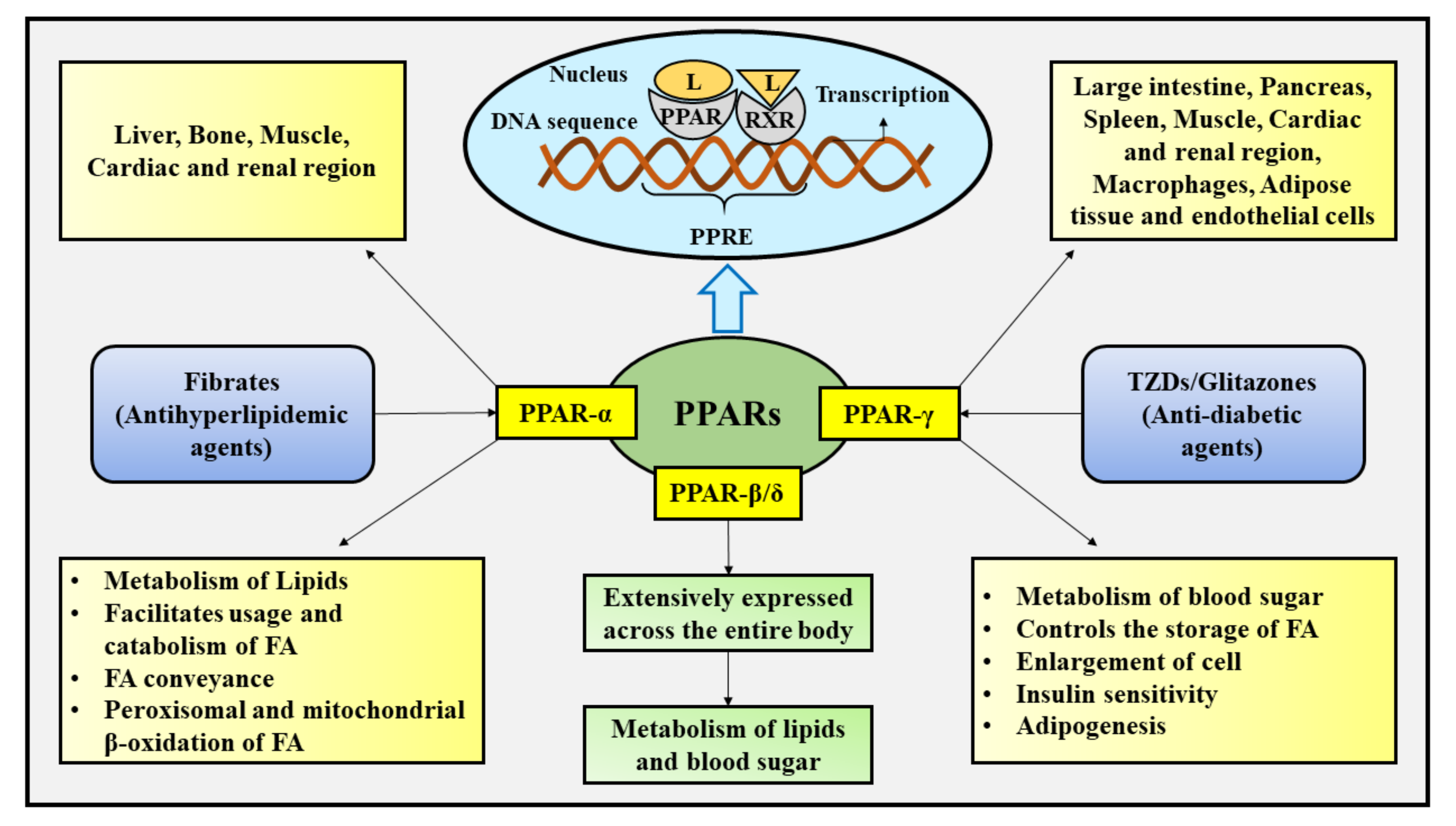
2. Cellular Influences of PPARs

The peroxisomes, otherwise known as microbodies, are sub-cellular structures spotted inside the building blocks of nearly all plants and animals that carry out varied biotransformation activities, such as fatty acid (FA) oxidation, hydrogen peroxide (H2O2)-reliant respiration, and metabolism of lipids [15]. PPARs are ligand-directed transcription factors that pertain to the class of thyroid, steroid, and retinoid receptors and often referred to as NHR [16,17]. These receptors play a pivotal role in the modulation of numerous genes, and biochemical pathways, such as the modulation of mitochondrial operation and redox equilibrium, for instance [18,19]. Up to the present, the trio, particularly PPAR-α (NR1C1), PPAR-β/δ (NR1C2), and PPAR-γ (glitazone receptor/NR1C3) receptor subtypes have been recognized, which are coded by specific genes positioned on the 22, 6, and 3 chromosomes of human beings, consecutively [20]. The three subtypes are effectively modulated via an additional group of genes designated as transcriptional co-activators [21]. Typically, small lipophilic molecules trigger the activation of PPARs, which then form heterodimers with their partner, namely RXR for their exhaustive cytoplasmic stimulation [22]. The amino (N)-terminus ligand self-reliant trans-activation portion, a deoxyribonucleic acid (DNA) interacting portion, and a carboxy (C)-terminus ligand-interacting portion and ligand-based actuation portion are entirely retained within the configuration of these varied subtypes of PPARs [23]. This C-terminus portion of the receptor is presumed to be actively engaged in creating heterodimers with its mate, the RXR. Within the modulatory portion of their target genes the RXR or PPAR heterodimer selectively interacts with DNA sequence elements referred as PPREs in order to control the transcriptional operation.
The stimulation of the plethora of gene cascades implicated in a wide range of body functions takes place following the selective interaction of PPAR with DNA sequences [24]. The interaction of PPAR as well as RXR heterodimers with the co-repressor complexes and the repression of transcription of genes occur during the non-appearance of ligands [25]. In contrast, the presence as well as interaction of natural (FA, and related compounds), and synthetic ([4-[3-(4-Acetyl-3-hydroxy-2-propylphenoxy)propoxy]phenoxy]acetic acid (L-165041), 2-[2-methyl-4-[[4-methyl-2-[4-(trifluoromethyl)phenyl]-1,3-thiazol-5-yl]methylsulfonyl]phenoxy]acetic acid (GW-501516), glitazones/thiazolidinediones (TZDs), and fibrates) and ligands provokes a configurational transition inside the PPAR, prompting co-repressor amino acid chain to detach and the co-activator amino acid chain to engage in order to elevate target gene transcription [16,23,26]. Several studies have revealed that certain NSAIDs, including indomethacin, ibuprofen, naproxen, and fenoprofen also stimulate PPAR-α, and PPAR-γ, relying upon their affinity to interact with PPAR [27,28].
The varied subtypes of PPAR are produced via a single PPAR gene, and their functioning is ascertained by the body tissues within which they remain embodied. These subtypes partake in distinct biological, therapeutic, and molecular processes, such as the modulation of thermogenesis, transcription, the metabolism of lipids, and mitochondrial FA β-oxidation, as well as display discrete particularities for ligands, relying upon their variable positioning and genes of target within the body tissues [15,16]. These subtypes differ in their extent of activity and distribution among various body organs and tissues. PPAR-α is chiefly found in the liver, and inferiorly in the muscle, bone, cardiac and renal region, and participate in facilitating FA usage and catabolism through the overexpression of genes implicated in the conveyance of FA, the metabolism of lipids, and peroxisomal and mitochondrial β-oxidation of FA [15,29,30]. Both natural FA and synthetic ligands (for instance, fibrates-antihyperlipidemic agents) hold the promise to activate PPAR-α [31]. PPAR-β/δ is widely displayed across the entire body, and is actively engaged in controlling the metabolism of blood sugar and lipids [15,31]. PPAR-γ is present in nearly all body tissues, including the large intestine, muscle, spleen, pancreas, the cardiac and renal regions, adipose tissue, macrophages, and endothelial cells, where it actively participates in the metabolism of blood sugar, the regulation of storage of FA, cell enlargement, adipogenesis, and insulin sensitivity [15,32]. Activation of PPAR-γ is triggered by a number of natural ligands and also the synthetic ligands (TZDs) [31].
The three subtypes of PPAR are known to exert the modulatory effect on the inflammatory pathways [33]. Besides the modulation of transcriptional action of nuclear factor kappa B (NF-κB), additional transcriptional factors associated with the inflammatory reactions are also controlled by PPARs as well as additional nuclear receptors. It involves the control of numerous transcriptional factors, namely activating transcription factor-1 (ATF-1), activating transcription factor-4 (ATF-4), and signal transducer and activator of transcription (STAT), along with the control of levels of various inflammatory mediators, for instance, cyclooxygenase-2 (COX-2), and nitric oxide (NO) synthase [33]. It has been reported that PPARs modulate the process of inflammation via various and numerous pathways. At the initial stage, PPAR contends with NF-κB to interact with a protruded collection of co-activators, in particular the cyclic AMP-response element binding protein (CREB), consequently suppressing the inflammatory reaction effectuated by NF-κB [33,34]. Further, the PPAR binds straightforward to the p50, REL-associated protein (RELA)/p65, and IkappaB alpha (IκBα), and results in the inhibition of ability of NF-κB to interact with DNA [33]. In addition, the PPAR suppresses NF-κB as well as activator protein-1 (AP-1) signal-reliant transcriptional stimulation of genes associated with inflammation via a well-renowned process termed as trans repression, by straightforward protein-protein interaction with promoter-interacted transcription factors as well as via the prevention of signal-reliant co-repressor complex elimination [26,33,34]. Figure 1 depicts the location, ligand-based activation, functions, and transcriptional activation of PPARs.
Currently there has been explosion in the exploration of effect of PPARs on the functioning of mitochondria. Pioglitazone, a PPAR-γ agonist, belonging to the class of TZDs has been reported to elevate the intake of oxygen (O2), mitochondrial DNA (mtDNA) levels, as well as the activity of several factors associated with the expansion and division of already existing mitochondria (mitochondrial biogenesis), namely mitochondrial transcription factor A (Tfam) and PPAR-gamma co-activator-1 alpha (PGC-1α) within the adipose tissue under the skin, and the neuronal NTERA-2 (NT2) cell line of humans [35,36,37]. In addition, pioglitazone also enhances blood sugar metabolism and mitochondrial activity within astroglia [38]. Additionally, MitoNEET, an iron-sulfur (2Fe-2S) comprising protein located externally to the mitochondrial membrane that plays a crucial role in modulating the oxidative capability, is balanced with the aid of pioglitazone [39,40]. Rosiglitazone, an aminopyridine pertaining to the TZDs category of drugs has been reported to induce the consumption of blood sugar and mitochondrial biogenesis within the brain of the experimental mouse model [41]. Moreover, glitazones raise mitochondrial membrane potential, and thereby assist in safeguarding the cells from undergoing programmed cell death subsequent to the elimination of growth factors [42]. The subtypes of PPAR actively participate in modulating evolution, inflammatory processes, healing of wounds, operation of mitochondria, tissue differentiation, and metabolism of blood sugar and lipids [43,44]. Owing to the effective participation of PPARs in various processes (such as mitochondrial biogenesis, inflammatory processes, metabolism of blood sugar and lipids, and cell differentiation) they furnish safeguarding outcomes in fatty liver disease [45], ischemic stroke [46], cancer [47], tumors [47,48], cardiac disorders [49], and type II diabetes [50].
Numerous studies have revealed that PPARs are also expressed in central nervous system (CNS) nerve cells and astroglia, which incentivized the researchers to scrutinize the PPAR agonists for their neuroprotective activity in a variety of neurodegenerative diseases [31,51]. PPAR agonists have been demonstrated to exert a protective action on tissues in the peripheral region, against oxidative injury, inflammatory processes, and programmed cell death. Moreover, these agonists have been revealed to possess a neuroprotective action in various CNS diseases [31,51]. Following short-term localized ischemia in rodents, pioglitazone and rosiglitazone restrain neuronal injury and inflammatory processes, thereby exhibiting neuroprotective action [52,53]. It has been reported that rosiglitazone, by elevating the expression of the B-cell lymphoma 2 (Bcl-2) protein defends the nerve cells of spinal ganglion and hippocampus from amyloid-beta (Aβ)-prompted mitochondrial injury [54]. Pioglitazone and rosiglitazone effectively restrain myelin deprivation, pain associated with neuropathy, neuronal injury, and inflammatory processes, and upgrade locomotor recuperation following damage to the rodent spinal column [55,56]. In cortical nerve cells and mixed glia cultures, agonism of PPAR-γ has been reported to elicit a pathway for the removal of Aβ peptide [57]. The PPAR family emerges to be a neoteric, propitious, and prospective target which holds the aptitude to assist in the therapy of neurodegenerative diseases, namely PD [31,58], Alzheimer’s disease (AD) [31,59], Huntington’s disease (HD) [31,60], Amyotrophic lateral sclerosis (ALS) [31,60], and Multiple sclerosis (MS) [61].

3. Parkinson’s Disease
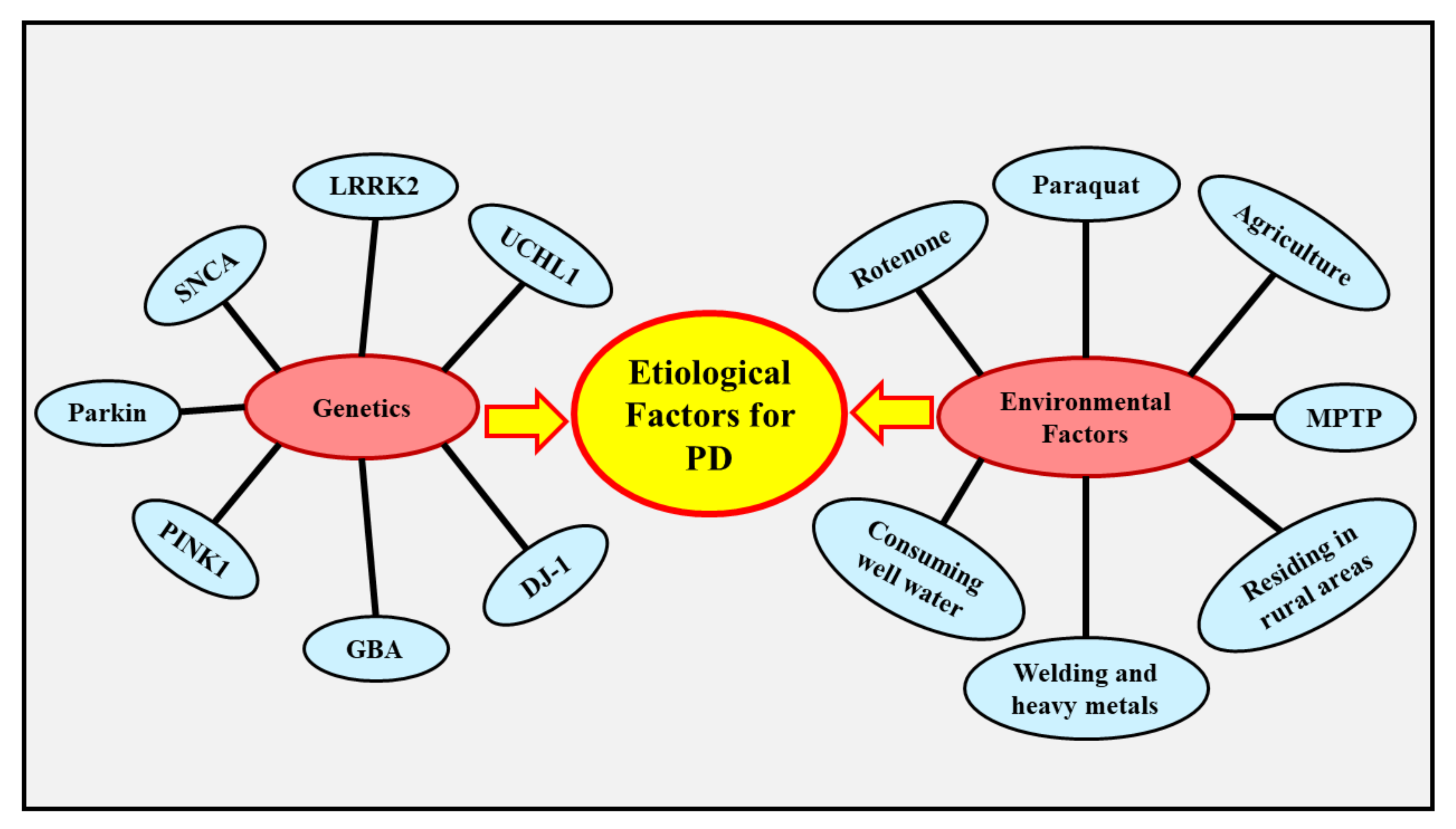
The term PD, otherwise known as paralysis agitans, was coined in 1817 by a British physician named James Parkinson [62]. PD is a common, persistent, intricate, progressive, and generally incapacitating neurodegenerative condition that is concerned with numerous motor and nonmotor brain systems, and predominantly affects elderly people [62,63]. The condition is portrayed by the loss of DA producing nerve cells in the SN-PC, culminating in striatal DA deficiency [58,64]. The build-up of α-synuclein protein in LBs and Lewy neurites located within the nerve cells is also regarded as a pathological feature of PD [65,66]. Following AD, PD is considered as the second most prevalent neurological disease closely related to substantial impairment and decline in well-being [64]. The chief manifestations of PD comprise tremor (shaking), rigor (stiffness), bradykinesia (slowness of movement), and postural abnormality (impairment in body posture and balance) [67].
In developed regions, the pervasiveness of PD is higher, and it has been proven to elevate with age [68]. PD is extremely uncommon in individuals below 40 years of age, whereas it significantly affects 1–2% of individuals beyond 65 years of age, and 4-5% of individuals beyond 85 years of age globally [69]. The occurrence of PD varies among genders, with women being less susceptible to developing PD in comparison to men, owing to the neuroprotective action exhibited by estrogen in women [70].
The underlying cause of PD is still obscure, however numerous genetic and environmental factors are presumed to be implicated in the evolution of the disease [67]. Mutations in genes, such as ubiquitin C-terminal hydrolase L1(UCHL1) [71], α-synuclein(SNCA) [72], leucine-rich repeat kinase 2(LRRK2) [73], Parkin RBR E3 ubiquitin-protein ligase(Parkin) [74], PTEN-induced kinase 1(PINK1) [74], protein deglycase(DJ-1) [75], and glucocerebrosidase(GBA) [76] can result in the development of PD. Several environmental factors, like exposure to pesticides (rotenone, and paraquat) [77], methanol (CH3OH) [78], injury to the head [79], and poisoning of carbon monoxide (CO) [80] are thought to be associated with the development of PD.

4. Etiology of PD

PD is an intricate and multifaceted condition in which genetic and environmental factors contribute profoundly. The preponderance of patients experience the sporadic (typically delayed commencement) form of PD instead of the familial (typically early commencement) form of PD, and they arise due to genetic, environmental, or both of these factors together. Mutations in genes have been found to be associated with approximately 15% of patients experiencing PD, particularly with the familial form [81]. Age is regarded as a major element of danger for PD, as 60 years of age is the mean age of commencement [82]. The occurrence of the condition escalates with aging, hitting 93.1 (every 100,000 person-years) in those aged 70–79 years [83,84]. There are also cultural diversity differences, with European, South American, and North American countries indicating elevated incidence in comparison to African, Arabic, and Asian countries [85] Figure 2 depicts the factors that contribute to the etiology of PD.

4.1. Genetics

Even though PD is predominantly a condition with an unknown cause, around 10–15% of individuals indicate a familial history, and around 5% of cases arise due to genetic inheritance [86]. Mutations in multitudinous genes, such as UCHL1, SNCA, LRRK2, Parkin, PINK1, DJ-1, and GBA have been linked to PD [71,72,73,74,75,76].
The UCHL1 gene, otherwise termed as protein gene product 9.5 (PGP9.5), appears to be a physiologically feasible predisposing gene for PD [71,87]. The articulation of UCHL1 is nerve cell specific and pervasive all over the brain, displaying notably robust in situ hybridization findings inside the SN-PC [88]. The UCHL1 enciphers a protein which constitutes nearly 1–2 % of the entire soluble protein present within the brain and is often discovered in LBs [89]. It has been proven that UCHL1 actively participates in ubiquitin-reliant cleavage of proteins/polypeptides via converting large structural repeating units of ubiquitin to a single unit of ubiquitin. In order to undergo degradation via proteasomes, ubiquitin effectuates activation, coupling, and joining with prejudicious proteins. The interruption of the entire ubiquitination as well as proteasomal degradation system and the consequent accumulation of SNCA within the cytoplasm has been postulated as an underlying mechanism for PD [90]. The gracile axonal dystrophy (gad) mouse involves the removal of UCHL1 within the gene, which in turn contributes to usual manifestations associated with neurodegeneration, for instance, deprivation of voluntary muscles balance, dying back type neuronal degeneration [91], and protein deposition in nerve endings [92]. The accumulation of UCHL1 and its isotypes related to PD, comprising UCHL1S18Y, and UCHL1I93M, is escalated within cultured cells, following the suppression of the UPS, thereby demonstrating a potential correlation between PD and UPS [93]. Moreover, mutations in UCHL1 isotypes, namely p.I93M, p.E7A, and p.S18Y are strongly linked with tremendous hazard towards PD [87]. These investigations disclose the substantial contribution of mutations in the UCHL1 gene and its isotypes to the evolution of PD.
The SNCA gene ciphers in order to produce a protein named α-synuclein which exists in nerve cells in the vicinity of presynaptic nerves as well as additional types of cells. This protein shares active involvement in synaptic transmission since it effectively controls the quantity and liberation of DA comprising neurotransmitter vesicles [94]. It has been reported that SNCA gene mutations can result in the build-up of this protein, which consecutively contributes to the anomalous amassing of DA. This results in making the body capable of splitting the profuse DA, which results in nerve cell death and the emergence of manifestations associated with PD [94].
The sporadic form of PD, which arises beyond 50 years of age, has been linked to LRRK2 gene mutations [95]. Dardarin, a protein possessing multiple domains, which is encoded by the LRRK2 gene, has been found to partake in transmission processes essential for protein-protein signaling and the operation of nerve cells [95]. The conformation and activity of dardarin proteins are greatly influenced by LRRK2 gene mutations. Several researchers have scrutinized and revealed that the dardarin mutant triggers programmed cell death, and its interaction with a protein termed parkin gives rise to an accumulation of cytoplasmic proteins [96]. Mutations in the LRRK2gene prompt breakdown and build-up of protein in an aberrant manner [97]. Elevated build-up of cytoplasmic proteins might promote programmed cell death, which in turn results in abnormalities in mobility and coordination that are often noticeable in patients experiencing PD, but the underlying pathways are still obscure [98].
The Parkin/PARK2 gene ciphers parkin (protein) that is speculated to direct proteins so as to effectuate breakdown with the aid of enzymes. Parkin has also been associated with the breakdown of impaired cell powerhouses/ energy factories (mitochondria). Autosomal recessive, early commencement forms of PD are found to be associated with PARK2 gene mutations [95]. As a consequence of PARK2 gene mutations, the parkin protein starts operating abnormally, and it has been noted that this deprivation of the usual functioning of parkin elicits the build-up of inappropriate proteins, which in turn could disrupt DA release and other usual cellular functions [99]. Owing to the profuse presence of parkin within the CNS, its abnormal functioning could result in the deprivation of DArgic nerve cells, which, as a result, contributes to the emanation of manifestations related to PD [98]. In addition, several investigations have reported that mutations in the PARK2 gene are also associated with diminished functioning of the powerhouse of the cell and elevated susceptibility towards substances that are harmful to the powerhouse of the cell, and in the case that the cells’ powerhouse in DArgic nerve cells is disrupted, it could impair the conveyance of DA, potentially contributing to the manifestation of PD [95].
Apart from this, mutations in the PINK1 gene are actively engaged in precipitating manifestations of PD. It has been elucidated that those mutations in the PINK1 gene are explicitly related to autosomal recessive, early commencement forms of PD [100]. PTEN, a protein encoded by the PINK1 gene, is expressed within the cellular energy factories across the body, and is presumed to exert a safeguarding action against oxidative damage [95]. The typical PTEN protein has been reported to suppress programmed cell death, whereas the mutant form of PTEN protein is powerless to suppress programmed cell death, and thereby might give rise to escalated nerve cell destruction.
The DJ-1 protein, otherwise termed as PARK7, which behaves as an antioxidant and safeguards nerve cells against oxidative damage, and restrains the α-synuclein build-up, is ciphered by the PARK7 gene. It has been elucidated that PARK7 gene mutations provoke the abnormal operation of DJ-1/PARK7 protein, eventually resulting in the build-up of α-synuclein as well as the accumulation and breakdown of profuse DA [99]. The abnormal operation of DJ-1/PARK7 induces oxidative damage, which consecutively evokes DArgic nerve cell destruction. In each of the aforementioned scenarios, the deprivation of DA is thought to play an integral role in the emergence of manifestations of PD [95].
It has been elucidated that the GBA gene ciphers the lysosomal enzyme named β-GBA, which effectuates the breakdown of sphingolipid, namely glucosylceramide (GluCer), as a means of producing a pair of components termed glucose (sugar), and ceramide (lipid molecule) [101]. It has been evaluated that nearly 12% of European patients experiencing PD, and 15 to 20% of Ashkenazi Jewish patients experiencing PD, are robustly linked with mutations and variations in the GBA gene, creating GBA as a critical genetic hazard for PD [102]. Patients who express mutations in the GBA gene are at a risk of developing PD earlier in life, as well as exhibiting cognitive disability [101]. In patients with sporadic forms of PD, the functioning of β-GBA is greatly diminished within the anterior cingulate cortex (ACC), and substantia nigra (SN) regions of the brain [103,104]. The disabled autophagy-lysosomal pathway (ALP) is presumed to be actively engaged in the α-synuclein build-up in an aberrant manner [101]. It has been reported that α-synuclein builds up and displays LBs attributes in physiological and experimental models possessing knocking down, knocking out or mutations in the β-GBA, and is associated with ALP disability [101]. Even though the precise pathway via which deprivation of β-GBA participates in the pathophysiology of PD is still poorly understood, it might comprise α-synuclein build-up, diminished lysosomal operation, and endoplasmic reticulum (ER)-related stress [105]. Considering homozygous mutations in the GBA gene, GluCer build-up within the lysosomes might provoke lysosomal abnormalities, whereas no such build-up of GluCer has been found in PD brains possessing heterozygous mutations in the GBA gene [105].

4.2. Environmental Factors

Besides the involvement of genetic factors in the evolution of PD, there are several environmental factors which significantly contribute to PD. The neurotoxin1-methyl-4-phenyl-1,2,3,6-tetrahydropyridine (MPTP), was initially recognized to be related to nigrostriatal degeneration, following the emergence of characteristic manifestations of PD in several individuals upon self-administration of narcotic substances contaminated with MPTP. MPTP is bio transformed into an active toxic metabolite named 1-methyl-4-phenylpyridinium ion (MPP+), which belongs to the family of mitochondrial complex-I suppressors, and is exclusively involved in devastating DArgic nerve cells within the SN [106,107]. The exploration of MPTP as a triggering factor for degeneration within the SN encouraged the postulation that PD might be precipitated by toxic substances present in the environment [108].
Thereafter, numerous investigations have revealed the significant relationship between exposure to pesticides and PD, particularly a single case-referent study demonstrating a strong correlation between occupational exposure to pesticides and delayed commencement forms of PD in men possessing an odds ratio of 2.2 [109]. It has been reported that other specific suppressors of mitochondrial complex-I, namely rotenone (a pesticide) [110], and paraquat (a herbicide exhibiting structural resemblance with MPP+) [111], provoke deprivation of DArgic nerve cells within experimental animal models experiencing PD.
Additionally, various epidemiological investigations have explored the association between subjection of such substances and the possibility of evolving PD. This eventually spurred the scrutiny of substitutional indicators, namely the relationship between agriculture, residing in rural regions, fertilizers [112], and consuming well water with the susceptibility of evolving PD. Subjection to welding and heavy metals comprising copper (Cu), zinc (Zn), iron (Fe), aluminum (Al), and lead (Pb), have likewise been examined, but the association between these factors and PD is still ambiguous [108].

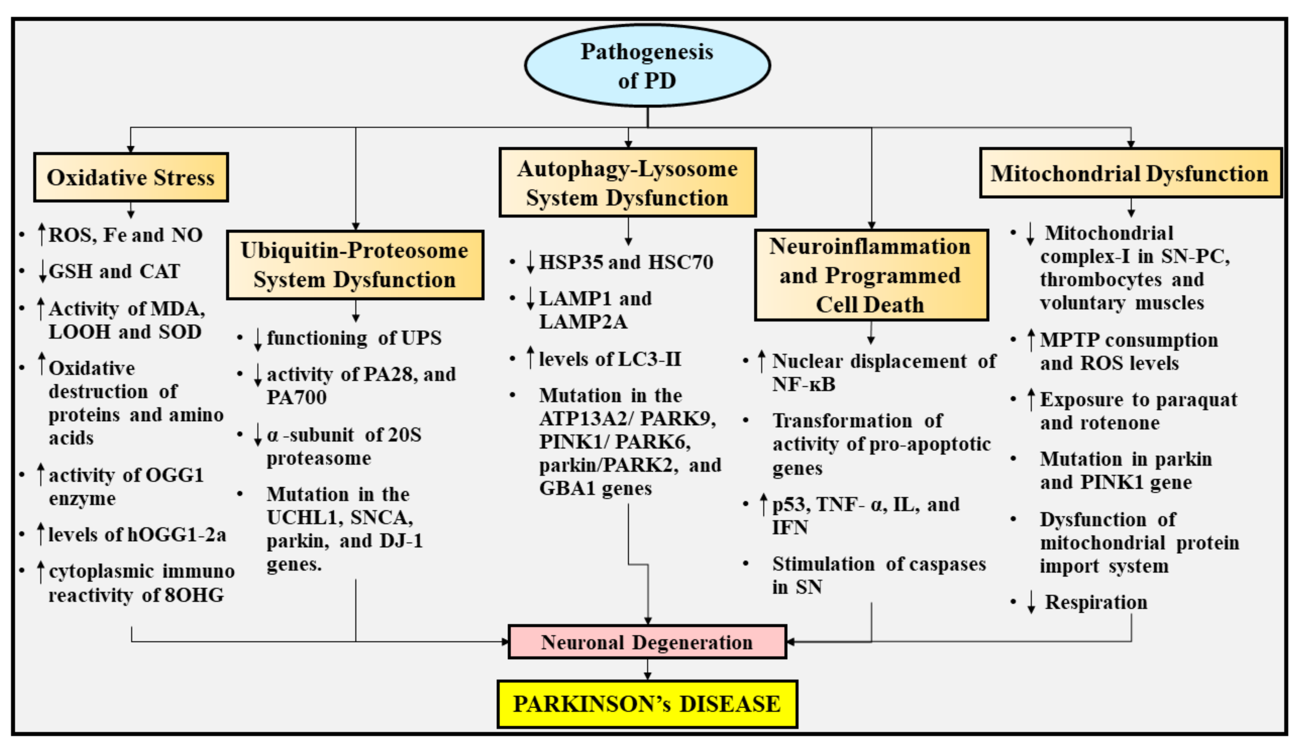
5. Pathogenesis of PD

The fundamental pathways implicated in the initiation and evolution of PD are still inexplicit, but elevated oxidative stress, UPS dysfunction, autophagy-lysosome system dysfunction, neuroinflammation, programmed cell death, and mitochondrial dysfunction probably contribute to the pathogenesis of PD. The various pathways involved in the pathogenesis of PD are depicted in Figure 3.

5.1. Oxidative Stress

Oxidative stress has acquired the utmost emphasis amid numerous pathogenic pathways speculated to partake in the death of DArgic nerve cells in PD. This is due to the fact that DA present within the specific areas of the brain experiences enzymatic and non-enzymatic metabolic processes in order to produce reactive oxygen species (ROS) in the SN pinpointed in the midbrain [58,113]. Elevated levels of Fe, a decline in the levels of an antioxidant named glutathione (GSH), raised quantities of a membrane polyunsaturated FA peroxidation end product termed malondialdehyde (MDA), and oxidative destruction of a long chain of amino acids and lipids have been revealed in postmortem examinations of the brains of patients experiencing PD, suggesting the significant involvement of oxidative stress in the evolution of the disease [114,115,116].
Likewise, the raised activity of the DNA repair enzyme termed 8-oxoguanine DNA glycosylase (OGG1) within the SN of patients experiencing PD clearly demonstrates elevated oxidation of DNA in the disease. Moreover, the levels of a subtype of human 8-oxoG DNA glycosylase (hOGG1) termed hOGG1 type 2a (hOGG1-2a) were found to be enhanced in SN of patients experiencing PD, presumably induced by mitochondrial oxidative damage [117]. Additionally, enhancement in the cytoplasmic immunoreactivity of a usual nucleic acid oxidation compound named 8-hydroxyguanosine (8OHG) has been reported in the SN of patients experiencing PD, in comparison to controlled age-matched individuals [118]. Apart from this, several other investigations have demonstrated an elevation in the lipid hydroperoxides (LOOH), functioning of superoxide dismutase (SOD), and reduction in the functioning of catalase (CAT) in patients experiencing PD. According to these researchers, MDA can indeed be the biosignature for PD, whereas LOOH and SOD are linked to delayed PD manifestations [119].
Furthermore, NO, a neurotransmitter that is produced through an amino acid, namely L-arginine, has been recognized to perform a distinctive physiological function [120]. Several investigations have revealed that NO exhibits a key function as a neurotoxic mediator related to mitochondrial impairment in a wide range of incapacitating neurodegenerative conditions, including PD [119]. In the case of diseased states, the expression of inducible nitric oxide synthase (iNOS), and nicotinamide adenine dinucleotide phosphate (NADPH) oxidase operation originates inside the microglia, culminating in extortionate synthesis of free radicals, namely NO and oxygen free radicals (O2). The interaction between NO and O2 prompts the generation of extremely reactive molecules, namely peroxynitrite radicals (ONOO), which might precipitate the destruction of DArgic nerve cells. Apart from this, nitration of tyrosine residues emerges as a well-renowned hallmark of oxidative stress in PD patients and is actually prompted by ONOO, demonstrating the significant contribution of NO generation and its secondary products in the pathogenesis of PD [121,122]. Another study has displayed that aldose reductase (AR) scarcity, a tyrosine hydroxylase (TH) cofactor actively engaged in the generation of DA, may elicit oxidative stress in animal models experiencing PD via elevating NO and nitrite (NO2), culminating in forfeiture of DArgic nerve cells and aberrations in the autophagy-lysosome system [123]. In contrast, it has been recognized that diminished blood serum NO metabolic products, namely nitrates (NOx), and NO2 are strongly related with cognitive dysfunction in patients experiencing PD, propounding NOx as an indicator of the early commencement form of PD [124]. Additionally, astrocytes exhibit elevated amounts of a heme-comprising peroxidase, namely myeloperoxidase (MPO), which in turn give rise to oxidative destruction via generating hypochlorous acid (HOCl) following the chemical reaction of H2O2 and chloride ions (Cl) [121]. Owing to the reason that HOCl may also react with O2, the existence of HOCl could raise the quantity of hydroxyl free radicals (OH.). It has been elucidated that MPO also evokes the transformation of non-reactive NO2 into its reactive free radical state (NO2), leading to escalation in protein destruction [125]. These findings propose the consequential participation of NO generation, iNOS activation, and the ONOO generation in nigrostriatal DArgic nerve cell degeneration, thus contributing to the pathogenesis of PD.

5.2. UPS Dysfunction

Various neurodegenerative diseases, which are marked by aberrant build-up of protein, comprise dysfunction in the proteasomal system as a prevalent hallmark [126]. The strongest proof of certain anomalies in PD emerges from postmortem investigations into the SN-PC, where the UPS enzymatic functioning has been reported to be considerably diminished in comparison to normal brains [127]. Afterwards, identical outcomes were observed in peripheral blood mononuclear cells (PBMCs) of patients suffering from PD, but no such effects were noticed in unaffected people [128]. In addition to declined functioning, the SN-PC of patients with PD has been reported to be associated with reduced activity of varied constituents of the proteasomal system. In particular, components which are implicated in the efficient operation of UPS, such as activators of the proteasomal system, namely proteasome activator 28 (PA28) and proteasome activator 700 (PA700) [129], and the α-subunit of the 20S proteasome are decreased [130]. Mutations in the UCHL1, SNCA, Parkin, and DJ-1 gene provide insight into the proteasomal abnormality in the evolution of PD [131]. It has been elucidated that the intra striatal administration of a specific proteasome suppressor named lactacystin provokes elevation in the heme oxygenase-1 concentrations, deposition of α-synuclein, and retrograde deterioration of nerve cells within the SN, demonstrating the participation of UPS impairment in PD [132].

5.3. Autophagy-Lysosome System Dysfunction

There are multifarious autophagy and lysosomal concomitant constituents which have been impaired/abnormally displayed in PD, in correspondence with outcomes in the UPS pathway. Upon postmortem investigation, various molecular chaperones pertaining to the heat-shock protein class, namely hereditary spastic paraplegia 35 (HSP35) and heat shock cognate protein 70 (HSC70), as well as extremely important lysosomal membrane proteins, such as lysosomal-associated membrane protein 1 (LAMP1) and lysosomal-associated membrane protein 2A (LAMP2A), were revealed to be diminished [133,134]. It has been demonstrated that an autophagosome indicator, namely microtubule-associated protein 1A/1B-light chain 3 (LC3)-II is elevated in the SN region of the brain in patients suffering from PD, demonstrating the build-up of autophagic vesicles [135,136]. In addition, mutations in the ATP13A2/PARK9 gene are thought to be strongly related to Kufor-Rakeb syndrome (KRS), an autosomal recessive kind of parkinsonian syndrome [137]. Also, it has been proven that mutations in a few additional PARK genes alter the operation of PINK1/PARK6[138] or Parkin/PARK2[139], which are both implicated in mitochondrial autophagy [140]. Furthermore, the emanation of mutations in the GBA1 gene, which precipitates the autophagy-lysosome system impairment, as a tremendous genetic hazard for PD, lends credence to the assumption regarding the substantial involvement of this system in the evolution of PD [141]. These findings indicate that abnormal functioning of the autophagy-lysosome system partakes in PD pathophysiology.

5.4. Neuroinflammation and Programmed Cell Death

Neuroinflammation and programmed cell death have been reported to actively participate in PD pathophysiology. Several investigations have shown that the postmortem studies on the brains of patients suffering from PD have been identified to possess programmed cell death and autophagy [142]. Within the DArgic nerve cells of patients suffering from PD, enhancement in the nuclear displacement of NF-κB was also spotted [143]. Inflammatory processes and programmed cell death in the PD brain are further supported by transformation of activity of pro-apoptotic genes, raised concentrations of a tumor suppressor protein termed p53, NF-κB, interferon gamma (IFNγ), and stimulation of caspases inside the SN region of individuals with PD [144,145,146,147]. Activation of microglia has been portrayed within the SN region of individuals experiencing PD, whereby they result in the emission of programmed cell death-prompting inflammatory mediators such as tumor necrosis factor-α (TNF-α), interleukins (IL), and interferons (IFN) [58]. Correspondingly, stimulation of microglia is also triggered by the build-up of α-synuclein, leading to prolonged and gradual degeneration of nerve cells in the SN of PD patients [148]. Although the pathways underlying microgliosis in PD are obscure, a catecholamine-reliant dark polymer pigment, namely neuromelanin (NM)-comprising DA nerve cells, has been displayed to be incredibly prone to inflammatory processes in the disease. Moreover, it remains questionable whether inflammatory processes in the neuronal region are the chief factor for inducing PD or merely an outcome of the condition.

5.5. Mitochondrial Dysfunction

The impairment in mitochondrial function is presumed to be actively engaged in the pathophysiology of PD having a genetic cause or an unknown cause [149]. It has been elucidated from the early postmortem findings that a fundamental constituent of the electron transport chain (ETC), namely mitochondrial complex-I, was found to be deprived within the SN-PC region of the brain of patients suffering from PD. These findings were probably the earliest ones highlighting the direct association between mitochondrial abnormalities and PD [150]. Apart from this, deprivation in mitochondrial complex-I was also detected in the thrombocytes and voluntary muscles of individuals suffering from PD in contrast to unaffected individuals [151,152].
Additionally, it has been found that uncontrolled consumption of MPTP precipitates the irreversible manifestations of PD [109], along with destruction of DArgic nerve cells revealed by postmortem analysis [153]. Several studies have disclosed that DA nerve cells recognize MPTP following its oxidation into a toxic metabolite termed MPP+, which then results in the suppression of mitochondrial complex-I [154]. Moreover, paraquat (a herbicide exhibiting a structural resemblance with MPP+), and rotenone (a pesticide) are two additional toxic substances that impede the operation of the mitochondrial complex-I, resulting in the emergence of manifestations of PD and DA cell destruction plausibly in human beings and animals [77,110,111]. Therefore, mitochondrial complex-I abnormality might partake in the destruction of DA cells owing to de-escalation in the levels of energy [149]. Furthermore, mutations in the Parkin and PINK1 genes provoke mitochondrial dysfunction, thereby eliciting an autosomal recessive form of PD [95,139].
In addition, it has been reported that α-synuclein following the binding with the membrane of mitochondria and deposition within the organelles deteriorates the operation of mitochondrial complex-I, which eventually contributes to escalated oxidative damage and mitochondrial abnormalities [155,156]. Moreover, the linkage between α-synuclein and the translocase of the inner mitochondrial membrane 20 (TOM20) evokes abnormality in the import system of the mitochondrial protein, profuse synthesis of ROS, and a decline in breathing [157]. These factors share their significant contribution to mitochondrial dysfunction.

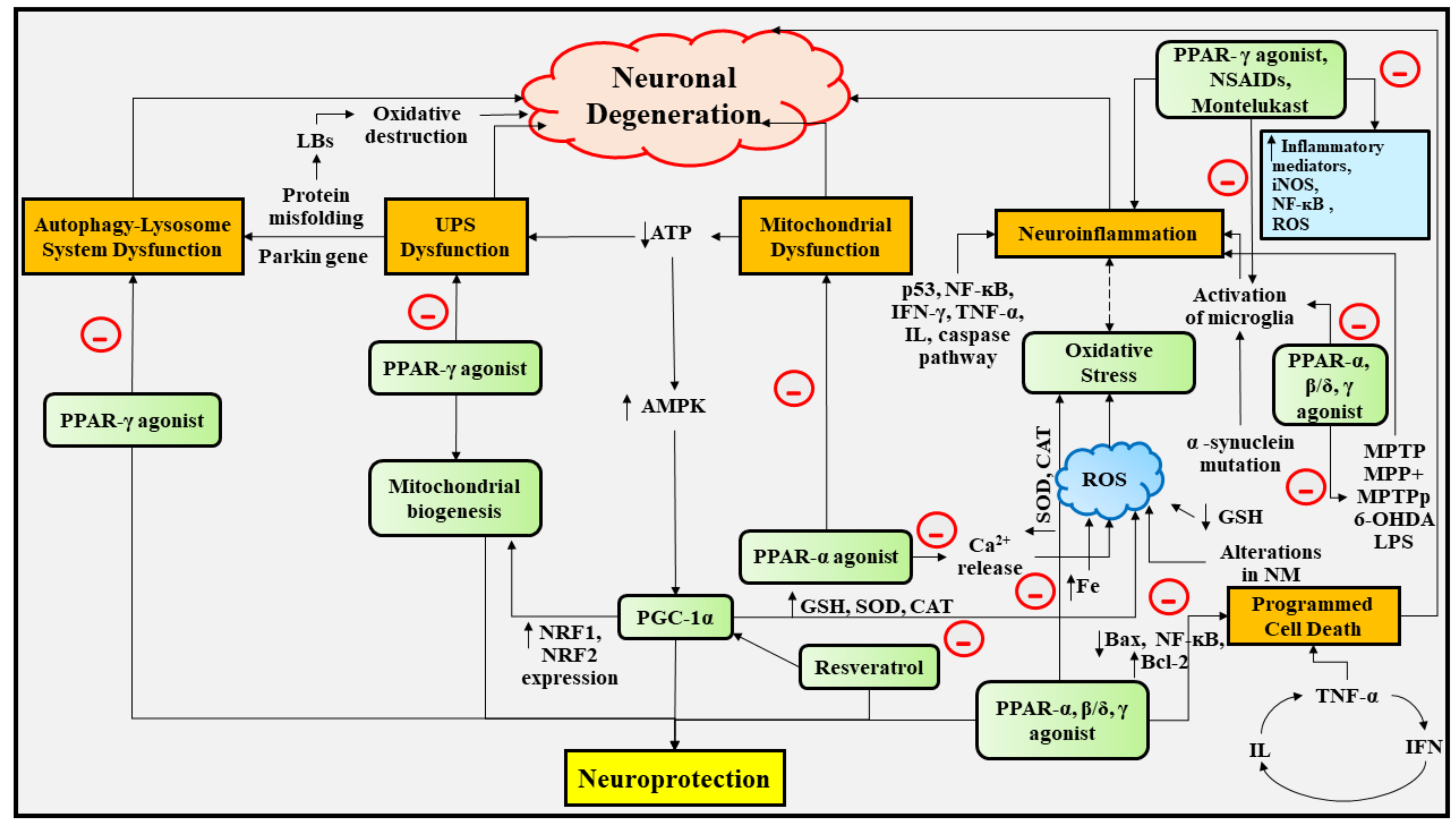
6. Experimental Studies Portraying the Deep Insights into the Neuroprotective Role of PPAR Agonists in PD

It has been elucidated that DArgic nerve cell degeneration is spurred by the generation of ROS, which in turn induces oxidative destruction, microglia-effectuated inflammation in the neuronal region, and mitochondrial abnormalities, and each of these in conjunction contributes to the stimulation of programmed cell death. Consequently, modulation of oxidative stress and mitochondrial abnormalities might assist in restraining the decline in functioning of nerve cells in PD [31,58]. In accordance with numerous investigations, it has been revealed that PPAR agonists exhibit neuroprotective actions in various in vivo and in vitro models experiencing PD.

6.1. Therapeutic Implications of PPAR-γ Agonists in PD

It has been reported that following the oral delivery of the PPAR-
γ agonist, namely pioglitazone (20 mg/kg) before i.p administration of MPTP (in a dose of 15 mg/kg) resulted in a reduction in MPTP-inebriation prompted microglia stimulation and precluded forfeiture of DArgic nerve cells within the SN-PC of an experimental model of mouse experiencing PD [158]. Furthermore, another investigation has revealed the safeguarding outcomes of pioglitazone in the case of MPTP-instigated neurotoxicity, which agrees with the results of previous investigations [159]. The safeguarding action of pioglitazone in the case of MPTP-prompted neurotoxicity is exerted via the inhibition of the transformation of MPTP to its deleterious metabolic product, MPP+, through monoamine oxidase B (MAOB) suppression [160,161]. It has been proven that after oral delivery of pioglitazone, significant shielding was extended towards MPTP-prompted nerve cell destruction in TH-immunoreactive SN nerve cells [159].
Therapy with the aid of pioglitazone gives rise to substantially decreased stimulation of microglia, nitro tyrosine activity in DArgic nerve cells, mediators of inflammatory processes, and the fraction of glial fibrillary acidic protein (GFAP) positive cells in the SN region and striate nucleus [159]. Several studies have reported that PPAR-γ agonists possess anti-inflammatory [15,17,162], and anti-neoplastic properties [15,17]. In experimental studies, oral delivery of pioglitazone rapidly following the emergence of PD in rhesus monkeys resulted in DA striatal fibers and SN nerve cells preservation, thereby exhibiting a neuroprotective role in PD [163]. Another investigation has shown that the oral introduction of pioglitazone (in a dose of 30 mg/kg) following 6-hydroxyDA(6-OHDA) lesions in male wistar rats experiencing PD resulted in rendering protection to DArgic nerve cells in the SN against neuronal destruction and offered a significant decrease in NF-κB and microglial stimulation [164]. A recent study has elaborated on the neuroprotective outcomes of pioglitazone in both in vivo and in vitro MPTP or MPP+ provoked PD models.
In vivo investigation has demonstrated that oral delivery of pioglitazone led to considerable improvement in behavioral manifestations destructed by MPTP, as well as the escalation in the lifespan of TH positive nerve cells, a fraction of cell powerhouses and improved mitochondrial ultrastructure, and enhanced activity of PGC-1α. In vitro investigation has revealed the modulation of activity of numerous proteins implicated in the operation of mitochondria, for instance, PGC-1α, mitochondrial fission 1 (Fis1), mitochondria fusion 2 (MFN2), nuclear respiratory factor 1 (NRF1), and nuclear respiratory factor 2 (NRF2) after oral treatment with pioglitazone [165].
Additionally, it has been stated that pioglitazone renders neuroprotection in intra-striatally delivered lipopolysaccharide (LPS) triggered inflammatory processes in an experimental model of rat possessing degeneration of DArgic nerve cells [166,167]. According to this investigation, treatment with the assistance of pioglitazone considerably evoked decline in LPS triggered microglial inflammatory processes, oxidative damage, upgraded the operation of mitochondria, incompletely reinstated DA concentrations, and enhanced DArgic neuroprotection.
Furthermore, a related study highlighted the neuroprotective pathway exhibited by pioglitazone towards LPS [168]. Through meddling with two processes, namely NF-κB and Jun N-terminal kinase (JNK), which reduce the expression of COX-2, pioglitazone safeguarded DArgic nerve cells against LPS prompted COX-2 and prostaglandin E2 (PGE2) driven intensification of stimulation of microglia in DArgic nerve cells-neuroglia co-cultures [168]. Apart from this, pioglitazone also safeguards DArgic nerve cells from LPS-prompted impairment via diminishing the expression of iNOS, and synthesis of NO through distinguishably controlling the p38 mitogen-activated protein kinase (MAPK), and phosphoionositide 3-kinase (PI3K)/Akt processes [169].
Moreover, i.p. administration of another PPAR-γ agonist, namely rosiglitazone (in a dose of 10 mg/kg) in the MPTP or probenecid (MPTPp) animal model of mice (C57BL/6J) experiencing PD resulted in a decline in the generation of inflammatory mediator, namely TNF-α in the microglia cells, and arrested MPTPp-prompted degeneration of nerve cells in the SN-PC [170]. Recently, it has been elaborated that rosiglitazone holds the aptitude to safeguard retinal nerve cells from the abnormalities precipitated by exposure to rotenone and escalates neuroprotection in the retina, and CNS of rotenone-prompted rat models of PD, following the systemic delivery of rosiglitazone in liposome-encapsulated form (1 mL/kg, i.p.) [171].
Furthermore, it has been discovered that rosiglitazone safeguards human neuroblastoma cells from a mitochondrial operation suppressor and neurotoxin, namely acetaldehyde (CH3CHO), and a neurotoxin, namely MPP+ [172,173]. Acetaldehyde elicits nerve cell destruction via promoting programmed cell death and intracellular release of ROS [172]. Through spurring the activity of antioxidant enzymes and controlling the expression of programmed cell death regulators, namely Bax and Bcl-2, rosiglitazone was capable of repressing acetaldehyde-prompted programmed cell death [172]. Rosiglitazone safeguards human neuroblastoma cells from MPP+ prompted cellular damage through the suppression of abnormalities in the functioning of mitochondria and ROS generation [173]. These findings indicate that apart from the anti-inflammatory property of PPAR agonists, they also safeguard nerve cells by modulating the activity of CAT and SOD (antioxidant enzymes) and maintaining an equilibrium between the activity of pro-apoptotic genes and anti-apoptotic genes [172,173].

6.2. Therapeutic Implications of PPAR-β/δ Agonists in PD

It has been demonstrated that in the rotenone-prompted rat model of PD, a PPAR-β/δ agonist, namely GW-501516, safeguards DArgic nerve cells from destruction provoked by deleterious substances, as well as upgrades behavioral performance by diminishing ER-related stress in PD. Moreover, in the MPTP instigated PD model of mice, a decline in nucleotide-binding domain and leucine-rich-repeat-protein 3 (NLRP3) inflammasome-related nerve cell inflammation is rendered by PPAR-β/δ agonists [174].
Another study revealed that PPAR-β/δ agonists, namely GW-501516 and L-165041, remarkably safeguarded SH-SY5Y neuroblastoma cells from staurosporine and MPP+ triggered programmed cell death primarily through suppressing the stimulation of the caspase-3 pathway [10]. Additionally, these agonists safeguarded against MPTP-instigated DA and its metabolic products deprivation in the SN-PC [10]. Besides, a recent investigation has revealed the outcomes of 2-[4-[[2-[3-fluoro-4-(trifluoromethyl) phenyl]-4-methyl-1,3-thiazol-5-yl] methyl sulfanyl-[2-methylphenoxy] acetic acid (GW0742), a PPAR-β/δ agonist in an experimental model of rat experiencing PD-related cognitive abnormalities. In accordance with this investigation, MPTP exposure to rats led to oxidative destruction and splitting-up of DNA strands into fragments. Thereafter, therapy with the aid of GW0742 (in a dose of 30 and 100 µg/kg) resulted in incomplete reinstatement of MPTP-damaged cognitive activities. Furthermore, it has been shown that GW0742 significantly minimized oxidative destruction and splitting-up of DNA strands into fragments in numerous assays, including immunochemical (Tunel), MDA, and GSH [175,176].

6.3. Therapeutic Implications of PPAR-α, and PPAR-α/γ Agonists in PD

In addition to PPAR-γ, and PPAR-β/δ agonists, several PPAR-α, and PPAR-α/γ agonists have been shown to exert a neuroprotective action in different PD models. Fenofibrate and benzafibrate are the two PPAR-α agonists that have been extensively examined in the MPTP-treated experimental mouse model of PD [177]. One study has revealed that fenofibrate potentially safeguarded DArgic nerve cells in the SN and TH-immunoreactive endings within the striatal region, however benzafibrate displayed no such safeguarding action, this can possibly be accounted by the fact that benzafibrate was administered at a 10 times lesser amount in comparison to fenofibrate [177].
Another study has reported that oral delivery of fenofibrate (in a dose of 100 mg/kg) in an experimental rat model of PD following one hour of MPTP exposure contributed to a significant decline in hypo-locomotion precipitated by MPTP, depressive behavioral patterns following neurotoxin administration, prevented both an escalation in ROS generation and a decline in DA concentrations following surgical procedures, thereby demonstrating neuroprotective action in the MPTP-prompted PD model [178]. These studies suggest that therapy with the assistance of fenofibrate holds the potential to impede the evolution of PD [59].
A recent study has stated that prior treatment with the aid of the PPAR-α/γ dual agonist, namely 2-[4-(5-chloro-1,3-benzothiazol-2-yl)phenoxy]-2-methylpropanoic acid (MHY908), had demonstrated neuroprotective action against MPTP-prompted experimental mouse models of PD [179]. The neuroprotection in this PD model was rendered via reduction in MPTP-prompted DArgic nerve cell deprivation and motor impairment, alleviation of MPTP-prompted stimulation of glial cells in the nigrostriatal region, suppression of MPP+ prompted stimulation of astroglia via inhibition of NF-κB signaling in primary cultured astrocytes, and suppression of MPP+ prompted cell destruction and ROS generation in SH-SY5Y neuroblastoma cells [179].
Table 1 presents the neuroprotective outcomes of PPAR agonists in PD and the pathways by which PPARs exhibit neuroprotective action in PD are depicted in Figure 4.

6.4. Therapeutic Implications of NSAIDs, Leukotriene Receptor Antagonist, and Vitamin E in PD

Apart from this, several commonly employed NSAIDs, for instance, ibuprofen, fenoprofen, flufenamic acid, naproxen, and indomethacin explicitly interact either with PPAR-α/PPAR-γ or both and culminate in their activation [27,190,191]. Numerous researchers have explored the efficacy of ibuprofen and indomethacin in rendering significant neuroprotection in PD and other neurodegenerative diseases [51,190,192]. According to one study, in mesencephalic cultured cells, paracetamol (1mM) and ibuprofen (0.1mM) effectively mitigated 6-OHDA, MPP+, and glutamate prompted DArgic nerve cell destruction [180,181].
Moreover, it has been stated that ibuprofen individually raised the quantity of DArgic nerve cells by approximately 47% [180]. A recent study has reported that multimodal therapy with an iminosugar, namely 1-deoxynojirimycin (1-DNJ) and a NSAID, namely ibuprofen, impedes destruction of mesencephalic DArgic nerve cells, minimizes the levels of inflammatory mediators like interleukin-6 (IL-6) and TNF-α, total microglia markers namely CD68+/Iba-1+ cells, and interaction between microglia cells and nerve cells in MPTP-subjected experimental mice model [182].
Additionally, a new investigation has demonstrated that co-treatment with a novel herbal mixture comprising 12 medicinal herbs, namely Gagam-Sipjeondaebo-Tang (GST) and ibuprofen, exhibited a synergistic action in ameliorating DArgic nerve cell destruction and reducing the activation of macrophages in the MPTP-prompted mouse model of PD [183]. Further, the levels of NO were considerably declined in LPS-activated macrophages following this co-treatment. According to this investigation, GST alone remarkably reduced DArgic nerve cell death, levels of IL-6, COX-2, iNOS, and interleukin-1 beta (IL-1β), and relieved PD-related behavioral abnormalities [183]. Another study revealed that in the MPTP prompted experimental model of mice, indomethacin extended safeguardance towards MPTP-prompted nerve cell destruction and diminished activation of microglia and the infiltration of lymphocytes [184].
Furthermore, several other agents have been proven to exert a neuroprotective action on PD, including celecoxib (a selective COX-2 inhibitor) [185], montelukast (a leukotriene receptor antagonist) [186,187,188,193], and tocopherol (vitamin E) [194,195]. Therapy with the aid of celecoxib (< 20 μM) has been shown to reinstate SH-SY5Y cells that had been potentially subjected to paraquat and 6-OHDA prompted damage [185]. Additionally, celecoxib therapy culminated in a significant and persistent overexpression of a lipocalin carrier of tiny hydrophobic molecules, namely apolipoprotein D (APOD), as well as a few of the microphthalmia transcription factors, namely microphthalmia-associated transcription factor (MITF) and transcription factor E-box binding (TFEB). Thus, celecoxib holds the aptitude to diminish the symptoms and evolution of PD by exerting its neuroprotective action by means of safeguarding the DArgic nerve cells from damage [185].
In an experimental mouse model of PD, montelukast exhibited safeguardance to DA nerve cells against the activation of microglia cells and reduced the generation of IL-1β and TNF-α [186]. Another study revealed that montelukast treatment resulted in a reduction in rotenone-prompted activation of microglia cells and safeguarded motor activities from impairment [187]. A more in-depth investigation into the role of montelukast in the rotenone-prompted PD rat model indicated a decline in activation of microglia cells and an upgradation in motor activities [188]. Moreover, administration of montelukast contributed to a significant reduction in p53 protein and decreased oxidative damage owing to montelukast’s ROS scavenging ability, thereby having a strong impact on the lifespan of nerve cells [188]. Vitamin E, owing to its antioxidant activity, may possess a neuroprotective action against PD, but the underlying pathways through which it exhibits neuroprotective action remain unclear [194]. These findings suggest that these agents can contribute to neuroprotection against PD via specific mechanisms.

6.5. Therapeutic Implications of PGC-1α in PD

The transcriptional coactivator, namely PGC-1α, is a fundamental modulator of mitochondrial biogenesis and operation, encompassing oxidative phosphorylation and elimination of ROS. PGC-1α is widely distributed in tissues that necessitate an enormous amount of energy [196]. The relationship between PD and variations in mitochondrial equilibrium has been observed [197]. Several investigations have been conducted in order to adequately scrutinize the involvement of PGC-1α in PD. It has been demonstrated that PGC-1α causes a significant decrease in oxidative stress via eliciting the activity of enzymes that possess ROS scavenging ability, such as glutathione peroxidase-1, SOD, and CAT [189]. PGC-1α genetically inactivated mice have displayed an elevated predisposition to MPTP-prompted degeneration of DArgic nerve cells in SN-PC, implying that PGC-1α possess remarkable neuroprotective effects. As a consequence, up-regulation of PGC-1α provoked mitochondrial biogenesis, and markedly safeguarded nerve cells from oxidative damage [189]. PGC-1α stimulation resulted in enhanced expression of nuclear-encoded ETC components as well as restrained DArgic nerve cell decline provoked by mutations in α-synuclein or exposure to rotenone in PD models [198]. Additionally, in human nerve cells, inactivation of PGC-1α raised the build-up of α-synuclein and eventually culminated in de-escalation of the Akt/GSK-3beta signaling mechanism [19,199]. The parkin-interacting substrate (PARIS), a parkin substrate, is a Zn-finger protein (ZFP) that is extensively located in the SN region. PARIS has been reported to suppress PGC-1α and NRF expression, and the connecting region between PARIS and PGC-1α is a pattern which actively participates in modulating metabolism of energy and pancreatic hormone (insulin) responsiveness. Experimental adult animals with a stipulatory inactivation of parkin experienced gradual destruction of DA nerve cells that was reliant upon the expression of PARIS. Furthermore, up-regulation in the expression of PARIS sparked specific DA nerve cell decline in the SN, which was rescued through the co-expression of Parkin/PGC-1α [200]. According to a new study, the mutations in the PINK1 gene disrupt parkin recruitment to energy factories in nerve cells, elevating mitochondrial copy numbers and PGC-1α overexpression [201]. Another investigation has revealed that up-regulating PGC-1α transgenicity or activating PGC-1α with the assistance of a polyphenol, namely resveratrol (an antioxidant), safeguards DArgic nerve cells in the MPTP animal model of PD [202]. These findings highlight that PGC-1α partakes in the pathogenesis of neurodegenerative diseases, and therefore could be a promising therapeutic target for such devastating and incapacitating diseases [19,203]. However, much research is essential to adequately unravel the molecular pathways by which PGC-1α modulates PPAR transcription in the CNS. Apart from the significant neuroprotective action of PPAR agonists in PD, these agonists also provide neuroprotection in numerous neurodegenerative diseases, such as AD, HD, and ALS.

6.6. Therapeutic Implications of Smoking, Caffeine, and Alcohol Consumption in PD

The consequences of smoking on PD have been eminently scrutinized, with relatively identical outcomes. The preponderance of epidemiological findings are case-referent studies that indicate a diminished possibility of acquiring PD, which is further confirmed by substantially bigger cohort studies [204,205,206]. An enormous meta-analyses comprising 8 cohort studies and 44 case-referent studies across twenty countries discovered an inversely proportional relationship between cigarette smoking and PD, with a cumulative hazard of 0.39 for active cigarette smokers [207]. Additionally, a few meta-analyses also revealed an inversely proportional relationship between cigarette smoking and PD, with a cumulative odds value varying between 0.23–0.70, implying a safeguarding approach towards PD [208,209].
Furthermore, researchers have also discovered an inversely proportional relationship between the total count of pack years, years of cigarette smoking, and the potential hazard of PD, with perennial or chronic cigarette smokers possessing a considerably decreased susceptibility to instigating PD in comparison to those who do not smoke [208]. There are numerous explanations suggesting the protective action of cigarette smoking on the susceptibility to developing PD, but they are still poorly understood [210,211]. Nicotine, a chiral alkaloid, which triggers the stimulation of DArgic nerve cells, alleviation of manifestations associated with PD, and also possesses a neuroprotective outcome, has spurred the most interest among the various chemical constituents present in cigarette smoke [211].
The influence of five distinct chemical constituents of cigarette smoke, namely anabasine, nicotine, hydroquinone, nomicotine, and cotinine upon the fibrillation of a protein named α-synuclein (which accumulates in LBs, and several other proteins in the case of PD), was explored in a recent investigation. It has been reported that nicotine and hydroquinone suppress the production of α-synuclein fibrils, with nicotine emerging as the more potent suppressor, implying that both the chemical constituents maintain soluble oligomeric forms of the protein [212]. However, nicotine can also induce DA release, which is implicated in reward processes, rendering it abstruse and perplexing to determine whether cigarette smoking aids in the prevention of PD or PD assists people to stop smoking. Patients experiencing PD might be less susceptible to compulsive actions, and therefore less probable to smoke cigarettes because of a decline in DA levels. This explanation is strengthened by the evidence that patients suffering from prefatory PD and PD hold the capability to cease cigarette smoking considerably more readily in comparison to controls, indicating that the diminished reactivity to nicotine may be liable for this correlation [213].
Numerous researchers have explored the action of caffeine (a most extensively utilized psychoactive agent) intake on the evolution of PD and discovered that individuals consuming coffee are less prone to the condition [214,215,216]. Caffeine belongs to the class of purinergic P1 adenosine (ADO) A2A receptor inhibitors, which are considered to exert a beneficial action on patients experiencing PD [217], and has been proven to exhibit a neuroprotective role in experimental mouse models experiencing PD [218]. Individuals consuming coffee possess a lower incidence of evolving PD, with a respective incidence varying from 0.45–0.80 in coffee consumers in comparison to individuals not consuming coffee, as per two large prospective epidemiological investigations [217,219], and numerous case-referent studies [220]. Moreover, according to a meta-analysis that comprised five cohort studies and eight case-referent studies, there is a substantially lower incidence of evolving PD (with a risk ratio of 0.69) in individuals consuming coffee [207]. Apart from this, consumption of tea has also been associated with a decreased incidence of evolving PD [216]. As with smoking, he mechanism by which caffeine exhibits protective action against PD is yet unknown. In addition, gender variations have been observed in several investigations. It has been reported that in 2 cohort studies, coffee has displayed a slightly elevated inversely proportional relationship in the evolution of PD in males as compared to females [217,219]. Moreover, the action of caffeine in post-menopausal women was reliant upon whether the women were or were not on estrogen-containing hormone replacement therapy (HRT). Because estrogen suppresses the metabolic processes that carry out degradation of caffeine, the interplay between estrogen and caffeine might elucidate the reason why/how HRT influences the incidence of PD in post-menopausal women [221].
According to a recent investigation and meta-analysis of case-referent studies, an inversely proportional relationship has been discovered amongst intake of alcohol and the vulnerability of evolving PD, while considering both chronic/modest consumption of alcohol with no/slight intake of alcohol, and “never” versus “ever” ingestion of alcohol [222]. This meta-analysis comprised 26 suitable retrospective case-referent studies, and five prospective longitudinal cohort studies on ingestion of alcohol and PD involving 8798 patients experiencing PD and15,699 control subjects, and 2404 patients experiencing PD and 600,592 control subjects, respectively. Retrospective studies have reported that following the comparison between patients experiencing PD and control subjects, the proportion of never drinkers was considerably greater than the proportion of chronic or/and modest drinkers (diagnostic odds ratios (95% confidence intervals) 1.33 (1.20–1.48), and 0.74 (0.64–0.85)), sequentially [222]. Contrastingly, prospective studies have revealed insignificant variations apart from a shift toward a substantially elevated prevalence of non-alcohol consumers in PD females than modest or/and chronic alcohol consumers in PD males among those investigations that distinguished results on the basis of gender [222]. This meta-analysis strongly demonstrated an inversely proportional relationship between alcohol intake and evolution of PD, which is corroborated by case-referent studies, but however not by prospective studies. Another meta-analysis of non-experimental studies examined the relationship between consumption of alcohol and evolution of PD. In accordance with this meta-analysis involving 32 studies, and comprising 677,550 patients, it has been elucidated that beer (risk ratio= 0.59, 95% confidence intervals: 0.39–0.90), but not wine or liquor, potentially safeguarded against the emergence of PD, particularly for men (risk ratio= 0.65, 95% confidence intervals: 0.47–0.90), although this did not extend to women [223,224]. However, there have been insufficient investigations performed on dose-response assessment and the interactions among beer, wine, and liquor. Owing to this obscurity, the outcomes of these investigations are contradictory. The association between consumption of alcohol and emergence of PD is intricate, and further research is essential in order to achieve evidenced based outcomes.

6.7. Therapeutic Implications of Physical Exercise in PD

It has been elucidated that physical exercise escalates mitochondrial energy generation, decreases inflammatory processes, triggers new blood vessel formation (angiogenesis), promotes antioxidant safeguardance, and elicits the synapse formation between nerve cells in the nervous system (synaptogenesis), and thereby could be a plausible strategy for the advancement of nerve cell protective and restorative therapy for PD [225]. Performing physical exercise is also accordant with a growing body of corroboration demonstrating the benefits of physical treatment in addressing motor deficits and upgrading cognitive and emotional well-being. The credence that physical exercise might assist in hindering the commencement and evolution of PD has incentivized healthcare practitioners to strongly suggest physical exercise to their patients. Furthermore, a growing body of corroboration reveals that physical exercise does assist patients experiencing PD in the same way as it assists people with other neurological conditions, for instance AD, HD, ALS, spinal column injury, and stroke [225]. It has been reported that switching to the utilization of running in a wheel or on a treadmill has gathered a great amount of corroboration that these kinds of physical exercises significantly de-escalate the behavioral repercussions of MPTP or 6-OHDA exposure in experimental models of monkey, rat, and mouse [225]. This is backed by several other investigations in the experimental rat model subjected to 6-OHDA [226,227,228,229], and an experimental mouse model subjected to MPTP [230]. Furthermore, mice of two to four months in age were provided with accessibility to a running wheel directly connected with their cages (for a duration of three months), the animals being sacrificed following one or two weeks of MPTP (in a dose of 4x20 mg/kg at 120 min intervals, i.p.) introduction; the quantity of TH immunoreactive cells in the SN region was assessed employing stereology. MPTP-subjected animals lacking accessibility to a running wheel had revealed a 42% decline in TH immunoreactive cells, while those exhibiting accessibility to running wheels had revealed just a 9% decline in TH immunoreactive cells [225]. Another investigation looked at adult male rats provided constant accessibility to a running wheel for a duration of 12 weeks, after which 6-OHDA was subjected into the nigrostriatal projection (in a dose of 0.6mg in 2 mL) and were then brought back to the same cage (rendering constant accessibility to a running wheel) for the next eight weeks, whereas control animals were not able to access the running wheel. 6-OHDA subjected rats in cages being inaccessible to running wheels dropped nearly 47% of TH immunoreactive cells, and 49% of TH immunoreactive cells in the striatal region, which was further escorted by a 36% decline in DA levels in the striatal region, each of these considerably decreased by means of physical exercise [225]. The relationship between physical exercise and PD emergence, and neuroprotective outcomes of physical exercise remains to be perplexed and equivocal, and additional research is required to attain novel propitious treatments for the therapy of PD.

7. Conclusions

Existing pharmacotherapy can palliate the manifestations associated with PD, but no therapy has been displayed to eradicate the disease evolution. Consequently, neurotherapeutic substances that can render neuronal protection are critically desired in order to treat this intricate and incapacitating condition. PD, being a multifaceted condition, emerges due to the contribution of genetic mutations and environmental factors. The molecular mechanisms, encompassing oxidative stress, UPS dysfunction, autophagy-lysosome system dysfunction, neuroinflammation and programmed cell death, and mitochondrial dysfunction are actively engaged in the pathogenesis of PD. Due to the single target regulation offered by existing pharmacotherapy, this makes the abolition of disease progression all but impossible. Thus, neuronal protection can be effectively accomplished with the aid of such pharmacological substances that hold the potential to regulate multiple molecular and pathogenic mechanisms at the same time. New expanding corroborations have revealed that PPAR agonists have the potential to alter and regulate multiple molecular mechanisms at the transcriptional level via prompting gene expression. Numerous PPAR agonists/substances, for instance, pioglitazone, rosiglitazone, GW-501516, L-165041, GW0742, fenofibrate, benzafibrate, and MHY908 have been reported to act at the transcriptional level and thereby emerge as neoteric and propitious targets in the therapy of neurodegenerative diseases. Further experimental studies are needed to gain an in-depth understanding of PPARs, their agonists, their neuroprotective outcomes, and their benefits and shortcomings in order to overcome neuronal degeneration. Eventually, a comprehensive knowledge of the molecular pathways through which PPARs render neuronal protection would assist in the development of a potentially effective treatment into clinical practice for the therapy of PD.

Author Contributions

Conceptualization, T.B. and P.M.; methodology, T.B. and S.G.B.; investigation, T.B. and P.M..; resources, T.B.; data curation, S.S., N.S. and S.B.; writing—original draft preparation, T.B., P.M. and S.G.B.; writing—review and editing, T.B., A.A.-H. and S.G.B.; visualization, A.S., S.C. and I.A.; supervision, T.B. and S.G.B. All authors have read and agreed to the published version of the manuscript.

Funding

This research received no external funding. University of Oradea, Oradea, Romania, Internal program.

Institutional Review Board Statement

Not applicable.

Informed Consent Statement

Not applicable.

Acknowledgments

The authors are thankful to Chitkara College of Pharmacy, Chitkara University, Punjab, India for providing the various resources for completion of the article and to University of Oradea, Oradea, Romania for supporting its publication.

Conflicts of Interest

The authors declare no conflict of interest.

Abbreviations

PD, Parkinson’s disease; DA, dopamine; SN-PC, substantia nigra pars compacta; UPS, ubiquitin-proteasome system; PPARs, Peroxisome proliferator-activated receptors; NHR, nuclear hormone receptors; NSAIDs, non-steroidal anti-inflammatory drugs; LBs, lewy bodies; RXR, retinoid X receptors; PPREs, peroxisome proliferator response elements; FA, fatty acids; H2O2, hydrogen peroxide; N, amino; DNA, deoxyribonucleic acid; C, carboxy; L-165041, [4-[3-(4-Acetyl-3-hydroxy-2-propylphenoxy)propoxy]phenoxy]acetic acid; GW-501516, 2-[2-methyl-4-[[4-methyl-2-[4-(trifluoromethyl)phenyl]-1,3-thiazol-5-yl]methylsulfonyl]phenoxy]acetic acid; TZDs, thiazolidinediones; NF-κB, nuclear factor kappa B; ATF-1, activating transcription factor-1; ATF-4, activating transcription factor-4; STAT, signal transducer and activator of transcription; COX-2, cyclooxygenase-2; NO, nitric oxide; CREB, cyclic AMP-response element binding protein; RELA, REL-associated protein; IκBα, IkappaB alpha; AP-1, activator protein-1; O2, oxygen; mtDNA, mitochondrial DNA; Tfam, mitochondrial transcription factor A; PGC-1α, PPAR-gamma co-activator-1 alpha; NT2, neuronal NTERA-2; 2Fe-2S, iron-sulfur; CNS, central nervous system; Bcl-2, B-cell lymphoma 2; Aβ, amyloid-beta; AD, Alzheimer’s disease; HD, Huntington’s disease; ALS, Amyotrophic lateral sclerosis; MS, Multiple sclerosis; LBs, lewy bodies; UCHL1, ubiquitin C-terminal hydrolase L1; SNCA, α-synuclein; LRRK2, leucine-rich repeat kinase 2; Parkin, Parkin RBR E3 ubiquitin-protein ligase; PINK1, PTEN-induced kinase 1; DJ-1, protein deglycase; GBA, glucocerebrosidase; CH3OH, methanol; CO, carbon monoxide; PGP9.5, protein gene product 9.5; gad, gracile axonal dystrophy; PTEN, protein encoded by the PINK1 gene; GluCer, glucosylceramide; ACC, anterior cingulate cortex; SN, substantia nigra; ALP, autophagy-lysosomal pathway; ER, endoplasmic reticulum; MPTP, 1-methyl-4-phenyl-1,2,3,6-tetrahydropyridine; MPP+, 1-methyl-4-phenylpyridinium ion; Cu, copper; Zn, zinc; Fe, iron; Al, aluminum; Pb, lead; ROS, reactive oxygen species; GSH, glutathione; MDA, malondialdehyde; OGG1, 8-oxoguanine DNA glycosylase; hOGG1, human 8-oxoG DNA glycosylase; hOGG1-2a, hOGG1 type 2a; 8OHG, 8-hydroxyguanosine; LOOH, lipid hydroperoxides; SOD, superoxide dismutase; CAT, catalase; iNOS, inducible nitric oxide synthase; NADPH, nicotinamide adenine dinucleotide phosphate; O2, oxygen free radicals; ONOO, peroxy-nitrite radicals; AR, aldose reductase; TH, tyrosine hydroxylase; NO2, nitrite; NOx, nitrates; MPO, myeloperoxidase; HOCl, hypochlorous acid; Cl, chloride ions; OH, hydroxyl free radicals; NO2, nitrite free radicals; PBMCs, peripheral blood mononuclear cells; PA28, proteasome activator 28; PA700, proteasome activator 700; HSP35, hereditary spastic paraplegia 35; HSC70, heat shock cognate protein 70; LAMP1, lysosomal-associated membrane protein 1; LAMP2A, lysosomal-associated membrane protein 2A; LC3, microtubule-associated protein 1A/1B-light chain 3; KRS, Kufor-Rakeb syndrome; IFNγ, interferon gamma; TNF-α, tumor necrosis factor-α; IL, interleukins; IFN, interferons; NM, neuromelanin; ETC, electron transport chain; TOM20, translocase of the inner mitochondrial membrane 20; MAOB, monoamine oxidase B; GFAP, glial fibrillary acidic protein; 6-OHDA, 6-hydroxyDA; Fis1, mitochondrial fission 1; MFN2, mitochondria fusion 2; NRF1, nuclear respiratory factor 1; NRF2, nuclear respiratory factor 2; LPS, lipopolysaccharide; JNK, Jun N-terminal kinase; PGE2, prostaglandin E2; MAPK, mitogen-activated protein kinase; PI3K, phosphoinositide 3-kinase; MPTPp, probenecid model of Parkinson’s disease; CH3CHO, acetaldehyde; NLRP3, nucleotide-binding domain and leucine-rich-repeat-protein 3; GW0742, 2-[4-[[2-[3-fluoro-4-(trifluoromethyl)phenyl]-4-methyl-1,3-thiazol-5-yl]methylsulfanyl]-2-methylphenoxy]acetic acid; MHY908, 2-[4-(5-chloro-1,3-benzothiazol-2-yl)phenoxy]-2-methylpropanoic acid; 1-DNJ, 1-deoxynojirimycin; IL-6, interleukin-6; GST, Gagam-Sipjeondaebo-Tang; IL-1β, interleukin-1 beta; APOD, apolipoprotein D; MITF, microphthalmia-associated transcription factor; TFEB, transcription factor E-box binding; PARIS, parkin-interacting substrate; ZFP, Zn-finger protein; L, ligand; SH-SY5Y, Human neuroblastoma cells; ATP, Adenosine triphosphate; AMPK, AMP-activated protein kinase; Ca2+, calcium; ADO, adenosine; HRT, hormone replacement therapy.

References

  1. Capriotti, T.; Terzakis, K. Parkinson Disease. Home Healthc. Now 2016, 34, 300–307. [Google Scholar] [CrossRef] [PubMed]
  2. Hayes, M. Parkinson’s Disease and Parkinsonism. Am J Med. 2019, 132, 802–807. [Google Scholar] [CrossRef]
  3. Tysnes, O.; Storstein, A. Epidemiology of Parkinson’s disease. J. Neural Transm 2017, 124, 901–905. [Google Scholar] [CrossRef] [PubMed]
  4. Picillo, M.; Nicoletti, A.; Fetoni, V.; Garavaglia, B.; Barone, P.; Pellecchia, M. The relevance of gender in Parkinson’s disease: A review. J. Neurol. 2017, 264, 1583–1607. [Google Scholar] [CrossRef]
  5. Vázquez-Vélez, G.; Zoghbi, H. Parkinson’s Disease Genetics and Pathophysiology. Annu. Rev. Neurosci. 2021, 44, 87–108. [Google Scholar] [CrossRef] [PubMed]
  6. Kakkar, A.; Dahiya, N. Management of Parkinson׳s disease: Current and future pharmacotherapy. Eur. J. Pharmacol. 2015, 750, 74–81. [Google Scholar] [CrossRef] [PubMed]
  7. Mal, S.; Dwivedi, A.; Kumar, V.; Kumar, N.; Kumar, B. Role of Peroxisome Proliferator-Activated Receptor Gamma (PPARγ) in Different Disease States: Recent Updates. Curr. Med. Chem. 2021, 28, 3193–3215. [Google Scholar] [CrossRef]
  8. Deb, R.; Joshi, N.; Nagotu, S. Peroxisomes of the Brain: Distribution, Functions, and Associated Diseases. Neurotox Res. 2021, 39, 986–1006. [Google Scholar] [CrossRef] [PubMed]
  9. Ciccocioppo, R.; Ubaldi, M. Nuclear peroxisome proliferator activated receptor-gamma (PPARγ) as a therapeutic target to treat neurodegeneration and dependence elicited by drugs of abuse. Neural Regen. Res. 2021, 16, 984–985. [Google Scholar] [CrossRef] [PubMed]
  10. Avarachan, J.; Augustine, A.; Shinde, P.M.; Gunasekaran, V. A Mechanistic approach of Peroxisome Proliferator-Activated Receptors and its subtypes on Clinical and preclinical model of Neurodegenerative disorders. Res. J. Pharm. Technol. 2021, 14, 3967–3975. [Google Scholar] [CrossRef]
  11. Upadhyay, A.; Amanullah, A.; Joshi, V.; Dhiman, R.; Prajapati, V.; Poluri, K.; Mishra, A. Ibuprofen-based advanced therapeutics: Breaking the inflammatory link in cancer, neurodegeneration, and diseases. Drug Metab. Rev. 2021, 53, 100–121. [Google Scholar] [CrossRef] [PubMed]
  12. Hain, E.; Sparenberg, M.; Rasińska, J.; Klein, C.; Akyüz, L.; Steiner, B. Indomethacin promotes survival of new neurons in the adult murine hippocampus accompanied by anti-inflammatory effects following MPTP-induced dopamine depletion. J. Neuroinflamm. 2018, 15, 162. [Google Scholar] [CrossRef] [Green Version]
  13. Michael, J.; Zirknitzer, J.; Unger, M.S.; Poupardin, R.; Rieß, T.; Paiement, N.; Zerbe, H.; Hutter-Paier, B.; Reitsamer, H.; Aigner, L. The Leukotriene Receptor Antagonist Montelukast Attenuates Neuroinflammation and Affects Cognition in Transgenic 5 × FAD Mice. Int. J. Mol. Sci. 2021, 22, 2782. [Google Scholar] [CrossRef] [PubMed]
  14. Mahalakshmi, B.; Maurya, N.; Lee, S.-D.; Bharath Kumar, V. Possible Neuroprotective Mechanisms of Physical Exercise in Neurodegeneration. Int. J. Mol. Sci. 2020, 21, 5895. [Google Scholar] [CrossRef] [PubMed]
  15. Tyagi, S.; Gupta, P.; Saini, A.S.; Kaushal, C.; Sharma, S. The peroxisome proliferator-activated receptor: A family of nuclear receptors role in various diseases. J. Adv. Pharm. Technol. Res. 2011, 2, 236. [Google Scholar] [CrossRef]
  16. Desvergne, B.; Wahli, W. Peroxisome proliferator-activated receptors: Nuclear control of metabolism. Endocr. Rev. 1999, 20, 649–688. [Google Scholar]
  17. Kostadinova, R.; Wahli, W.; Michalik, L. PPARs in diseases: Control mechanisms of inflammation. Curr. Med. Chem. 2005, 12, 2995–3009. [Google Scholar] [CrossRef] [PubMed]
  18. Corona, J.C.; Duchen, M.R. PPARγ as a therapeutic target to rescue mitochondrial function in neurological disease. Free Radic. Biol. Med. 2016, 100, 153–163. [Google Scholar] [CrossRef] [Green Version]
  19. Corona, J.C.; Duchen, M.R. PPARγ and PGC-1α as therapeutic targets in Parkinson’s. Neurochem. Res. 2015, 40, 308–316. [Google Scholar] [CrossRef] [PubMed] [Green Version]
  20. Chen, Y.-C.; Wu, J.-S.; Tsai, H.-D.; Huang, C.-Y.; Chen, J.-J.; Sun, G.Y.; Lin, T.-N. Peroxisome proliferator-activated receptor gamma (PPAR-γ) and neurodegenerative disorders. Mol. Neurobiol. 2012, 46, 114–124. [Google Scholar] [CrossRef]
  21. Puigserver, P. Tissue-specific regulation of metabolic pathways through the transcriptional coactivator PGC1-α. Int. J. Obes. 2005, 29, S5–S9. [Google Scholar] [CrossRef] [PubMed] [Green Version]
  22. Van Neerven, S.; Mey, J. RAR/RXR and PPAR/RXR signaling in spinal cord injury. PPAR Res. 2007, 2007, 29275. [Google Scholar] [CrossRef] [PubMed] [Green Version]
  23. Zoete, V.; Grosdidier, A.; Michielin, O. Peroxisome proliferator-activated receptor structures: Ligand specificity, molecular switch and interactions with regulators. Biochim. Biophys. Acta Mol. Cell Biol. Lipids 2007, 1771, 915–925. [Google Scholar] [CrossRef] [PubMed]
  24. Qi, C.; Zhu, Y.; Reddy, J.K. Peroxisome proliferator-activated receptors, coactivators, and downstream targets. Cell Biochem. Biophys. 2000, 32, 187–204. [Google Scholar] [CrossRef]
  25. Ziouzenkova, O.; Plutzky, J. Retinoid metabolism and nuclear receptor responses: New insights into coordinated regulation of the PPAR–RXR complex. FEBS Lett. 2008, 582, 32–38. [Google Scholar] [CrossRef] [PubMed] [Green Version]
  26. Straus, D.S.; Glass, C.K. Anti-inflammatory actions of PPAR ligands: New insights on cellular and molecular mechanisms. Trends Immunol. 2007, 28, 551–558. [Google Scholar] [CrossRef]
  27. Lehmann, J.M.; Lenhard, J.M.; Oliver, B.B.; Ringold, G.M.; Kliewer, S.A. Peroxisome proliferator-activated receptors α and γ are activated by indomethacin and other non-steroidal anti-inflammatory drugs. J. Biol. Chem. 1997, 272, 3406–3410. [Google Scholar] [CrossRef] [Green Version]
  28. Jaradat, M.S.; Wongsud, B.; Phornchirasilp, S.; Rangwala, S.M.; Shams, G.; Sutton, M.; Romstedt, K.J.; Noonan, D.J.; Feller, D.R. Activation of peroxisome proliferator-activated receptor isoforms and inhibition of prostaglandin H2 synthases by ibuprofen, naproxen, and indomethacin. Biochem. Pharmacol. 2001, 62, 1587–1595. [Google Scholar] [CrossRef]
  29. Marion-Letellier, R.; Savoye, G.; Ghosh, S. Fatty acids, eicosanoids and PPAR gamma. Eur. J. Pharmacol. 2016, 785, 44–49. [Google Scholar] [CrossRef] [PubMed]
  30. Mottillo, E.P.; Bloch, A.E.; Leff, T.; Granneman, J.G. Lipolytic products activate peroxisome proliferator-activated receptor (PPAR) α and δ in brown adipocytes to match fatty acid oxidation with supply. J. Biol. Chem. 2012, 287, 25038–25048. [Google Scholar] [CrossRef] [PubMed] [Green Version]
  31. Agarwal, S.; Yadav, A.; Chaturvedi, R.K. Peroxisome proliferator-activated receptors (PPARs) as therapeutic target in neurodegenerative disorders. Biochem. Biophys. Res. Commun. 2017, 483, 1166–1177. [Google Scholar] [CrossRef]
  32. Janani, C.; Kumari, B.R. PPAR gamma gene–a review. Diabetes Metab. Syndr. Clin. Res. Rev. 2015, 9, 46–50. [Google Scholar] [CrossRef]
  33. Chung, J.H.; Seo, A.Y.; Chung, S.W.; Kim, M.K.; Leeuwenburgh, C.; Yu, B.P.; Chung, H.Y. Molecular mechanism of PPAR in the regulation of age-related inflammation. Ageing Res. Rev. 2008, 7, 126–136. [Google Scholar] [CrossRef]
  34. Pascual, G.; Glass, C.K. Nuclear receptors versus inflammation: Mechanisms of transrepression. Trends Endocrinol. Metab. 2006, 17, 321–327. [Google Scholar] [CrossRef]
  35. Brown, G.C.; Murphy, M.P.; Jornayvaz, F.R.; Shulman, G.I. Regulation of mitochondrial biogenesis. Essays Biochem. 2010, 47, 69–84. [Google Scholar] [CrossRef] [PubMed] [Green Version]
  36. Bogacka, I.; Xie, H.; Bray, G.A.; Smith, S.R. Pioglitazone induces mitochondrial biogenesis in human subcutaneous adipose tissue in vivo. Diabetes 2005, 54, 1392–1399. [Google Scholar] [CrossRef] [PubMed] [Green Version]
  37. Ghosh, S.; Patel, N.; Rahn, D.; McAllister, J.; Sadeghi, S.; Horwitz, G.; Berry, D.; Wang, K.X.; Swerdlow, R.H. The thiazolidinedione pioglitazone alters mitochondrial function in human neuron-like cells. Mol. Pharmacol. 2007, 71, 1695–1702. [Google Scholar] [CrossRef] [Green Version]
  38. Russo, C.D.; Gavrilyuk, V.; Weinberg, G.; Almeida, A.; Bolanos, J.P.; Palmer, J.; Pelligrino, D.; Galea, E.; Feinstein, D.L. Peroxisome proliferator-activated receptor γ thiazolidinedione agonists increase glucose metabolism in astrocytes. J. Biol. Chem. 2003, 278, 5828–5836. [Google Scholar] [CrossRef] [PubMed] [Green Version]
  39. Geldenhuys, W.J.; Leeper, T.C.; Carroll, R.T. mitoNEET as a novel drug target for mitochondrial dysfunction. Drug Discov. Today 2014, 19, 1601–1606. [Google Scholar] [CrossRef] [PubMed]
  40. Paddock, M.L.; Wiley, S.E.; Axelrod, H.L.; Cohen, A.E.; Roy, M.; Abresch, E.C.; Capraro, D.; Murphy, A.N.; Nechushtai, R.; Dixon, J.E. MitoNEET is a uniquely folded 2Fe–2S outer mitochondrial membrane protein stabilized by pioglitazone. Proc. Natl. Acad. Sci. USA 2007, 104, 14342–14347. [Google Scholar] [CrossRef] [PubMed] [Green Version]
  41. Strum, J.C.; Shehee, R.; Virley, D.; Richardson, J.; Mattie, M.; Selley, P.; Ghosh, S.; Nock, C.; Saunders, A.; Roses, A. Rosiglitazone induces mitochondrial biogenesis in mouse brain. J. Alzheimer’s Dis. 2007, 11, 45–51. [Google Scholar] [CrossRef]
  42. Wang, Y.L.; Frauwirth, K.A.; Rangwala, S.M.; Lazar, M.A.; Thompson, C.B. Thiazolidinedione activation of peroxisome proliferator-activated receptor γ can enhance mitochondrial potential and promote cell survival. J. Biol. Chem. 2002, 277, 31781–31788. [Google Scholar] [CrossRef] [PubMed] [Green Version]
  43. Reddy, J.K.; Hashimoto, T. Peroxisomal β-oxidation and peroxisome proliferator–activated receptor α: An adaptive metabolic system. Annu. Rev. Nutr. 2001, 21, 193–230. [Google Scholar] [CrossRef] [PubMed]
  44. Wan, Y.; Chong, L.-W.; Evans, R.M. PPAR-γ regulates osteoclastogenesis in mice. Nat. Med. 2007, 13, 1496–1503. [Google Scholar] [CrossRef] [PubMed]
  45. Wang, X.; Chen, D. Effects of rosiglitazone on Kruppul-like factor 6 (KLF6) signaling in the livers of rats with nonalcoholic fatty liver fibrosis. Zhonghua Gan Zang Bing Za Zhi/Zhonghua Ganzangbing Zazhi/Chin. J. Hepatol. 2007, 15, 649–653. [Google Scholar]
  46. Lee, J.; Reding, M. Effects of thiazolidinediones on stroke recovery: A case-matched controlled study. Neurochem. Res. 2007, 32, 635–638. [Google Scholar] [CrossRef]
  47. Gou, Q.; Gong, X.; Jin, J.; Shi, J.; Hou, Y. Peroxisome proliferator-activated receptors (PPARs) are potential drug targets for cancer therapy. Oncotarget 2017, 8, 60704. [Google Scholar] [CrossRef] [PubMed] [Green Version]
  48. Han, S.; Roman, J. Peroxisome proliferator-activated receptor γ: A novel target for cancer therapeutics? Anti-Cancer Drugs 2007, 18, 237–244. [Google Scholar] [CrossRef] [PubMed]
  49. Khuchua, Z.; Glukhov, A.I.; Strauss, A.W.; Javadov, S. Elucidating the beneficial role of PPAR agonists in cardiac diseases. Int. J. Mol. Sci. 2018, 19, 3464. [Google Scholar] [CrossRef] [PubMed] [Green Version]
  50. Gross, B.; Staels, B. PPAR agonists: Multimodal drugs for the treatment of type-2 diabetes. Best Pract. Res. Clin. Endocrinol. Metab. 2007, 21, 687–710. [Google Scholar] [CrossRef]
  51. Heneka, M.T.; Landreth, G.E. PPARs in the brain. Biochim. Biophys. Acta Mol. Cell Biol. Lipids 2007, 1771, 1031–1045. [Google Scholar] [CrossRef] [PubMed]
  52. Tureyen, K.; Kapadia, R.; Bowen, K.K.; Satriotomo, I.; Liang, J.; Feinstein, D.L.; Vemuganti, R. Peroxisome proliferator-activated receptor-γ agonists induce neuroprotection following transient focal ischemia in normotensive, normoglycemic as well as hypertensive and type-2 diabetic rodents. J. Neurochem. 2007, 101, 41–56. [Google Scholar] [CrossRef]
  53. Luo, Y.; Yin, W.; Signore, A.P.; Zhang, F.; Hong, Z.; Wang, S.; Graham, S.H.; Chen, J. Neuroprotection against focal ischemic brain injury by the peroxisome proliferator-activated receptor-γ agonist rosiglitazone. J. Neurochem. 2006, 97, 435–448. [Google Scholar] [CrossRef] [PubMed] [Green Version]
  54. Fuenzalida, K.; Quintanilla, R.; Ramos, P.; Piderit, D.; Fuentealba, R.A.; Martinez, G.; Inestrosa, N.C.; Bronfman, M. Peroxisome proliferator-activated receptor γ up-regulates the Bcl-2 anti-apoptotic protein in neurons and induces mitochondrial stabilization and protection against oxidative stress and apoptosis. J. Biol. Chem. 2007, 282, 37006–37015. [Google Scholar] [CrossRef] [PubMed] [Green Version]
  55. Park, S.-W.; Yi, J.-H.; Miranpuri, G.; Satriotomo, I.; Bowen, K.; Resnick, D.K.; Vemuganti, R. Thiazolidinedione class of peroxisome proliferator-activated receptor γ agonists prevents neuronal damage, motor dysfunction, myelin loss, neuropathic pain, and inflammation after spinal cord injury in adult rats. J. Pharmacol. Exp. Ther. 2007, 320, 1002–1012. [Google Scholar] [CrossRef] [PubMed]
  56. McTigue, D.M.; Tripathi, R.; Wei, P.; Lash, A.T. The PPAR gamma agonist Pioglitazone improves anatomical and locomotor recovery after rodent spinal cord injury. Exp. Neurol. 2007, 205, 396–406. [Google Scholar] [CrossRef] [PubMed] [Green Version]
  57. Camacho, I.E.; Serneels, L.; Spittaels, K.; Merchiers, P.; Dominguez, D.; De Strooper, B. Peroxisome proliferator-activated receptor γ induces a clearance mechanism for the amyloid-β peptide. J. Neurosci. 2004, 24, 10908–10917. [Google Scholar] [CrossRef] [Green Version]
  58. Chaturvedi, R.K.; Beal, M.F. PPAR: A therapeutic target in Parkinson’s disease. J. Neurochem. 2008, 106, 506–518. [Google Scholar] [CrossRef]
  59. Wójtowicz, S.; Strosznajder, A.K.; Jeżyna, M.; Strosznajder, J.B. The novel role of PPAR alpha in the brain: Promising target in therapy of Alzheimer’s disease and other neurodegenerative disorders. Neurochem. Res. 2020, 45, 972–988. [Google Scholar] [CrossRef] [PubMed] [Green Version]
  60. Kiaei, M. Peroxisome Proliferator-Activated Receptor-in Amyotrophic Lateral Sclerosis and Huntington’s Disease. PPAR Res. 2008, 2008, 418765. [Google Scholar] [CrossRef] [PubMed] [Green Version]
  61. Bright, J.; Walline, C.; Kanakasabai, S.; Chakraborty, S. Targeting PPAR as a therapy to treat multiple sclerosis. Expert Opin. Ther. Targets 2008, 12, 1565–1575. [Google Scholar] [CrossRef] [PubMed]
  62. Lee, T.; Yankee, E. A review on Parkinson’s disease treatment. Neuroimmunol. Neuroinflamm. 2021, 8. [Google Scholar] [CrossRef]
  63. McGregor, M.M.; Nelson, A.B. Circuit mechanisms of Parkinson’s disease. Neuron 2019, 101, 1042–1056. [Google Scholar] [CrossRef] [Green Version]
  64. Poewe, W.; Seppi, K.; Tanner, C.M.; Halliday, G.M.; Brundin, P.; Volkmann, J.; Schrag, A.-E.; Lang, A.E. Parkinson disease. Nat. Rev. Dis. Primers 2017, 3, 1–21. [Google Scholar] [CrossRef] [PubMed]
  65. Gómez-Benito, M.; Granado, N.; García-Sanz, P.; Michel, A.; Dumoulin, M.; Moratalla, R. Modeling Parkinson’s disease with the alpha-synuclein protein. Front. Pharmacol. 2020, 11, 356. [Google Scholar] [CrossRef] [PubMed]
  66. Xu, L.; Pu, J. Alpha-synuclein in Parkinson’s disease: From pathogenetic dysfunction to potential clinical application. Parkinson’s Dis. 2016, 2016. [Google Scholar] [CrossRef] [PubMed] [Green Version]
  67. Váradi, C. Clinical features of Parkinson’s disease: The evolution of critical symptoms. Biology 2020, 9, 103. [Google Scholar] [CrossRef]
  68. Hirsch, L.; Jette, N.; Frolkis, A.; Steeves, T.; Pringsheim, T. The incidence of Parkinson’s disease: A systematic review and meta-analysis. Neuroepidemiology 2016, 46, 292–300. [Google Scholar] [CrossRef]
  69. Driver, J.A.; Logroscino, G.; Gaziano, J.M.; Kurth, T. Incidence and remaining lifetime risk of Parkinson disease in advanced age. Neurology 2009, 72, 432–438. [Google Scholar] [CrossRef] [PubMed]
  70. Cerri, S.; Mus, L.; Blandini, F. Parkinson’s disease in women and men: What’s the difference? J. Parkinson’s Dis. 2019, 9, 501–515. [Google Scholar] [CrossRef] [PubMed] [Green Version]
  71. Maraganore, D.M.; Lesnick, T.G.; Elbaz, A.; Chartier-Harlin, M.C.; Gasser, T.; Krüger, R.; Hattori, N.; Mellick, G.D.; Quattrone, A.; Satoh, J.I. UCHL1 is a Parkinson’s disease susceptibility gene. Ann. Neurol. 2004, 55, 512–521. [Google Scholar] [CrossRef]
  72. Siddiqui, I.J.; Pervaiz, N.; Abbasi, A.A. The Parkinson Disease gene SNCA: Evolutionary and structural insights with pathological implication. Sci. Rep. 2016, 6, 1–11. [Google Scholar]
  73. Rui, Q.; Ni, H.; Li, D.; Gao, R.; Chen, G. The role of LRRK2 in neurodegeneration of Parkinson disease. Curr. Neuropharmacol. 2018, 16, 1348–1357. [Google Scholar] [CrossRef] [PubMed]
  74. Quinn, P.M.; Moreira, P.I.; Ambrósio, A.F.; Alves, C.H. PINK1/PARKIN signalling in neurodegeneration and neuroinflammation. Acta Neuropathol. Commun. 2020, 8, 1–20. [Google Scholar] [CrossRef] [PubMed]
  75. Repici, M.; Giorgini, F. DJ-1 in Parkinson’s disease: Clinical insights and therapeutic perspectives. J. Clin. Med. 2019, 8, 1377. [Google Scholar] [CrossRef] [Green Version]
  76. O’Regan, G.; DeSouza, R.-M.; Balestrino, R.; Schapira, A.H. Glucocerebrosidase mutations in Parkinson disease. J. Parkinson’s Dis. 2017, 7, 411–422. [Google Scholar] [CrossRef] [Green Version]
  77. Tanner, C.M.; Kamel, F.; Ross, G.W.; Hoppin, J.A.; Goldman, S.M.; Korell, M.; Marras, C.; Bhudhikanok, G.S.; Kasten, M.; Chade, A.R. Rotenone, paraquat, and Parkinson’s disease. Environ. Health Perspect. 2011, 119, 866–872. [Google Scholar] [CrossRef] [PubMed] [Green Version]
  78. Lock, E.A.; Zhang, J.; Checkoway, H. Solvents and Parkinson disease: A systematic review of toxicological and epidemiological evidence. Toxicol. Appl. Pharmacol. 2013, 266, 345–355. [Google Scholar] [CrossRef] [Green Version]
  79. Kenborg, L.; Rugbjerg, K.; Lee, P.-C.; Ravnskjær, L.; Christensen, J.; Ritz, B.; Lassen, C.F. Head injury and risk for Parkinson disease: Results from a Danish case-control study. Neurology 2015, 84, 1098–1103. [Google Scholar] [CrossRef] [PubMed] [Green Version]
  80. Lai, C.-Y.; Chou, M.-C.; Lin, C.-L.; Kao, C.-H. Increased risk of Parkinson disease in patients with carbon monoxide intoxication: A population-based cohort study. Medicine 2015, 94, e869. [Google Scholar] [CrossRef]
  81. Gopalakrishna, A.; Alexander, S.A. Understanding Parkinson disease: A complex and multifaceted illness. J. Neurosci. Nurs. 2015, 47, 320–326. [Google Scholar] [CrossRef]
  82. VanItallie, T.B. Parkinson disease: Primacy of age as a risk factor for mitochondrial dysfunction. Metabolism 2008, 57, S50–S55. [Google Scholar] [CrossRef] [PubMed]
  83. De Rijk, M.; Breteler, M.; Graveland, G.; Ott, A.; Grobbee, D.; Van der Meche, F.; Hofman, A. Prevalence of Parkinson’s disease in the elderly: The Rotterdam Study. Neurology 1995, 45, 2143–2146. [Google Scholar] [CrossRef]
  84. Bower, J.H.; Maraganore, D.M.; McDonnell, S.K.; Rocca, W.A. Incidence and distribution of parkinsonism in Olmsted County, Minnesota, 1976–1990. Neurology 1999, 52, 1214. [Google Scholar] [CrossRef]
  85. Sauerbier, A.; Aris, A.; Lim, E.W.; Bhattacharya, K.; Ray Chaudhuri, K. Impact of ethnicity on the natural history of Parkinson disease. Med. J. Aust. 2018, 208, 410–414. [Google Scholar] [CrossRef] [Green Version]
  86. Deng, H.; Wang, P.; Jankovic, J. The genetics of Parkinson disease. Ageing Res. Rev. 2018, 42, 72–85. [Google Scholar] [CrossRef] [PubMed]
  87. C Lee, Y.-T.; D Hsu, S.-T. Familial mutations and post-translational modifications of UCH-L1 in Parkinson’s disease and neurodegenerative disorders. Curr. Protein Pept. Sci. 2017, 18, 733–745. [Google Scholar] [CrossRef] [PubMed]
  88. Solano, S.M.; Miller, D.W.; Augood, S.J.; Young, A.B.; Penney Jr, J.B. Expression of α-synuclein, parkin, and ubiquitin carboxy-terminal hydrolase L1 mRNA in human brain: Genes associated with familial Parkinson’s disease. Ann. Neurol. Off. J. Am. Neurol. Assoc. Child. Neurol. Soc. 2000, 47, 201–210. [Google Scholar] [CrossRef]
  89. Jackson, P.; Thompson, R. The demonstration of new human brain-specific proteins by high-resolution two-dimensional polyacrylamide gel electrophoresis. J. Neurol. Sci. 1981, 49, 429–438. [Google Scholar] [CrossRef]
  90. McNaught, K.S.P.; Olanow, C.W. Proteolytic stress: A unifying concept for the etiopathogenesis of Parkinson’s disease. Ann. Neurol. Off. J. Am. Neurol. Assoc. Child. Neurol. Soc. 2003, 53, S73–S86. [Google Scholar] [CrossRef] [PubMed]
  91. Coleman, M. Axon degeneration mechanisms: Commonality amid diversity. Nat. Rev. Neurosci. 2005, 6, 889–898. [Google Scholar] [CrossRef]
  92. Saigoh, K.; Wang, Y.-L.; Suh, J.-G.; Yamanishi, T.; Sakai, Y.; Kiyosawa, H.; Harada, T.; Ichihara, N.; Wakana, S.; Kikuchi, T. Intragenic deletion in the gene encoding ubiquitin carboxy-terminal hydrolase in gad mice. Nat. Genet. 1999, 23, 47–51. [Google Scholar] [CrossRef] [PubMed]
  93. Ardley, H.C.; Scott, G.B.; Rose, S.A.; Tan, N.G.; Robinson, P.A. UCH-L1 aggresome formation in response to proteasome impairment indicates a role in inclusion formation in Parkinson’s disease. J. Neurochem. 2004, 90, 379–391. [Google Scholar] [CrossRef] [PubMed] [Green Version]
  94. Recchia, A.; Debetto, P.; Negro, A.; Guidolin, D.; Skaper, S.D.; Giusti, P. α-Synuclein and Parkinson’s disease. FASEB J. 2004, 18, 617–626. [Google Scholar] [CrossRef] [PubMed] [Green Version]
  95. Nuytemans, K.; Theuns, J.; Cruts, M.; Van Broeckhoven, C. Genetic etiology of Parkinson disease associated with mutations in the SNCA, PARK2, PINK1, PARK7, and LRRK2 genes: A mutation update. Hum. Mutat. 2010, 31, 763–780. [Google Scholar] [CrossRef] [PubMed] [Green Version]
  96. Smith, W.W.; Pei, Z.; Jiang, H.; Moore, D.J.; Liang, Y.; West, A.B.; Dawson, V.L.; Dawson, T.M.; Ross, C.A. Leucine-rich repeat kinase 2 (LRRK2) interacts with parkin, and mutant LRRK2 induces neuronal degeneration. Proc. Natl. Acad. Sci. USA 2005, 102, 18676–18681. [Google Scholar] [CrossRef] [Green Version]
  97. Lesage, S.; Brice, A. Parkinson’s disease: From monogenic forms to genetic susceptibility factors. Hum. Mol. Genet. 2009, 18, R48–R59. [Google Scholar] [CrossRef]
  98. Gandhi, P.N.; Chen, S.G.; Wilson-Delfosse, A.L. Leucine-rich repeat kinase 2 (LRRK2): A key player in the pathogenesis of Parkinson’s disease. J. Neurosci. Res. 2009, 87, 1283–1295. [Google Scholar] [CrossRef] [PubMed] [Green Version]
  99. Tan, E.K.; Skipper, L.M. Pathogenic mutations in Parkinson disease. Hum. Mutat. 2007, 28, 641–653. [Google Scholar] [CrossRef]
  100. Hilker, R.; Pilatus, U.; Eggers, C.; Hagenah, J.; Roggendorf, J.; Baudrexel, S.; Klein, J.C.; Neumaier, B.; Fink, G.R.; Steinmetz, H. The bioenergetic status relates to dopamine neuron loss in familial PD with PINK1 mutations. PLoS ONE 2012, 7, e51308. [Google Scholar] [CrossRef] [PubMed] [Green Version]
  101. Sanchez-Martinez, A.; Beavan, M.; Gegg, M.E.; Chau, K.-Y.; Whitworth, A.J.; Schapira, A.H. Parkinson disease-linked GBA mutation effects reversed by molecular chaperones in human cell and fly models. Sci. Rep. 2016, 6, 1–12. [Google Scholar] [CrossRef] [Green Version]
  102. Maple-Grødem, J.; Dalen, I.; Tysnes, O.-B.; Macleod, A.D.; Forsgren, L.; Counsell, C.E.; Alves, G. Association of GBA Genotype With Motor and Functional Decline in Patients With Newly Diagnosed Parkinson Disease. Neurology 2021, 96, e1036–e1044. [Google Scholar] [PubMed]
  103. Rocha, E.M.; Smith, G.A.; Park, E.; Cao, H.; Brown, E.; Hallett, P.; Isacson, O. Progressive decline of glucocerebrosidase in aging and P arkinson’s disease. Ann. Clin. Transl. Neurol. 2015, 2, 433–438. [Google Scholar] [CrossRef]
  104. Murphy, K.E.; Gysbers, A.M.; Abbott, S.K.; Tayebi, N.; Kim, W.S.; Sidransky, E.; Cooper, A.; Garner, B.; Halliday, G.M. Reduced glucocerebrosidase is associated with increased α-synuclein in sporadic Parkinson’s disease. Brain 2014, 137, 834–848. [Google Scholar] [CrossRef] [PubMed] [Green Version]
  105. Sardi, S.P.; Cheng, S.H.; Shihabuddin, L.S. Gaucher-related synucleinopathies: The examination of sporadic neurodegeneration from a rare (disease) angle. Prog. Neurobiol. 2015, 125, 47–62. [Google Scholar] [CrossRef] [Green Version]
  106. Bajpai, P.; Sangar, M.C.; Singh, S.; Tang, W.; Bansal, S.; Chowdhury, G.; Cheng, Q.; Fang, J.-K.; Martin, M.V.; Guengerich, F.P. Metabolism of 1-methyl-4-phenyl-1, 2, 3, 6-tetrahydropyridine by mitochondrion-targeted cytochrome P450 2D6: Implications in Parkinson disease. J. Biol. Chem. 2013, 288, 4436–4451. [Google Scholar] [CrossRef] [PubMed] [Green Version]
  107. Mustapha, M.; Taib, C.N.M. MPTP-induced mouse model of Parkinson’s disease: A promising direction for therapeutic strategies. Bosn. J. Basic Med. Sci. 2021, 21, 422. [Google Scholar] [PubMed]
  108. Behl, T.; Kaur, G.; Sehgal, A.; Bhardwaj, S.; Singh, S.; Buhas, C.; Judea-Pusta, C.; Uivarosan, D.; Munteanu, M.A.; Bungau, S. Multifaceted Role of Matrix Metalloproteinases in Neurodegenerative Diseases: Pathophysiological and Therapeutic Perspectives. Int. J. Mol. Sci. 2021, 22. [Google Scholar] [CrossRef] [PubMed]
  109. Elbaz, A.; Clavel, J.; Rathouz, P.J.; Moisan, F.; Galanaud, J.P.; Delemotte, B.; Alpérovitch, A.; Tzourio, C. Professional exposure to pesticides and Parkinson disease. Ann. Neurol. 2009, 66, 494–504. [Google Scholar] [CrossRef]
  110. Xiong, N.; Long, X.; Xiong, J.; Jia, M.; Chen, C.; Huang, J.; Ghoorah, D.; Kong, X.; Lin, Z.; Wang, T. Mitochondrial complex I inhibitor rotenone-induced toxicity and its potential mechanisms in Parkinson’s disease models. Crit. Rev. Toxicol. 2012, 42, 613–632. [Google Scholar] [CrossRef] [PubMed]
  111. Di Monte, D.; Sandy, M.S.; Ekström, G.; Smith, M.T. Comparative studies on the mechanisms of paraquat and 1-methyl-4-phenylpyridine (MPP+) cytotoxicity. Biochem. Biophys. Res. Commun. 1986, 137, 303–309. [Google Scholar] [CrossRef]
  112. Bungau, S.; Behl, T.; Aleya, L.; Bourgeade, P.; Aloui-Sosse, B.; Purza, A.L.; Abid, A.; Samuel, A.D. Expatiating the impact of anthropogenic aspects and climatic factors on long-term soil monitoring and management. Environ. Sci. Pollut. Res. 2021, 28, 30528–30550. [Google Scholar] [CrossRef]
  113. Federoff, H.J.; Burke, R.E.; Fahn, S.; Fiskum, G. Parkinson’s disease (the life cycle of the dopamine neuron). Ann. N. Y. Acad. Sci. 2003, 991, 1–360. [Google Scholar]
  114. Wang, J.-Y.; Zhuang, Q.-Q.; Zhu, L.-B.; Zhu, H.; Li, T.; Li, R.; Chen, S.-F.; Huang, C.-P.; Zhang, X.; Zhu, J.-H. Meta-analysis of brain iron levels of Parkinson’s disease patients determined by postmortem and MRI measurements. Sci. Rep. 2016, 6, 1–13. [Google Scholar] [CrossRef] [Green Version]
  115. Aoyama, K. Glutathione in the Brain. Int. J. Mol. Sci. 2021, 22, 5010. [Google Scholar] [CrossRef] [PubMed]
  116. Wei, Z.; Li, X.; Li, X.; Liu, Q.; Cheng, Y. Oxidative stress in Parkinson’s disease: A systematic review and meta-analysis. Front. Mol. Neurosci. 2018, 11, 236. [Google Scholar] [CrossRef] [PubMed]
  117. Fukae, J.; Takanashi, M.; Kubo, S.-i.; Nishioka, K.-i.; Nakabeppu, Y.; Mori, H.; Mizuno, Y.; Hattori, N. Expression of 8-oxoguanine DNA glycosylase (OGG1) in Parkinson’s disease and related neurodegenerative disorders. Acta Neuropathol. 2005, 109, 256–262. [Google Scholar] [CrossRef]
  118. Zhang, J.; Perry, G.; Smith, M.A.; Robertson, D.; Olson, S.J.; Graham, D.G.; Montine, T.J. Parkinson’s disease is associated with oxidative damage to cytoplasmic DNA and RNA in substantia nigra neurons. Am. J. Pathol. 1999, 154, 1423–1429. [Google Scholar] [CrossRef]
  119. Percário, S.; Da Silva Barbosa, A.; Varela, E.L.P.; Gomes, A.R.Q.; Ferreira, M.E.S.; De Nazaré Araújo Moreira, T.; Dolabela, M.F. Oxidative stress in parkinson’s disease: Potential benefits of antioxidant supplementation. Oxidative Med. Cell. Longev. 2020, 2020, 2360872. [Google Scholar] [CrossRef] [PubMed]
  120. Subedi, L.; Gaire, B.P.; Kim, S.-Y.; Parveen, A. Nitric Oxide as a Target for Phytochemicals in Anti-Neuroinflammatory Prevention Therapy. Int. J. Mol. Sci. 2021, 22, 4771. [Google Scholar] [CrossRef]
  121. Yao, D.; Gu, Z.; Nakamura, T.; Shi, Z.Q.; Ma, Y.; Gaston, B.; Palmer, L.A.; Rockenstein, E.M.; Zhang, Z.; Masliah, E.; et al. Nitrosative stress linked to sporadic Parkinson’s disease: S-nitrosylation of parkin regulates its E3 ubiquitin ligase activity. Proc. Natl. Acad. Sci. USA 2004, 101, 10810–10814. [Google Scholar] [CrossRef] [PubMed] [Green Version]
  122. Kavya, R.; Saluja, R.; Singh, S.; Dikshit, M. Nitric oxide synthase regulation and diversity: Implications in Parkinson’s disease. Nitric Oxide 2006, 15, 280–294. [Google Scholar] [CrossRef]
  123. Yeung, P.K.K.; Lai, A.K.W.; Son, H.J.; Zhang, X.; Hwang, O.; Chung, S.S.M.; Chung, S.K. Aldose reductase deficiency leads to oxidative stress-induced dopaminergic neuronal loss and autophagic abnormality in an animal model of Parkinson’s disease. Neurobiol Aging. 2017, 50, 119–133. [Google Scholar] [CrossRef]
  124. Rathnayake, D.; Chang, T.; Udagama, P. Selected serum cytokines and nitric oxide as potential multi-marker biosignature panels for Parkinson disease of varying durations: A case-control study. BMC Neurol. 2019, 19, 56. [Google Scholar] [CrossRef] [Green Version]
  125. Hirsch, E.C.; Hunot, S. Neuroinflammation in Parkinson’s disease: A target for neuroprotection? Lancet Neurol. 2009, 8, 382–397. [Google Scholar] [CrossRef]
  126. McKinnon, C.; Tabrizi, S.J. The ubiquitin-proteasome system in neurodegeneration. Antioxid. Redox Signal. 2014, 21, 2302–2321. [Google Scholar] [CrossRef] [PubMed]
  127. McNaught, K.S.P.; Jenner, P. Proteasomal function is impaired in substantia nigra in Parkinson’s disease. Neurosci. Lett. 2001, 297, 191–194. [Google Scholar] [CrossRef]
  128. Ullrich, C.; Mlekusch, R.; Kuschnig, A.; Marksteiner, J.; Humpel, C. Ubiquitin enzymes, ubiquitin and proteasome activity in blood mononuclear cells of MCI, Alzheimer and Parkinson patients. Curr. Alzheimer Res. 2010, 7, 549–555. [Google Scholar] [CrossRef]
  129. McNaught, K.S.P.; Belizaire, R.; Isacson, O.; Jenner, P.; Olanow, C.W. Altered proteasomal function in sporadic Parkinson’s disease. Exp. Neurol. 2003, 179, 38–46. [Google Scholar] [CrossRef]
  130. McNaught, K.S.P.; Belizaire, R.; Jenner, P.; Olanow, C.W.; Isacson, O. Selective loss of 20S proteasome α-subunits in the substantia nigra pars compacta in Parkinson’s disease. Neurosci. Lett. 2002, 326, 155–158. [Google Scholar] [CrossRef]
  131. Dawson, T.M.; Dawson, V.L. Rare genetic mutations shed light on the pathogenesis of Parkinson disease. J. Clin. Investig. 2003, 111, 145–151. [Google Scholar] [CrossRef] [PubMed]
  132. Miwa, H.; Kubo, T.; Suzuki, A.; Nishi, K.; Kondo, T. Retrograde dopaminergic neuron degeneration following intrastriatal proteasome inhibition. Neurosci. Lett. 2005, 380, 93–98. [Google Scholar] [CrossRef] [PubMed]
  133. Alvarez-Erviti, L.; Rodriguez-Oroz, M.C.; Cooper, J.M.; Caballero, C.; Ferrer, I.; Obeso, J.A.; Schapira, A.H. Chaperone-mediated autophagy markers in Parkinson disease brains. Arch. Neurol. 2010, 67, 1464–1472. [Google Scholar] [CrossRef] [PubMed] [Green Version]
  134. Chu, Y.; Dodiya, H.; Aebischer, P.; Olanow, C.W.; Kordower, J.H. Alterations in lysosomal and proteasomal markers in Parkinson’s disease: Relationship to alpha-synuclein inclusions. Neurobiol. Dis. 2009, 35, 385–398. [Google Scholar] [CrossRef] [PubMed]
  135. Tanji, K.; Mori, F.; Kakita, A.; Takahashi, H.; Wakabayashi, K. Alteration of autophagosomal proteins (LC3, GABARAP and GATE-16) in Lewy body disease. Neurobiol. Dis. 2011, 43, 690–697. [Google Scholar] [CrossRef] [PubMed]
  136. Dehay, B.; Bové, J.; Rodríguez-Muela, N.; Perier, C.; Recasens, A.; Boya, P.; Vila, M. Pathogenic lysosomal depletion in Parkinson’s disease. J. Neurosci. 2010, 30, 12535–12544. [Google Scholar] [CrossRef] [PubMed] [Green Version]
  137. Yang, X.; Xu, Y. Mutations in the ATP13A2 gene and Parkinsonism: A preliminary review. BioMed Res. Int. 2014, 2014. [Google Scholar] [CrossRef] [PubMed] [Green Version]
  138. Valente, E.M.; Salvi, S.; Ialongo, T.; Marongiu, R.; Elia, A.E.; Caputo, V.; Romito, L.; Albanese, A.; Dallapiccola, B.; Bentivoglio, A.R. PINK1 mutations are associated with sporadic early-onset parkinsonism. Ann. Neurol. 2004, 56, 336–341. [Google Scholar] [CrossRef] [PubMed]
  139. Kitada, T.; Asakawa, S.; Hattori, N.; Matsumine, H.; Yamamura, Y.; Minoshima, S.; Yokochi, M.; Mizuno, Y.; Shimizu, N. Mutations in the parkin gene cause autosomal recessive juvenile parkinsonism. Nature 1998, 392, 605–608. [Google Scholar] [CrossRef]
  140. Pickrell, A.M.; Youle, R.J. The roles of PINK1, parkin, and mitochondrial fidelity in Parkinson’s disease. Neuron 2015, 85, 257–273. [Google Scholar] [CrossRef] [Green Version]
  141. Ryan, E.; Seehra, G.; Sharma, P.; Sidransky, E. GBA1-associated parkinsonism: New insights and therapeutic opportunities. Curr. Opin. Neurol. 2019, 32, 589–596. [Google Scholar] [CrossRef] [PubMed]
  142. Anglade, P.; Vyas, S.; Javoy-Agid, F.; Herrero, M.; Michel, P.; Marquez, J.; Mouatt-Prigent, A.; Ruberg, M.; Hirsch, E.; Agid, Y. Apoptosis and autophagy in nigral neurons of patients with Parkinson’s disease. Histol. Histopathol. 1997, 12, 25–31. [Google Scholar] [PubMed]
  143. Hunot, S.; Brugg, B.; Ricard, D.; Michel, P.P.; Muriel, M.-P.; Ruberg, M.; Faucheux, B.A.; Agid, Y.; Hirsch, E.C. Nuclear translocation of NF-κB is increased in dopaminergic neurons of patients with Parkinson disease. Proc. Natl. Acad. Sci. USA 1997, 94, 7531–7536. [Google Scholar] [CrossRef] [Green Version]
  144. Mogi, M.; Harada, M.; Kondo, T.; Mizuno, Y.; Narabayashi, H.; Riedere, P.; Nagatsu, T. bcl-2 protein is increased in the brain from parkinsonian patients. Neurosci. Lett. 1996, 215, 137–139. [Google Scholar] [CrossRef]
  145. Hartmann, A.; Hunot, S.; Michel, P.P.; Muriel, M.-P.; Vyas, S.; Faucheux, B.A.; Mouatt-Prigent, A.; Turmel, H.; Srinivasan, A.; Ruberg, M. Caspase-3: A vulnerability factor and final effector in apoptotic death of dopaminergic neurons in Parkinson’s disease. Proc. Natl. Acad. Sci. USA 2000, 97, 2875–2880. [Google Scholar] [CrossRef] [Green Version]
  146. Hartmann, A.; Mouatt-Prigent, A.; Vila, M.; Abbas, N.; Perier, C.; Faucheux, B.A.; Vyas, S.; Hirsch, E.C. Increased expression and redistribution of the antiapoptotic molecule Bcl-xL in Parkinson’s disease. Neurobiol. Dis. 2002, 10, 28–32. [Google Scholar] [CrossRef] [PubMed] [Green Version]
  147. Mogi, M.; Kondo, T.; Mizuno, Y.; Nagatsu, T. p53 protein, interferon-γ, and NF-κB levels are elevated in the parkinsonian brain. Neurosci. Lett. 2007, 414, 94–97. [Google Scholar] [CrossRef]
  148. Zhang, W.; Wang, T.; Pei, Z.; Miller, D.S.; Wu, X.; Block, M.L.; Wilson, B.; Zhang, W.; Zhou, Y.; Hong, J.-S. Aggregated α-synuclein activates microglia: A process leading to disease progression in Parkinson’s disease. FASEB J. 2005, 19, 533–542. [Google Scholar] [CrossRef] [PubMed]
  149. Moon, H.E.; Paek, S.H. Mitochondrial dysfunction in Parkinson’s disease. Exp. Neurobiol. 2015, 24, 103. [Google Scholar] [CrossRef] [PubMed]
  150. Schapira, A.; Cooper, J.; Dexter, D.; Clark, J.; Jenner, P.; Marsden, C. Mitochondrial complex I deficiency in Parkinson’s disease. J. Neurochem. 1990, 54, 823–827. [Google Scholar] [CrossRef]
  151. Krige, D.; Carroll, M.T.; Cooper, J.M.; Marsden, C.D.; Schapira, A.H.; Group), R.K.Q.P.D.R. Platelet mitochondria function in Parkinson’s disease. Ann. Neurol. 1992, 32, 782–788. [Google Scholar] [CrossRef]
  152. Bindoff, L.; Birch-Machin, M.A.; Cartlidge, N.; Parker Jr, W.; Turnbull, D. Respiratory chain abnormalities in skeletal muscle from patients with Parkinson’s disease. J. Neurol. Sci. 1991, 104, 203–208. [Google Scholar] [CrossRef]
  153. Langston, J.; Forno, L.; Tetrud, J.; Reeves, A.; Kaplan, J.; Karluk, D. Evidence of active nerve cell degeneration in the substantia nigra of humans years after 1-methyl-4-phenyl-1, 2, 3, 6-tetrahydropyridine exposure. Ann. Neurol. Off. J. Am. Neurol. Assoc. Child. Neurol. Soc. 1999, 46, 598–605. [Google Scholar] [CrossRef]
  154. Nicklas, W.J.; Vyas, I.; Heikkila, R.E. Inhibition of NADH-linked oxidation in brain mitochondria by 1-methyl-4-phenyl-pyridine, a metabolite of the neurotoxin, 1-methyl-4-phenyl-1, 2, 5, 6-tetrahydropyridine. Life Sci. 1985, 36, 2503–2508. [Google Scholar] [CrossRef]
  155. Devi, L.; Raghavendran, V.; Prabhu, B.M.; Avadhani, N.G.; Anandatheerthavarada, H.K. Mitochondrial import and accumulation of α-synuclein impair complex I in human dopaminergic neuronal cultures and Parkinson disease brain. J. Biol. Chem. 2008, 283, 9089–9100. [Google Scholar] [CrossRef] [Green Version]
  156. Luth, E.S.; Stavrovskaya, I.G.; Bartels, T.; Kristal, B.S.; Selkoe, D.J. Soluble, prefibrillar α-synuclein oligomers promote complex I-dependent, Ca2+-induced mitochondrial dysfunction. J. Biol. Chem. 2014, 289, 21490–21507. [Google Scholar] [CrossRef] [Green Version]
  157. Di Maio, R.; Barrett, P.J.; Hoffman, E.K.; Barrett, C.W.; Zharikov, A.; Borah, A.; Hu, X.; McCoy, J.; Chu, C.T.; Burton, E.A. α-Synuclein binds to TOM20 and inhibits mitochondrial protein import in Parkinson’s disease. Sci. Transl. Med. 2016, 8, ra342–ra378. [Google Scholar] [CrossRef] [PubMed] [Green Version]
  158. Breidert, T.; Callebert, J.; Heneka, M.; Landreth, G.; Launay, J.; Hirsch, E. Protective action of the peroxisome proliferator-activated receptor-γ agonist pioglitazone in a mouse model of Parkinson’s disease. J. Neurochem. 2002, 82, 615–624. [Google Scholar] [CrossRef] [PubMed]
  159. Dehmer, T.; Heneka, M.T.; Sastre, M.; Dichgans, J.; Schulz, J.B. Protection by pioglitazone in the MPTP model of Parkinson’s disease correlates with IκBα induction and block of NFκB and iNOS activation. J. Neurochem. 2004, 88, 494–501. [Google Scholar] [CrossRef]
  160. Geldenhuys, W.J.; Darvesh, A.S.; Funk, M.O.; Van der Schyf, C.J.; Carroll, R.T. Identification of novel monoamine oxidase B inhibitors by structure-based virtual screening. Bioorg. Med. Chem. Lett. 2010, 20, 5295–5298. [Google Scholar] [CrossRef]
  161. Ridder, D.A.; Schwaninger, M. In search of the neuroprotective mechanism of thiazolidinediones in Parkinson’s disease. Exp. Neurol. 2012, 238, 133–137. [Google Scholar] [CrossRef]
  162. Carta, A.R.; Pisanu, A.; Carboni, E. Do PPAR-gamma agonists have a future in Parkinson’s disease therapy? Parkinson’s Dis. 2011, 2011. [Google Scholar] [CrossRef] [PubMed] [Green Version]
  163. Swanson, C.R.; Joers, V.; Bondarenko, V.; Brunner, K.; Simmons, H.A.; Ziegler, T.E.; Kemnitz, J.W.; Johnson, J.A.; Emborg, M.E. The PPAR-γ agonist pioglitazone modulates inflammation and induces neuroprotection in parkinsonian monkeys. J. Neuroinflammation 2011, 8, 1–14. [Google Scholar] [CrossRef] [PubMed] [Green Version]
  164. Machado, M.M.F.; Bassani, T.B.; Cóppola-Segovia, V.; Moura, E.L.R.; Zanata, S.M.; Andreatini, R.; Vital, M.A.B.F. PPAR-γ agonist pioglitazone reduces microglial proliferation and NF-κB activation in the substantia nigra in the 6-hydroxydopamine model of Parkinson’s disease. Pharmacol. Rep. 2019, 71, 556–564. [Google Scholar] [CrossRef]
  165. Wang, Y.; Zhao, W.; Li, G.; Chen, J.; Guan, X.; Chen, X.; Guan, Z. Neuroprotective effect and mechanism of thiazolidinedione on dopaminergic neurons in vivo and in vitro in Parkinson’s disease. PPAR Res. 2017, 2017. [Google Scholar] [CrossRef] [PubMed] [Green Version]
  166. Hunter, R.L.; Dragicevic, N.; Seifert, K.; Choi, D.Y.; Liu, M.; Kim, H.C.; Cass, W.A.; Sullivan, P.G.; Bing, G. Inflammation induces mitochondrial dysfunction and dopaminergic neurodegeneration in the nigrostriatal system. J. Neurochem. 2007, 100, 1375–1386. [Google Scholar] [CrossRef] [PubMed]
  167. Hunter, R.L.; Choi, D.-Y.; Ross, S.A.; Bing, G. Protective properties afforded by pioglitazone against intrastriatal LPS in Sprague–Dawley rats. Neurosci. Lett. 2008, 432, 198–201. [Google Scholar] [CrossRef] [PubMed] [Green Version]
  168. Xing, B.; Liu, M.; Bing, G. Neuroprotection with pioglitazone against LPS insult on dopaminergic neurons may be associated with its inhibition of NF-κB and JNK activation and suppression of COX-2 activity. J. Neuroimmunol. 2007, 192, 89–98. [Google Scholar] [CrossRef] [PubMed]
  169. Xing, B.; Xin, T.; Hunter, R.L.; Bing, G. Pioglitazone inhibition of lipopolysaccharide-induced nitric oxide synthase is associated with altered activity of p38 MAP kinase and PI3K/Akt. J. Neuroinflamm. 2008, 5, 1–11. [Google Scholar] [CrossRef] [Green Version]
  170. Carta, A.; Frau, L.; Pisanu, A.; Wardas, J.; Spiga, S.; Carboni, E. Rosiglitazone decreases peroxisome proliferator receptor-gamma levels in microglia and inhibits TNF-alpha production: New evidences on neuroprotection in a progressive Parkinson’s disease model. Neuroscience 2011, 194, 250–261. [Google Scholar] [CrossRef]
  171. Normando, E.M.; Davis, B.M.; De Groef, L.; Nizari, S.; Turner, L.A.; Ravindran, N.; Pahlitzsch, M.; Brenton, J.; Malaguarnera, G.; Guo, L. The retina as an early biomarker of neurodegeneration in a rotenone-induced model of Parkinson’s disease: Evidence for a neuroprotective effect of rosiglitazone in the eye and brain. Acta Neuropathol. Commun. 2016, 4, 1–15. [Google Scholar] [CrossRef] [PubMed] [Green Version]
  172. Jung, T.W.; Lee, J.Y.; Shim, W.S.; Kang, E.S.; Kim, S.K.; Ahn, C.W.; Lee, H.C.; Cha, B.S. Rosiglitazone protects human neuroblastoma SH-SY5Y cells against acetaldehyde-induced cytotoxicity. Biochem. Biophys. Res. Commun. 2006, 340, 221–227. [Google Scholar] [CrossRef] [PubMed]
  173. Jung, T.W.; Lee, J.Y.; Shim, W.S.; Kang, E.S.; Kim, S.K.; Ahn, C.W.; Lee, H.C.; Cha, B.S. Rosiglitazone protects human neuroblastoma SH-SY5Y cells against MPP+ induced cytotoxicity via inhibition of mitochondrial dysfunction and ROS production. J. Neurol. Sci. 2007, 253, 53–60. [Google Scholar] [CrossRef]
  174. Altinoz, M.A.; Elmaci, I.; Hacimuftuoglu, A.; Ozpinar, A.; Hacker, E.; Ozpinar, A. PPARδ and its ligand erucic acid may act anti-tumoral, neuroprotective, and myelin protective in neuroblastoma, glioblastoma, and Parkinson’s disease. Mol. Asp. Med. 2021, 78, 100871. [Google Scholar] [CrossRef] [PubMed]
  175. Das, N.R.; Gangwal, R.P.; Damre, M.V.; Sangamwar, A.T.; Sharma, S.S. A PPAR-β/δ agonist is neuroprotective and decreases cognitive impairment in a rodent model of Parkinson’s disease. Curr. Neurovasc. Res. 2014, 11, 114–124. [Google Scholar] [CrossRef] [PubMed]
  176. Strosznajder, A.K.; Wójtowicz, S.; Jeżyna, M.J.; Sun, G.Y.; Strosznajder, J.B. Recent Insights on the Role of PPAR-β/δ in Neuroinflammation and Neurodegeneration, and Its Potential Target for Therapy. NeuroMol. Med. 2021, 23, 86–98. [Google Scholar] [CrossRef]
  177. Kreisler, A.; Gelé, P.; Wiart, J.-F.; Lhermitte, M.; Destée, A.; Bordet, R. Lipid-lowering drugs in the MPTP mouse model of Parkinson’s disease: Fenofibrate has a neuroprotective effect, whereas bezafibrate and HMG-CoA reductase inhibitors do not. Brain Res. 2007, 1135, 77–84. [Google Scholar] [CrossRef]
  178. Barbiero, J.K.; Santiago, R.; Tonin, F.S.; Boschen, S.; Da Silva, L.M.; De Paula Werner, M.F.; Da Cunha, C.; Lima, M.M.; Vital, M.A. PPAR-α agonist fenofibrate protects against the damaging effects of MPTP in a rat model of Parkinson’s disease. Prog. Neuro-Psychopharmacol. Biol. Psychiatry 2014, 53, 35–44. [Google Scholar] [CrossRef] [PubMed]
  179. Lee, Y.; Cho, J.-H.; Lee, S.; Lee, W.; Chang, S.-C.; Chung, H.Y.; Moon, H.R.; Lee, J. Neuroprotective effects of MHY908, a PPAR α/γ dual agonist, in a MPTP-induced Parkinson’s disease model. Brain Res. 2019, 1704, 47–58. [Google Scholar] [CrossRef]
  180. Casper, D.; Yaparpalvi, U.; Rempel, N.; Werner, P. Ibuprofen protects dopaminergic neurons against glutamate toxicity in vitro. Neurosci. Lett. 2000, 289, 201–204. [Google Scholar] [CrossRef]
  181. Singh, A.; Tripathi, P.; Singh, S. Neuroinflammatory responses in Parkinson’s disease: Relevance of Ibuprofen in therapeutics. Inflammopharmacology 2021, 29, 5–14. [Google Scholar] [CrossRef] [PubMed]
  182. Costa, T.; Fernandez-Villalba, E.; Izura, V.; Lucas-Ochoa, A.; Menezes-Filho, N.; Santana, R.; De Oliveira, M.; Araújo, F.; Estrada, C.; Silva, V. Combined 1-deoxynojirimycin and ibuprofen treatment decreases Microglial activation, phagocytosis and dopaminergic degeneration in MPTP-treated mice. J. Neuroimmune Pharmacol. 2021, 16, 390–402. [Google Scholar] [CrossRef] [PubMed]
  183. Won, S.; Ko, J.H.; Jeon, H.; Park, S.-S.; Kim, S.-N. Co-Administration of Gagam-Sipjeondaebo-Tang and Ibuprofen Alleviates the Inflammatory Response in MPTP-Induced Parkinson’s Disease Mouse Model and RAW264. 7 Macrophages. Pathogens 2021, 10, 268. [Google Scholar] [CrossRef] [PubMed]
  184. Kurkowska-Jastrzębska, I.; Babiuch, M.; Joniec, I.; Przybyłkowski, A.; Członkowski, A.; Członkowska, A. Indomethacin protects against neurodegeneration caused by MPTP intoxication in mice. Int. Immunopharmacol. 2002, 2, 1213–1218. [Google Scholar] [CrossRef]
  185. Dassati, S.; Schweigreiter, R.; Buechner, S.; Waldner, A. Celecoxib promotes survival and upregulates the expression of neuroprotective marker genes in two different in vitro models of Parkinson’s disease. Neuropharmacology 2021, 194, 108378. [Google Scholar] [CrossRef] [PubMed]
  186. Jang, H.; Kim, S.; Lee, J.M.; Oh, Y.-S.; Park, S.M.; Kim, S.R. Montelukast treatment protects nigral dopaminergic neurons against microglial activation in the 6-hydroxydopamine mouse model of Parkinson’s disease. Neuroreport 2017, 28, 242–249. [Google Scholar] [CrossRef] [PubMed]
  187. Nagarajan, V.B.; Marathe, P.A. Effect of montelukast in experimental model of Parkinson’s disease. Neurosci. Lett. 2018, 682, 100–105. [Google Scholar] [CrossRef]
  188. Mansour, R.M.; Ahmed, M.A.; El-Sahar, A.E.; El Sayed, N.S. Montelukast attenuates rotenone-induced microglial activation/p38 MAPK expression in rats: Possible role of its antioxidant, anti-inflammatory and antiapoptotic effects. Toxicol. Appl. Pharmacol. 2018, 358, 76–85. [Google Scholar] [CrossRef]
  189. St-Pierre, J.; Drori, S.; Uldry, M.; Silvaggi, J.M.; Rhee, J.; Jäger, S.; Handschin, C.; Zheng, K.; Lin, J.; Yang, W. Suppression of reactive oxygen species and neurodegeneration by the PGC-1 transcriptional coactivators. Cell 2006, 127, 397–408. [Google Scholar] [CrossRef] [Green Version]
  190. Heneka, M.T.; Landreth, G.E.; Hüll, M. Drug insight: Effects mediated by peroxisome proliferator-activated receptor-γ in CNS disorders. Nat. Clin. Pract. Neurol. 2007, 3, 496–504. [Google Scholar] [CrossRef] [PubMed]
  191. Sastre, M.; Dewachter, I.; Rossner, S.; Bogdanovic, N.; Rosen, E.; Borghgraef, P.; Evert, B.O.; Dumitrescu-Ozimek, L.; Thal, D.R.; Landreth, G. Nonsteroidal anti-inflammatory drugs repress β-secretase gene promoter activity by the activation of PPARγ. Proc. Natl. Acad. Sci. USA 2006, 103, 443–448. [Google Scholar] [CrossRef] [PubMed] [Green Version]
  192. Esposito, E.; Di Matteo, V.; Benigno, A.; Pierucci, M.; Crescimanno, G.; Di Giovanni, G. Non-steroidal anti-inflammatory drugs in Parkinson’s disease. Exp. Neurol. 2007, 205, 295–312. [Google Scholar] [CrossRef] [PubMed] [Green Version]
  193. Wallin, J.; Svenningsson, P. Potential Effects of Leukotriene Receptor Antagonist Montelukast in Treatment of Neuroinflammation in Parkinson’s Disease. Int. J. Mol. Sci. 2021, 22, 5606. [Google Scholar] [CrossRef]
  194. Chang, M.C.; Kwak, S.G.; Kwak, S. Effect of dietary vitamins C and E on the risk of Parkinson’s disease: A meta-analysis. Clin. Nutr. 2021, 40, 3922–3930. [Google Scholar] [CrossRef] [PubMed]
  195. Makkar, R.; Behl, T.; Bungau, S.; Zengin, G.; Mehta, V.; Kumar, A.; Uddin, M.S.; Ashraf, G.M.; Abdel-Daim, M.M.; Arora, S.; et al. Nutraceuticals in Neurological Disorders. Int. J. Mol. Sci. 2020, 21. [Google Scholar] [CrossRef]
  196. Rius-Pérez, S.; Torres-Cuevas, I.; Millán, I.; Ortega, Á.L.; Pérez, S. PGC-1α, inflammation, and oxidative stress: An integrative view in metabolism. Oxidative Med. Cell. Longev. 2020, 2020, 1452696. [Google Scholar] [CrossRef] [Green Version]
  197. Piccinin, E.; Sardanelli, A.M.; Seibel, P.; Moschetta, A.; Cocco, T.; Villani, G. PGC-1s in the Spotlight with Parkinson’s Disease. Int. J. Mol. Sci. 2021, 22, 3487. [Google Scholar] [CrossRef] [PubMed]
  198. Zheng, B.; Liao, Z.; Locascio, J.J.; Lesniak, K.A.; Roderick, S.S.; Watt, M.L.; Eklund, A.C.; Zhang-James, Y.; Kim, P.D.; Hauser, M.A. PGC-1α, a potential therapeutic target for early intervention in Parkinson’s disease. Sci. Transl. Med. 2010, 2, 52ra73. [Google Scholar] [CrossRef] [PubMed] [Green Version]
  199. Ebrahim, A.S.; Ko, L.-w.; Yen, S.-H. Reduced expression of peroxisome-proliferator activated receptor gamma coactivator-1α enhances α-synuclein oligomerization and down regulates AKT/GSK3β signaling pathway in human neuronal cells that inducibly express α-synuclein. Neurosci. Lett. 2010, 473, 120–125. [Google Scholar] [CrossRef] [PubMed] [Green Version]
  200. Shin, J.-H.; Ko, H.S.; Kang, H.; Lee, Y.; Lee, Y.-I.; Pletinkova, O.; Troconso, J.C.; Dawson, V.L.; Dawson, T.M. PARIS (ZNF746) repression of PGC-1α contributes to neurodegeneration in Parkinson’s disease. Cell 2011, 144, 689–702. [Google Scholar] [CrossRef] [Green Version]
  201. Seibler, P.; Graziotto, J.; Jeong, H.; Simunovic, F.; Klein, C.; Krainc, D. Mitochondrial Parkin recruitment is impaired in neurons derived from mutant PINK1 induced pluripotent stem cells. J. Neurosci. 2011, 31, 5970–5976. [Google Scholar] [CrossRef] [PubMed]
  202. Mudo, G.; Mäkelä, J.; Di Liberto, V.; Tselykh, T.V.; Olivieri, M.; Piepponen, P.; Eriksson, O.; Mälkiä, A.; Bonomo, A.; Kairisalo, M. Transgenic expression and activation of PGC-1α protect dopaminergic neurons in the MPTP mouse model of Parkinson’s disease. Cell. Mol. Life Sci. 2012, 69, 1153–1165. [Google Scholar] [CrossRef] [PubMed]
  203. Lin, M.T.; Beal, M.F. Mitochondrial dysfunction and oxidative stress in neurodegenerative diseases. Nature 2006, 443, 787–795. [Google Scholar] [CrossRef] [PubMed]
  204. Grandinetti, A.; Morens, D.M.; Reed, D.; MacEachern, D. Prospective study of cigarette smoking and the risk of developing idiopathic Parkinson’s disease. Am. J. Epidemiol. 1994, 139, 1129–1138. [Google Scholar] [CrossRef]
  205. Hernán, M.A.; Zhang, S.M.; Rueda-DeCastro, A.M.; Colditz, G.A.; Speizer, F.E.; Ascherio, A. Cigarette smoking and the incidence of Parkinson’s disease in two prospective studies. Ann. Neurol. 2001, 50, 780–786. [Google Scholar] [CrossRef]
  206. Paganini-Hill, A. Risk factors for Parkinson’s disease: The leisure world cohort study. Neuroepidemiology 2001, 20, 118–124. [Google Scholar] [CrossRef]
  207. Hernán, M.A.; Takkouche, B.; Caamaño-Isorna, F.; Gestal-Otero, J.J. A meta-analysis of coffee drinking, cigarette smoking, and the risk of Parkinson’s disease. Ann. Neurol. 2002, 52, 276–284. [Google Scholar] [CrossRef]
  208. Breckenridge, C.B.; Berry, C.; Chang, E.T.; Sielken Jr, R.L.; Mandel, J.S. Association between Parkinson’s disease and cigarette smoking, rural living, well-water consumption, farming and pesticide use: Systematic review and meta-analysis. PLoS ONE 2016, 11, e0151841. [Google Scholar]
  209. Ritz, B.; Ascherio, A.; Checkoway, H.; Marder, K.S.; Nelson, L.M.; Rocca, W.A.; Ross, G.W.; Strickland, D.; Van Den Eeden, S.K.; Gorell, J. Pooled analysis of tobacco use and risk of Parkinson disease. Arch. Neurol. 2007, 64, 990–997. [Google Scholar] [CrossRef] [Green Version]
  210. Morens, D.; Grandinetti, A.; Reed, D.; White, L.; Ross, G. Cigarette smoking and protection from Parkinson’s disease: False association or etiologic clue? Neurology 1995, 45, 1041–1051. [Google Scholar] [CrossRef] [PubMed]
  211. Quik, M. Smoking, nicotine and Parkinson’s disease. Trends Neurosci. 2004, 27, 561–568. [Google Scholar] [CrossRef]
  212. Hong, D.-P.; Fink, A.L.; Uversky, V.N. Smoking and Parkinson’s disease: Does nicotine affect α-synuclein fibrillation? Biochim. Biophys. Acta Proteins Proteom. 2009, 1794, 282–290. [Google Scholar] [CrossRef] [Green Version]
  213. Ritz, B.; Lee, P.-C.; Lassen, C.F.; Arah, O.A. Parkinson disease and smoking revisited: Ease of quitting is an early sign of the disease. Neurology 2014, 83, 1396–1402. [Google Scholar] [CrossRef] [PubMed] [Green Version]
  214. Munoz, D.G.; Fujioka, S. Caffeine and Parkinson disease: A possible diagnostic and pathogenic breakthrough. Neurology 2018, 90, 205–206. [Google Scholar] [CrossRef] [PubMed]
  215. Ren, X.; Chen, J.-F. Caffeine and Parkinson’s Disease: Multiple Benefits and Emerging Mechanisms. Front. Neurosci. 2020, 14, 1334. [Google Scholar] [CrossRef]
  216. Hong, C.T.; Chan, L.; Bai, C.-H. The effect of caffeine on the risk and progression of Parkinson’s disease: A meta-analysis. Nutrients 2020, 12, 1860. [Google Scholar] [CrossRef] [PubMed]
  217. Ross, G.W.; Abbott, R.D.; Petrovitch, H.; Morens, D.M.; Grandinetti, A.; Tung, K.-H.; Tanner, C.M.; Masaki, K.H.; Blanchette, P.L.; Curb, J.D. Association of coffee and caffeine intake with the risk of Parkinson disease. JAMA 2000, 283, 2674–2679. [Google Scholar] [CrossRef] [PubMed]
  218. Chen, J.-F.; Xu, K.; Petzer, J.P.; Staal, R.; Xu, Y.-H.; Beilstein, M.; Sonsalla, P.K.; Castagnoli, K.; Castagnoli, N.; Schwarzschild, M.A. Neuroprotection by caffeine and A2A adenosine receptor inactivation in a model of Parkinson’s disease. J. Neurosci. 2001, 21, RC143. [Google Scholar] [CrossRef] [Green Version]
  219. Ascherio, A.; Zhang, S.M.; Hernán, M.A.; Kawachi, I.; Colditz, G.A.; Speizer, F.E.; Willett, W.C. Prospective study of caffeine consumption and risk of Parkinson’s disease in men and women. Ann. Neurol. Off. J. Am. Neurol. Assoc. Child. Neurol. Soc. 2001, 50, 56–63. [Google Scholar] [CrossRef]
  220. Benedetti, M.; Bower, J.; Maraganore, D.; McDonnell, S.; Peterson, B.; Ahlskog, J.; Schaid, D.; Rocca, W. Smoking, alcohol, and coffee consumption preceding Parkinson’s disease: A case-control study. Neurology 2000, 55, 1350–1358. [Google Scholar] [CrossRef] [PubMed]
  221. Xu, K.; Xu, Y.; Brown-Jermyn, D.; Chen, J.-F.; Ascherio, A.; Dluzen, D.E.; Schwarzschild, M.A. Estrogen prevents neuroprotection by caffeine in the mouse 1-methyl-4-phenyl-1, 2, 3, 6-tetrahydropyridine model of Parkinson’s disease. J. Neurosci. 2006, 26, 535–541. [Google Scholar] [CrossRef] [PubMed]
  222. Jiménez-Jiménez, F.J.; Alonso-Navarro, H.; García-Martín, E.; Agúndez, J.A.G. Alcohol consumption and risk for Parkinson’s disease: A systematic review and meta-analysis. J. Neurol. 2019, 266, 1821–1834. [Google Scholar] [CrossRef]
  223. Zhang, D.; Jiang, H.; Xie, J. Alcohol intake and risk of Parkinson’s disease: A meta-analysis of observational studies. Mov. Disord. 2014, 29, 819–822. [Google Scholar] [CrossRef]
  224. Peng, B.; Yang, Q.; Joshi, R.B.; Liu, Y.; Akbar, M.; Song, B.J.; Zhou, S.; Wang, X. Role of Alcohol Drinking in Alzheimer’s Disease, Parkinson’s Disease, and Amyotrophic Lateral Sclerosis. Int. J. Mol. Sci. 2020, 21, 2316. [Google Scholar] [CrossRef] [Green Version]
  225. Zigmond, M.J.; Smeyne, R.J. Exercise: Is it a neuroprotective and if so, how does it work? Parkinsonism Relat Disord. 2014, 20, S123–S127. [Google Scholar] [CrossRef]
  226. Jadavji, N.M.; Kolb, B.; Metz, G.A. Enriched environment improves motor function in intact and unilateral dopamine-depleted rats. Neuroscience 2006, 140, 1127–1138. [Google Scholar] [CrossRef]
  227. Mabandla, M.; Kellaway, L.; St Clair Gibson, A.; Russell, V.A. Voluntary running provides neuroprotection in rats after 6-hydroxydopamine injection into the medial forebrain bundle. Metab. Brain Dis. 2004, 19, 43–50. [Google Scholar] [CrossRef] [PubMed]
  228. O’Dell, S.J.; Gross, N.B.; Fricks, A.N.; Casiano, B.D.; Nguyen, T.B.; Marshall, J.F. Running wheel exercise enhances recovery from nigrostriatal dopamine injury without inducing neuroprotection. Neuroscience 2007, 144, 1141–1151. [Google Scholar] [CrossRef] [PubMed]
  229. Steiner, B.; Winter, C.; Hosman, K.; Siebert, E.; Kempermann, G.; Petrus, D.S.; Kupsch, A. Enriched environment induces cellular plasticity in the adult substantia nigra and improves motor behavior function in the 6-OHDA rat model of Parkinson’s disease. Exp. Neurol. 2006, 199, 291–300. [Google Scholar] [CrossRef]
  230. Fisher, B.E.; Petzinger, G.M.; Nixon, K.; Hogg, E.; Bremmer, S.; Meshul, C.K.; Jakowec, M.W. Exercise-induced behavioral recovery and neuroplasticity in the 1-methyl-4-phenyl-1,2,3,6-tetrahydropyridine-lesioned mouse basal ganglia. J. Neurosci. Res. 2004, 77, 378–390. [Google Scholar] [CrossRef] [PubMed]
Figure 1. Location, ligand-based activation, functions, and transcriptional activation of PPARs. FA, fatty acids; L, ligand; PPARs, Peroxisome proliferator-activated receptors; RXR, retinoid X receptors; DNA, deoxyribonucleic acid; PPRE, peroxisome proliferator response element; TZDs, thiazolidinediones.
Ijms 22 10161 g001
Figure 2. Etiological factors for Parkinson’s disease. UCHL1, ubiquitin C-terminal hydrolase L1; LRRK2, leucine-rich repeat kinase 2; SNCA, α-synuclein; Parkin, Parkin RBR E3 ubiquitin-protein ligase; PINK1, PTEN-induced kinase 1; GBA, glucocerebrosidase; DJ-1, protein deglycase; PD, Parkinson’s disease; MPTP, 1-methyl-4-phenyl-1,2,3,6-tetrahydropyridine.
Ijms 22 10161 g002
Figure 3. Pathogenesis of Parkinson’s disease. PD, Parkinson’s disease; ROS, reactive oxygen species; Fe, iron; NO, nitric oxide; GSH, glutathione; CAT, catalase; MDA, malondialdehyde; LOOH, lipid hydroperoxides; SOD, superoxide dismutase; OGG1, 8-oxoguanine DNA glycosylase; hOGG1-2a, hOGG1 type 2a; 8OHG, 8-hydroxyguanosine; UPS, ubiquitin-proteasome system; PA28, proteasome activator 28; PA700, proteasome activator 700; UCHL1, ubiquitin C-terminal hydrolase L1; SNCA, α-synuclein; Parkin, Parkin RBR E3 ubiquitin-protein ligase; DJ-1, protein deglycase; HSP35, hereditary spastic paraplegia 35; HSC70, heat shock cognate protein 70; LAMP1, lysosomal-associated membrane protein 1; LAMP2A, lysosomal-associated membrane protein 2A; LC3, microtubule-associated protein 1A/1B-light chain 3; PINK1, PTEN-induced kinase 1; NF-κB, nuclear factor kappa B; TNF-α, tumor necrosis factor-α; IL, interleukins; IFN, interferons; SN, substantia nigra; SN-PC, substantia nigra pars compacta; MPTP, 1-methyl-4-phenyl-1,2,3,6-tetrahydropyridine; ↑, increasing/activating/enhancing; ↓, decreasing/inhibiting/reducing.
Ijms 22 10161 g003
Figure 4. Pathways implicated in neuroprotection by means of PPARs in Parkinson’s disease. Genetic mutations as well as environmental factors play a pivotal role in the evolution of PD. Oxidative stress, UPS dysfunction, autophagy-lysosome system dysfunction, mitochondrial dysfunction, neuroinflammation, and programmed cell death contribute to the pathogenesis of PD. Each of these contributing factors may not be present simultaneously, and their association appears to participate in PD pathogenesis. Exposure to environmental toxins provokes oxidative stress, and activation of microglia. In addition, α-synuclein mutation also leads to the activation of microglia, and further provokes neuroinflammation. Activation of microglia in turn triggers the liberation of mediators of inflammation and ROS generation, which eventually prompt nerve cell degeneration via programmed cell death or necrosis. Analogously, apoptotic nerve cells liberate ROS and a variety of pernicious substances, which might aggravate the inflammatory processes precipitated via the activation of microglia. Ca2+ release, increased Fe levels, decreased GSH, and alterations in NM promote ROS generation, which leads to oxidative stress. Decrease in ATP as a result of mitochondrial dysfunction results in UPS dysfunction, aberrant protein build-up, and oxidative destruction effectuated neuronal degeneration. Parkin gene mutations result in autophagy-lysosome system dysfunction and mitochondrial dysfunction, which consecutively promotes neuronal degeneration. PPAR agonists render neuronal protection through the suppression of autophagy-lysosome system dysfunction, UPS dysfunction, mitochondrial dysfunction, oxidative stress, neuroinflammation, and programmed cell death. PPAR agonists, by elevating the levels of SOD and CAT decrease the oxidative stress. These agonists suppress the toxin exposure triggered activation of microglia. Further, PPAR agonists, NSAIDs, and montelukast diminish the expression of iNOS, NF-κB, ROS, inflammatory mediators, microglia activation, and neuroinflammation. Analogously, they also decrease the programmed cell death via suppressing the phosphorylation of Bax and NF-κB expression, and by prompting the expression of Bcl-2. Besides, a PPAR-γ-coactivator, namely PGC-1α decreases oxidative stress by elevating the levels of GSH, SOD, and CAT, and provokes the NRF1 and NRF2 (downstream target genes) expression implicated in mitochondrial biogenesis, thereby rendering neuronal protection. Apart from this, an antioxidant, namely resveratrol, also exhibits neuroprotective action by activating PGC-1α. Minus sign denotes suppressing/diminishing action, whereas arrow denotes promoting action. LBs, lewy bodies; Parkin, Parkin RBR E3 ubiquitin-protein ligase; UPS, ubiquitin-proteasome system; NRF1, nuclear respiratory factor 1; NRF2, nuclear respiratory factor 2; ATP, Adenosine triphosphate; AMPK, AMP-activated protein kinase; PGC-1α, PPAR-gamma co-activator-1 alpha; GSH, glutathione; SOD, superoxide dismutase; CAT, catalase; Ca2+, calcium; Fe, iron; NM, neuromelanin; ROS, reactive oxygen species; NF-κB, nuclear factor kappa B; IFNγ, interferon gamma; TNF-α, tumor necrosis factor-α; IL, interleukins; NSAIDs, non-steroidal anti-inflammatory drugs; iNOS, inducible nitric oxide synthase; MPTP, 1-methyl-4-phenyl-1,2,3,6-tetrahydropyridine; MPTPp, probenecid model of Parkinson’s disease; MPP+, 1-methyl-4-phenylpyridinium ion; 6-OHDA, 6-hydroxyDA; LPS, lipopolysaccharide; Bcl-2, B-cell lymphoma 2; IFN, interferons; PD, Parkinson’s disease; ↑, increasing/activating/enhancing; ↓, decreasing/inhibiting/reducing.
Ijms 22 10161 g004
Table 1. The neuroprotective outcomes of PPAR agonists in Parkinson’s disease.
PPAR Agent/
Ligand
Compound/
Toxin Utilized
Animal Model/
Cell Type
OutcomesRef.
1. PPAR-γ agonist
Pioglitazone
(oral, 20 mg/kg)
MPTP
(i.p, 15 mg/kg)
MouseReduced MPTP-inebriation prompted microglia stimulation.
Precluded forfeiture of DArgic nerve cells within the SN-PC.
[158]
Pioglitazone
(oral)
MPTPMouseExtended safeguardance towards MPTP-prompted nerve cell destruction in TH-immunoreactive SN nerve cells.
Decreased stimulation of microglia, inflammatory mediators, nitro tyrosine activity in DArgic nerve cells, and the fraction of GFAP positive cells in the SN and striate nucleus.
[159]
MPTPRhesus monkeysPreserved DA striatal fibers and SN nerve cells.[163]
Pioglitazone
(oral, 30 mg/kg)
6-OHDAMale Wistar
Rats
Rendered protection to DArgic nerve cells in the SN against neuronal destruction.
Offered a significant reduction in NF-κB and microglial stimulation.
[164]
Pioglitazone LPS (i.c.v)RatDecreased LPS triggered microglial inflammatory processes, and oxidative damage.
Upgraded mitochondrial operation, incompletely reinstated DA levels, and enhanced DArgic neuroprotection.
[166,167]
LPSDArgic nerve
cells-neuroglia co-culture
Safeguarded DA nerve cells via suppression of microglial activation, diminished phosphorylation of NF-κB and JNK, and reduced expression of COX-2.
Diminished expression of iNOS, and synthesis of NO through distinguishably modulating the p38 MAPK and PI3K/Akt processes.
[168,169]
Rosiglitazone
(i.p, 10 mg/kg)
MPTP/MPTPpMice (C57BL/6J)Decreased generation of TNF-α in the microglia cells, and arrested MPTPp-instigated nerve cell degeneration in the SN-PC.[170]
Rosiglitazone (liposome-encapsulated form), (i.p, 1mL/kg)RotenoneRatCan safeguard retinal nerve cells from the abnormalities provoked by subjection to rotenone, and elevated neuroprotection in the retina and CNS.[171]
Rosiglitazone AcetaldehydeHuman neuroblastoma SH-SY5Y cells Safeguarded DA nerve cells from acetaldehyde prompted programmed cell death via enhancing the activity of antioxidant enzymes, and by controlling the expression of Bax and Bcl-2.[172]
MPP+Safeguards SH-SY5Y cells from MPP+ prompted cellular damage via the suppression of impairment in the functioning of mitochondria and ROS generation.
Raised CAT, SOD, Bcl-2 expression, and diminished Bax expression.
[173]
2. PPAR-β/δ agonist
GW-501516RotenoneRatSafeguards DArgic nerve cells from damage caused by deleterious substances and upgrades behavioral performance by diminishing ER-related stress.[174]
GW-501516 and
L-165041
Staurosporine, and MPP+SH-SY5Y cellsSafeguarded SH-SY5Y cells from staurosporine and MPP+ elicited programmed cell death via suppressing the caspase-3 pathway activation.[10]
GW0742
(30 and 100 μg/kg)
MPTPRatIncomplete reinstatement of MPTP-damaged cognitive activities.
Reduced oxidative destruction and splitting-up of DNA strands into fragments.
[175,176]
3. PPAR-α agonist
Fenofibrate
(0.2% in diet) and Benzafibrate
(0.02% in diet)
MPTPMouseFenofibrate safeguarded DArgic nerve cells in the SN and TH-immunoreactive endings within the striatal region, however benzafibrate displayed no such safeguarding action.[177]
Fenofibrate
(oral, 100 mg/kg)
RatReduced MPTP provoked hypo locomotion, and depressive behavioral patterns after neurotoxin administration.
Safeguarded from elevation in the ROS generation and decrease in levels of DA following surgical procedure.
[178]
4. PPAR-α/γ dual agonist
MHY908MPTPMouseReduced MPTP-prompted DArgic nerve cell deprivation, and motor impairment.
Alleviation of MPTP-instigated activation of glial cells in the nigrostriatal region.
Suppression of MPP+ prompted activation of astroglia by inhibition of NF-signaling in primary cultured astrocytes.
Suppression of MPP+ provoked cellular damage and ROS generation in SH-SY5Y neuroblastoma cells.
[179]
5. NSAIDs
Paracetamol
(1 mM), and Ibuprofen
(0.1 mM)
6-OHDA, MPP+, and glutamateMesencephalic cultured cellsEffectively mitigated 6-OHDA, MPP+, and glutamate prompted DArgic nerve cell death.
Ibuprofen individually elevated the quantity of DArgic nerve cells by nearly 47%.
[180,181]
1-DNJ + IbuprofenMPTP MiceImpedes mesencephalic DArgic nerve cell death.
Minimizes the levels of IL-6, TNF-α, total microglia markers namely CD68+/Iba-1+ cells, and interaction between microglia cells and nerve cells.
[182]
GST + IbuprofenMouseExhibited a synergistic action in ameliorating
DArgic nerve cell death and reducing the activation of macrophages.
Reduced NO levels in LPS-activated macrophages.
GST alone reduced DArgic nerve cell death, levels of iNOS, IL-6, IL-1β, and COX-2, and relieved PD-concerned behavioral impairment.
[183]
IndomethacinMiceExtended safeguard towards MPTP-prompted nerve cell destruction.
Diminished infiltration of lymphocytes, and microglia activation.
[184]
Celecoxib (< 20 uM)Paraquat, and
6-OHDA
SH-SY5Y cellsReinstated SH-SY5Y cells from damage caused by exposure to paraquat and 6-OHDA.
Resulted in prolonged overexpression of APOD, MITF, and TFEB, and safeguarded DArgic nerve cell from damage.
[185]
6. Leukotriene receptor antagonist
Montelukast6-OHDAMouseSafeguarded DA nerve cells against microglia cells activation, and reduced the generation of IL-1β and TNF-α.[186]
MontelukastRotenoneRatReduced microglia cells activation and upgraded motor activities.
Decreased p53 protein, oxidative damage, thereby strongly influences life span of nerve cells.
[187,188]
7. PGC-1α
PGC-1αMPTPPGC-1α genetically inactivated miceElevated proneness to MPTP prompted degeneration of DArgic nerve cells in SN-PC.
Up-regulation of PGC-1α provoked mitochondrial biogenesis, and safeguarded nerve cells from oxidative damage.
[189]
Legend: PPAR, Peroxisome proliferator-activated receptor; GW-501516, 2-[2-methyl-4-[[4-methyl-2-[4-(trifluoromethyl)phenyl]-1,3-thiazol-5-yl]methylsulfonyl]phenoxy]acetic acid; L-165041, [4-[3-(4-Acetyl-3-hydroxy-2-propylphenoxy)propoxy]phenoxy]acetic acid; GW0742, 2-[4-[[2-[3-fluoro-4-(trifluoromethyl)phenyl]-4-methyl-1,3-thiazol-5-yl]methylsulfanyl]-2-methylphenoxy]acetic acid; MHY908, 2-[4-(5-chloro-1,3-benzothiazol-2-yl)phenoxy]-2-methylpropanoic acid; NSAIDs, non-steroidal anti-inflammatory drugs; 1-DNJ, 1-deoxynojirimycin; GST, Gagam-Sipjeondaebo-Tang; PGC-1α, PPAR-gamma co-activator-1 alpha; MPTP, 1-methyl-4-phenyl-1,2,3,6-tetrahydropyridine; 6-OHDA, 6-hydroxyDA; LPS, lipopolysaccharide; MPTPp, probenecid model of Parkinson’s disease; MPP+, 1-methyl-4-phenylpyridinium ion; SH-SY5Y, Human neuroblastoma cells; SN-PC, substantia nigra pars compacta; TH, tyrosine hydroxylase; SN, substantia nigra; GFAP, glial fibrillary acidic protein; DA, dopamine; NF-κB, nuclear factor kappa B; JNK, Jun N-terminal kinase; COX-2, cyclooxygenase-2; iNOS, inducible nitric oxide synthase, NO, nitric oxide; MAPK, mitogen-activated protein kinase; PI3K, phosphoionositide 3-kinase; TNF-α, tumor necrosis factor-α; CNS, central nervous system; Bcl-2, B-cell lymphoma 2; ROS, reactive oxygen species; CAT, catalase; SOD, superoxide dismutase; ER, endoplasmic reticulum; DNA, deoxyribonucleic acid; IL-6, interleukin-6; IL-1β, interleukin-1 beta; PD, Parkinson’s disease; APOD, apolipoprotein D; MITF, microphthalmia-associated transcription factor; TFEB, transcription factor E-box binding.
Publisher’s Note: MDPI stays neutral with regard to jurisdictional claims in published maps and institutional affiliations.

