Article

Disulfide Competition for Phosphine Gold(I) Thiolates: Phosphine Oxide Formation vs. Thiolate Disulfide Exchange

by
Gamage S. P. Garusinghe
,
S. Max Bessey
,
Mostapha Aghamoosa
,
Meaghan McKinnon
,
Alice E. Bruce
and
Mitchell R. M. Bruce
*
Department of Chemistry, University of Maine, Orono, ME 04469-5706, USA
*
Author to whom correspondence should be addressed.
Submission received: 19 November 2014 / Revised: 6 February 2015 / Accepted: 9 February 2015 / Published: 27 February 2015
(This article belongs to the Special Issue Frontiers in Gold Chemistry)

Abstract

:
Phosphine gold(I) thiolate complexes react with aromatic disulfides via two pathways: either thiolate–disulfide exchange or a pathway that leads to formation of phosphine oxide. We have been investigating the mechanism of gold(I) thiolate–disulfide exchange. Since the formation of phosphine oxide is a competing reaction, it is important for our kinetic analysis to understand the conditions under which phosphine oxide forms. 1H and 31P{1H} NMR, and GC-MS techniques were employed to study the mechanism of formation of phosphine oxide in reactions of R3PAu(SRʹ) (R = Ph, Et; SRʹ = SC6H4CH3, SC6H4Cl, SC6H4NO2, or tetraacetylthioglucose (TATG)) and R*SSR* (SR* = SC6H4CH3, SC6H4Cl, SC6H4NO2, or SC6H3(COOH)(NO2)). The phosphine oxide pathway is most significant for disulfides with strongly electron withdrawing groups and in high dielectric solvents, such as DMSO. Data suggest that phosphine does not dissociate from gold(I) prior to reaction with disulfide. 2D (1H-1H) NMR ROESY experiments are consistent with an intermediate in which the disulfide and phosphine gold(I) thiolate are in close proximity. Water is necessary but not sufficient for formation of phosphine oxide since no phosphine oxide forms in acetonitrile, a solvent, which frequently contains water.

Graphical Abstract

1. Introduction

Thiol–disulfide exchange reactions are essential for a number of biochemical transformations. The mechanism of this reaction in solution (in the absence of metals) is described as SN2 between thiolate and disulfide [1,2]. We have been studying how metals, such as a phosphine gold(I) thiolate complexes, alter the mechanistic reaction pathways for thiol–disulfide exchange. Our work to date has shown that the phosphine gold(I) thiolate–disulfide exchange reaction is overall second order; first order in gold–thiolate and first order in disulfide (see Equation (1)) [3,4,5]. The initial products of exchange are the unsymmetrical disulfide (i.e., R*SSRʹ) and a new gold–thiolate complex (i.e., R3PAuSR*). In a second step, the unsymmetrical disulfide reacts with the gold–thiolate starting material forming the symmetrical disulfide (RʹSSRʹ) (see Equation (2)). Furthermore, neither free thiolate nor gold(III) (from possible oxidative addition of disulfide) [6] appear to be involved in the reaction.
R3PAuSRʹ + R*SSR* ⇄ R3PAuSR* + R*SSRʹ
R3PAuSRʹ + R*SSRʹ ⇄ R3PAuSR* + RʹSSRʹ
Our focus has been on understanding how the phosphine and thiolate ligands, disulfide, and solvent influence the kinetics of the exchange reaction. During the course of exchange studies, we have found that the nature of the solvent has a profound effect on the outcome of the reaction. When the reaction is carried out in solvents of low dielectric constant (e.g., methylene chloride), thiolate–disulfide exchange is clean but occurs very slowly (e.g., hours to days timescale). In solvents with higher dielectric constants (e.g., acetonitrile), thiolate–disulfide exchange is faster (e.g., min to hours timescale).
We have reported our preliminary findings on exchange reactions [3,4,5,7,8,9,10], but several issues to date have complicated the data analysis. One has been trace impurities in certain solvents that persist at the low mM concentration and complicate kinetic analysis when metal thiolate disulfide exchange reactions are studied at µM to mM concentrations. This issue will be reported separately [11]. The other issue has been a competing reaction leading to formation of phosphine oxide and a bis-thiolate gold(I) complex. The production of phosphine oxide becomes evident when the exchange reactions in high dielectric solvents are monitored for longer periods of time. We report here a series of experiments conducted to investigate reaction of disulfide with phosphine gold(I) thiolates in order to gain mechanistic insight into the competing pathways of gold(I) thiolate–disulfide exchange and phosphine oxide formation.

2. Results and Discussion

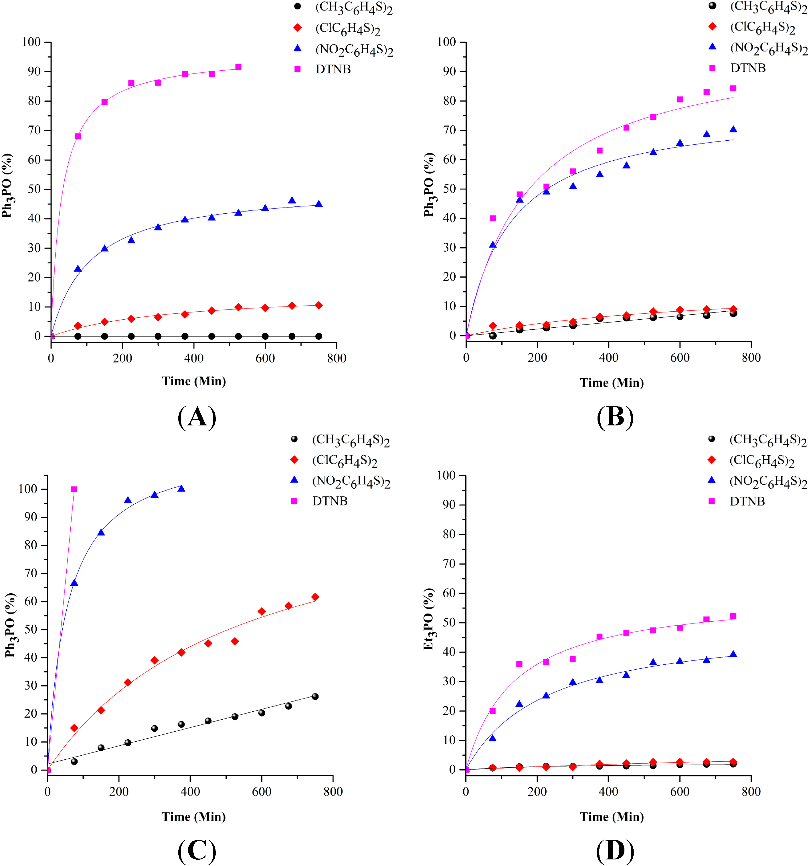
2.1. Phosphine Oxide Formation in DMSO

Reactions of Ph3PAu(SC6H4CH3) and (SC6H4NO2)2 were monitored by using 1H and 31P NMR spectroscopy. We observed formation of R3P=O in DMSO and DMF, but not in tetrahydrofuran, methylene chloride, acetone, or acetonitrile. We investigated the reaction in DMSO in more detail to determine the influence of the phosphine, thiolate and disulfide on the amount of phosphine oxide formed. The reactions of four phosphine gold(I) thiolates (10 mM) with four disulfides (10 mM) were carried out in DMSO-d6 and monitored by 31P{1H} NMR for 12 h. The results are plotted in Figure 1A–D for reactions of Ph3PAuSC6H4CH3, Ph3PAuSC6H4Cl, Ph3PAuSC6H4NO2, and Et3PAuTATG (auranofin), respectively. The percentage of R3P=O was calculated from the ratio of integrals for R3P=O (at 26.5 ppm for R = Ph or 50.8 ppm for R = Et) and R3PAuSRʹ (at 39.2 ppm for R = Ph or 40.3 ppm for auranofin). These are the only two signals observed in the 31P NMR spectra during the course of each reaction. Table 1 lists the reactions in decreasing order of extent of phosphine oxide formation at the 75 min. time point for each combination shown in Figure 1. The results show that disulfides with more strongly electron withdrawing groups [DTNB and (SC6H4NO2)2] produce more phosphine oxide. Electron withdrawing groups on the thiolate ligands also increase the extent of R3P=O formation. However the nature of the disulfide appears to have a larger effect. We have noted some variability in reaction rates with different batches of DMSO-d6, which may originate from differences in water content in the solvent. The experimental results reported in Table 1 were obtained employing the same batch of DMSO-d6 solvent.
Figure 1. Formation of R3PO in DMSO-d6 during reaction of 10 mM disulfides with 10 mM (A) Ph3PAu(SC6H4CH3); (B) Ph3PAu(SC6H4Cl); (C) Ph3PAu(SC6H4NO2); (D) Et3PAu(TATG) (auranofin). The percentage of R3PO was calculated as the ratio of R3PO to the total phosphorus in the 31P{1H} NMR.
Inorganics 03 00040 g001
Table 1. Comparison of the percentage of R3P=O formed from the reaction of 10 mM PR3AuSRʹ and 10 mM R*SSR* in DMSO-d6 (t = 75 min).
R3PAuSRʹR*SSR*R3P=O (%)
Ph3PAuSC6H4NO2DTNB 1100
Ph3PAuSC6H4CH3DTNB68
Ph3PAuSC6H4NO2(SC6H4NO2)267
Ph3PAuSC6H4ClDTNB40
Ph3PAuSC6H4Cl(SC6H4NO2)231
Ph3PAuSC6H4CH3(SC6H4NO2)223
Et3PAu(TATG) 2DTNB20
Ph3PAuSC6H4NO2(SC6H4Cl)215
Et3PAu(TATG)(SC6H4NO2)211
Ph3PAuSC6H4CH3(SC6H4Cl)24
Ph3PAuSC6H4Cl(SC6H4Cl)23
Ph3PAuSC6H4NO2(SC6H4CH3)23
Et3PAu(TATG) (SC6H4Cl)2<1
Et3PAu(TATG)(SC6H4CH3)2<1
Ph3PAuSC6H4CH3(SC6H4CH3)20
Ph3PAuSC6H4Cl(SC6H4CH3)20
1 DTNB = 5,5'-dithiobis-(2-nitrobenzoic acid); 2 TATG = tetraacetylthioglucose.

2.2. Possible Mechanistic Pathways for Phosphine Oxide Formation in DMSO

Two possible explanations for formation of phosphine oxide are illustrated in Scheme 1. The first involves dissociation of the phosphine ligand from gold(I), followed by nucleophilic attack on disulfide. The second involves reaction of the intact phosphine gold(I) thiolate complex in which the Au-P bond acts as the nucleophile. Dissociation of phosphine from the gold(I) complex was considered likely because of the well-known reaction of triarylphosphines with disulfides. Mechanistic studies reveal that the reaction of Ph3P and disulfide in dioxane/water mixtures is second order and proceeds via nucleophilic attack of Ph3P on the S–S bond in disulfide, resulting in a thiophosphonium cationic intermediate ([R′SPR3]+), which is subsequently hydrolyzed by water to produce phosphine oxide and thiol [12,13]. Disulfides with electron withdrawing groups react faster than those with electron donating groups. We have seen a similar trend for reactions of free Ph3P with disulfides in DMSO-d6 solution. For example, reactions of ca. 5 mM Ph3P and 10 mM R*SSR* (R* = C6H4NO2, C6H4Cl, C6H4CH3) were monitored by 31P NMR and the relative rates of formation of Ph3P=O are NO2 >> Cl > CH3.
Scheme 1. (A) Dissociation of phosphine and nucleophilic attack on disulfide; (B) Nucleophilic attack of Au–P bond on disulfide.
Inorganics 03 00040 g004aInorganics 03 00040 g004b
It is also interesting to note that Et3P=O is known to be an in vivo metabolite of auranofin. Interactions of auranofin with albumin have been studied in vitro. Shaw, et al. proposed that Et3P, from the Et3PAu(I) moiety bound to the cysteine-34 in albumin, is substituted by an additional cysteine thiolate. The released phosphine reacts with a protein disulfide bond, followed by hydrolysis to give Et3P=O [14,15].

2.3. No Evidence for Dissociation of Phosphine from Phosphine Gold(I) Thiolate in DMSO

To check for dissociation of phosphine from the phosphine gold(I) thiolate complexes in Table 1, the stability of each gold complex in DMSO-d6 was monitored by 1H and 31P NMR. The complexes are stable in this solvent and there is no evidence for dissociation of phosphine over a period of 24 h. An additional check on the stability of the gold–phosphorus bond was performed by adding benzyl azide as a trapping agent [16,17]. In a control reaction, a few drops of neat benzyl azide were added to a 5 mM solution of PPh3 in DMSO-d6 and the reaction was observed by 31P NMR. One hour after mixing, all of the phosphine was converted to a mixture of Ph3P=O and C6H5CH2N=PPh3. In contrast, when a few drops of neat benzyl azide were added to separate solutions of Ph3PAuSC6H4CH3 and Ph3PAuSC6H4NO2 (5 mM in DMSO-d6), the only peak observed in the 31P NMR was at ca. 39 ppm for the Ph3P ligand in Ph3PAu(SR′). These results suggest that the gold(I) complexes are stable in DMSO-d6 and phosphine does not dissociate, i.e., the reaction sequence depicted in Scheme 1A is not likely.

2.4. Water Is the Source of Oxygen for the Formation of Ph3P=O

Water is critical to the formation of R3P=O in the reaction of free phosphine with disulfide. The next set of experiments were carried out to determine whether water is also critical for the formation of R3P=O in the gold(I)/disulfide system. Dimethyl sulfoxide is a hygroscopic solvent and NMR solutions prepared in the atmosphere typically contain water. We therefore prepared a solution of 10 mM Ph3PAuSC6H4NO2 and 10 mM NO2C6H4SSC6H4NO2 in DMSO-d6 in an argon-filled drybox to minimize the amount of water present. (We used sealed ampoules of 99.5% DMSO-d6 with a water content of ca. 60 ppm, or 3.3 mM). The solution was divided into two portions and a drop of water was added to one of these solutions. Each solution was observed immediately (within 5 min) by 31P NMR. In the solution without added water, approximately 35% of the Ph3P had been converted to Ph3P=O. Addition of a drop of water (which is an excess) to the second sample caused complete conversion of the phosphine in Ph3PAuSC6H4NO2 to Ph3P=O. This demonstrates that water accelerates the formation of phosphine oxide in the gold(I)/disulfide system. In addition, GC-MS experiments using labeled water (H218O) demonstrate the formation of Ph3P=18O, confirming that water is the source of the oxygen in Ph3P=O (see Experimental Section).

2.5. What Is the Fate of Gold(I)?

When a solution of 7.5 mM Ph3PAuSC6H4CH3 and 5.0 mM NO2C6H4SSC6H4NO2 in DMSO-d6 was monitored by 1H NMR, a new peak at 7.9 ppm (broad doublet) was observed. This peak was assigned to the aromatic protons in [Au(SC6H4NO2)2] on the basis of comparison to a sample prepared independently by reaction of Au(tht)Cl with two equivalents of the sodium salt of para-nitrophenylthiolate (Na[SC6H4NO2]).

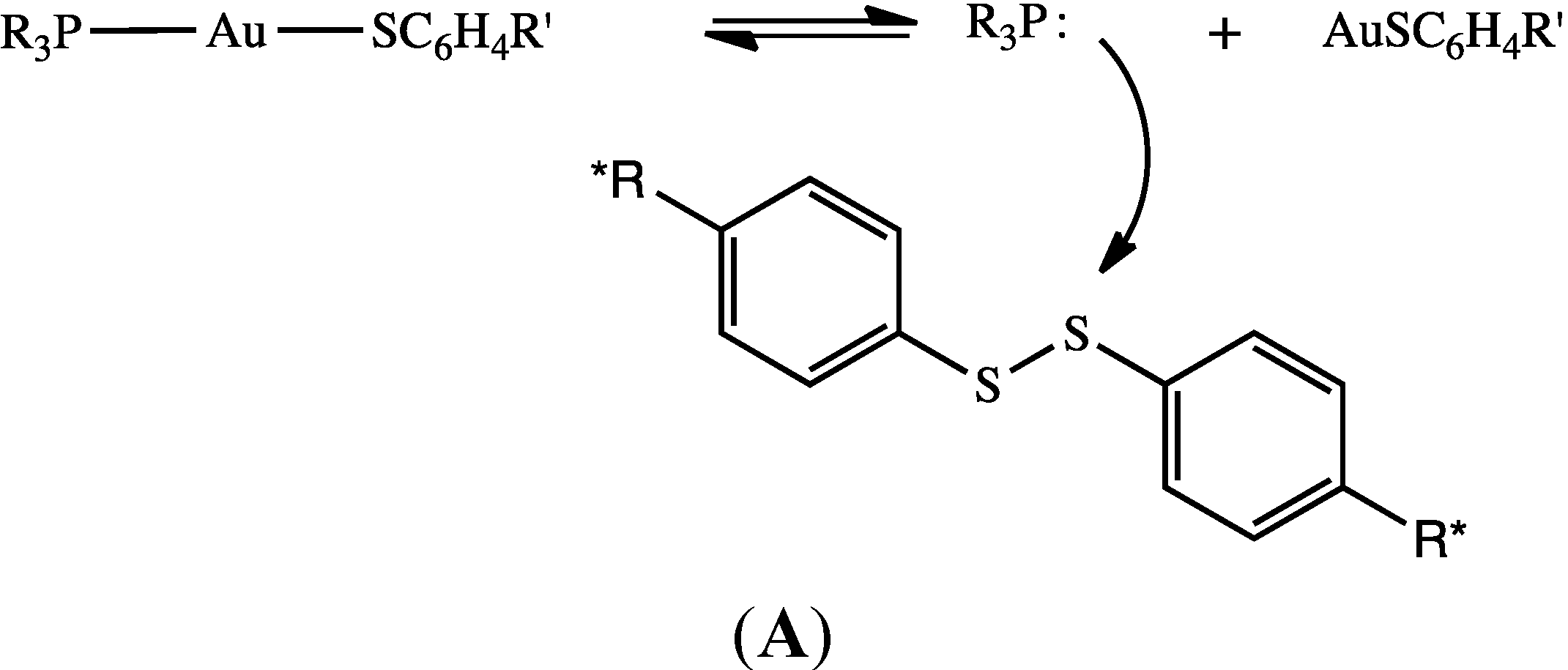
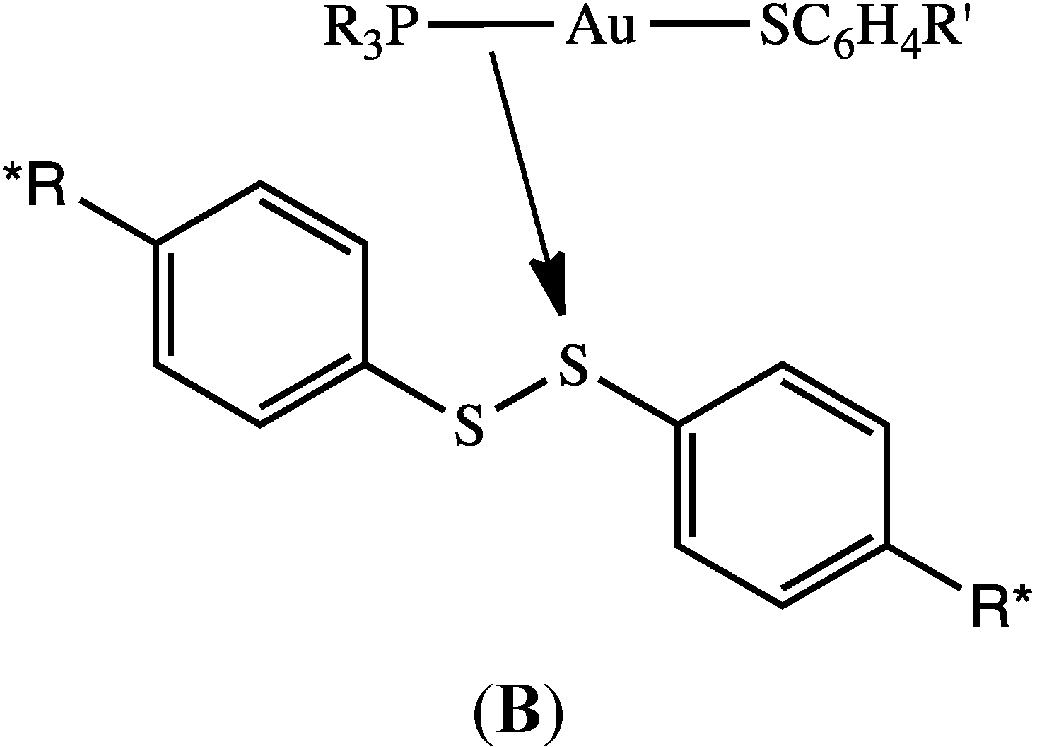
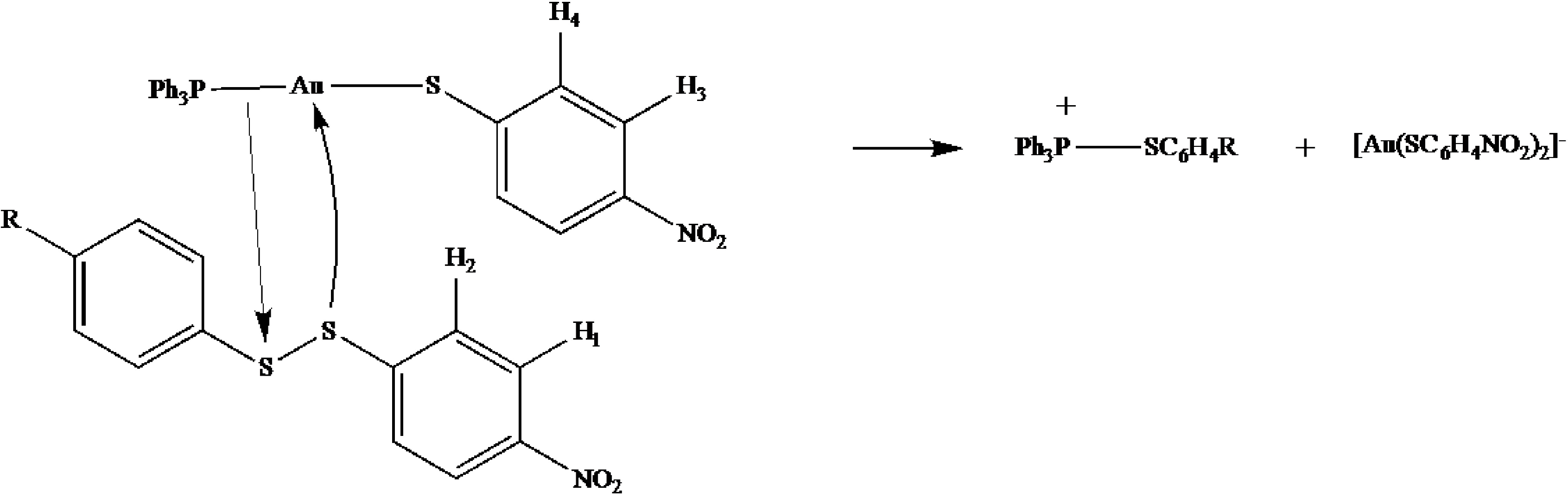
2.6. Mechanistic Reaction Sequence of Disulfide Competition for Phosphine Gold(I) Thiolates

The competing pathways for formation of thiolate–disulfide exchange and formation of phosphine are summarized in Equations (3)–(7), using as an example the reaction of Ph3PAuSC6H4CH3 and (SC6H4NO2)2.
Thiolate–disulfide exchange:
Ph3PAuSC6H4CH3 + (SC6H4NO2)2 ⇄ Ph3PAuSC6H4NO2 + NO2C6H4SSC6H4CH3
Phosphine oxide formation:
Ph3PAuSC6H4CH3 + (SC6H4NO2)2 ⇄ [Ph3PSC6H4NO2]+ + [(NO2C6H4S)Au(SC6H4CH3) ]
[Ph3PSC6H4NO2]+ + H2O ⇄ Ph3PO + HSC6H4NO2 + H+
HSC6H4NO2 ⇄ H+ + [SC6H4NO2]
[SC6H4NO2] + [(NO2C6H4S)Au(SC6H4CH3) ] ⇄ [Au(SC6H4NO2)2] + [SC6H4CH3]

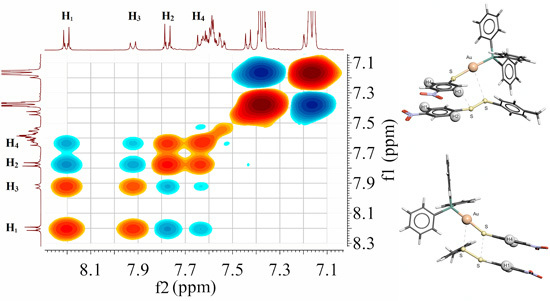
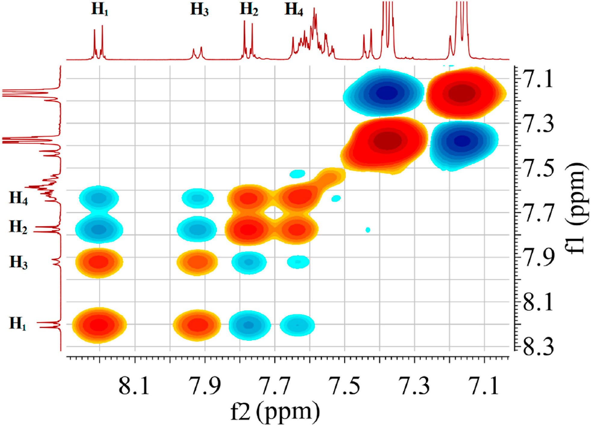
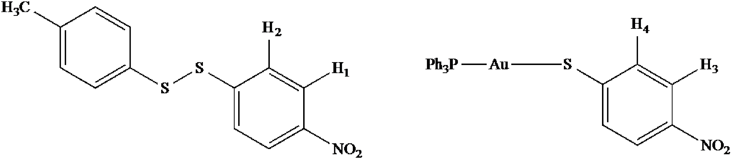
2.7. Scheme 1B Is Supported by 2D (1H-1H) NMR Experiments

The second possibility shown in Scheme 1B is that the Au-P bond acts as nucleophile toward the disulfide. Indirect support for this possibility is provided by 2D (1H-1H) NMR experiments, described below.
As part of our studies on the mechanism of thiolate–disulfide exchange, we have carried out a series of 2D (1H-1H) NMR ROESY experiments. For the self-exchange reaction between (SC6H4NO2)2 and Ph3PAuSC6H4NO2 in CD3CN during a 12 h ROESY experiment (for which no R3PO is detected, vide supra), analysis of the NOE peaks suggests a close approach of the disulfide and the gold(I)-thiolate moiety, which places the disulfide in a position to undergo exchange with the gold(I) bound thiolate [5]. In addition, on the related zinc(II)-thiolate, disulfide exchange reaction, there is experimental data suggesting close association [5,18], as well as DFT calculations which indicate close association of the disulfide with the zinc(II)-thiolate in the transition state [19].

2.8. 2D (1H-1H) NMR ROESY Experiments

2D (1H-1H) NMR ROESY experiments were also carried out in DMSO-d6. Since formation of phosphine oxide competes with thiolate–disulfide exchange in this solvent, and leads to eventual decomposition of the gold(I) thiolate complex, reactants were chosen to minimize the formation of R3PO. On the basis of the results shown in Table 1, we reasoned that the more electron donating methyl substituent on the thiolate ligand and the disulfide would produce the least amount of phosphine oxide. However, a nitro substituent on at least one aromatic ring is desirable because these peaks are well separated from the aromatic ring protons in the thiolate and phosphine ligands (i.e., SC6H4CH3 and PPh3) allowing for easier visualization and assignment of the NOEs. Thus we used Ph3PAuSC6H4CH3 in combination with the unsymmetrical disulfide, NO2C6H4SSC6H4CH3. (The unsymmetrical disulfide was prepared in situ by mixing NO2C6H4SSC6H4NO2 with an excess of CH3C6H4SSC6H4CH3, prior to addition of the gold(I) thiolate complex; see experimental section.)
The 2D (1H-1H) NMR ROESY spectrum shown in Figure 2 was acquired over a 4-h period. In this time frame, the starting gold(I) thiolate complex has undergone thiolate–disulfide exchange according to Equation (8). This is evident from the peaks labeled H3 and H4, which belong to the thiolate aromatic ring in Ph3PAuSC6H4NO2, and H1 and H2 for the nitro aromatic ring in NO2C6H4SSC6H4CH3 (see Scheme 2 for labels).
Ph3PAuSC6H4CH3 + NO2C6H4SSC6H4CH3 ⇄ Ph3PAuSC6H4NO2 + CH3C6H4SSC6H4CH3
The remaining peaks in the 1D 1H-NMR spectra are assigned as follows, starting at the high field side. The intense doublets at 7.38 and 7.18 ppm are due to the large excess of CH3C6H4SSC6H4CH3. The doublets at 7.42 and 7.2ppm (partially hidden by the symmetrical disulfide peak at 7.18 ppm) are assigned to the methyl aromatic ring in NO2C6H4SSC6H4CH3. The complex multiplet at 7.6 ppm is assigned to PPh3 and a small amount of Ph3PO. (The 31P NMR spectrum collected at the end of the 2D (1H-1H) experiment, shows ca. 3% Ph3P=O formation, which indicates that the conditions minimize the formation of phosphine oxide.)
Figure 2. 2D (1H-1H) ROESY spectrum in the aromatic region showing interaction between Ph3PAuSC6H4NO2 and CH3C6H4SSC6H4NO2 (DMSO-d6). See Scheme 2 for assignment of aromatic protons labeled H1, H2, H3, and H4. (The intense peaks at 7.16 and 7.37 ppm are due to the excess CH3C6H4S-SC6H4CH3; see Experimental Section.)
Inorganics 03 00040 g002
Scheme 2. Labels for aromatic ring protons in the disulfide and gold(I) thiolate complex.
Inorganics 03 00040 g005
The off diagonal contours in the 2D (1H-1H) ROESY spectrum provide important conformational information. Cross peaks with the same phasing as along the diagonal (red), indicate molecular exchange while the oppositely phased contours (blue) indicate long-range spatial interactions. The red, cross peaks centered at 7.9 and 8.2 ppm indicate thiolate–disulfide exchange between H3 on the thiolate in Ph3PAuSC6H4NO2 and H1 on the nitro end of the unsymmetrical disulfide, NO2C6H4SSC6H4CH3. (This is a nonproductive exchange since no new molecules are formed.) Intramolecular, long-range interactions between protons on the same ring (ortho to each other) are indicated by blue cross peaks for H1 and H2 in the disulfide, and also for H3 and H4 on the thiolate.
The most interesting aspect of the 2D (1H-1H) ROESY spectrum is the set of blue cross peaks, which indicate intermolecular long-range interactions between H4 on the thiolate ligand and H1 on the unsymmetrical disulfide as well as H3 on thiolate and H2 on disulfide. The distance between spatially close nuclei can be estimated according to the Equation (9) [20], where ηab is the ROE cross-peak volume (intensity) and rab is the inter-proton distance of the two protons “a” and “b”. The term, rref is a known distance between two protons and its ROE volume is given by ηref [20].
r ab r ref =   ( η ref η ab ) 1 6
For our calculation, rref was chosen as the intramolecular distances between H1 and H2 in the disulfide. The crystal structure of the symmetrical disulfide, (SC6H4NO2)2, is available in the Cambridge Structural Database (CSD Refcode: NIPHSS) [21,22] and the average distance between these protons is 2.365 Å. Using this value and the experimental ROE volumes, the spatial separation between H1…H4, and H2…H3 is estimated to be 2.6 Å.

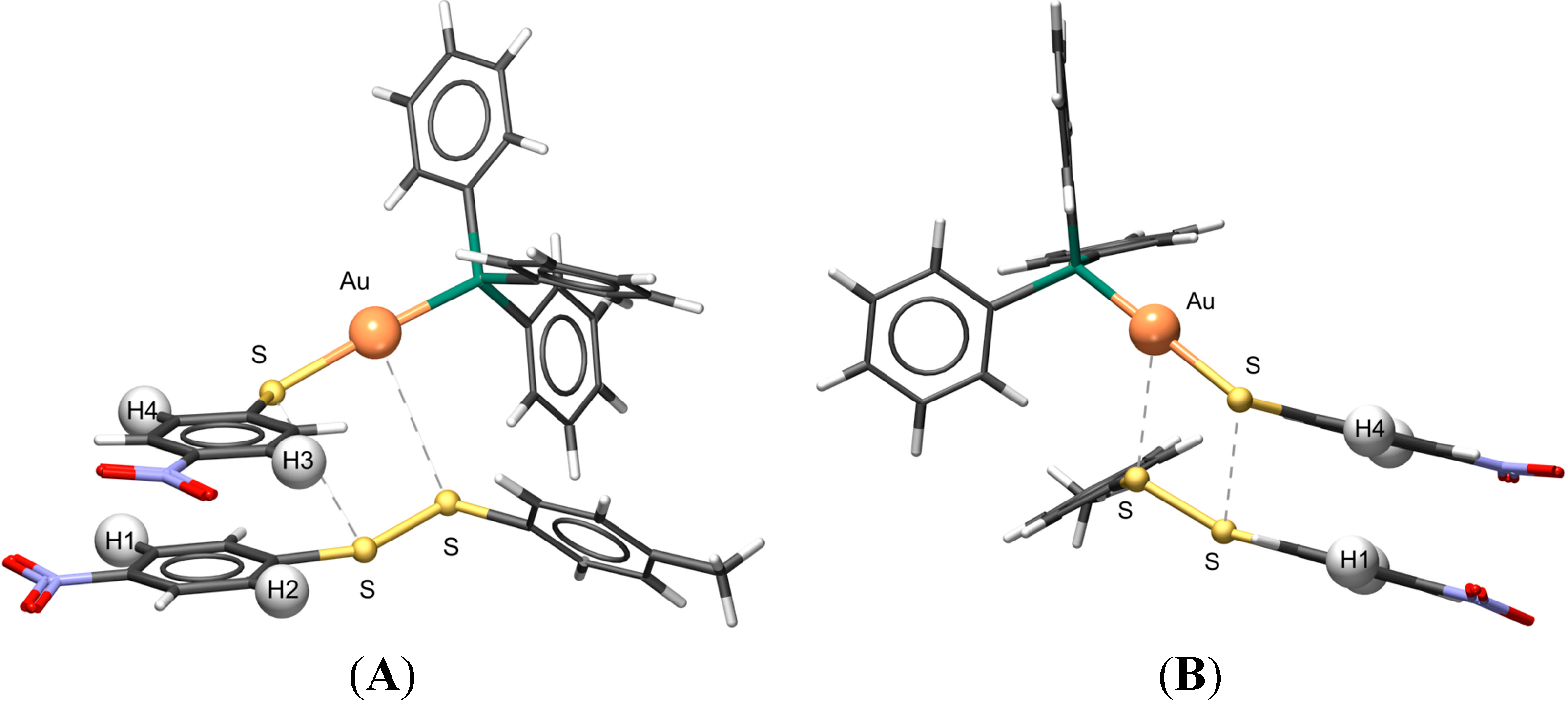
2.9. Visualizing the Association of Disulfide and the Phosphine Gold(I) Thiolate Complex

Figure 3 shows two views of a cartoon drawing to help visualize the association (and exchange) between disulfide and the phosphine gold(I) thiolate complex. The figure was constructed in Autodesk using typical bond lengths and angles for the disulfide and the gold(I) complex (e.g., the S–S, Au–S, and Au–P bonds are 2.02, 2.30, and 2.26 Å, respectively) [21,23]. The spatial orientations of the two molecules were adjusted to achieve intermolecular H1…H4, and H2…H3 distances of 2.6 Å, the distance estimated by analysis of the ROESY data. The S–S bond is oriented approximately parallel to the Au–S bond and the intermolecular S…S, S…Au and S…P distances are 3.66, 3.60, and 4.44 Å, respectively. We emphasize that this cartoon is not the result of a calculation but is useful to envision a possible conformational arrangement of the disulfide and gold(I) thiolate complex that is consistent with the ROESY data.
Figure 3. Cartoon drawings in two different views (A and B) to illustrate a possible orientation of Ph3PAu(SC6H4NO2) and the unsymmetrical disulfide, CH3C6H4SSC6H4NO2. The distance between the aromatic protons, H1 and H4, and H2 and H3 was set at 2.60 Å. The S…S and Au…S distances are 3.66 and 3.60 Å, respectively. The drawings were generated by using Autodesk 3DS MAX 2013.
Inorganics 03 00040 g003
Figure 3 also is useful for visualizing the competing reaction that leads to phosphine oxide. Sliding the disulfide along the linear S–Au–P moiety toward the Au–P bond would appear to require a relatively small spatial reorganization and would set up the disulfide to react across the Au–P bond rather than the Au–S bond (see Scheme 3). Reaction of gold(I) with the more electron withdrawing half of the unsymmetrical disulfide is consistent with the known chemistry of gold(I) [24]. The reaction is promoted by DMSO, a solvent with a high dielectric constant (ε = 46.6), which may help to stabilize the charged species that form following scission of the S–S bond.
Scheme 3. Competing reaction that leads to phosphine oxide.
Inorganics 03 00040 g006

3. Experimental Section

3.1. Chemicals and Instrumentation

All NMR solvents were purchased from Cambridge Isotope Laboratories, Inc. (Tewksbury, MA, USA) Only for 2D (1H-1H) NMR, solvents in ampules were used as received and solvents in the bottles were used after degassing by 5–10 freeze-pump cycles. Triphenylphosphine oxide (Ph3PO), triethylphosphine oxide (Et3PO), triphenylphosphine (PPh3), benzyl azide (BzN3), bis(4-chlorophenyl) disulfide [(ClC6H4S)2], water (H218O) and Ellman’s reagent [5,5′-dithiobis-(2-nitrobenzoic acid) or DTNB] were purchased from Sigma-Aldrich (St. Louis, MO, USA) and used as received. Bis(4-nitrophenyl) disulfide [(O2NC6H4S)2] and bis(4-methylphenyl) disulfide [(CH3C6H4S)2] were purchased from Sigma-Aldrich (St. Louis, MO, USA) and used after recrystallization. Auranofin [(Et3P)Au(TATG); TATG = 2,3,4,6-tetraacetyl-1-thio-D-glucopyranosato] was purchased from Enzo Life Sciences (Farmingdale, NY, USA). Syntheses of Au(tht)Cl [25], and phosphine gold(I) thiolates [23], were carried out according to previously reported procedures. Experiments were carried out under atmospheric conditions unless otherwise stated.
31P{1H} NMR spectra were obtained at 26 °C in deuterated solvents on a Varian (Palo Alto, CA, USA) UNITYplus-400 MHz “Oxford” NMR spectrometer operating at 161.9 MHz. 31P{1H} NMR spectra were referenced to 85% H3PO4 (external standard).
The sample tubes for 2D (1H-1H) ROESY (rotating frame Overhauser enhancement spectroscopy) NMR experiments were prepared in a Vacuum Atmospheres dry box under an argon environment and were capped with an air-tight seal. Two-dimensional NMR spectra were acquired at a constant temperature (30 °C) on a Varian (Palo Alto, CA, USA) UNITYplus-400 MHz “Oxford” NMR spectrometer (399.96 MHz for 1H) equipped with 4-nuc probes. Chemical shifts were referenced to residual 1H signals of the deuterated solvents.
GC-MS analyses were performed using a Thermo Scientific Trace (Waltham, MA, USA) GC Ultra gas chromatograph and Thermo Scientific (Waltham, MA, USA) ISQ mass spectrometer. Compound separation was achieved with a Rtx5-MS column (30 m × 0.25 mm i.d., crossbond 5% diphenyl/95% dimethyl polysiloxane× 0.25 µm (df) capacity Film thickness, Restek Corp., Bellefonte, PA, USA). The column flow rate was 2 mL/min. The oven temperature was held at 50 °C for 1 min, then increased to 350 °C (at a rate of 20 °C/min) and held at that temperature for 1 min. Injection, MS transfer line, and ion source temperatures were 270, 250, and 200 °C, respectively. Electron ionization mass spectrometric data from m/z 50 to 300 amu were collected using an ionization voltage of 70 eV. Compound identifications were made by comparing mass spectral patterns with those from known samples and the use of NIST mass spectra database.

3.2. Calibration of 31P NMR Integrals for a Mixture of Ph3PAuSC6H4CH3 and Ph3PO

The amount of 0.5 mL of a 9.96 mM solution of Ph3PAuSC6H4CH3 in DMSO-d6 was mixed in a vial with 0.5 mL of a 10.42 mM solution of Ph3PO in DMSO-d6. The resulting solution mixture containing 4.98 mM of Ph3PAuSC6H4CH3 and 5.21 mM Ph3PO was transferred into a NMR tube and investigated by 31P{1H}NMR experiments. The integral ratio for the peak at 39.2 ppm (Au–P) to the peak at 26.5 (P=O) was ca. 1:1.

3.3. Procedure for Measuring % of Phosphine Oxide

The amount of 0.4 mL of a 20.0 mM solution of Ph3PAuSC6H4R (R = CH3, Cl, NO2) or Et3PAu(TATG) in DMSO-d6 was mixed in a vial with 0.4 mL of a 20.0 mM solution of (R′C6H4S)2 (R′ = CH3, Cl, NO2) or DTNB in DMSO-d6. The solution mixture containing 10.0 mM Ph3PAuSC6H4R and 10.0 mM (R′C6H4S)2 or DTNB was transferred into a NMR tube and investigated by 31P{1H} NMR array experiments. The same experimental procedure was carried out for the reaction between Et3PAuSC6H4NO2 and (O2NC6H4S)2 in DMSO-d6. The Au–PR3 peak and R3P=O peaks were integrated and the % R3PO is based on the ratio of the R3P=O integral to the total phosphorus signal.

3.4. Test for Free Phosphine; Reaction of PPh3 and N3Bz in DMSO-d6

Four drops of benzyl azide (N3Bz) (~1.5 mmol) were added into 0.75 mL of a 5 mM solution of Ph3PAuSC6H4CH3 in DMSO-d6. 31P{1H} NMR spectra were observed over the time. The same reaction was carried out for Ph3PAuSC6H4NO2 in DMSO-d6. As a control, four drops of benzyl azide (N3Bz) (~1.5 mmol) were added into 0.75 mL of 5 mM solution of PPh3 in DMSO-d6. The 31P{1H} NMR spectra were observed over the time.

3.5. Test for Influence of Water

Solutions of Ph3PAuSC6H4NO2 (20 mM) and (SC6H4NO2)2 (20 mM) in DMSO-d6 were prepared in an argon-filled drybox. Equal volumes of solutions were mixed and the mixture was divided into two NMR tubes. The 31P{1H} NMR spectrum was obtained for one sample while keeping the second sample in the drybox. A drop of water was added to the second sample and the 31P{1H} NMR spectrum was obtained.

3.6. GC-MS Experiment for Source of Water

The amount of 0.2 mL of H218O was transferred into 2 mL of DMSO-d6, which contained 90 ppm (5 mM) water as received from the manufacturer. This solution was used to prepare solutions in the drybox for 31P {1H} NMR and GC-MS analysis as follows.
A solution of Ph3PAuSC6H4CH3 (5.8 mg in 1.1 mL, 10.0 mM) in DMSO-d6 was mixed in a vial with a solution of (O2NC6H4S)2 (3.1 mg in 1.1 mL, 10.0 mM) in DMSO-d6. Then 0.75 mL of the solution mixture was transferred into an NMR tube and investigated by 31P NMR. A separate sample (1 mL) of the same solution was transferred into a vial for GC-MS analysis.
A solution of Ph3PO in DMSO-d6 (1.3 mg in 1.0 mL, 4.7 mM) was prepared and analyzed by GC-MS as a reference.

3.7. Reaction of Au(tht)Cl and Na[SC6H4NO2]

The reaction was carried out under N2 using using Schlenk techniques. A solution of sodium methoxide (0.09 g, 1.56 mmol) in CH3OH (10 mL) was transferred via cannula into a solution of 4-nitrobenzenethiol (0.24 g, 1.56 mmol) in CH2Cl2 (20 mL). After stirring at 0 °C for five min., the mixture was transferred into a solution of Au(tht)Cl (0.25 g, 0.78 mmol) in CH2Cl2 (20 mL). The final mixture was stirred for 30 mins at 0 °C. The product was obtained by reducing the solvent in vacuo and then filtering through a sintered glass crucible in air. The product was washed thoroughly with EtOH and dried under a stream of N2. Yield calculated for Na[Au(SC6H4NO2)2]: 0.36 g, 90.12%. The sample was dissolved in DMSO-d6 to obtain a 1H NMR spectrum. There are two sets of doublets; the set with lower intensity centered at 7.89 and 7.60 ppm appears to convert to a more intense set centered at 7.87 and 7.55 ppm. Over time, the more intense set decreases and no new peaks grow in as a pale yellow solid precipitates, which is likely a gold(I) thiolate polymer since it is only sparingly soluble in DMSO and insoluble in other solvents.

3.8. 2D (1H-1H) ROESY Experiment

All solutions were prepared in an argon-filled drybox. Preparation of unsymmetrical disulfide: the two symmetrical disulfides, (SC6H4NO2)2 and (SC6H4CH3)2 were combined in an ~1:4 ratio and dissolved in 1 mL of DMSO-d6. (Complete conversion of (SC6H4NO2)2 to CH3C6H4SSC6H4NO22 under these conditions was confirmed by 1H NMR in a separate experiment.) After 30 min, the disulfide mixture was transferred into a vial containing Ph3PAu(SC6H4CH3). All solids were completely dissolved. The final concentrations of Ph3PAu(SC6H4NO2), CH3C6H4SSC6H4NO22, and (SC6H4CH3)2 at the end of the 2D experiment (~3 h) were estimated by integration to be 20, 30 and 160 mM, respectively. The reaction mixture was transferred into a screw cap NMR tube and a ROESY experiment was carried out with the π/2–t1–π/2–tm(spin lock)–π/2-FID pulse sequence by setting the previously measured PW90 and T1 for a dummy sample in DMSO-d6. An 8 s mixing time was applied. The spectra were recorded with 128 increments in t1values and 512 data points for the t2 dimension with 4 scans per t1 increment with a relaxation delay of 3 s. 2D (1H-1H) NMR data were processed with MestReNova NMR software (8.1.1, MestRelab Research, Santiago de Compostela, Spain, 2013).
The cartoon drawing showing a possible orientation of the disulfide and phosphine gold(I) thiolate, which is consistent with the 2D (1H-1H) ROESY experimental results was constructed in Autodesk 3ds MAX (2013, Autodesk, San Rafael, CA, USA).

4. Conclusions

The formation of phosphine oxide in the phosphine gold(I) thiolate–disulfide system shares some features with the reaction of free phosphine and disulfide; i.e., disulfides with electron withdrawing substituents react faster, and water is the source of oxygen. The presence of water is necessary but not sufficient in the gold(I)thiolate–disulfide system since phosphine oxide was not observed in acetonitrile, which also contains mM concentrations of water. There is no evidence for dissociation of the phosphine ligand from the gold(I) complex prior to reaction with disulfide. This suggests that the Au–P bond acts as a nucleophile to attack the S–S bond. 2D (1H-1H) NMR ROESY experiments provide evidence for a close association between disulfide and the gold(I) complex, which we have interpreted as a possible intermediate in the thiolate–disulfide exchange reaction. In addition, we suggest that a small spatial reorganization would allow the disulfide to react with the Au–P bond. The two disulfides which produce the most phosphine oxide are (SC6H4NO2)2 and DTNB. The corresponding thiols of these disulfides have low pKa’s (5.5 and 2.95, respectively) [12,26] and the thiolates would be expected to have a strong affinity for gold(I) [24]. This may be an important factor for the formation of the bis-thiolate gold(I) anion shown in Scheme 3. However, the relative affinity of thiolates for gold(I) would also be expected to drive the thiolate–disulfide exchange reaction. Thus the factors controlling the competition between thiolate–disulfide exchange and phosphine oxide formation are subtle and need to be more thoroughly investigated. Finally we have established that phosphine oxide formation happens on a longer time scale than thiolate–disulfide exchange, which is important for kinetic studies aimed at understanding the latter reaction.

Acknowledgments

The authors wish to thank Brian Frederick for his assistance with GC-MS experiments.

Author Contributions

The syntheses, 1H, 31P and 2D NMR experiments were performed by Gamage S. P. Garusinghe. Mostapha Aghamoosa contributed to experiments involving auranofin. Meaghan McKinnon carried reactions of free triphenylphosphine and disulfide. S. Max Bessey constructed Figure 3 and contributed to discussions of the association of disulfide and the phosphine gold(I) thiolate complex. Mitchell R. M. Bruce and Alice E. Bruce supervised the research. Gamage S. P. Garusinghe, Mitchell R. M. Bruce and Alice E. Bruce are responsible for the discussion, analysis of data and writing of the manuscript.

Conflicts of Interest

The authors declare no conflict of interest.

References

  1. Whitesides, G.M.; Lilburn, J.E.; Szajewski, R.P. Rates of thiol-disulfide interchange reactions between mono- and dithiols and Ellman’s reagent. J. Org. Chem. 1977, 42, 332–338. [Google Scholar] [CrossRef]
  2. Fernandes, P.A.; Ramos, M.J. Theoretical insights into the mechanism for thiol/disulfide exchange. Chemistry 2004, 10, 257–266. [Google Scholar] [CrossRef] [PubMed]
  3. DiLorenzo, M.; Ganesh, S.; Tadayon, L.; Chen, J.; Bruce, M.R.M.; Bruce, A.E. Reactions of organic disulfides and gold(I) complexes. Met.-Based Drugs 1999, 6, 247–253. [Google Scholar] [CrossRef] [PubMed]
  4. Mohamed, A.A.; Abdou, H.E.; Chen, J.; Bruce, A.E.; Bruce, M.R.M. Perspectives in Inorganic and Bioinorganic Gold Sulfur Chemistry. Comments Inorg. Chem. 2002, 23, 321–334. [Google Scholar] [CrossRef]
  5. Garusinghe, G.S.P. Kinetic Analysis of Metal Assisted-Thiolate Disulfide Exchange (M = Au, Zn). Ph.D. Thesis, The University of Maine, Orono, ME, USA, May 2013. [Google Scholar]
  6. Bachman, R.E.; Bodolosky-Bettis, S.A.; Pyle, C.J.; Gray, M.A. Reversible Oxidative Addition and Reductive Elimination of Fluorinated Disulfides at Gold(I) Thiolate Complexes: A New Ligand Exchange Mechanism. J. Am. Chem. Soc. 2008, 130, 14303–14310. [Google Scholar] [CrossRef] [PubMed]
  7. Garusinghe, G.S.P.; Bessey, S.M.; Bruce, A.E.; Bruce, M.R.M. Interactions between Metal Thiolate, R3PAuSC6H4NO2 (R = Ph and Et) and Bis(4-nitrophenyl) Disulfide. In Proceedings of the 36th Northeast Regional Meeting of the American Chemical Society, Hartford, CT, USA, 7–10 October 2009.
  8. Chandrasoma, A.; Bruce, A.; Bruce, M. Insight into Metal-Mediated Thiol–Disulfide Exchange. In Proceedings of the 37th Northeast Regional Meeting of the American Chemical Society, Burlington, VT, USA, 29 June–2 July 2008.
  9. Aghamoosa, M.; Briggs, B.; Harriman, E.; Cashman, A.; Bruce, A.; Bruce, M. Gold(I)-Mediated Disulfide Exchange Kinetics as a Function of Solvent Dielectric Constant. In Proceedings of the 37th Northeast Regional Meeting of the American Chemical Society, Burlington, VT, USA, 29 June–2 July 2008.
  10. DiLorenzo, M.A.; Ganesh, S.; Bruce, A.E.; Bruce, M.R.M. Gold(I) mediated thiolate/disulfide exchange reactions. In Proceedings of the 213th ACS National Meeting, San Francisco, CA, USA, 13–17 April 1997.
  11. Garusinghe, G.S.P.; Bessey, S.M.; Boyd, C.; Aghamoosa, M.; Frederick, B.; Bruce, M.R.M.; Bruce, A.E.; (University of Maine, Orono, ME, USA). Determination of dimethyl sulfide in dimethyl sulfoxide. Unpublished work. 2015. [Google Scholar]
  12. Overman, L.E.; Matzinge, D.; Oconnor, E.M.; Overman, J.D. Nucleophilic Cleavage of Sulfur–Sulfur Bond by Phosphorus Nucleophiles—Kinetic Study of Reduction of Aryl Disulfides with Triphenylphosphine and Water. J. Am. Chem. Soc. 1974, 96, 6081–6089. [Google Scholar] [CrossRef]
  13. Dmitrenko, O.; Thorpe, C.; Bach, R.D. Mechanism of SN2 Disulfide Bond Cleavage by Phosphorus Nucleophiles. Implications for Biochemical Disulfide Reducing Agents. J. Org. Chem. 2007, 72, 8298–8307. [Google Scholar] [CrossRef] [PubMed]
  14. Shaw, C.F.; Isab, A.A.; Hoeschele, J.D.; Starich, M.; Locke, J.; Schulteis, P.; Xiao, J. Oxidation of the phosphine from the auranofin analog, triisopropylphosphine(2,3,4,6-tetra-O-acetyl-1-thio-β-D-glucopyranosato-S)gold(I), via a protein-bound phosphonium intermediate. J. Am. Chem. Soc. 1994, 116, 2254–2260. [Google Scholar] [CrossRef]
  15. Hill, D.T.; Isab, A.A.; Griswold, D.E.; DiMartino, M.J.; Matz, E.D.; Figueroa, A.L.; Wawro, J.E.; DeBrosse, C.; Reiff, W.M.; Elder, R.C.; et al. Seleno-Auranofin (Et3PAuSe-tagl): Synthesis, Spectroscopic (EXAFS, 197Au Mossbauer, 31P, 1H, 13C, and 77Se NMR, ESI-MS) Characterization, Biological Activity, and Rapid Serum Albumin-Induced Triethylphosphine Oxide Generation. Inorg. Chem. 2010, 49, 7663–7675. [Google Scholar] [CrossRef] [PubMed]
  16. Lin, F.L.; Hoyt, H.M.; van Halbeek, H.; Bergman, R.G.; Bertozzi, C.R. Mechanistic investigation of the Staudinger ligation. J. Am. Chem. Soc. 2005, 127, 2686–2695. [Google Scholar] [CrossRef] [PubMed]
  17. Woehrle, G.H.; Hutchison, J.E. Thiol-functionalized undecagold clusters by ligand exchange: Synthesis, mechanism, and properties. Inorg. Chem. 2005, 44, 6149–6158. [Google Scholar] [CrossRef] [PubMed]
  18. Boerzel, H.; Koeckert, M.; Bu, W.; Spingler, B.; Lippard, S.J. Zinc-Bound Thiolate-Disulfide Exchange: A Strategy for Inhibiting Metallo-β-Lactamases. Inorg. Chem. 2003, 42, 1604–1615. [Google Scholar] [CrossRef] [PubMed] [Green Version]
  19. Acharige, A. Reactions of Sulfur Compounds with Zn(II), Au(I), and Hg(II) and Metal Linker Polyaniline Electrodes for Energy Storage. Ph.D. Thesis, University of Maine, Orono, ME, USA, May 2011. [Google Scholar]
  20. Binotti, B.; Macchioni, A.; Zuccaccia, C.; Zuccaccia, D. Application of NOE and PGSE NMR methodologies to investigate non-covalent intimate inorganic adducts in solution. Comments Inorg. Chem. 2002, 23, 417–450. [Google Scholar] [CrossRef]
  21. Ricci, J.S.; Bernal, I. Crystal structure of α-bis(p-nitrophenyl) disulfide. J. Am. Chem. Soc. 1969, 91, 4078–4082. [Google Scholar] [CrossRef]
  22. Allen, F. The Cambridge Structural Database: A quarter of a million crystal structures and rising. Acta Crystallogr. Sect. B 2002, 58, 380–388. [Google Scholar] [CrossRef]
  23. Narayanaswamy, R.; Young, M.A.; Parkhurst, E.; Ouellette, M.; Kerr, M.E.; Ho, D.M.; Elder, R.C.; Bruce, A.E.; Bruce, M.R.M. Synthesis, structure, and electronic spectroscopy of neutral, dinuclear gold(I) complexes. Gold(I)–gold(I) interactions in solution and in the solid state. Inorg. Chem. 1993, 32, 2506–2517. [Google Scholar] [CrossRef]
  24. Isab, A.A.; Sadler, P.J. A 13C Nuclear Magnetic-Resonance Study of Thiol-Exchange Reactions of Gold(I) Thiomalate (Myocrisin) Including Applications to Cysteine Derivatives. Dalton Trans. 1982, 135–141. [Google Scholar] [CrossRef]
  25. Uson, R.; Laguna, A.; Laguna, M.; Briggs, D.A.; Murray, H.H.; Fackler, J.P. (Tetrahydrothiophene) Gold(I) or Gold(III) Complexes. In Inorganic Syntheses; John Wiley & Sons, Inc.: Hoboken, NJ, USA, 2007; pp. 85–91. [Google Scholar]
  26. Bordwell, F.G.; Hughes, D.L. Thiol Acidities and Thiolate Ion Reactivities toward Butyl Chloride in Dimethylsulfoxide Solution—The Question of Curvature in Bronsted Plots. J. Org. Chem. 1982, 47, 3224–3232. [Google Scholar] [CrossRef]

