Article

Upregulation of Transferrin and Major Royal Jelly Proteins in the Spermathecal Fluid of Mated Honeybee (Apis mellifera) Queens

1
College of Natural Resources and Life Science, Dong-A University, Busan 49315, Korea
2
Department of Agricultural Biology, National Academy of Agricultural Science, Wanju 55365, Korea
*
Authors to whom correspondence should be addressed.
These authors contributed equally to the work.
Submission received: 30 June 2021 / Revised: 26 July 2021 / Accepted: 29 July 2021 / Published: 31 July 2021
(This article belongs to the Section Insect Molecular Biology and Genomics)

Abstract

:

Simple Summary

To understand the mechanisms underlying long-term storage and survival of sperm in honeybee Apis mellifera queens, previous studies have elucidated the components of honeybee spermathecal fluid. However, the expression profiles of transferrin (Tf) and major royal jelly proteins 1–9 (MRJPs 1–9) in the spermatheca and spermathecal fluid of mated honeybee queens have still not been characterized. In this study, we confirmed upregulation of Tf and MRJPs in the spermatheca and spermathecal fluid of mated honeybee queens by using RNA sequencing, reverse transcription-polymerase chain reaction, and Western blot analyses. The levels of Tf and antioxidant enzymes were elevated in the spermathecal fluid of the mated queens, paralleling the levels of reactive oxygen species, H2O2, and iron. The increased levels of MRJPs, especially MRJP1, MRJP4, and MRJP6, in the spermathecal fluid of mated queens may be responsible for energy provision during sperm storage in honeybee queens. Overall, our findings indicate that Tf and MRJPs are upregulated in the spermatheca and spermathecal fluid of mated honeybee queens, providing a novel insight into antioxidant defense and energy metabolism for stored sperm in honeybee queens.

Abstract

Sperm storage in the spermathecae of honeybee (Apis mellifera) queens is vital for reproduction of honeybees. However, the molecular mechanisms whereby queens store sperm in a viable state over prolonged periods in the spermatheca are not fully understood. Here, we conducted RNA sequencing analysis of the spermathecae in both virgin and mated A. mellifera queens 24 h after mating and observed that the genes encoding transferrin (Tf) and major royal jelly proteins (MRJPs) were differentially expressed in the spermathecae of mated queens. The concentrations of Tf and antioxidant proteins such as superoxide dismutase 1, catalase, and glutathione peroxidase as well as the levels of reactive oxygen species, H2O2, and iron were higher in the spermathecal fluid of the mated queens than in virgin queens. Tf upregulation is likely to perform a protective role against the Fenton reaction occurring between iron and H2O2 in the antioxidant pathway in the mated queen’s spermathecal fluid. Furthermore, MRJPs—especially MRJP1, MRJP4, and MRJP6—were upregulated in the mated queen’s spermathecal fluid, indicating that they may serve as antimicrobial and antioxidant agents as well as an energy source for stored sperm in the spermathecal fluid of honeybee queens. Together, our findings show that Tf and MRJPs are upregulated in the spermatheca and spermathecal fluid of mated honeybee queens.

1. Introduction

Honeybee (Apis mellifera) queens mate during the early stages of their lives, store sperm in the spermatheca, and subsequently use the stored sperm throughout their lifetimes [1,2]. Sperm storage in the spermatheca of honeybee queens plays a vital role in the reproduction of honeybees, but the molecular mechanisms whereby queens can maintain sperm in a viable state in the spermatheca over prolonged periods are still not completely understood. Spermathecal fluid, which is secreted by the spermathecal glands of honeybee queens, contains proteins and metabolites [2,3,4,5] and has been shown to facilitate long-term sperm storage [2,4,6,7]. Several proteins, such as the glycolytic enzyme triosephosphate isomerase [3] and antioxidant enzymes [8,9,10], have been proposed to be responsible for sperm viability in the spermathecal fluid of honeybee queens.
Extensive studies, including proteomic, metabolomic, and transcriptomic analyses, have been conducted on the storage and survival of sperm in A. mellifera [1,2,4,5,10,11,12,13,14]. A previous study identified >100 proteins as major spermathecal fluid constituents in A. mellifera queens and analyzed the association of spermathecal fluid proteins with energy metabolism and antioxidant defense [2]. Recent studies have identified >10,000 transcripts and 2778 proteins in the mated or virgin A. mellifera queen’s spermathecae via transcriptomic and proteomic analyses, respectively [13,14]. The results from the transcriptomic analyses revealed the gene expression patterns during sperm storage after mating and showed that Kielin/chordin-like and trehalase transcripts are highly expressed in the spermathecae of the mated queens, in comparison with their levels in virgin queens [14]. The glycolytic metabolite glyceraldehyde-3-phosphate reportedly facilitates the long-term survival and energy (ATP) production of honeybee sperm during their storage in the spermathecae, because this metabolite functions independently of oxygen and prevents sperm damage caused by reactive oxygen species (ROS) [4]. Moreover, the activities of catalase and glutathione S-transferase (GST) in the mated A. mellifera queen’s spermathecae were higher than those in the virgin queen’s spermathecae [8]. The upregulation of antioxidant genes such as catalase, thioredoxin 2, and thioredoxin reductase 1 after mating is associated with sperm viability within the mated A. mellifera queen’s spermathecae [10]. Moreover, stored sperm showed a reduced metabolic rate and lower ROS levels [15].
The metabolic energy profiles of sperm and spermathecae were characterized on the basis of the activities of enzymes involved in the aerobic and anaerobic carbohydrate metabolisms during the life cycles of honeybee queens [11]. Glycolytic enzymes such as glyceraldehyde-3-phosphate dehydrogenase (GAPDH) and enolase are abundant in the stored sperm [16] and spermathecal fluid of honeybee queens [2]. Heat-induced changes in GAPDH expression influence the viability of sperm in the spermathecae of honeybee queens [13]. Glycolytic enzymes in the spermathecal fluid of honeybee queens have been linked to sperm energy metabolism [4], whereas antioxidant enzymes reportedly protect the stored sperm against ROS activity [8,9,10].
Previous studies have investigated the energy metabolism and antioxidant defense during sperm storage in the spermathecae of honeybee queens [4.15]. Moreover, honeybee spermathecal fluid components have been elucidated to clarify the mechanisms underlying long-term storage and survival of sperm in the spermathecae of A. mellifera queens [1,2]. Proteomic analysis has also demonstrated that the spermathecal fluid of A. mellifera queens contains transferrin (Tf) and two major royal jelly proteins (MRJPs 8 and 9) [2]. However, the expression patterns and roles of Tf and MRJPs 1–9 in the spermathecae of mated honeybee queens have still not been described in detail. Moreover, the expression patterns of MRJPs 1–9 in the spermathecal fluid of mated queens have not been characterized, and the potential roles of increased levels of Tf in protecting the sperm from ROS action remain unclear. Therefore, characterization of the expression patterns of MRJPs and Tf in the spermathecal fluid of mated queens is essential to understand the mechanisms underlying sperm storage in honeybee queens.
In this study, we aimed to address these questions by using RNA sequencing (RNA-seq) analyses to compare the spermathecae of virgin and mated A. mellifera queens 24 h after mating. We found that unlike the spermathecae of virgin queens, the spermathecae of mated queens showed upregulation of Tf, which functions as an antioxidant protein with a protective role against the Fenton reaction occurring between Fe2+ and H2O2 [17,18,19,20,21,22], while the mated queen’s spermathecal fluid showed increased levels of ROS, H2O2, and iron. We also showed that MRJPs 1–9—especially MRJP1, MRJP4, and MRJP6—are upregulated in the spermathecae and spermathecal fluid of mated queens. These results suggest that upregulation of MRJPs and Tf may be involved in energy metabolism and antioxidant defense during sperm storage in the mated queen’s spermathecal fluid.

2. Materials and Methods

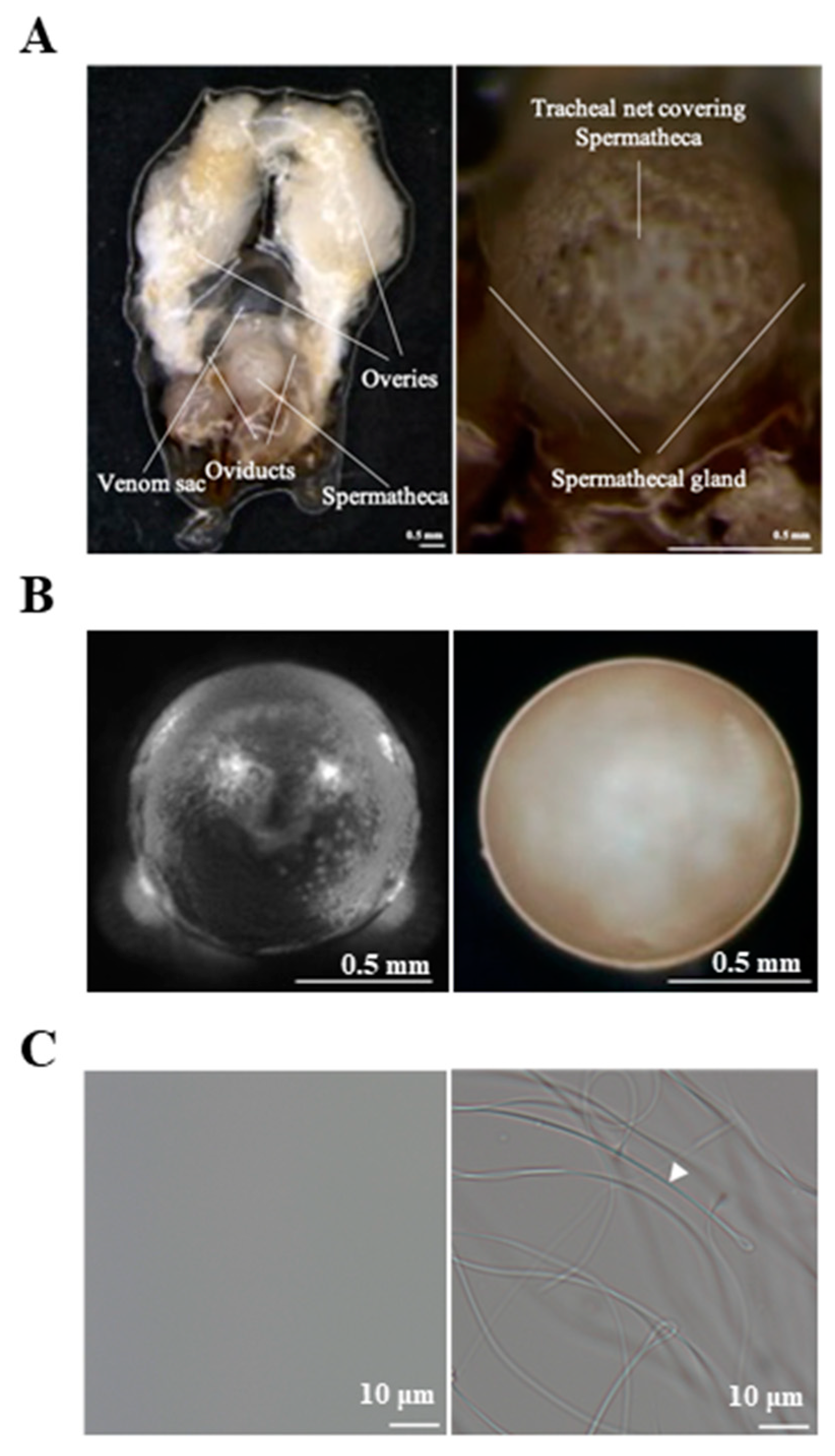
2.1. Sample Preparation

The bees used for the experiments were reared at Dong-A University. To produce honeybee queens, one-day-old larvae were grafted into queen cells, placed on frames, and introduced into a de-queened colony [23]. Virgin queens were kept in nucleus colonies, and six-day-old virgin queens were artificially inseminated using a previously developed method to produce mated queens [24]. Spermathecae from six-day-old virgin queens after emergence were dissected on ice by using a stereomicroscope (Zeiss, Jena, Germany), while spermathecae from mated queens were dissected 24 h after artificial insemination. The spermathecae were punctured to extract the spermathecal fluid, which was subsequently centrifuged at 14,000× g for 10 min to remove tissue debris and sperm (in the fluid extracted from mated queens). The spermathecae of virgin and mated queens and the sperm in the spermathecal fluid of mated queens were observed using a stereomicroscope. The supernatant (spermathecal fluid) was stored at −70 °C until further use. Spermathecal samples, including the spermathecal glands from virgin queens (without spermathecal fluid) and mated queens (without spermathecal fluid and sperm), were collected and washed with phosphate-buffered saline (PBS: 140 mM NaCl, 27 mM KCl, 8 mM Na2HPO4, and 1.5 mM KH2PO4, pH 7.4) for subsequent molecular analyses. All samples were collected from three biological replicates: three pooled spermatheca or spermathecal fluid samples (n = 9 virgin or mated queens).

2.2. RNA Sequencing and Data Analysis

Total RNA was isolated from the spermatheca samples (without spermathecal fluid or sperm) from virgin or mated queens by using TRIzol reagent (Invitrogen, Carlsbad, CA, USA) according to the manufacturer’s protocol. The RNA samples for RNA sequencing were prepared from three biological replicates: three pooled spermatheca samples (n = 9 virgin or mated queens). RNA sequencing was performed using the NovaSeq 6000 Sequencing System (Illumina, San Diego, CA, USA) at Macrogen Inc. (Seoul, Korea). Briefly, sequence libraries were constructed using the TruSeq RNA Library Preparation Kit (Illumina) by following the manufacturer’s protocol. The sequences were qualified using both the FastQC (www.bioinformatics.babraham.ac.uk/projects/fastqc, accessed on 30 July 2021) and Trimmomatic programs (www.usadellab.org/cms/index/php?page=trimmomatic, accessed on 30 July 2021). RNA reads were mapped to a reference genome (GCF_000002195.4_Amel_4.5) [14] by using the HISAT2 aligner [25]. Transcript counts, representing gene expression levels, were determined as fragments per kilobase of exon per million fragments mapped (FPKM). The analysis of differentially expressed genes (DEGs) was performed using the Database for Annotation, Visualization and Integrated Discovery (DAVID) platform [26] (https://david.ncifcrf.gov/home.jsp, accessed on 30 July 2021), and log2 values were calculated and normalized through quantile normalization. Fold changes were calculated as the mean log2 (FPKM+1) value, and an independent t-test was used to determine the statistical significance of the differences between the values obtained from mated and virgin queens. Differences with p-values < 0.05 were considered statistically significant.

2.3. Reverse Transcription-PCR (RT-PCR)

To examine the expression patterns of genes encoding antioxidant proteins and MRJPs in the spermathecae, the cDNAs for catalase (GenBank accession number GB41427), glutathione peroxidase (GTPX) (GenBank accession number GB47478), transferrin (Tf) (GenBank accession number GB50226), superoxide dismutase 1 (SOD1) (GenBank accession number GB47880), and MRJPs (MRJP1, MRJP2, MRJP3, MRJP4, MRJP5, MRJP6, MRJP7, MRJP8, and MRJP9) were amplified from the total RNA by using RT-PCR. The PCR primers for MRJPs 1–9 have been described in our previous studies [27,28]: MRJP1 (GenBank accession number NM_001011579), MRJP2 (NM_001011580), MRJP3 (NM_001011601), MRJP4 (NM_001011610), MRJP5 (NM_001011599), MRJP6 (NM_001011622), MRJP7 (NM_001014429), MRJP8 (NM_001011564), and MRJP9 (NM_001024697). The PCR primer sets used in this study were designed to amplify full sequences and are listed in Table 1. β–Actin (GenBank accession number NM_001185145) was used as the internal control. PCR was performed using a thermocycler (Takara Bio, Otsu, Japan) as follows: 94 °C for 2 min, 35 cycles of amplification (94 °C for 1 min, 53 °C for 1 min, and 72 °C for 2 min), and 72 °C for 10 min. The resulting fragments were observed by electrophoresis with a 1.0% agarose gel, and the cDNA concentrations were measured [29]. Finally, the resulting fragments were verified by DNA sequence analysis using a 3730XL DNA Analyzer (Applied Biosystems, Foster City, CA, USA).

2.4. Recombinant Protein Expression and Purification

Recombinant catalase—GTPX, Tf, and SOD1—were produced using a baculovirus expression system [30]. The protein-coding sequences of catalase, GTPX, Tf, and SOD1 were PCR-amplified, and a His-tag sequence was included in each cDNA sequence. The cDNA fragments corresponding to the protein sequences were inserted into the pBacPAK8 transfer vector (Clontech, Palo Alto, CA, USA), in which gene expression is under the control of the polyhedrin promoter of Autographa californica nucleopolyhedrovirus (AcNPV). For the expression experiments, 500 ng of the constructs and 100 ng of AcNPV viral DNA [30] were co-transfected into 1.0–1.5 × 10⁶ Spodoptera frugiperda (Sf9) cells for 5 h by using Lipofectin transfection reagent (Gibco BRL, Gaithersburg, MD, USA). The transfected cells were cultured in TC100 medium (Gibco BRL) with 10% fetal bovine serum (FBS, Gibco BRL) at 27 °C for 5 d. Recombinant baculoviruses were propagated in Sf9 cells cultured in TC100 medium at 27 °C to produce the recombinant proteins, which were then purified using the MagneHis Protein Purification System (Promega, Madison, WI, USA). Protein concentrations were determined using the Bio-Rad Protein Assay Kit (Bio-Rad, Hercules, CA, USA). The recombinant MRJP proteins (MRJPs 1–9) produced in baculovirus-infected insect cells were used as described in our previous studies [27,28].

2.5. Polyclonal Antibody Production and Western Blotting

Purified recombinant proteins, such as catalase, GTPX, Tf, and SOD1, were mixed with an equal volume of Freund’s complete adjuvant (a total of 200 μL) and injected into BALB/c mice (Samtako Bio Korea Co., Osan, Korea). Two successive injections with a one-week interval were administered using antigens mixed with equal volumes of Freund’s incomplete adjuvant (a total of 200 μL). Blood was collected 3 d after the second injection and centrifuged at 10,000× g for 5 min. The supernatants were used as the antibodies for the Western blot analyses. The anti-MRJP 1–9 antibodies raised against recombinant MRJPs 1–9 were used as described in our previous studies [27,28].
Western blot analysis was performed using an enhanced chemiluminescence (ECL) western blot system (Amersham Biosciences, Piscataway, NJ, USA). Protein samples were mixed with the sample buffer (0.0625 M Tri-HCl, pH 6.8, 2% sodium dodecyl sulfate [SDS], 10% glycerol, 5% β-mercaptoethanol, and 0.125% bromophenol blue) and then boiled for 5 min. The protein samples (5 μg/lane) from the spermathecae (without spermathecal fluid or sperm) and the spermathecal fluid (without sperm) of virgin and mated queens were separated using SDS-polyacrylamide gel electrophoresis (SDS-PAGE) with a 12% gel. After the electrophoresis, the proteins were transferred onto a nitrocellulose membrane (Schleicher & Schuell, Dassel, Germany), which was subsequently blocked with 1% bovine serum albumin. The membranes were then incubated with the antiserum solution (1:1000 v/v) at room temperature for 1 h and washed in TBST (10 mM Tri-HCl, pH 8.0, 100 mM NaCl, and 0.005% (v/v) Tween 20). They were incubated with a 1:5000 (v/v) diluted anti-mouse IgG horseradish peroxidase (HRP) conjugate and HRP–streptavidin complex. After repeated washes with TBST, the membranes were incubated with ECL detection reagents (Amersham Biosciences) and exposed to an autoradiography film. The western blot images were analyzed by determining the integrated density of each band area by using a computerized image analysis system (Alpha Innotech Co., San Leandro, CA, USA) and AlphaEaseFC (ver. 4.0). The relative levels of each protein are shown as the mean values from three measurements, which were calculated relative to the levels in the spermathecal fluid of virgin queens (shown as 100%).

2.6. Measurement of ROS and Iron Levels

The ROS, H2O2, and iron levels in the spermathecal fluid of virgin and mated honeybee queens were quantified using the OxiSelect In Vitro ROS/RNS Assay Kit (Green Fluorescence; Cell Biolabs, Inc., San Diego, CA, USA), Hydrogen Peroxide Assay Kit (Abcam, Cambridge, UK), and Iron Colorimetric Assay Kit (BioVision Inc., Milpitas, CA, USA) according to the instructions of the manufacturers. Experiments were performed three times by using independent samples.

2.7. Enzymatic Activity Assay

The catalase, peroxidase, and SOD enzyme activities in the spermathecal fluid were assayed using a Catalase Activity Colorimetric/Fluorometric Assay Kit (BioVision Inc.), Peroxidase Activity Colorimetric/Fluorometric Assay Kit (BioVision Inc.), and SOD Activity Assay Kit (BioVision Inc.) according to the manufacturer’s instructions.

2.8. Statistical Analysis

Data are presented as the mean ± SD. To determine the statistical significance of the differences in values between the mated and virgin queens, unpaired two-tailed Student’s t-test and the statistical software SPSS version 22.0 (IBM Inc., Chicago, IL, USA) were used. We set statistical significance at p-values of <0.05, <0.01, and <0.001.

3. Results

3.1. Identification of Genes Differentially Expressed between the Mated and Virgin Queens’ Spermathecae

We collected spermathecae and spermathecal fluid from mated queens 24 h after mating and compared them with those obtained from virgin queens (Figure 1). At 24 h after mating, the color of the spermathecae of mated queens changed, and sperms were observed in their spermathecal fluid.
The spermathecal fluid was collected from the dissected spermathecae of mated queens and virgin queens (Figure 1); subsequently, the spermathecal tissues, including the spermathecal glands, were used for total RNA extraction. Using RNA-seq analysis, we compared the transcriptome profiles of the spermathecae of mated queens with those of the virgins. We identified genes that were differentially expressed in the spermathecae of mated queens. A total of 13,064 genes showed differential expression between the mated and virgin queens’ spermathecae. Of these, 749 genes were considered significant with an independent t-test p < 0.05 and a mated queen/virgin queen expression ratio >2.0 (Tables S1 and S2). A list of the selected genes that encode antioxidant proteins and MRJPs is shown in Table 2. Of these genes encoding antioxidant proteins, catalase, GTPX, and Tf were upregulated in the spermathecae of the mated queens (Table 2), whereas SOD1 was not differentially expressed. Moreover, MRJPs, especially MRJP1, MRJP4, and MRJP6, were upregulated in the spermathecae of the mated queens in comparison with their levels in the virgin queens (Table 2). We focused on MRJPs and the antioxidant proteins SOD1, catalase, GTPX, and Tf for further investigation.

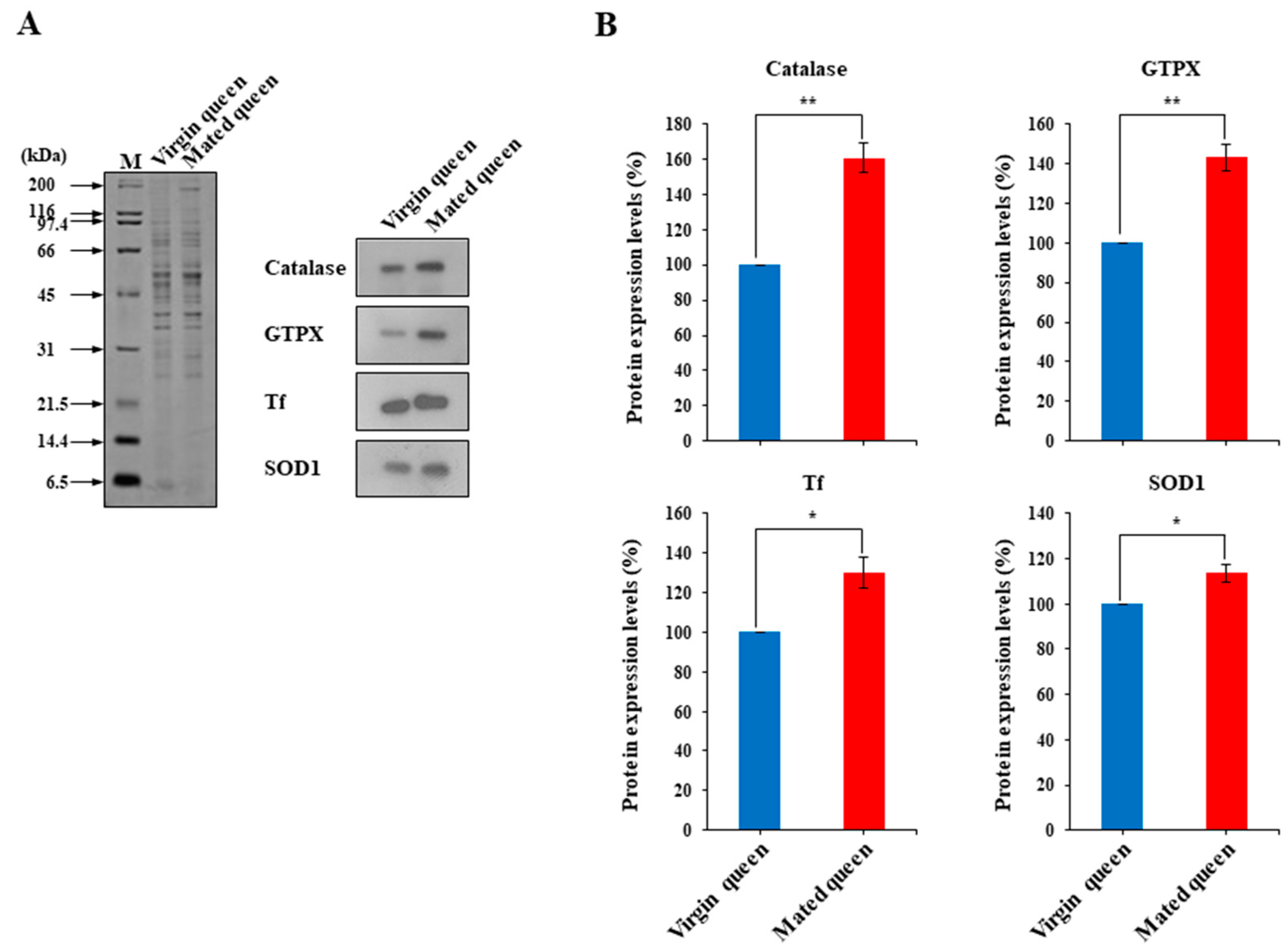
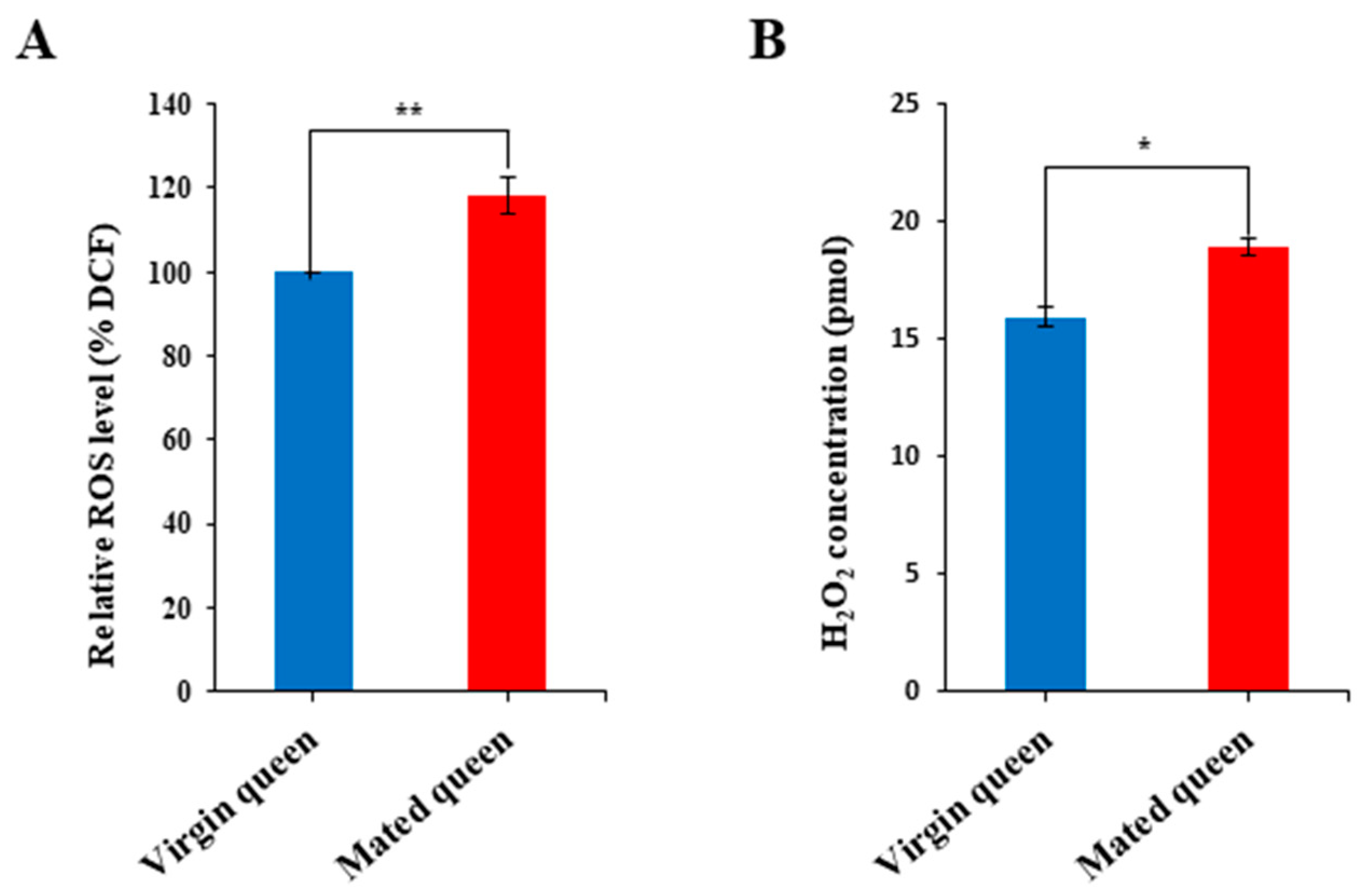
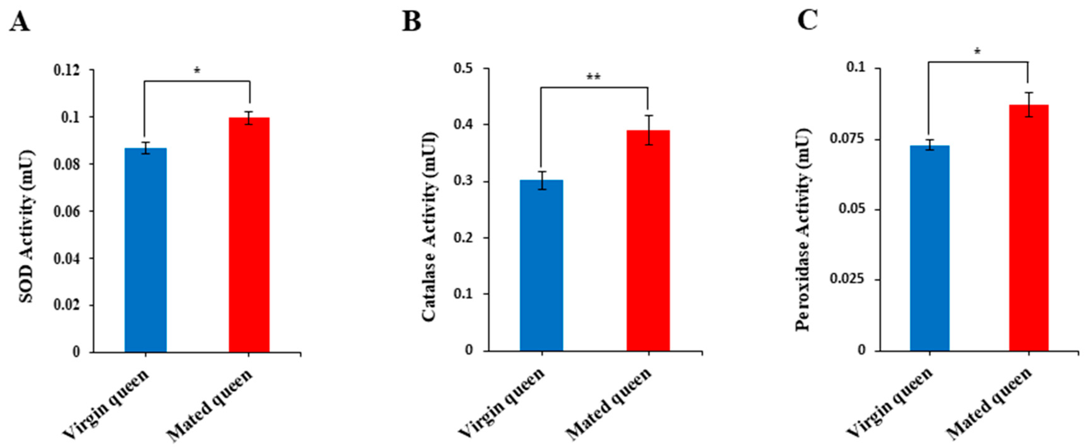
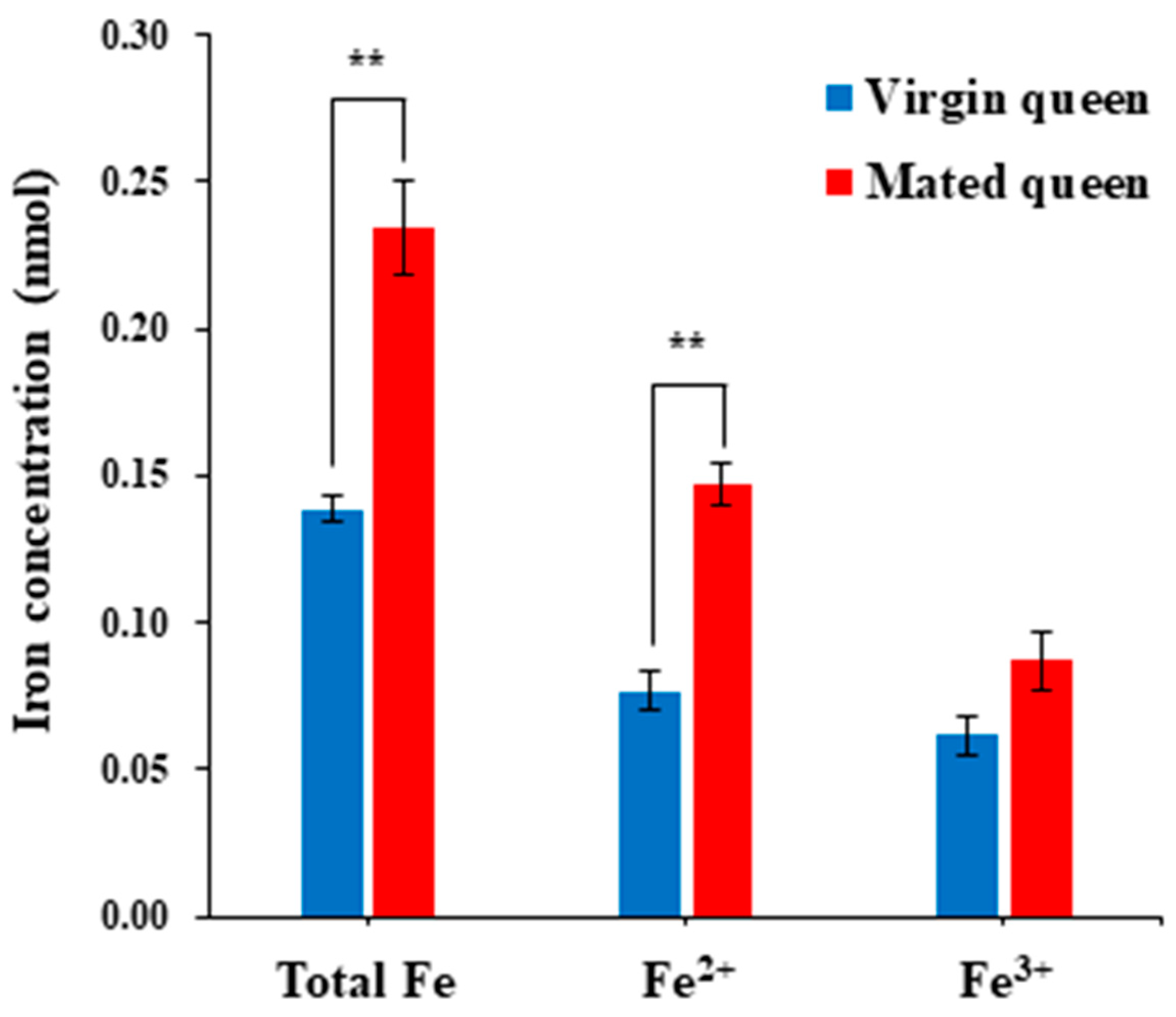
3.2. Antioxidant Defense in the Spermathecae of Mated Queens and Virgin Queens

We found that Tf as well as catalase and GTPX were differentially expressed in the spermathecae of the mated queens. Therefore, we examined the transcript levels of the antioxidant proteins catalase, GTPX, and Tf in the spermathecae of mated and virgin queens (Figure 2A). Considering the antioxidant pathway, SOD1, which catalyzes the dismutation of superoxide to H2O2 [31,32], was examined in this study (Figure 2A). We confirmed that catalase, GTPX, and Tf transcript levels are upregulated in the mated queen’s spermathecae in comparison with the levels in the virgin queens; however, the SOD1 transcript level did not significantly differ between the mated and virgin queens’ spermathecae (Figure 2B). We also measured the catalase, GTPX, Tf, and SOD1 protein levels in the spermathecal fluid of mated and virgin queens by using antibodies generated against these proteins recombinantly produced in baculovirus-infected insect cells (Figure 3A,B). Notably, these results revealed that the SOD1, catalase, GTPX, and Tf protein levels were significantly higher in the spermathecal fluid of mated queens than in the spermathecal fluid of virgin queens.
Because Tf, catalase, GTPX, and SOD1 were upregulated in the mated queens’ spermathecal fluid (Figure 3), we hypothesized that ROS and H2O2 levels must be relatively higher in the spermathecal fluid of mated queens than in that of virgin queens, and thus antioxidant enzyme activities must also show the same trend as that of ROS and H2O2 levels. Notably, ROS and H2O2 levels were significantly higher in the spermathecal fluid of the mated queens than in the spermathecal fluid of the virgin queens (Figure 4A,B). Moreover, the activities of antioxidant enzymes—such as SOD1, catalase, and peroxidase—were increased, paralleling the ROS and H2O2 levels, in the mated queens’ spermathecal fluid (Figure 5A–C). These results are consistent with the hypothesis that antioxidant enzyme activities are higher in the mated queen’s spermathecal fluid owing to increased ROS and H2O2 levels; this phenomenon, in turn, indicates that an increase in antioxidant enzyme activity is an antioxidant defense mechanism against ROS in the spermathecal fluid of mated queens [10].
As previously described, Tf, an iron-binding protein, is upregulated in the spermathecae and spermathecal fluid of mated queens as opposed to those of virgin queens. Therefore, we compared the level of iron in the spermathecal fluid of mated queens with that of the virgin queens. Notably, iron content—including Fe2+ and Fe3+ concentrations—was higher in the mated queens than in the virgin queens (Figure 6), indicating that the upregulation of Tf in the spermathecal fluid of the mated queens is likely due to an increase in the iron level. This result is consistent with the increased H2O2 level in the spermathecal fluid of the mated queens and suggests that the Tf upregulation is a protective response to the Fenton reaction, which involves the production of HO˙ from H2O2 via Fe2+-catalyzed reactions [17,18,19,20,21,22]. Thus, these results suggest that the increased levels of antioxidant proteins, including Tf, in the mated queen’s spermathecal fluid play protective roles against ROS.

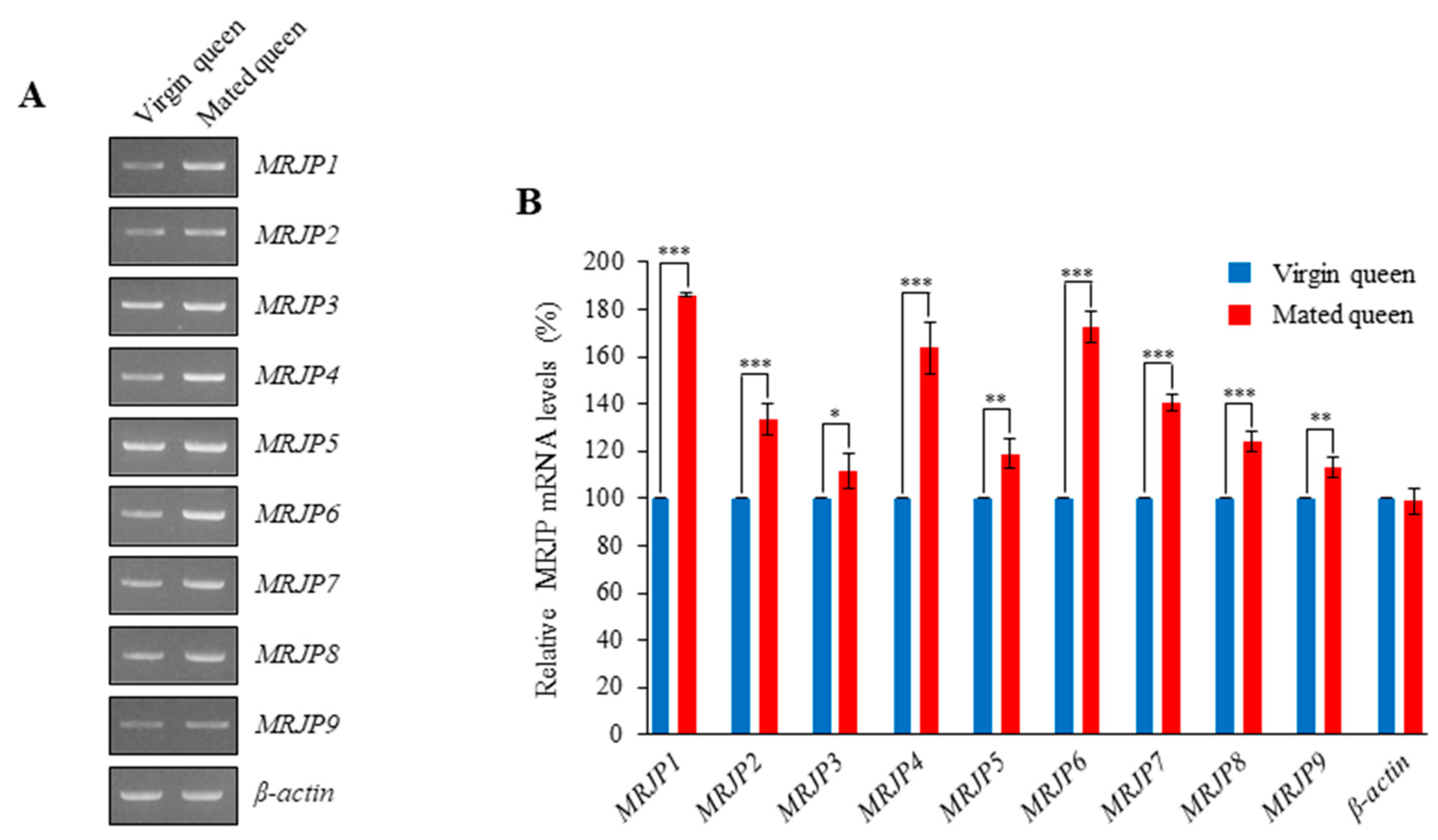
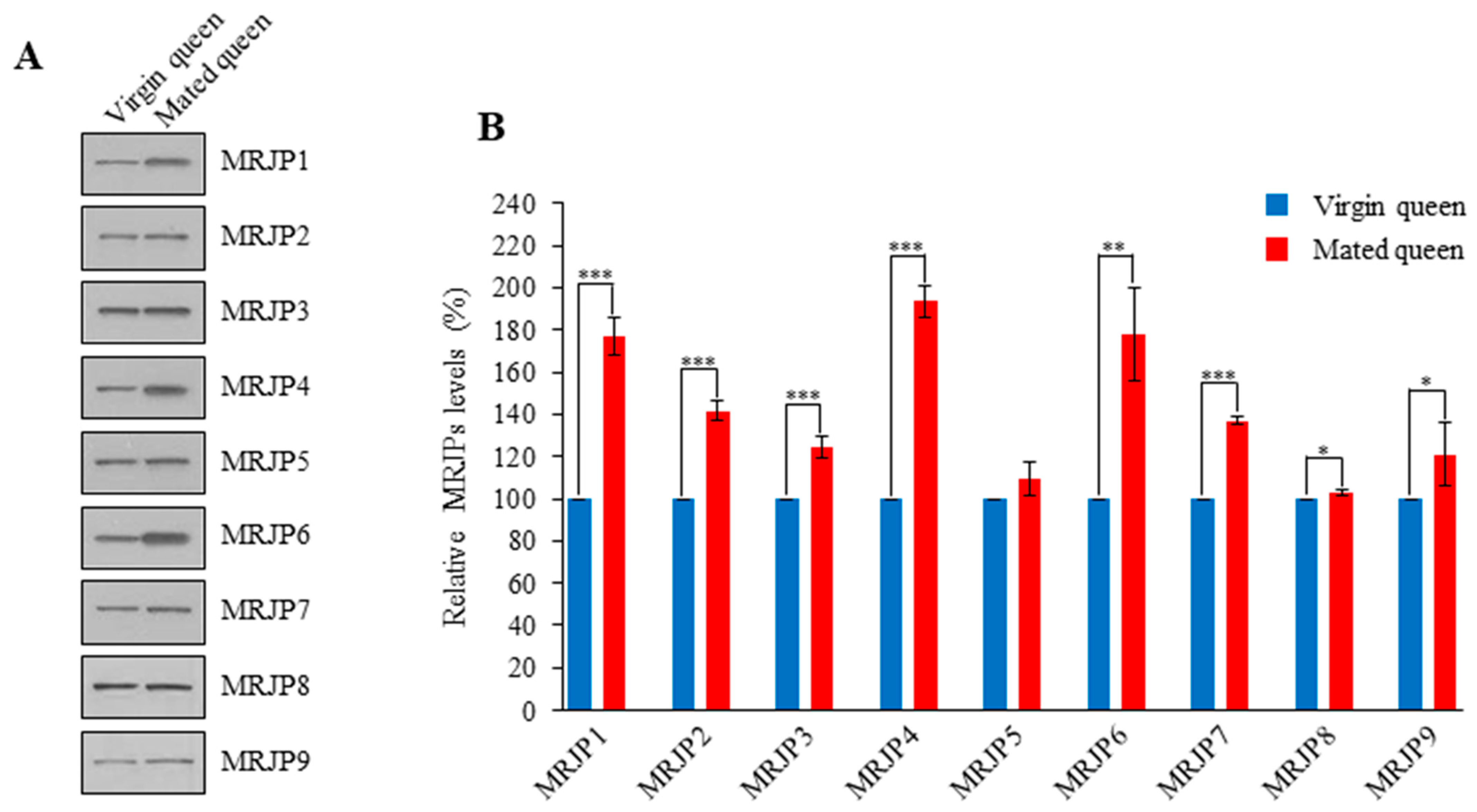
3.3. MRJPs Are Differentially Expressed in the Spermathecae of Mated Queens

Interestingly, we found that MRJPs 1–9 are expressed in the mated and virgin queens’ spermathecae and that these genes are differentially expressed in the spermathecae of mated queens (Figure 7). Among MRJPs 1–9, the transcript levels of MRJP1, MRJP4, and MRJP6 were highly upregulated in the spermathecae of mated queens (Figure 7). The higher transcript levels were consistent with higher protein levels in the spermathecal fluid of mated queens (Figure 8, Figure S1). These results indicate that MRJPs were upregulated in the spermathecal fluid of mated queens and in particular, the upregulated levels of MRJP1, MRJP4, and MRJP6 were much higher than those of the other MRJPs.

4. Discussion

In this study, we examined the sperm in the mated honeybee queen’s spermathecal fluid 24 h after mating and performed transcriptomic analysis of the spermathecae of mated and virgin queens. The aim of these analyses was to characterize the differential gene expression patterns in honeybee queen spermathecae after mating and assess changes in the levels of Tf and MRJPs in the spermathecal fluid of mated honeybee queens. The findings have helped broaden and strengthen our understanding of the molecular basis of spermathecal fluid proteins in the spermathecae of honeybee queens. We first evaluated differential expression of Tf and MRJPs in the spermathecae of mated queens by conducting an RNA-seq analysis of the spermathecae extracted from both mated and virgin queens. Although extensive studies on the molecular basis of sperm storage in the spermathecae and spermathecal fluid of A. mellifera queens have been conducted [2,10,13,14], little is known about the upregulation of Tf and MRJPs in the spermathecae and spermathecal fluid of mated queens. In this study, therefore, we focused on the expression profiles of Tf and MRJPs, which were differentially expressed in the spermathecae and spermathecal fluid of mated queens.
The energy metabolism of the sperm stored in the spermathecae of mated queens is essential for their survival. However, these sperm also simultaneously produce ROS, which may decrease their viability during their storage in the spermathecae of honeybee queens [10]. As a strategy to minimize the ROS-induced damage to sperm, the ROS-production levels are minimized through reduction of the metabolic rate during storage [15]. A second strategy is to use an oxygen-independent pathway, which may minimize the ROS-induced damage in the stored sperm [4]. Honeybee sperm can maximize ATP production through aerobic metabolism and carry out non-aerobic energy production via the glycolytic pathway [4]. Another strategy is to minimize ROS production by producing antioxidant proteins in the mated queen’s spermathecal fluid. Various studies have revealed that antioxidant enzymes are detected in higher quantities in the mated queen’s spermathecal fluid than in that of virgin queens and have thereby suggested that antioxidant proteins protect the stored sperm from ROS [1,2,8−10]. The mRNA levels of antioxidant protein genes, such as catalase, thioredoxin 2, and thioredoxin reductase 1, were upregulated in the mated queen’s spermatheca, whereas no difference was observed in the mRNA level of the SOD1 gene between the spermathecae of mated and virgin queens [10]. Moreover, Tf was detected in the spermathecal fluid and seminal fluid of honeybee queens [2,33]. Here, we observed that the antioxidant protein genes, such as catalase, GTPX, and Tf, showed a two-fold higher expression in the spermathecae of mated queens than in that of virgin queens, whereas the SOD1 mRNA levels were not significantly different; this observation for SOD1 is consistent with the findings of a previous study [10]. Notably, our results revealed that antioxidant proteins—such as catalase, GTPX, and Tf—were detected in higher quantities in the mated queen’s spermathecal fluid than in that of the virgin queens. Additionally, enzyme activity assays and western blot analyses revealed that the spermathecal fluid of mated queens exhibits higher SOD1 levels than the spermathecal fluid of virgin queen, as observed in the case of SOD enzyme activity in a previous study [8]. These results were consistent with increased ROS and H2O2 levels in the mated queen’s spermathecal fluid. Of the abovementioned antioxidant proteins, Tf upregulation is required for decreasing the enhanced iron level in the spermathecal fluid of mated queens, which suggests that Tf plays a role in suppressing toxic hydroxyl radical production via the Fenton reaction between Fe2+ and H2O2 [17,18,19,20,21,22]. Thus, our data suggest that Tf, which is upregulated in the spermathecal fluid of mated queens, may be responsible for antioxidant defense against ROS during sperm storage in the spermathecae of honeybee queens.
Energy metabolism is essential for sperm storage in the spermathecae of mated queens. Sperm energy metabolism is coupled with ATP production by the mitochondria, which are required for complete oxidation of substrates such as sugar, fat, and proteins [11]. The advantage of carbohydrate-based energy metabolism is the production of ATP in the absence of oxygen (anaerobic metabolism) [11]. Moreover, GAPDH expression has been shown to be significantly higher in the stored sperm [4]. MRJP8 and MRJP9 have been identified as spermathecal fluid proteins in honeybee queens [2]. In this study, we found that MRJPs 1–9 are differentially expressed in the spermathecae of mated queens. Since MRJPs 1–7 are expressed in the hypopharyngeal gland and MRJPs 8 and 9 are expressed in the venom gland [34,35], it is somewhat surprising that MRJPs 1–9 are detected in the spermathecal fluid of honeybee queens. Notably, these results provide the first evidence that MRJPs, especially MRJP1, MRJP4, and MRJP6, are detected in higher quantities in the mated queen’s spermathecal fluid than in virgin queens. Considering the nutritional role of MRJPs in the queen bee diet [36,37], our results suggest that highly expressed MRJPs in the spermathecae of mated queens may serve as a protein source for energy metabolism of the stored sperm.
In contrast, previous studies have demonstrated that MRJPs exhibit antimicrobial and antioxidant activities [27,38,39,40,41]. Thus, the present study could not exclude the possibility that MRJPs may function as antimicrobial and antioxidant agents and a protein source in the spermathecal fluid of honeybee queens. Further studies should attempt to define the functional roles of MRJPs in the spermathecal fluid of honeybee queens.
Additionally, we performed RNA-seq analysis, RT-PCR, and western blotting to reveal the differential expression of Tf and MRJPs in the spermathecal fluid and spermathecae of honeybee queens after mating. However, RT-PCR and Western blot data showed some discrepancies relative to the RNA-seq data, which was also described in a transcriptomic analysis of the honeybee queen spermathecae [14]. Our study indicates that the expressions of Tf and MRJPs are upregulated to various levels in the spermathecal fluid and spermathecae of honeybee queens after mating, in comparison with the corresponding expression levels in virgin queens, which suggests that the differential expression of Tf and MRJPs in the spermathecae and spermathecal fluid of mated queens may be involved in sperm storage [2,14].
In conclusion, our data provide evidence for the upregulation of Tf and MRJPs in the spermathecal fluid of mated queens, indicating their role in antioxidant defense and energy provision for the sperm stored in the spermathecal fluid of honeybee queens. Together, our findings elucidate the molecular-level changes in Tf and MRJP expression in the spermathecae and spermathecal fluid of mated honeybee queens, providing novel insights into the antioxidant defense and energy metabolism for stored sperm in honeybee queens.

Supplementary Materials

The following are available online at https://0-www-mdpi-com.brum.beds.ac.uk/article/10.3390/insects12080690/s1, Table S1: The genes were differentially expressed in the spermathecae between mated queens and virgin queens, Table S2: The lists of genes related to ontology and pathway in the spermathecae using the online tool DAVID, Figure S1: Detection of MRJPs in the spermathecal fluid of virgin and mated queens.

Author Contributions

Conceptualization, K.-S.L. and B.-R.J.; Methodology, K.-S.L., Y.-S.C. and H.-J.Y.; Validation, K.-S.L., B.-R.J., Y.-S.C. and H.-J.Y.; Formal analysis and investigation: H.-G.P., B.-Y.K., K.-S.L. and J.-M.K.; Data collection and curation, H.-G.P., B.-Y.K. and K.-S.L.; Writing—original draft preparation, K.-S.L. and B.-R.J.; Writing—review and editing, K.-S.L. and B.-R.J. All authors have read and agreed to the published version of the manuscript.

Funding

This work was supported by a grant from the Next-Generation BioGreen 21 Program (project no. PJ01326401), Rural Development Administration, Basic Science Research Program through the National Research Foundation of Korea (NRF) funded by the Ministry of Education (grant no. 2020R1A6A1A03047729), and the National Research Foundation of Korea (project no. NRF-2020R1F1A1074240), Republic of Korea.

Institutional Review Board Statement

Not applicable.

Informed Consent Statement

Excluded.

Data Availability Statement

Excluded.

Conflicts of Interest

The authors declare no conflict of interest.

References

  1. Collins, A.M.; Caperna, T.J.; Williams, V.; Garrett, W.M.; Evans, J.D. Proteomic analyses of male contributions to honey bee sperm storage and mating. Insect Mol. Biol. 2006, 15, 541–549. [Google Scholar] [CrossRef] [PubMed] [Green Version]
  2. Baer, B.; Eubel, H.; Taylor, N.L.; O’Toole, N.; Millar, A.H. Insights into female sperm storage from the spermathecal fluid proteome of the honeybee Apis mellifera. Genome Biol. 2009, 10, R67. [Google Scholar] [CrossRef] [PubMed] [Green Version]
  3. Klenk, M.; Koeniger, G.; Koeniger, N.; Fasold, H. Proteins in spermathecal gland secretion and spermathecal fluid and the properties of a 29 kDa protein in queens of Apis mellifera. Apidologie 2004, 35, 371–381. [Google Scholar] [CrossRef] [Green Version]
  4. Paynter, E.; Millar, A.H.; Welch, M.; Baer-Imhoof, B.; Cao, D.; Baer, B. Insights into the molecular basis of long-term storage and survival of sperm in the honeybee (Apis mellifera). Sci. Rep. 2017, 7, 40236. [Google Scholar] [CrossRef] [Green Version]
  5. Liu, Z.; Liu, F.; Li, G.; Chi, X.; Wang, Y.; Wang, H.; Ma, L.; Han, K.; Zhao, G.; Guo, X.; et al. Metabolite support of long-term storage of sperm in the spermatheca of honeybee (Apis mellifera) queens. Front. Physiol. 2020, 11, 574856. [Google Scholar] [CrossRef]
  6. den Boer, S.P.; Boomsma, J.J.; Baer, B. Seminal fluid enhances sperm viability in the leafcutter ant Atta colombica. Behav. Ecol. Sociobiol. 2008, 62, 1843–1849. [Google Scholar] [CrossRef] [Green Version]
  7. den Boer, S.P.; Boomsma, J.J.; Baer, B. Honey bee males and queens use glandular secretions to enhance sperm viability before and after storage. J. Insect Physiol. 2009, 55, 538–543. [Google Scholar] [CrossRef] [Green Version]
  8. Weirich, G.F.; Collins, A.M.; Williams, V.P. Antioxidant enzymes in the honey bee, Apis mellifera. Apidologie 2002, 33, 3–14. [Google Scholar] [CrossRef] [Green Version]
  9. Collins, A.M.; Williams, V.; Evans, J.D. Sperm storage and antioxidative enzyme expression in the honey bee, Apis mellifera. Insect Mol. Biol. 2004, 13, 141–146. [Google Scholar] [CrossRef]
  10. Gonzalez, A.N.; Ing, N.; Rangel, J. Upregulation of antioxidant genes in the spermathecae of honey bee (Apis mellifera) queens after mating. Apidologie 2018, 49, 1–11. [Google Scholar] [CrossRef] [Green Version]
  11. Al-Lawati, H.; Kamp, G.; Bienefeld, K. Characteristics of the spermathecal contents of old and young honeybee queens. J. Insect Physiol. 2009, 55, 117–122. [Google Scholar] [CrossRef] [PubMed]
  12. Zareie, R.; Eubel, H.; Millar, A.H.; Baer, B. Long-term survival of high quality sperm: Insights into the sperm proteome of the honeybee Apis mellifera. J. Proteome Res. 2013, 12, 5180–5188. [Google Scholar] [CrossRef] [Green Version]
  13. McAfee, A.; Chapman, A.; Higo, H.; Underwood, R.; Milone, J.; Foster, L.J.; Guarna, M.M.; Tarpy, D.R.; Pettis, J.S. Vulnerability of honey bee queens to heat-induced loss of fertility. Nat. Sustain. 2020, 3, 367–376. [Google Scholar] [CrossRef]
  14. Rangel, J.; Shepherd, T.F.; Gonzalez, A.N.; Hillhouse, A.; Konganti, K.; Ing, N.H. Transcriptomic analysis of the honey bee (Apis mellifera) queen spermathecae reveals genes that may be involved in sperm storage after mating. PLoS ONE 2021, 16, e0244648. [Google Scholar] [CrossRef] [PubMed]
  15. Ribou, A.C.; Reinhardt, K. Reduced metabolic rate and oxygen radicals production in stored insect sperm. Proc. R. Soc. Lond. Ser. B Biol. Sci. 2012, 279, 2196–2203. [Google Scholar] [CrossRef]
  16. Poland, V.; Eubel, H.; King, M.; Solheim, C.; Harvey Millar, A.; Baer, B. Stored sperm differs from ejaculated sperm by proteome alterations associated with energy metabolism in the honeybee Apis mellifera. Mol. Ecol. 2011, 20, 2643–2654. [Google Scholar] [CrossRef] [PubMed] [Green Version]
  17. Crichton, R.R.; Wilmet, S.; Legssyer, R.; Ward, R.J. Molecular and cellular mechanisms of iron homeostasis and toxicity in mammalian cells. J. Inorg. Biochem. 2002, 91, 9–18. [Google Scholar] [CrossRef]
  18. Fenoglio, I.; Tomatis, M.; Lison, D.; Muller, J.; Fonseca, A.; Nagy, J.B.; Fubini, B. Reactivity of carbon nanotubes: Free radical generation of scavenging activity? Free Radic. Biol. Med. 2006, 40, 1227–1233. [Google Scholar] [CrossRef]
  19. Lee, K.S.; Kim, B.Y.; Kim, H.J.; Seo, S.J.; Yoon, H.J.; Choi, Y.S.; Kim, I.; Han, Y.S.; Je, Y.H.; Lee, S.M.; et al. Transferrin inhibits stress-induced apoptosis in a beetle. Free. Radic. Biol. Med. 2006, 41, 1151–1161. [Google Scholar] [CrossRef] [PubMed]
  20. Kim, B.Y.; Lee, K.S.; Choo, Y.M.; Kim, I.; Je, Y.H.; Woo, S.D.; Lee, S.M.; Park, H.C.; Son, H.D.; Jin, B.R. Insect transferrin functions as an antioxidant protein in a beetle larva. Comp. Biochem. Physiol. B Biochem. Mol. Biol. 2008, 150, 161–169. [Google Scholar] [CrossRef]
  21. Kim, B.Y.; Lee, K.S.; Yoon, H.J.; Kim, I.; Li, J.H.; Sohn, H.D.; Jin, B.R. Expression profile of the iron binding proteins transferrin and ferritin heavy chain subunit in the bumblebee Bombus ignitus. Comp. Biochem. Physiol. B Biochem. Mol. Biol. 2009, 153, 165–170. [Google Scholar] [CrossRef] [PubMed]
  22. Geiser, D.L.; Winzerling, J.J. Insect transferrins: Multifunctional proteins. Biochim. Biophys. Acta 2012, 1820, 437–451. [Google Scholar] [CrossRef] [PubMed]
  23. Harmen, P.H.; John, N.C.; Willem, J.B. Stimulating natural supersedure of honeybee queens, Apis mellifera. Proc. Neth. Entomol. Soc. 2004, 15, 29–33. [Google Scholar]
  24. Schley, P. Einführung in die Technik der Instrumentellen Besamung von Bienenköniginnen, 2nd ed.; Köhler Offset KG: Giessen, Germany, 1987; p. 103. [Google Scholar]
  25. Kim, D.; Landmead, B.; Salzberg, S.L. HISAT: A fast spliced aligner with low memory requirements. Nat. Methods 2015, 12, 357–360. [Google Scholar] [CrossRef] [PubMed] [Green Version]
  26. Huang, D.W.; Sherman, B.T.; Lempicki, R.A. Systematic and integrative analysis of large gene lists using DAVID Bioinformatics Resources. Nat. Protoc. 2009, 4, 44–57. [Google Scholar] [CrossRef]
  27. Park, H.G.; Kim, B.Y.; Park, M.J.; Deng, Y.; Choi, Y.S.; Lee, K.S.; Jin, B.R. Antibacterial activity of major royal jelly proteins of the honeybee (Apis mellifera) royal jelly. J. Asia Pac. Entomol. 2019, 22, 737–741. [Google Scholar] [CrossRef]
  28. Kim, B.Y. Antiapoptotic role of major royal jelly protein 8 of honeybee (Apis mellifera) venom. J. Asia Pac. Entomol. 2021, 24. [Google Scholar] [CrossRef]
  29. Park, H.G.; Lee, K.S.; Kim, B.Y.; Yoon, H.J.; Choi, Y.S.; Lee, K.Y.; Wan, H.; Li, J.; Jin, B.R. Honeybee (Apis cerana) vitellogenin acts as an antimicrobial and antioxidant agent in the body and venom. Dev. Comp. Immunol. 2018, 85, 51–60. [Google Scholar] [CrossRef]
  30. Je, Y.H.; Chang, J.H.; Kim, M.H.; Roh, J.Y.; Jin, B.R.; O’Reilly, D.R.; Kang, S.K. A defective viral genome maintained in Escherichia coli for the generation of baculovirus expression vectors. Biotechnol. Lett. 2001, 23, 575–582. [Google Scholar] [CrossRef]
  31. Liochev, S.I.; Fridovich, I. The effects of superoxide dismutase on H2O2 formation. Free Radic. Biol. Med. 2007, 42, 1465–1469. [Google Scholar] [CrossRef]
  32. Liochev, S.I.; Fridovich, I. Mechanism of the peroxidase activity of Cu, Zn superoxide dismutase. Free Radic. Biol. Med. 2010, 48, 1565–1569. [Google Scholar] [CrossRef]
  33. Baer, B.; Heazlewood, J.L.; Taylor, N.L.; Eubel, H.; Millar, A.H. The seminal fluid proteome of the honeybee Apis mellifera. Proteomics 2009, 9, 2085–2097. [Google Scholar] [CrossRef] [PubMed]
  34. Blank, S.; Bantleon, F.I.; McIntyre, M.; Oliert, M.; Spiliner, E. The major royal jelly proteins 8 and 9 (Api m 11) are glycosylated components of Apis mellifera venom with allergenic potential beyond carbohydrate-based reactivity. Clin. Exp. Allergy 2012, 42, 976–985. [Google Scholar] [CrossRef] [PubMed]
  35. Fratini, F.; Cilia, G.; Mancini, S.; Felicioli, A. Royal jelly: An ancient remedy with remarkable antibacterial properties. Microbiol. Res. 2016, 192, 130–141. [Google Scholar] [CrossRef]
  36. Tamura, S.; Amano, S.; Kono, T.; Kondoh, J.; Yamaguchi, K.; Kobayashi, S.; Atabe, T.; Moriyama, T. Molecular characteristics and physiological functions of major royal jelly protein 1 oligomer. Proteomics 2009, 9, 5534–5543. [Google Scholar] [CrossRef]
  37. Ramanathan, A.N.K.G.; Nair, A.J.; Sugunan, V.S. A review on royal jelly proteins and peptides. J. Funct. Foods 2018, 44, 255–264. [Google Scholar] [CrossRef]
  38. Bílikova, K.; Wub, G.; Šimuth, J. Isolation of a peptide fraction from honeybee royal jelly as a potential antifoulbrood factor. Apidologie 2001, 32, 275–283. [Google Scholar] [CrossRef] [Green Version]
  39. Kim, B.Y.; Lee, K.S.; Jung, B.; Choi, Y.S.; Kim, H.K.; Yoon, H.J.; Gui, Z.Z.; Lee, J.; Jin, B.R. Honeybee (Apis cerana) major royal jelly protein 4 exhibits antimicrobial activity. J. Asia Pac. Entomol. 2019, 22, 175–182. [Google Scholar] [CrossRef]
  40. Park, M.J.; Kim, B.Y.; Park, H.G.; Deng, Y.; Yoon, H.J.; Choi, Y.S.; Lee, K.S.; Jin, B.R. Major royal jelly protein 2 acts as an antimicrobial and antioxidant in royal jelly. J. Asia Pac. Entomol. 2019, 22, 684–689. [Google Scholar] [CrossRef]
  41. Park, M.J.; Kim, B.Y.; Deng, Y.; Park, H.G.; Choi, Y.S.; Lee, K.S.; Jin, B.R. Antioxidant capacity of major royal jelly proteins of honeybee (Apis mellifera) royal jelly. J. Asia Pac. Entomol. 2020, 23, 445–448. [Google Scholar] [CrossRef]
Figure 1. The reproductive organs, spermathecae, and sperm of A. mellifera queens. (A) The reproductive organs of the A. mellifera queen 24 h after mating. (B) The spermathecae of the virgin queen (Left) and the mated queen 24 h after mating (Right). (C) The spermathecal fluid of the virgin queen (Left) and the mated queen 24 h after mating (Right). The sperm in the spermathecal fluid of mated queens is indicated by arrowheads.
Insects 12 00690 g001
Figure 2. Transcript levels of genes encoding antioxidant proteins in the spermathecae of virgin and mated queens. (A) The transcripts of the antioxidant proteins catalase, GTPX, Tf, and SOD1 in the spermathecae of virgin and mated queens were quantified using RT-PCR. β-Actin was used as the internal control. (B) The mRNA levels are shown relative to their levels in the spermathecae of virgin queens (shown as 100%). The bars represent the mean ± SD values. Significant differences (p < 0.01) are indicated with two asterisks.
Insects 12 00690 g002
Figure 3. Detection of antioxidant proteins in the spermathecal fluid of virgin and mated queens. (A) Antioxidant proteins (catalase, GTPX, Tf, or SOD1) in the spermathecal fluid of virgin and mated queens were detected using SDS-PAGE (Left), and Western blot analysis was conducted using antibodies against catalase, GTPX, Tf, or SOD1 (Right). (B) The levels of catalase, GTPX, Tf, and SOD1 proteins are shown relative to their levels in the spermathecal fluid of virgin queens (shown as 100%). The bars represent the mean ± SD values. * p < 0.05 and ** p < 0.01.
Insects 12 00690 g003
Figure 4. ROS (A) and H2O2 (B) levels in the spermathecal fluid of virgin and mated queens. The levels of ROS and H2O2 are shown relative to their levels in the spermathecal fluid of virgin queens (shown as 100%). The bars represent the mean ± SD values. * p < 0.05 and ** p < 0.01.
Insects 12 00690 g004
Figure 5. SOD (A), catalase (B), and peroxidase (C) activities in the spermathecal fluid of virgin and mated queens. The bars represent the mean ± SD values. * p < 0.05 and ** p < 0.01.
Insects 12 00690 g005
Figure 6. Iron concentrations in the spermathecal fluid of virgin and mated queens. The bars represent the mean ± SD values. Significant differences (p < 0.01) are indicated with two asterisks.
Insects 12 00690 g006
Figure 7. Transcript levels of MRJP genes in the spermathecae of virgin and mated queens. (A) The transcripts of MRJP genes (MRJPs 1-9) in the spermathecae of virgin and mated queens were quantitated using RT-PCR. β-actin was used as an internal control. (B) The MRJP mRNA levels are shown relative to their levels in the virgin queen’s spermathecae (shown as 100%). The bars represent the mean ± SD values. * p < 0.05, ** p < 0.01, and *** p < 0.001.
Insects 12 00690 g007
Figure 8. Detection of MRJPs in the spermathecal fluid of virgin and mated queens. (A) The MRJPs in the spermathecal fluid of virgin and mated queens were detected using western blot analysis conducted with antibodies against MRJPs. (B) The levels of MRJPs are shown relative to their levels in the spermathecal fluid of virgin queens (shown as 100%). The bars represent the mean ± SD values. * p < 0.05, ** p < 0.01, and *** p < 0.001.
Insects 12 00690 g008
Table 1. Primers used in the RT-PCR analysis of the genes encoding antioxidant proteins and MRJPs in the spermathecae of virgin and mated queens.
GenePrimer SequenceProduct Length (bp)
Catalaseforward 5ʹ-ATGACTGAAATAAAACGA-3ʹ1542
reverse 5ʹ-TCACAATCTAGCTGTCTTGCCATAA-3ʹ
GTPXforward 5′-ATGAGTGGAAACGACAAC-3′507
reverse 5′-TTAAAAATATTTTTCAAGATGATTT-3′
Tfforward 5′-ATGATGCTCCGATGCAAT-3′2139
reverse 5′-TTAAGCAGCACCGTAATTATTT-3′
SOD1forward 5′-ATGACTAAAGCAGTGTGC-3′459
reverse 5′-TTAGACTTTTGTAATTCCAA-3′
MRJP1forward 5′-ATGACAAGATTGTTTATG-3′1299
reverse 5′-TTACAAATGGATTGAAATTTTGAAA-3′
MRJP2forward 5′-ATGACAAGGTGGTTGTTC-3′1359
reverse 5′-TTAATTATCATTCTGATTGTTATTC-3′
MRJP3forward 5′-ATGACAAAGTGGTTGTTG-3′1635
reverse 5′-TTAATGTAATTTTGAAGAATGATGA-3′
MRJP4forward 5′-ATGACAAAATGGTTGCTG-3′1395
reverse 5′-TTAATCGTTATTGTTATGCCGATTG-3′
MRJP5forward 5′-ATGACAACTTGGTTGTTG-3′1797
reverse 5′-TTAATTATTATGCTTATTTTGATTG-3′
MRJP6forward 5′-ATGACAAATTGGTTACTG-3′1314
reverse 5′-CTAATCTAAATGAGCTTGATTCTTA-3′
MRJP7forward 5′-ATGACAAGGTGGTTGTTT-3′1332
reverse 5′-CTAATTAAATGAAGCGTCTATTGTG-3′
MRJP8forward 5′-ATGATAAGATGGTTGCTG-3′1251
reverse 5′-TCAAGGAGATATGCAACGAGTATTC-3′
MRJP9forward 5′-ATGTCTTTCAATATCTGG-3′1272
reverse 5′-TCAAAGGAAAATTGAGAAAAAATTT-3′
β–Actinforward 5′-ATGTCTGACGAAGAAGTT-3′1131
reverse 5′-TTAGAAGCACTTCCTGTGGA-3′
Table 2. Antioxidant proteins and MRJPs differentially expressed in the spermathecae of mated queens.
ProteinGene IDFold Change
Mated Queen/Virgin Queen
TfGB502262.51
CatalaseGB414272.77
GTPXGB474782.07
MRJP1GB552052.78
MRJP4GB552063.00
MRJP6GB552074.42
Publisher’s Note: MDPI stays neutral with regard to jurisdictional claims in published maps and institutional affiliations.

