Review

The Role of Malassezia spp. in Atopic Dermatitis

by
Martin Glatz
*,
Philipp P. Bosshard
,
Wolfram Hoetzenecker
and
Peter Schmid-Grendelmeier
*
Allergy Unit, Department of Dermatology, University Hospital of Zurich, Gloriastrasse 31, 8091 Zurich, Switzerland
*
Authors to whom correspondence should be addressed.
J. Clin. Med. 2015, 4(6), 1217-1228; https://0-doi-org.brum.beds.ac.uk/10.3390/jcm4061217
Submission received: 12 March 2015 / Revised: 19 May 2015 / Accepted: 22 May 2015 / Published: 29 May 2015
(This article belongs to the Special Issue Epidemiology and Treatment of Atopic Eczema)

Abstract

:
Malassezia spp. is a genus of lipophilic yeasts and comprises the most common fungi on healthy human skin. Despite its role as a commensal on healthy human skin, Malassezia spp. is attributed a pathogenic role in atopic dermatitis. The mechanisms by which Malassezia spp. may contribute to the pathogenesis of atopic dermatitis are not fully understood. Here, we review the latest findings on the pathogenetic role of Malassezia spp. in atopic dermatitis (AD). For example, Malassezia spp. produces a variety of immunogenic proteins that elicit the production of specific IgE antibodies and may induce the release of pro-inflammatory cytokines. In addition, Malassezia spp. induces auto-reactive T cells that cross-react between fungal proteins and their human counterparts. These mechanisms contribute to skin inflammation in atopic dermatitis and therefore influence the course of this disorder. Finally, we discuss the possible benefit of an anti-Malassezia spp. treatment in patients with atopic dermatitis.

1. Introduction

Atopic dermatitis (AD) is a frequent chronic relapsing inflammatory skin disorder. It is characterized by intensely itchy skin eczema and is frequently associated with allergic rhino-conjunctivitis and allergic asthma. The prevalence of AD in industrialized countries has tripled during the past 30 years [1], affecting 15%–30% of children and up to 10% of adults [2]. Despite its frequency and impact on public health, we do not fully understand the pathogenesis of AD. It appears to be in parts genetically determined and several factors seem to contribute to the development of AD. For example, the skin of AD patients is characterized by an impaired skin barrier function with increased trans-epidermal water loss, increased surface pH, reduced stratum corneum hydration and reduced expression of tight junction components [3]. Second, the skin immune system in AD patients is altered compared to healthy individuals [4]. For example, AD patients have increased levels of the cytokines interleukin (IL-) 4, IL-10 and IL-13 in their skin compared to healthy controls. These cytokines reduce the production of the antimicrobial peptides LL-37, human beta defensin (hBD)-2, and hBD-3 in keratinocytes [5,6,7], which are important components of the skin’s innate immune system in the defense against microorganisms. It can be speculated that the impaired skin barrier function and the altered skin immune system may play intertwining roles and contribute to colonization and growth of microorganisms on the skin of AD patients [3,7]. The altered skin colonization with microorganisms in AD patients versus healthy individuals has been extensively investigated for bacteria, in particular Staphylococcus aureus. During recent years, AD research has also focused on the possible pathogenetic correlation between eczema and the skin commensal fungus Malassezia spp. because AD patients are often sensitized to Malassezia spp.; and AD patients may benefit from an antifungal therapy that is effective against Malassezia spp. This led to the publication of a plethora of studies on the possible role of Malassezia spp. in the development and course of AD. Here, we will review the biology of Malassezia spp. on human skin and the current state of research on the role of Malassezia spp. in AD.

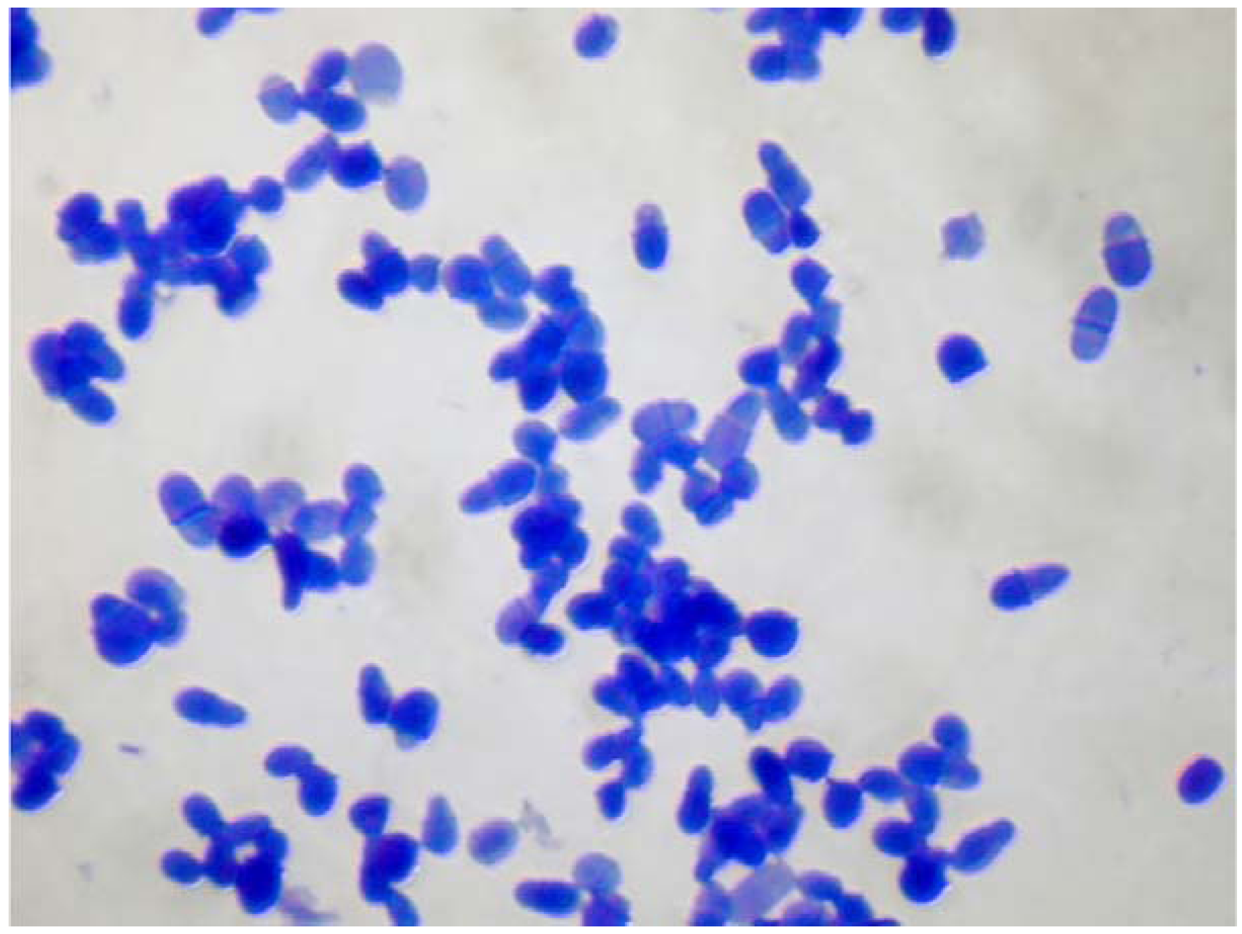
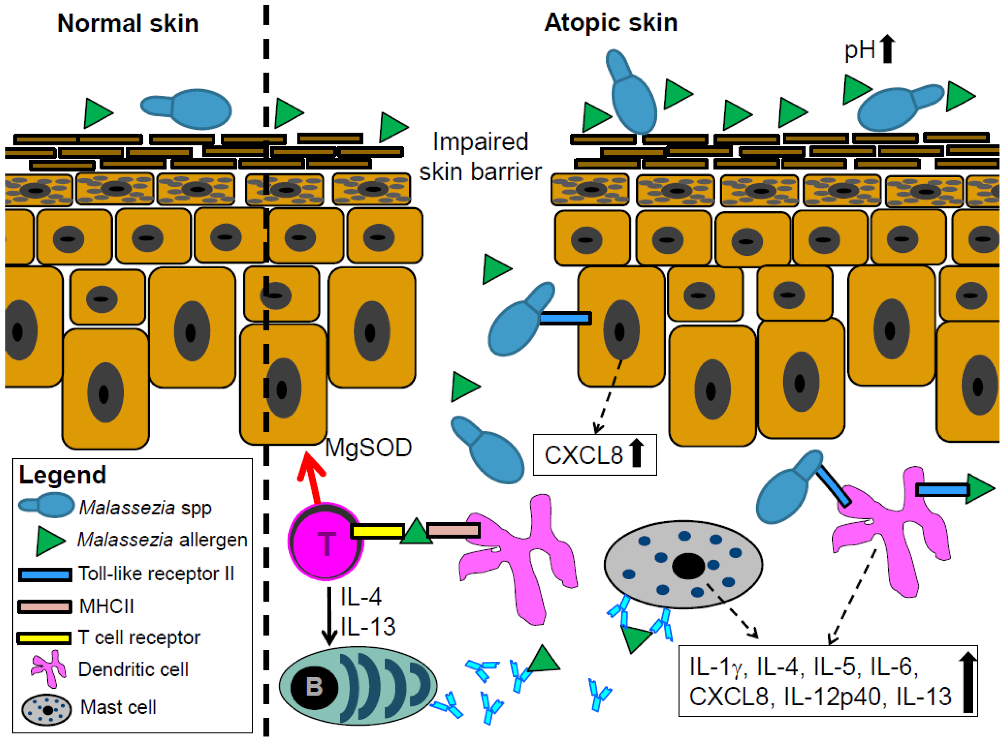
2. Malassezia spp. As Part of the Normal and Atopic Skin Flora

The skin is an ecosystem and harbors diverse and body site-specific microbial communities, which have been termed the skin microbiome. The phylogenetic profiling of the skin microbiome revealed that fungi are part of the normal skin flora at all body sites and comprise 1%–22% of the phylogenetic composition of the skin microbiome [8]. The fungal flora of the healthy skin almost exclusively comprises Malassezia spp., and Malassezia spp. is therefore the main eukaryotic member of the microbial flora of the skin [8,9]. Malassezia spp. is a genus of lipophilic yeasts (Figure 1). Most of the species within this genus lack the genes for fatty acid synthase genes and therefore rely on exogenous fatty acid sources to satisfy their nutritive requirement [10]. M. pachydermatis, a species isolated from dogs and other animals [11], is the only known Malassezia species that grows in the absence of exogenous lipids [10]. Their need for exogenous lipids explains the predilection of Malassezia species for seborrheic skin sites, such as the head and neck.
The taxonomy of Malassezia spp. has been controversial since its recognition as a member of the human skin flora in the mid-19th century. The taxonomy was defined in its current form in 1996, based on morphology, ultrastructure, physiology and molecular biology [12]. Malassezia spp. belongs to the phylum Basidiomycota and currently encompasses 14 species that have been isolated from human and animal skin. Two of these species, M. globosa and M. restricta, predominate on human skin and are identified in almost all individuals and body sites [9,13]. Several studies investigated the epidemiology of Malassezia spp. in healthy and diseased skin by culture and molecular methods such as polymerase chain reaction [14,15,16,17,18,19,20,21,22,23]. These studies obtained variable results presumably owing to methodical inconsistencies between the studies; for example, the sampling sites were inconsistent between these studies. Next generation sequencing revealed that the skin fungal microbiome is highly site specific between body sites [13]. Therefore, comparing the prevalence of Malassezia species between different body sites sampled in different studies will give unreliable results. In addition, the epidemiological studies used different culture media to detect Malassezia species, and it was shown that different culture media favor the growth of particular Malassezia species [24,25].Therefore, the use of only one or a few types of culture media does not necessarily depict the whole spectrum of Malassezia species present in a sample. Despite these methodical considerations, epidemiological studies indicated a geographical variation in the distribution of particular Malassezia species, presumably owing to climate factors. For example, M. sympodialis has been reported in studies from Canada, Russia and Sweden as the most frequent species, whereas in Japan M. furfur was the most common species [14,15,16,17,18,19,20,21,22,23]. Of importance, studies comparing healthy individuals and AD patients did not reveal a difference in the frequency of skin colonization with Malassezia spp. between both groups [14,15,16,17,18,19,20,21,22,23].
Figure 1. Microscopic image of a Malassezia spp. culture. Methylene blue staining.
Jcm 04 01217 g001

3. Sensitization to Malassezia spp. May Correlate with the Severity of Atopic Dermatitis

Malassezia spp. as part of the healthy skin flora regularly interacts with the skin immune system. Therefore Malassezia spp.-specific IgG and IgM antibodies can be detected in healthy individuals [10]. However, healthy individuals are usually not sensitized to Malassezia spp., while a high proportion of AD patients is sensitized to this yeast [26]. This was demonstrated by positive atopy patch tests, skin prick tests or the presence of Malassezia spp.-specific IgE antibodies [26]. For example, 30%–80% of adult AD patients have a positive skin prick test with Malassezia spp. extract [27,28,29,30]. Unfortunately, standardized skin test extracts for Malassezia spp. are not yet commercially available, making it difficult to compare the results of different studies. In contrast, Malassezia spp.-specific serum IgE can be measured using a commercial and standardized kit (ImmunoCAP® m70, Phadia) based on M. sympodialis (ATCC strain 42,132). Recently, a new kit containing several species of Malassezia has been introduced (ImmunoCAP® m227) with a slightly increased sensitivity compared to the single species test according to our experience [31]. Using these commercial kits, Malassezia spp.-specific IgE are found in 5%–27% of children [27,32,33,34] and 29%–65% of adults with AD [27,31,34,35,36], which is consistent with the rates found by skin prick tests. Sensitization rates against Malassezia spp. are particularly higher in patients with head and neck types of AD [31]. Therefore, some authors assume that Malassezia spp. plays a pathogenetic role, particularly in this type of AD [37].
The reason for the high frequency of Malassezia spp.-sensitization in AD patients compared to healthy individuals is still unclear but is attributed to a combination of dysfunctional skin barrier, genetic background, and environmental factors [38]. Several recent studies investigated a possible correlation between AD severity and the IgE-mediated sensitization to Malassezia spp. Our group analyzed 132 children and 67 adults with AD and found a significant correlation between the severity of AD and sensitization to Malassezia spp.-specific IgE in adults but not in children [34]. These results substantiated previous findings in 61 adult AD patients from Japan [39]. The lower frequency of Malassezia spp. sensitization in children compared to adults and the missing correlation between AD severity and Malassezia spp.-specific IgE in children might owe to the poor growth conditions for Malassezia spp. in children compared to adults. The lipid content of sebum, which is a prerequisite for skin colonization with most Malassezia spp. is low in children but rises during puberty and is high until the age of 50 [40]. Accordingly, sensitization to Malassezia spp. seems to occur preferably in adulthood, while sensitization to food allergens and aeroallergens frequently occurs during childhood [34]. Several allergens of Malassezia spp. elucidate a specific IgE response (Figure 2). To date, 13 allergens from two Malassezia species, M. furfur and M. sympodialis, are listed in the official allergen nomenclature list (http://www.allergen.org). In vitro experiments confirmed that Malassezia spp. release more allergens in the less acidic environment of pH 6.0 that represents conditions of atopic skin, than in the more acidic environment of pH 5.5 of healthy skin [41] (Figure 2). However, despite the frequent sensitization of adult AD patients against Malassezia spp., it is unclear if the IgE response plays a pathogenetic role in AD or rather serves as a marker for the severity of AD.
Figure 2. Proposed mechanisms by which Malassezia spp. contributes to skin inflammation in atopic dermatitis (AD) patients. The increased pH in atopic skin contributes to increased allergen release by Malassezia spp. These allergens, supposedly together with whole Malassezia spp. cells, penetrate the epidermis through the disturbed skin barrier in AD patients. Malassezia spp. cells and their allergens may be recognized by toll-like receptor 2 expressed on keratinocytes and dendritic cells which elicits the release of pro-inflammatory cytokines. Malassezia spp. components elicit the production of Malassezia spp.-specific IgE antibodies through the dendritic cells and T cell-mediated activation of B cells. These IgE antibodies may also contribute, possibly through mast cells, to the inflammation in atopic skin. Finally, autoreactive T cells can cross react between fungal and human manganese-dependent superoxide dismutase (MgSOD) and hence sustain skin inflammation.
Jcm 04 01217 g002

4. Malassezia spp. Interacts with the Skin Immune System

The interaction between Malassezia spp. and the skin immune system supposedly contributes to skin inflammation in AD patients. For example, the Malassezia spp. allergen Mala s 13 is a fungal thioredoxin that is very similar to its human homolog. CD4+ T cells that react against the fungal thioredoxin of Malassezia spp. are fully cross-reactive to the human enzyme. Therefore, besides recognizing the fungal enzyme, these T cells cross-react with the human enzyme, what may contribute to skin inflammation in AD [42]. A similar induction of autoreactive T cells was observed for a different Malassezia spp. allergen; Mala s 11 is a manganese-dependent superoxide dismutase with a high degree of sequence identity to the corresponding human enzyme. Similar to Mala s 13, Mala s 11 activates T cells that then react against the human homolog and sustain skin inflammation (Figure 2). These findings were substantiated by the strong correlation between AD severity and sensitization to Mala s 11 [43]. A protein from M. globosa, MGL_1304, was recently identified in the sweat of AD patients and is a potential allergen that may contribute to skin inflammation in AD [44]. The sensitization to this protein also positively correlates with the severity of AD [45].
Prior studies indicated that Malassezia spp. cells interact with various types of human skin and immune cells. This induces a pro-inflammatory immune response by the skin and immune cells, which may maintain skin inflammation in AD. It is still unclear how this interaction between Malassezia spp. cells and host cells occurs, and at least two possible ways of interaction are hypothesized. First, the impaired skin barrier facilitates the physical encounter between Malassezia spp. cells and cells of the epidermis and dermis, such as keratinocytes, Langerhans cells, dermal dendritic cells, natural killer cells and fibroblasts [46]. A second possible mechanism of Malassezia spp.—human cell interaction—might be mediated by immunogenic proteins of Malassezia spp. These proteins can be released in nanovesicles [47] and it was demonstrated that these proteins that might be present in these nanovesicles induce an increased release of TNF-alpha, IL-6, IL-8, IL-10 and IL-12p70 by dendritic cells and mast cells [48,49]. Other proteins such as MGL_1304 induce the degranulation of mast cells and the release of IL-4 by basophils [44].
Some authors suggest that Toll-like receptors (TLRs) mediate the communication between whole Malassezia spp. cells or their immunogenic proteins and human cells. TLRs are members of the large family of pattern recognition receptors, which play a key role in the innate immune system as they recognize molecules that are commonly shared by pathogens. It is known that particularly TLR2 recognizes components of yeast such as Malassezia spp. [7]. Some recent findings substantiated the relevance of TLRs for the immune response of human cells against Malassezia spp. For example, Malassezia spp. induces the expression of TLR2 and TLR4 on human keratinocytes [50,51], which mediate the increased production of the antimicrobial peptide human beta defensin 2 and the chemokine CXLC8 [51]. Others attribute the pro-inflammatory response of dendritic cells against Malassezia spp. to TLR-mediated mechanisms [46] (Figure 2).
The interaction between Malassezia spp. cells and human cells elicits a cytokine release by the human cells [3,52,53]. For example, various Malassezia species activate the NLRP3 inflammasome in skin dendritic cells and induce the production of IL-1β, IL-4, IL-5, IL-13, and IL-18 [3,52,53]. Additionally, mast cells of AD patients release increased amounts of IL-6 in response to M. sympodialis exposure [54]. This cytokine release from human cells in response to Malassezia spp. is suggested to contribute to the skin inflammation in AD (Figure 2).

Therapeutic Approaches with Antifungals in AD

The most effective therapy for AD patients comprises excellent skin care to reconstitute the impaired skin barrier, anti-inflammatory treatment most commonly with topical steroids or calcineurin inhibitors, and the identification and elimination of trigger factors [38]. The significance of an antifungal therapy for AD has been discussed since many years. Azole antifungals are the most common class of antifungal drugs prescribed for AD patients. In vitro, azole antifungals are effective against Malassezia spp. [55,56] but susceptibility testing of M. pachydermatis, a species most commonly isolated from dogs, showed that strains isolated from dogs with AD were less susceptible to azole antifungals than strains isolated from healthy dogs [57]. The relevance of this finding for Malassezia isolates from humans remains to be elucidated.
In a clinical routine, the topical application of ketoconazole on the face of patients with AD of the head and neck type frequently improves eczema, presumably due to a partial overlap with seborrhoic dermatitis in some cases. However, a placebo-controlled study with topical miconazole-hydrocortisone cream and ketoconazole shampoo in AD of the head and neck type did not show any difference to treatment with hydrocortisone alone [58]. The effect of topical antifungals alone in AD compared with topical steroids or calcineurin inhibitors has not yet been investigated to date. The benefit of a systemic antifungal treatment for AD patients has been assessed in several randomized, placebo-controlled trials. A study compared 36 AD patients treated with ketoconazole with 39 AD patients treated with placebo. AD severity improved significantly in the ketoconazole group but not in the placebo group [59]. In another trial, a total of 53 AD patients were treated with either two different dosages of itraconazole or placebo. The improvement of AD severity was significantly higher in itraconazole treated patients than in the placebo group [60]. These positive effects of antifungals were not confirmed by another study comparing 15 AD patients treated with ketoconazole with 14 AD patients treated with placebo. Notably, both treatment groups received topical steroids. Although AD severity improved in both treatment groups, this improvement was not correlated to ketoconazole but rather to the topical steroids [61]. The ambiguous results of these clinical trials might be attributed to a selection bias. It can be speculated that antifungal therapies are more effective in one subgroup of AD patients than in another, for example in patients with a head-neck type of eczema. More recently published studies were of less quality, for example they comprised retrospective observations and lacked a standardized scoring system to assess the severity of AD [62]. Such trials are not appropriate to finally decide on a positive benefit of antifungal therapy in AD patients. Therefore, more randomized, placebo-controlled studies to assess the benefit of antifungal therapy in AD are needed. These studies should be designed to determine the subgroup of AD patients that benefits most from an antifungal treatment and to determine the optimal treatment regimen.
Interestingly, azole antifungals such as ketoconazole or itraconazole have anti-inflammatory properties. It was shown that these drugs inhibit the production of IL-4 and IL-5 by T cells, which might reduce skin inflammation and therefore contribute to improvement of eczema during antifungal treatment [63].
Another therapeutic approach for AD patients specifically against Malassezia spp. might be photodynamic therapy. M. furfur was cultured in the presence of a cationic photosensitizer. The irradiation with a 670-nm diode laser significantly reduced the viablity of M. furfur culture, depending on irradiation duration and photosensitizer dosage [64]. However, the relevance of these results in clinical routine still needs to be proved.

5. Conclusions

In summary, there is little doubt that Malassezia spp. plays a role in AD. This yeast may interact with the skin immune system, which is facilitated by the impaired barrier function of atopic skin, and sensitization against Malassezia spp. correlates with the activity of AD. In addition, antifungal therapy shows beneficial effects in some patients. However, the pathogenetic mechanism and mutual interaction between Malassezia and AD still remain partly unclear and need further investigation.

Acknowledgments

We are grateful to Jürg Meyer, Department of Dermatology, University Hospital Zurich, Switzerland for providing Figure 1.

Author Contributions

Martin Glatz searched for the latest literature and wrote the first draft of the manuscript.
Philipp P. Bosshard discussed the manuscript and gave essential input on the microbiology of Malassezia spp. and Figure 2.
Wolfram Hoetzenecker discussed the manuscript and gave essential input on the clinical significance of Malassezia spp. for atopic dermatitis.
Peter Schmid-Grendelmeier had the idea for the manuscript and coordinated the manuscript. He finally edited the manuscript.

Conflicts of Interest

The authors declare no conflict of interest.

References

  1. Bieber, T. Atopic dermatitis. N. Engl. J. Med. 2008, 358, 1483–1494. [Google Scholar] [CrossRef] [PubMed]
  2. Williams, H.; Flohr, C. How epidemiology has challenged 3 prevailing concepts about atopic dermatitis. J. Allergy Clin. Immunol. 2006, 118, 209–213. [Google Scholar] [CrossRef] [PubMed]
  3. De Benedetto, A.; Kubo, A.; Beck, L.A. Skin barrier disruption: A requirement for allergen sensitization? J. Investig. Dermatol. 2012, 132, 949–963. [Google Scholar] [CrossRef] [PubMed]
  4. Kuo, I.H.; Yoshida, T.; De Benedetto, A.; Beck, L.A. The cutaneous innate immune response in patients with atopic dermatitis. J. Allergy Clin. Immunol. 2013, 131, 266–278. [Google Scholar] [CrossRef] [PubMed]
  5. Howell, M.D.; Gallo, R.L.; Boguniewicz, M.; Jones, J.F.; Wong, C.; Streib, J.E.; Leung, D.Y. Cytokine milieu of atopic dermatitis skin subverts the innate immune response to vaccinia virus. Immunity 2006, 24, 341–348. [Google Scholar] [CrossRef] [PubMed]
  6. McGirt, L.Y.; Beck, L.A. Innate immune defects in atopic dermatitis. J. Allergy Clin. Immunol. 2006, 118, 202–208. [Google Scholar] [CrossRef] [PubMed]
  7. Baker, B.S. The role of microorganisms in atopic dermatitis. Clin. Exp. Immunol. 2006, 144, 1–9. [Google Scholar] [CrossRef] [PubMed]
  8. Oh, J.; Byrd, A.L.; Deming, C.; Conlan, S.; Kong, H.H.; Segre, J.A. Biogeography and individuality shape function in the human skin metagenome. Nature 2014, 514, 59–64. [Google Scholar] [CrossRef] [PubMed]
  9. Gaitanis, G.; Magiatis, P.; Hantschke, M.; Bassukas, I.D.; Velegraki, A. The Malassezia genus in skin and systemic diseases. Clin. Microbiol. Rev. 2012, 25, 106–141. [Google Scholar] [CrossRef] [PubMed]
  10. Saunders, C.W.; Scheynius, A.; Heitman, J. Malassezia fungi are specialized to live on skin and associated with dandruff, eczema, and other skin diseases. PLoS Pathog. 2012, 8, e1002701. [Google Scholar] [CrossRef] [PubMed]
  11. Chen, T.A.; Hill, P.B. The biology of Malassezia organisms and their ability to induce immune responses and skin disease. Vet. Dermatol. 2005, 16, 4–26. [Google Scholar] [CrossRef] [PubMed]
  12. Gueho, E.; Midgley, G.; Guillot, J. The genus Malassezia with description of four new species. Antonie Van Leeuwenhoek 1996, 69, 337–355. [Google Scholar] [CrossRef] [PubMed]
  13. Findley, K.; Oh, J.; Yang, J.; Conlan, S.; Deming, C.; Meyer, J.A.; Schoenfeld, D.; Nomicos, E.; Park, M.; Kong, H.H.; et al. Topographic diversity of fungal and bacterial communities in human skin. Nature 2013, 498, 367–370. [Google Scholar] [CrossRef] [PubMed]
  14. Zomorodian, K.; Mirhendi, H.; Tarazooie, B.; Zeraati, H.; Hallaji, Z.; Balighi, K. Distribution of Malassezia species in patients with psoriasis and healthy individuals in Tehran, Iran. J. Cutan. Pathol. 2008, 35, 1027–1031. [Google Scholar] [CrossRef] [PubMed]
  15. Prohic, A. Distribution of Malassezia species in seborrhoeic dermatitis: Correlation with patients’ cellular immune status. Mycoses 2010, 53, 344–349. [Google Scholar] [PubMed]
  16. Gupta, A.K.; Kohli, Y. Prevalence of Malassezia species on various body sites in clinically healthy subjects representing different age groups. Med. Mycol. 2004, 42, 35–42. [Google Scholar] [CrossRef] [PubMed]
  17. Ben Salah, S.; Makni, F.; Marrakchi, S.; Sellami, H.; Cheikhrouhou, F.; Bouassida, S.; Zahaf, A.; Ayadi, A. Identification of Malassezia species from Tunisian patients with pityriasis versicolor and normal subjects. Mycoses 2005, 48, 242–245. [Google Scholar] [CrossRef] [PubMed]
  18. Gupta, A.K.; Kohli, Y.; Summerbell, R.C.; Faergemann, J. Quantitative culture of Malassezia species from different body sites of individuals with or without dermatoses. Med. Mycol. 2001, 39, 243–251. [Google Scholar] [CrossRef] [PubMed]
  19. Yim, S.M.; Kim, J.Y.; Ko, J.H.; Lee, Y.W.; Choe, Y.B.; Ahn, K.J. Molecular analysis of Malassezia microflora on the skin of the patients with atopic dermatitis. Ann. Dermatol. 2010, 22, 41–47. [Google Scholar] [CrossRef] [PubMed]
  20. Sandstrom Falk, M.H.; Tengvall Linder, M.; Johansson, C.; Bartosik, J.; Back, O.; Sarnhult, T.; Wahlgren, C.F.; Scheynius, A.; Faergemann, J. The prevalence of Malassezia yeasts in patients with atopic dermatitis, seborrhoeic dermatitis and healthy controls. Acta Derm. Venereol. 2005, 85, 17–23. [Google Scholar] [CrossRef] [PubMed]
  21. Tajima, M.; Sugita, T.; Nishikawa, A.; Tsuboi, R. Molecular analysis of Malassezia microflora in seborrheic dermatitis patients: Comparison with other diseases and healthy subjects. J. Investig. Dermatol. 2008, 128, 345–351. [Google Scholar] [CrossRef] [PubMed]
  22. Kaga, M.; Sugita, T.; Nishikawa, A.; Wada, Y.; Hiruma, M.; Ikeda, S. Molecular analysis of the cutaneous Malassezia microbiota from the skin of patients with atopic dermatitis of different severities. Mycoses 2011, 54. [Google Scholar] [CrossRef] [PubMed]
  23. Oh, B.H.; Song, Y.C.; Lee, Y.W.; Choe, Y.B.; Ahn, K.J. Comparison of Nested PCR and RFLP for Identification and Classification of Malassezia Yeasts from Healthy Human Skin. Ann. Dermatol. 2009, 21, 352–357. [Google Scholar] [CrossRef] [PubMed]
  24. Kaneko, T.; Makimura, K.; Abe, M.; Shiota, R.; Nakamura, Y.; Kano, R.; Hasegawa, A.; Sugita, T.; Shibuya, S.; Watanabe, S.; et al. Revised culture-based system for identification of Malassezia species. J. Clin. Microbiol. 2007, 45, 3737–3742. [Google Scholar] [CrossRef] [PubMed]
  25. Kaneko, T.; Makimura, K.; Sugita, T.; Yamaguchi, H. Tween 40-based precipitate production observed on modified chromogenic agar and development of biological identification kit for Malassezia species. Med. Mycol. 2006, 44, 227–231. [Google Scholar] [CrossRef] [PubMed]
  26. Johansson, C.; Sandstrom, M.H.; Bartosik, J.; Sarnhult, T.; Christiansen, J.; Zargari, A.; Back, O.; Wahlgren, C.F.; Faergemann, J.; Scheynius, A.; et al. Atopy patch test reactions to Malassezia allergens differentiate subgroups of atopic dermatitis patients. Br. J. Dermatol. 2003, 148, 479–488. [Google Scholar] [CrossRef] [PubMed]
  27. Scalabrin, D.M.; Bavbek, S.; Perzanowski, M.S.; Wilson, B.B.; Platts-Mills, T.A.; Wheatley, L.M. Use of specific IgE in assessing the relevance of fungal and dust mite allergens to atopic dermatitis: A comparison with asthmatic and nonasthmatic control subjects. J. Allergy Clin. Immunol. 1999, 104, 1273–1279. [Google Scholar] [CrossRef]
  28. Scheynius, A.; Johansson, C.; Buentke, E.; Zargari, A.; Linder, M.T. Atopic eczema/dermatitis syndrome and Malassezia. Int. Arch. Allergy Immunol. 2002, 127, 161–169. [Google Scholar] [CrossRef] [PubMed]
  29. Zargari, A.; Eshaghi, H.; Back, O.; Johansson, S.; Scheynius, A. Serum IgE reactivity to Malassezia furfur extract and recombinant M. furfur allergens in patients with atopic dermatitis. Acta Derm. Venereol. 2001, 81, 418–422. [Google Scholar] [CrossRef] [PubMed]
  30. Johansson, C.; Eshaghi, H.; Linder, M.T.; Jakobson, E.; Scheynius, A. Positive atopy patch test reaction to Malassezia furfur in atopic dermatitis correlates with a T helper 2-like peripheral blood mononuclear cells response. J. Investig. Dermatol. 2002, 118, 1044–1051. [Google Scholar] [CrossRef] [PubMed]
  31. Brodska, P.; Panzner, P.; Pizinger, K.; Schmid-Grendelmeier, P. IgE-mediated sensitization to Malassezia in atopic dermatitis: More common in male patients and in head and neck type. Dermatitis 2014, 25, 120–126. [Google Scholar] [CrossRef] [PubMed]
  32. Lange, L.; Alter, N.; Keller, T.; Rietschel, E. Sensitization to Malassezia in infants and children with atopic dermatitis: Prevalence and clinical characteristics. Allergy 2008, 63, 486–487. [Google Scholar] [CrossRef] [PubMed]
  33. Kekki, O.M.; Scheynius, A.; Poikonen, S.; Koskinen, A.; Kautiainen, H.; Turjanmaa, K. Sensitization to Malassezia in children with atopic dermatitis combined with food allergy. Pediatr. Allergy Immunol. 2013, 24, 244–249. [Google Scholar] [CrossRef] [PubMed]
  34. Glatz, M.; Buchner, M.; von Bartenwerffer, W.; Schmid-Grendelmeier, P.; Worm, M.; Hedderich, J.; Folster-Holst, R. Malassezia spp.-specific Immunoglobulin E Level is a Marker for Severity of Atopic Dermatitis in Adults. Acta Derm. Venereol. 2015, 95, 191–196. [Google Scholar] [CrossRef] [PubMed]
  35. Sandstrom Falk, M.H.; Faergemann, J. Atopic dermatitis in adults: Does it disappear with age? Acta Derm. Venereol. 2006, 86, 135–139. [Google Scholar] [PubMed]
  36. Ramirez de Knott, H.M.; McCormick, T.S.; Kalka, K.; Skandamis, G.; Ghannoum, M.A.; Schluchter, M.; Cooper, K.D.; Nedorost, S.T. Cutaneous hypersensitivity to Malassezia sympodialis and dust mite in adult atopic dermatitis with a textile pattern. Contact Dermat. 2006, 54, 92–99. [Google Scholar]
  37. Faergemann, J. Atopic dermatitis and fungi. Clin. Microbiol. Rev. 2002, 15, 545–563. [Google Scholar] [CrossRef] [PubMed]
  38. Akdis, C.A.; Akdis, M.; Bieber, T.; Bindslev-Jensen, C.; Boguniewicz, M.; Eigenmann, P.; Hamid, Q.; Kapp, A.; Leung, D.Y.; Lipozencic, J.; et al. Diagnosis and treatment of atopic dermatitis in children and adults: European Academy of Allergology and Clinical Immunology/American Academy of Allergy, Asthma and Immunology/PRACTALL Consensus Report. Allergy 2006, 61, 969–987. [Google Scholar] [CrossRef] [PubMed]
  39. Zhang, E.; Tanaka, T.; Tajima, M.; Tsuboi, R.; Kato, H.; Nishikawa, A.; Sugita, T. Anti-Malassezia-Specific IgE Antibodies Production in Japanese Patients with Head and Neck Atopic Dermatitis: Relationship between the Level of Specific IgE Antibody and the Colonization Frequency of Cutaneous Malassezia Species and Clinical Severity. J. Allergy 2011, 2011. [Google Scholar] [CrossRef] [PubMed]
  40. Cotterill, J.A.; Cunliffe, W.J.; Williamson, B.; Bulusu, L. Age and sex variation in skin surface lipid composition and sebum excretion rate. Br. J. Dermatol. 1972, 87, 333–340. [Google Scholar] [CrossRef] [PubMed]
  41. Selander, C.; Zargari, A.; Mollby, R.; Rasool, O.; Scheynius, A. Higher pH level, corresponding to that on the skin of patients with atopic eczema, stimulates the release of Malassezia sympodialis allergens. Allergy 2006, 61, 1002–1008. [Google Scholar] [CrossRef] [PubMed]
  42. Balaji, H.; Heratizadeh, A.; Wichmann, K.; Niebuhr, M.; Crameri, R.; Scheynius, A.; Werfel, T. Malassezia sympodialis thioredoxin-specific T cells are highly cross-reactive to human thioredoxin in atopic dermatitis. J. Allergy Clin. Immunol. 2011, 128. [Google Scholar] [CrossRef] [PubMed]
  43. Schmid-Grendelmeier, P.; Fluckiger, S.; Disch, R.; Trautmann, A.; Wuthrich, B.; Blaser, K.; Scheynius, A.; Crameri, R. IgE-mediated and T cell-mediated autoimmunity against manganese superoxide dismutase in atopic dermatitis. J. Allergy Clin. Immunol. 2005, 115, 1068–1075. [Google Scholar] [CrossRef] [PubMed]
  44. Hiragun, T.; Ishii, K.; Hiragun, M.; Suzuki, H.; Kan, T.; Mihara, S.; Yanase, Y.; Bartels, J.; Schroder, J.M.; Hide, M.; et al. Fungal protein MGL_1304 in sweat is an allergen for atopic dermatitis patients. J. Allergy Clin. Immunol. 2013, 132, 608–615. [Google Scholar] [CrossRef] [PubMed]
  45. Hiragun, M.; Hiragun, T.; Ishii, K.; Suzuki, H.; Tanaka, A.; Yanase, Y.; Mihara, S.; Haruta, Y.; Kohno, N.; Hide, M.; et al. Elevated serum IgE against MGL_1304 in patients with atopic dermatitis and cholinergic urticaria. Allergol. Int. 2014, 63, 83–93. [Google Scholar] [CrossRef] [PubMed]
  46. Buentke, E.; Scheynius, A. Dendritic cells and fungi. Apmis 2003, 111, 789–796. [Google Scholar] [CrossRef] [PubMed]
  47. Gehrmann, U.; Qazi, K.R.; Johansson, C.; Hultenby, K.; Karlsson, M.; Lundeberg, L.; Gabrielsson, S.; Scheynius, A. Nanovesicles from Malassezia sympodialis and host exosomes induce cytokine responses—Novel mechanisms for host-microbe interactions in atopic eczema. PLoS ONE 2011, 6, e21480. [Google Scholar] [CrossRef] [PubMed]
  48. Vilhelmsson, M.; Johansson, C.; Jacobsson-Ekman, G.; Crameri, R.; Zargari, A.; Scheynius, A. The Malassezia sympodialis allergen Mala s 11 induces human dendritic cell maturation, in contrast to its human homologue manganese superoxide dismutase. Int. Arch. Allergy Immunol. 2007, 143, 155–162. [Google Scholar] [CrossRef] [PubMed]
  49. Selander, C.; Engblom, C.; Nilsson, G.; Scheynius, A.; Andersson, C.L. TLR2/MyD88-Dependent and -independent activation of mast cell IgE responses by the skin commensal yeast Malassezia sympodialis. J. Immunol. 2009, 182, 4208–4216. [Google Scholar] [CrossRef] [PubMed]
  50. Brasch, J.; Morig, A.; Neumann, B.; Proksch, E. Expression of antimicrobial peptides and toll-like receptors is increased in tinea and pityriasis versicolor. Mycoses 2014, 57, 147–152. [Google Scholar] [CrossRef] [PubMed]
  51. Baroni, A.; Orlando, M.; Donnarumma, G.; Farro, P.; Iovene, M.R.; Tufano, M.A.; Buommino, E. Toll-like receptor 2 (TLR2) mediates intracellular signalling in human keratinocytes in response to Malassezia furfur. Arch. Dermatol. Res. 2006, 297, 280–288. [Google Scholar] [CrossRef] [PubMed]
  52. Novak, N.; Leung, D.Y. Advances in atopic dermatitis. Curr. Opin. Immunol. 2011, 23, 778–783. [Google Scholar] [CrossRef] [PubMed]
  53. Kistowska, M.; Fenini, G.; Jankovic, D.; Feldmeyer, L.; Kerl, K.; Bosshard, P.; Contassot, E.; French, L.E. Malassezia yeasts activate the NLRP3 inflammasome in antigen-presenting cells via Syk-kinase signaling. Exp. Dermatol. 2014, 23, 884–889. [Google Scholar] [CrossRef] [PubMed]
  54. Ribbing, C.; Engblom, C.; Lappalainen, J.; Lindstedt, K.; Kovanen, P.T.; Karlsson, M.A.; Lundeberg, L.; Johansson, C.; Nilsson, G.; Lunderius-Andersson, C.; et al. Mast cells generated from patients with atopic eczema have enhanced levels of granule mediators and an impaired Dectin-1 expression. Allergy 2011, 66, 110–119. [Google Scholar] [CrossRef] [PubMed]
  55. Darabi, K.; Hostetler, S.G.; Bechtel, M.A.; Zirwas, M. The role of Malassezia in atopic dermatitis affecting the head and neck of adults. J. Am. Acad. Dermatol. 2009, 60, 125–136. [Google Scholar] [CrossRef] [PubMed]
  56. Sugita, T.; Tajima, M.; Ito, T.; Saito, M.; Tsuboi, R.; Nishikawa, A. Antifungal activities of tacrolimus and azole agents against the eleven currently accepted Malassezia species. J. Clin. Microbiol. 2005, 43, 2824–2829. [Google Scholar] [CrossRef] [PubMed]
  57. Watanabe, S.; Koike, A.; Kano, R.; Nagata, M.; Chen, C.; Hwang, C.Y.; Hasegawa, A.; Kamata, H. In vitro susceptibility of Malassezia pachydermatis isolates from canine skin with atopic dermatitis to ketoconazole and itraconazole in East Asia. J. Vet. Med. Sci. 2014, 76, 579–581. [Google Scholar] [CrossRef] [PubMed]
  58. Broberg, A.; Faergemann, J. Topical antimycotic treatment of atopic dermatitis in the head/neck area. A double-blind randomised study. Acta Derm. Venereol. 1995, 75, 46–49. [Google Scholar] [PubMed]
  59. Lintu, P.; Savolainen, J.; Kortekangas-Savolainen, O.; Kalimo, K. Systemic ketoconazole is an effective treatment of atopic dermatitis with IgE-mediated hypersensitivity to yeasts. Allergy 2001, 56, 512–517. [Google Scholar] [CrossRef] [PubMed]
  60. Svejgaard, E.; Larsen, P.O.; Deleuran, M.; Ternowitz, T.; Roed-Petersen, J.; Nilsson, J. Treatment of head and neck dermatitis comparing itraconazole 200 mg and 400 mg daily for 1 week with placebo. J. Eur. Acad. Dermatol. Venereol. 2004, 18, 445–449. [Google Scholar] [CrossRef] [PubMed]
  61. Back, O.; Bartosik, J. Systemic ketoconazole for yeast allergic patients with atopic dermatitis. J. Eur. Acad. Dermatol. Venereol. 2001, 15, 34–38. [Google Scholar] [CrossRef] [PubMed]
  62. Kaffenberger, B.H.; Mathis, J.; Zirwas, M.J. A retrospective descriptive study of oral azole antifungal agents in patients with patch test-negative head and neck predominant atopic dermatitis. J. Am. Acad. Dermatol. 2014, 71, 480–483. [Google Scholar] [CrossRef] [PubMed]
  63. Kanda, N.; Enomoto, U.; Watanabe, S. Anti-mycotics suppress interleukin-4 and interleukin-5 production in anti-CD3 plus anti-CD28-stimulated T cells from patients with atopic dermatitis. J. Investig. Dermatol. 2001, 117, 1635–1646. [Google Scholar] [CrossRef] [PubMed]
  64. Takahashi, H.; Nakajima, S.; Sakata, I.; Iizuka, H. Antifungal effect of TONS504-photodynamic therapy on Malassezia furfur. J. Dermatol. 2014, 41, 895–897. [Google Scholar] [CrossRef] [PubMed]

