Article

Different Ectopic Hoxa2 Expression Levels in Mouse Cranial Neural Crest Cells Result in Distinct Craniofacial Anomalies and Homeotic Phenotypes

1
Friedrich Miescher Institute for Biomedical Research, Maulbeerstrasse 66, 4058 Basel, Switzerland
2
INSERM UMR 1121, Faculté de Chirurgie Dentaire, Université de Strasbourg, 8 rue Sainte Elisabeth, 67 000 Strasbourg, France
3
Departement Biozentrum, University of Basel, Spitalstrasse 41, 4056 Basel, Switzerland
*
Author to whom correspondence should be addressed.
Submission received: 20 December 2021 / Revised: 26 January 2022 / Accepted: 27 January 2022 / Published: 31 January 2022
(This article belongs to the Special Issue 2021 Feature Papers by JDB’s Editorial Board Members)

Abstract

:
Providing appropriate positional identity and patterning information to distinct rostrocaudal subpopulations of cranial neural crest cells (CNCCs) is central to vertebrate craniofacial morphogenesis. Hox genes are not expressed in frontonasal and first pharyngeal arch (PA1) CNCCs, whereas a single Hox gene, Hoxa2, is necessary to provide patterning information to second pharyngeal arch (PA2) CNCCs. In frog, chick and mouse embryos, ectopic expression of Hoxa2 in Hox-negative CNCCs induced hypoplastic phenotypes of CNCC derivatives of variable severity, associated or not with homeotic transformation of a subset of PA1 structures into a PA2-like identity. Whether these different morphological outcomes are directly related to distinct Hoxa2 overexpression levels is unknown. To address this issue, we selectively induced Hoxa2 overexpression in mouse CNCCs, using a panel of mouse lines expressing different Hoxa2 ectopic expression levels, including a newly generated Hoxa2 knocked-in mouse line. While ectopic Hoxa2 expression at only 60% of its physiological levels was sufficient for pinna duplication, ectopic Hoxa2 expression at 100% of its normal level was required for complete homeotic repatterning of a subset of PA1 skeletal elements into a duplicated set of PA2-like elements. On the other hand, ectopic Hoxa2 overexpression at non-physiological levels (200% of normal levels) led to an almost complete loss of craniofacial skeletal structures. Moreover, ectopic Hoxa5 overexpression in CNCCs, while also resulting in severe craniofacial defects, did not induce homeotic changes of PA1-derived CNCCs, indicating Hoxa2 specificity in repatterning a subset of Hox-negative CNCCs. These results reconcile some discrepancies in previously published experiments and indicate that distinct subpopulations of CNCCs are differentially sensitive to ectopic levels of Hox expression.

1. Introduction

The multipotent neural crest cells delaminate dorsally along the rostrocaudal axis of the forming neural tube and migrate towards various regions of the developing embryo, where they locally differentiate into a broad range of cell lineages [1]. In the head, cranial neural crest cells (CNCCs) colonize the frontonasal process (FNP) and pharyngeal arches (PAs), providing the main source of skeletogenic head mesenchyme, central to craniofacial and pharyngeal morphogenesis. Rostral CNCCs give rise to the frontonasal skeleton and make extensive contributions to the membranous bones of the skull. More posterior CNCCs fill the PAs, forming the cartilages and bones of the upper and lower jaws, middle ear ossicles, outer ear, hyoid and thyroid structures [1,2,3,4]. Defects of regional specification and patterning of different CNCC subpopulations lead to distinct craniofacial abnormalities [5].
Rostrocaudally, a fundamental molecular difference among CNCC subpopulations concerns their patterns of Hox gene expression. CNCCs contributing to the FNP and first pharyngeal arch (PA1) do not express Hox genes, whereas CNCCs contributing to the second and more posterior arches display nested combinatorial Hox expression patterns, providing each PA with distinct regional identities along the rostrocaudal axis [3,4,6,7,8]. The role of Hox genes in establishing rostrocaudal identity of CNCC derivatives first became evident with the targeted inactivation of Hoxa2 in the mouse, which resulted in homeotic transformation of PA2 into a subset of PA1-like derivatives [9,10]. Namely, in Hoxa2 knockout mice, the PA2-derived stapes, styloid process and lesser horns of hyoid bone were absent and replaced by proximal PA1-like structures including the malleus, incus, gonial bone and tympanic ring in a mirror image pattern of their normal counterparts [9,10]. Furthermore, the outer ear or pinna, a PA2-derived structure [11], was absent as well and replaced by a duplication of the PA1-derived external auditory canal [10,11,12]. Hoxa2 downregulation in Xenopus [13] and zebrafish [14] also induced PA2 to PA1-like homeotic transformation of skeletal elements, indicating an essential and conserved role of Hoxa2 for PA2 CNCC identity specification.
In contrast, distinct experiments analyzing Hoxa2 ectopic expression in Hox-negative CNCCs led to different morphological phenotypes. Several studies reported a repatterning of proximal PA1 into PA2-like derivatives [11,14,15,16,17], while this phenotype was not observed by others [18]. Rather, it was reported that forced expression of Hoxa2 in chick Hox-negative CNCCs abolishes the development of the entire facial skeleton, including frontonasal structures and jaws [18]. These dramatic phenotypes might be caused by abnormal persistent expression of Hoxa2 in the chondrogenic regions [19]. To explain these discrepancies, it was initially proposed that a morphological transformation could only be obtained following global overexpression of Hoxa2, both in the CNCCs and surrounding tissues. When only CNCCs were targeted in chick embryos, the loss of facial structures was observed but not homeotic repatterning of PA1 elements [15,18].
However, recent studies in the mouse have shown that Wnt1-Cre-dependent conditional ectopic expression of Hoxa2 in CNCCs is alone sufficient to induce a repatterning of proximal PA1 into PA2-like skeletal elements, including mirror image duplication of the pinna [11,16]. Notably, the development of more anterior skeletal structures, including frontonasal elements and jaws, was impaired as well [11,16]. Namely, short snout and exencephaly were observed, and skull vault (frontal), facial (nasal, premaxillary), skull base (nasal septum, vomer, palatine, pterygoid, alisphenoid), lower jaw (dentary) and upper jaw (maxillary, jugal, squamous) skeletal elements became hypoplastic and malformed. These findings indicated a differential ability of Hoxa2 to repattern Hox-negative CNCC subpopulations [11,16].
Lastly, little is known about the specificity of Hox genes in the repatterning of Hox-negative CNCC derivatives. In the chick, Creuzet et al., 2002 [18], showed that ectopic expression of Hoxa2, Hoxa3 or Hoxb4 in Hox-negative CNCCs had negative effects on facial skeleton formation, albeit at different extents. While ectopic Hoxa2 expression caused the absence of the entire Hox-negative CNCC derivatives, the effects of Hoxa3 and Hoxb4 were more restricted [18]. However, since Creuzet et al., 2002 [18], did not observe homeotic repatterning of a subset of Hox-negative CNCCs following Hoxa2 overexpression, it is still unclear whether ectopic expression of Hox genes other than Hoxa2 in the mouse might also induce repatterning into PA2-like derivatives.
One possibility to reconcile the distinct morphological phenotypes reported by the different studies is that different threshold levels of ectopic Hoxa2 expression may differentially affect the morphogenesis of Hox-negative CNCC subpopulations. Here, we evaluated how distinct ectopic Hoxa2 expression levels differentially affect Hox-negative CNCC subpopulation morphogenesis. To this aim, we induced Wnt1::Cre-dependent conditional Hoxa2 overexpression in CNCCs using mouse lines that generate different Hoxa2 ectopic expression levels. Namely, we used our previously reported mouse allele [11] and showed that it achieves Hoxa2 ectopic expression at only 60% of endogenous levels in PA2, and a newly generated allele, which expresses high, non-physiological, levels of ectopic Hoxa2, namely 200% of endogenous levels. We further compared the obtained phenotypes with those described in Kitazawa et al., 2015 [16], which achieved Wnt1::Cre-dependent conditional ectopic Hoxa2 expression in Hox-negative CNCCs comparable to physiological endogenous levels (100%) in PA2. In this study, a repatterning of proximal PA1 into a full set of PA2-like derivatives (skeletal elements and pinna) was observed alongside with a severe impairment of more anterior skeletal structures [16]. Here, we found that Hoxa2 overexpression at 60% of its physiological level induced only a partial repatterning of proximal PA1 into PA2-like skeletal elements, including full duplication of the pinna, and relatively modest hypoplasia of more anterior skeletal structures. In contrast, high, non-physiological, ectopic levels (200%) of Hoxa2 overexpression caused dramatic hypoplasia and/or aplasia of craniofacial elements including PA2 and proximal PA1 derivatives, without clear homeotic phenotype. Lastly, we assessed Hox gene specificity in repatterning of craniofacial elements by analyzing mice displaying Wnt1::Cre-dependent Hoxa5 conditional overexpression in CNCCs. We found that overexpression of Hoxa5 in CNCCs induced a loss of proximal PA1 and PA2–PA4 structures without homeotic transformation, indicating specificity of Hoxa2 in the repatterning of a subset of PA1 into PA2-like CNCC derivatives.

2. Materials and Methods

2.1. Animals

All animal experiments were performed in accordance with Guide for Care and Use of Laboratory Animals and were approved by the Veterinary Department of the Canton of Basel-Stadt.

2.2. Generation of the ROSA26CAG::(lox-stop-lox)-Hoxa2-IRES-EGFP-WPRE Mouse Line

We generated a mouse line that utilizes CAG promoter and WPRE sequence to induce high levels of Hoxa2 overexpression (ROSA26CAG::(lox-stop-lox)-Hoxa2-IRES-EGFP-WPRE) in a Cre-dependent manner, with a similar strategy as for the ROSA26CAG::(lox-stop-lox)-Hoxa5-IRES-EGFP-WPRE mouse line [20]. By homologous recombination, we introduced in the ROSA26 locus the targeting vector pR26-CAG-lsl-3xFLAG-Hoxa2-IRES-GFP, consisting of a CAG promoter, a lox-stop-lox cassette, a Hoxa2 tagged with a 3xFLAG, an IRES-GFP, a WPRE element, a bGH poly(A) and a PGK-Neo cassette. To generate this plasmid, we used pR26-CAG-lsl-Kir (kind gift from Guillermina López-Bendito; [21]), in which we replaced the insert located between the two FseI restriction sites by the cassette 3xFLAG-Hoxa2-IRES-GFP (PCR amplified from the plasmid pROSA26-FLAG-Hoxa2-IRES-EGFP [22] and cloned into the TOPO vector pCRII (Invitrogen) with insertion of a 3xFLAG tag). This vector was linearized with PvuI and electroporated into the E14 embryonic stem cell (ESC) line. After G418 resistance-selection and screening by PCR, positive ESC clones were aggregated with morula-stage embryos obtained from inbred (C57BL/6 X DBA/2) F1 mice. Germline transmission of the ROSA26CAG::(lox-stop-lox)-Hoxa2-IRES-EGFP-WPRE allele was obtained. Heterozygous and homozygous mice were viable and fertile.

2.3. Mouse Lines and Mating Scheme

ROSA26(lox-stop-lox)-Hoxa2-IRES-EGFP [22], ROSA26CAG::(lox-stop-lox)-Hoxa5-IRES-EGFP-WPRE [20] and newly generated ROSA26CAG::(lox-stop-lox)-Hoxa2-IRES-EGFP-WPRE alleles were initially generated in the background of E14 ESCs. Germline-transmitted alleles were crossed with the CD1 mouse line and maintained as homozygous mice. Homozygous ROSA26(lox-stop-lox)-Hoxa2-IRES-EGFP, ROSA26CAG::(lox-stop-lox)-Hoxa2-IRES-EGFP-WPRE or ROSA26CAG::(lox-stop-lox)-Hoxa5-IRES-EGFP-WPRE females were crossed with the Wnt1::Cre transgenic mouse line [23] on C57B/6 background (JAX stock #02250) to generate Wnt1::Cre;ROSA26(lox-stop-lox)-Hoxa2-IRES-EGFP (Wnt1-Hoxa2-low), Wnt1::Cre; ROSA26CAG::(lox-stop-lox)-Hoxa2-IRES-EGFP-WPRE (Wnt1-Hoxa2-high) or Wnt1::Cre;ROSA26CAG::(lox-stop-lox)-Hoxa5-IRES-EGFP-WPRE (Wnt1-Hoxa5) mice, respectively.

2.4. Skeletal Staining

Skeletal staining of mouse E18.5 Wnt1-Hoxa2-low (n = 4), Wnt1-Hoxa2-high (n = 6) and Wnt1-Hoxa5 (n = 9) fetuses was performed according to a previously described protocol with minor modifications [16,24]. Additionally, we re-analyzed E18.5 skeletons from our previous study [11] generated by crossing ROSA26 (lox-stop-lox)-Hoxa2-IRES-EGFP mice with a distinct Wnt1::Cre line [25] (n = 6), showing undistinguishable phenotype from the Wnt1-Hoxa2-low mice reported here. Samples were fixed in 95% ethanol for 5–7 days. Subsequently, embryos were incubated with 0.015% alcian blue 8GS, 0.005% alizarin red S and 5% acetic acid for 3 days with agitation at 37 °C. Samples were cleared in 1% KOH for several days and in 1% KOH/glycerol series until surround tissues turned transparent. The samples were stored in glycerol for a longer term.

2.5. RT-qPCR Analysis of mRNA Expression Levels

We carried out RNA extraction from PA1 and PA2 in E10.5 wild-type, Wnt1-Hoxa2-low and Wnt1-Hoxa2-high mice in a comparable manner with previous Wnt1-Hoxa2-medium mice analysis [16]. The PA1 mandibular and PA2 were manually dissected from E10.5 embryos, and 2–3 pairs of PAs were pooled. Subsequently, total RNA was purified by RNeasy Mini Kit (Qiagen, Germantown, MD 20874, USA, #74104) with genomic DNA digestion using RNase-Free DNase I Set (Qiagen, Germantown, MD, USA, #79254) according to the manufactures’ protocol. 1 μg of total RNA was reverse-transcribed using Superscript III Reverse Transcriptase (Thermo Fisher, Waltham, MA, USA, #18080093) and oligo(dT) primer (Thermo Fisher, Waltham, MA, USA, #SO131). Quantification of mRNA was performed by real-time PCR using StepOnePlus Real-Time PCR System with SYBR Green PCR Master Mix. Quantification of Hoxa2 and Gapdh was performed by ΔΔCt using Gapdh as an internal control. Statistical analysis was performed by one-way analysis of variance (ANOVA) followed by Tukey’s honest significant difference (HSD) post hoc tests.

3. Results

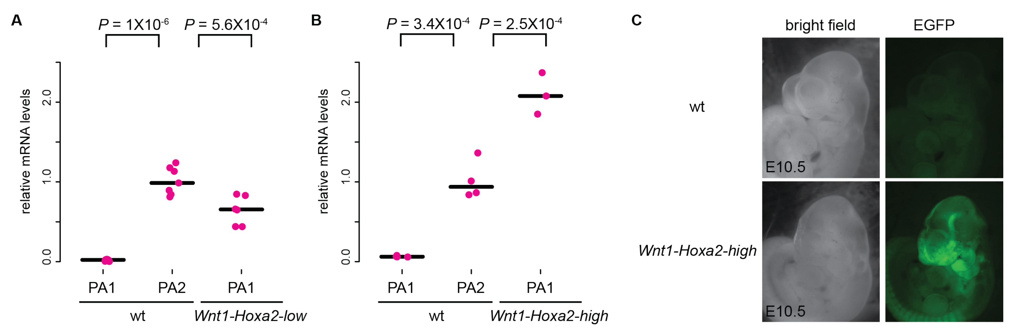
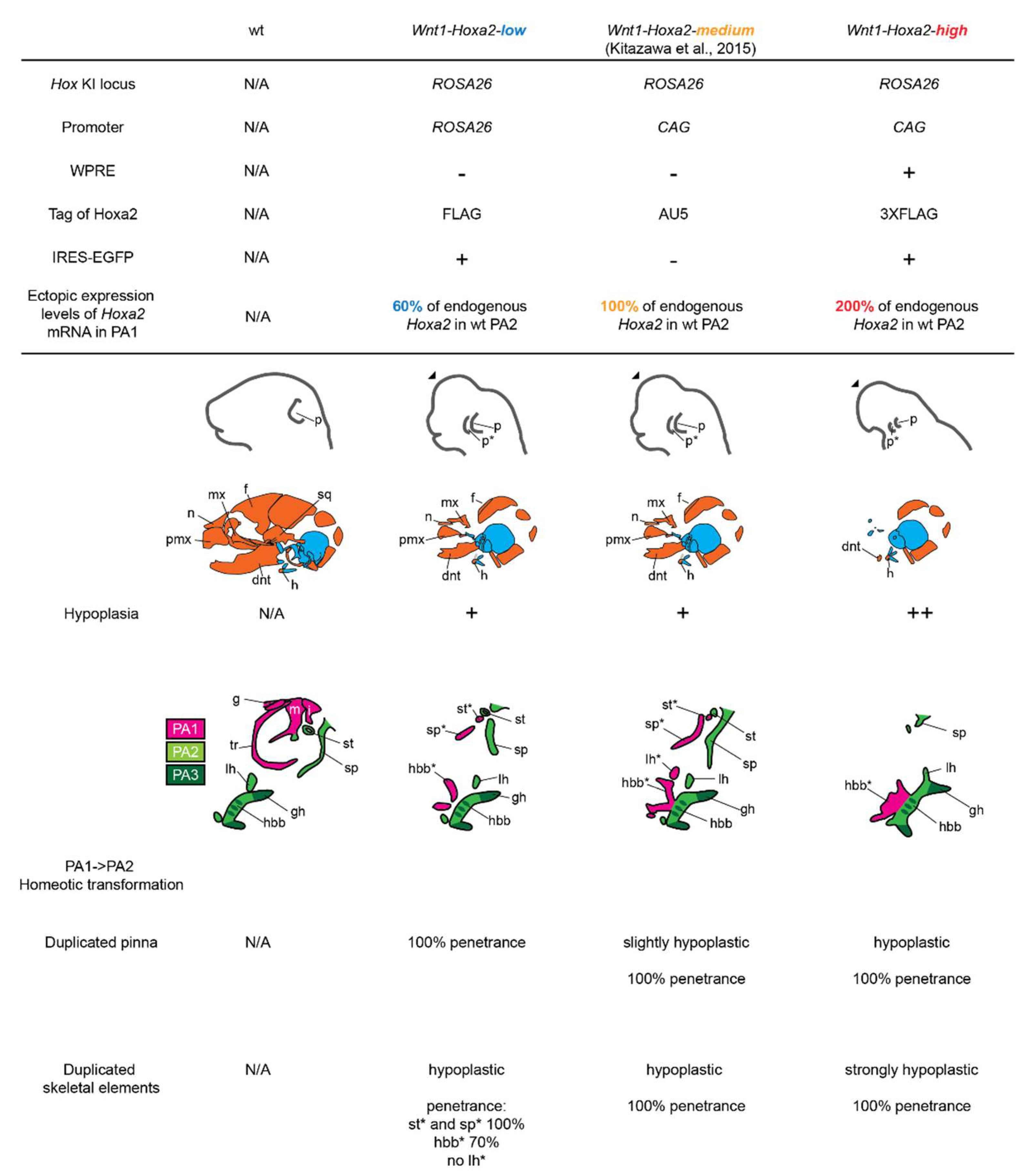
3.1. Analysis of Mouse Phenotypes Induced by Low Levels of Conditional Ectopic Hoxa2 Expression in CNCCs

In a previous study [16], Wnt1::Cre [25]-dependent conditional ectopic Hoxa2 overexpression was achieved in mouse Hox-negative premigratory CNCC progenitors by knock-in of a CAG promoter-(lox-stop-lox)-Hoxa2 cassette into ROSA26 to generate the ROSACAG::(lox-stop-lox)-Hoxa2 mouse line. RT-qPCR analysis in E10.5 embryos showed that ectopic Hoxa2 expression in PA1 was at a level comparable to endogenous Hoxa2 expression in wild-type PA2 (100%) [16]. We refer, hereafter, to this line as Wnt1-Hoxa2-medium.
In Wnt1-Hoxa2-medium fetuses, ectopic Hoxa2 expression resulted in two categories of phenotypes ([16]; final summary schemes of this study). The proximal part of PA1 was homeotically transformed into PA2-like skeletal elements. Namely, malleus, incus, tympanic ring and gonial elements were repatterned into ectopic lesser horn and body of hyoid bone, styloid process and stapes elements in a mirror image pattern of their normal counterparts, with a slight hypoplasia. Wnt1-Hoxa2-medium fetuses also displayed a duplication of the pinna, even though both normal and ectopic pinnae were mildly hypoplastic as compared to wild-type pinna. Moreover, the development of the other Hox-negative CNCC subpopulations, including the FNP and distal part of PA1, was severely impaired.
To assess how differential ectopic Hoxa2 expression levels affect Hox-negative CNCCs morphogenesis, we firstly reinvestigated our previous ROSA(lox-stop-lox)-Hoxa2-IRES-EGFP mouse line [11,22], where a (lox-stop-lox)-Hoxa2-IRES-EGFP cassette was knocked-in to the ROSA26 locus. To conditionally overexpress Hoxa2 in CNCCs, this line was crossed with the Wnt1::Cre transgenic mouse line [23]. RT-qPCR analysis of PA1 mRNA in E10.5 embryos revealed that the ectopic Hoxa2 expression driven by the ROSA26 promoter was significantly lower, about 60%, as compared to the endogenous Hoxa2 expression in wild-type PA2 (Figure 1A), likely because the ROSA26 promoter is weaker than the CAG promoter in Kitazawa et al., 2015 [16]. We refer, hereafter, to the Wnt1::Cre; ROSA (lox-stop-lox)-Hoxa2-IRES-EGFP line as Wnt1-Hoxa2-low. We also re-analyzed ROSA26(lox-stop-lox)-Hoxa2-IRES-EGFP mice from our previous study [11] crossed with a distinct Wnt1::Cre line [25], showing undistinguishable phenotype from Wnt1-Hoxa2-low specimen (see below).
Wnt1-Hoxa2-low E18.5 fetuses displayed a fully duplicated pinna, as previously reported [11] (Figure 2A–E). They also display a duplication of the styloid process and stapes elements in a mirror image pattern of their normal counterparts (Figure 2N,Q; final summary schemes ), even though both the ectopic and normal elements were hypoplastic. However, repatterning of the other proximal PA1 skeletal structures into PA2-like morphology was incomplete (Figure 2J,K,M,N,P,Q). An ectopic body of hyoid bone was observed in only 70% (7/10) of samples (Figure 2K,N,Q) and duplication of the lesser horn of hyoid bone was never observed (Figure 2K,N,Q). Thus, lower than physiological levels of ectopic Hoxa2 expression can induce pinna duplication, while not sufficient for the complete homeotic transformation of skeletal elements. Moreover, in E18.5 Wnt1-Hoxa2-low fetuses, structures derived from the FNP and distal PA1, e.g., facial, nasal, premaxillary, nasal septum, vomer, palatine, pterygoid, alisphenoid, dentary, maxillary, jugal and squamous, were reduced or absent. This suggests that the severity of hypoplasia and malformation or aplasia of individual elements derived from rostral Hox-negative CNCC subpopulations is differentially dependent on local levels of ectopic Hoxa2 expression.

3.2. Analysis of Mouse Phenotypes Induced by High Levels of Conditional Ectopic Hoxa2 Expression in CNCCs

To assess how high, non-physiological, levels of Hoxa2 overexpression affect the morphogenesis of Hox-negative CNCCs, we generated a novel Hoxa2 overexpression mouse line by knocking-in a CAG promoter-(lox-stop-lox)-Hoxa2-IRES-EGFP-woodchuck hepatitis virus posttranscriptional regulatory element (WPRE) cassette into the mouse ROSA26 locus to generate ROSACAG::(lox-stop-lox)-Hoxa2-IRES-EGFP-WPRE mice. The CAG promoter is shared with the ROSACAG::(lox-stop-lox)-Hoxa2 mouse line [16], but the WPRE sequence further enhances the expression of the transgene by increasing nuclear and cytoplasmic mRNA transcript levels [26,27]. To overexpress Hoxa2 in CNCCs, we crossed this line with the Wnt1::Cre transgenic mouse line [23]. In E10.5 embryos, we confirmed expression of EGFP in CNCCs, allowed by the IRES sequence (Figure 1C). In addition, RT-qPCR analysis of Hoxa2 mRNA in PA1 revealed that its ectopic expression level was significantly higher, about 200%, as compared with wild-type PA2 endogenous Hoxa2 expression (Figure 1B). We refer, hereafter, to the Wnt1::Cre; ROSACAG::(lox-stop-lox)-Hoxa2-IRES-EGFP-WPRE line as Wnt1-Hoxa2-high.
E18.5 Wnt1-Hoxa2-high fetuses showed extensive hypoplasia and/or aplasia of craniofacial structures derived from FNP and distal PA1 (Figure 2C,I; final summary schemes). In particular, most of the lower jaw (dentary bone) and nasal septum were largely absent and replaced by several cartilage nodules (Figure 2I), which might include mesoderm-derived hypochiasmatic cartilage [28]. In addition, E18.5 Wnt1-Hoxa2-high fetuses showed strong hypoplasia and malformation of the craniofacial elements derived from the PA2 and proximal PA1 CNCCs (Figure 2F,L,O,R; final summary schemes). Consequently, no homeotic transformation could be observed in the middle ear: the malleus, incus, gonial, tympanic ring and stapes elements were absent, and the styloid process was strongly malformed (Figure 2O,R). In the hyoid region, an ectopic body of hyoid bone was present but strongly malformed, and an ectopic lesser horn of hyoid bone was not observed (Figure 2L,R). Moreover, the PA2-derived body and lesser horns of hyoid bone were malformed and fused (Figure 2L,R). While we could still observe duplication of the pinna, both PA1- and PA2-derived pinnae were much smaller than wild-type pinna (Figure 2F).
In summary, malformation or absence of craniofacial structures was evidently more severe in Wnt1-Hoxa2-high mice as compared with Wnt1-Hoxa2-low and Wnt1-Hoxa2-medium mice ([16]; Figure 2; final summary schemes), with no clear homeotic phenotype (except for pinna duplication—a structure lacking in chick embryos), thus mimicking the dramatically hypoplastic craniofacial phenotypes of Hoxa2-overexpressing chick embryos in Creuzet et al., 2002 [18].

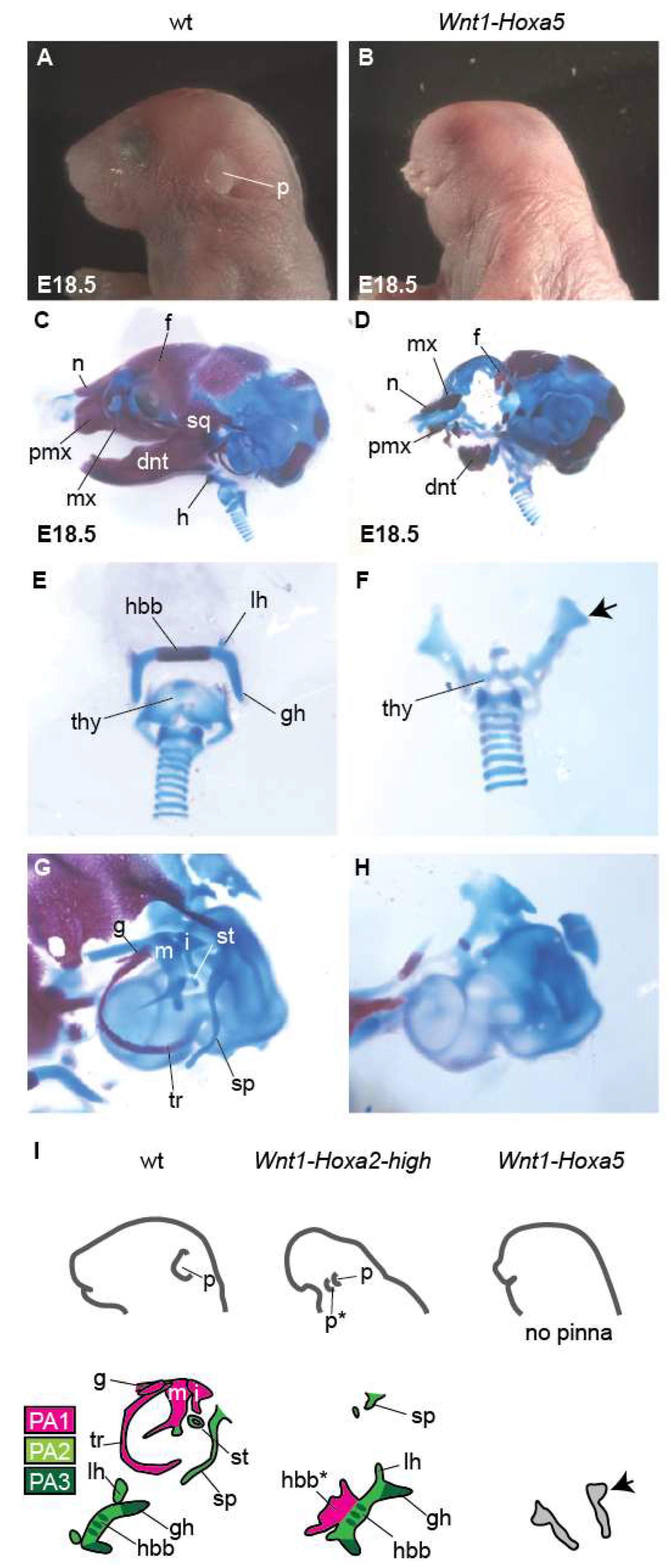
3.3. Analysis of Mouse Phenotypes Induced by Conditional Ectopic Hoxa5 Overexpression in CNCCs

To assess the specificity of ectopically-expressed Hoxa2 in the repatterning of CNCC derivatives, we induced conditional overexpression in CNCCs of Hoxa5, a Hox gene not expressed in neural crest cells contributing to craniofacial structures. To this aim, similar to the construction of the Wnt1-Hoxa2-high allele, a CAG promoter-(lox-stop-lox)-Hoxa5-IRES-EGFP-WPRE cassette, driving high levels of Hoxa5 expression, was knocked-in to the ROSA26 locus to generate the ROSACAG::(lox-stop-lox)-Hoxa5-IRES-EGFP-WPRE mouse line [20]. This line was crossed with the Wnt1::Cre transgenic mouse line [23]. We refer, hereafter, to the Wnt1::Cre; ROSACAG::(lox-stop-lox)-Hoxa5-IRES-EGFP-WPRE line as Wnt1-Hoxa5.
As a proxy for Hoxa5 overexpression from the Hoxa5-IRES-EGFP-WPRE allele, we detected EGFP expression at E18.5 (Figure S1). E18.5 Wnt1-Hoxa5 fetuses showed hypoplasia and malformation of craniofacial structures derived from FNP and distal PA1 CNCCs, albeit milder as compared with Wnt1-Hoxa2-high fetuses (Figure 2 and Figure 3A–D). In particular, the short snout and exencephaly were less severe. In addition, many skeletal elements including facial, nasal, premaxillary, nasal septum, alisphenoid, dentary and maxillary elements were reduced but still present in Wnt1-Hoxa5 fetuses (Figure 3A–D).
On the other hand, in E18.5 Wnt1-Hoxa5 fetuses, PA2 and proximal PA1-derived structures were severely affected. The pinna was absent (Figure 3A,B,I). Furthermore, hyoid bone (body, greater and lesser horn) and middle ear elements (malleus, incus, gonial bone, tympanic ring, stapes and styloid process) were absent, without homeotic transformation into PA2-like derivatives (Figure 3E–I). In the hyoid region, ectopic rod-like cartilage nodules were formed (Figure 3F,I, arrows). In addition, PA4-derived thyroid cartilage, which was not affected by Hoxa2 overexpression (Figure 2J–L), was hypoplastic (Figure 3E,F).
In summary, Hoxa5 ectopic expression in CNCCs caused more severe hypoplasia and malformation of proximal PA1 and PA2–PA4 than FNP and distal PA1 CNCC derivatives and could not induce homeotic transformation of PA1 into PA2-like elements. These results indicate a specificity of Hoxa2 in the ability to selectively repattern proximal PA1 elements.

4. Discussion

4.1. Homeotic Repatterning of Hox-Negative CNCCs by Hoxa2 is Dependent on Its Ectopic Expression Levels and CNCC Position within the Craniofacial Complex

In mouse PA2 CNCCs, Hoxa2 regulates the spatial distribution of chondrocytes and inhibits intra-membranous ossification [19,29], providing critical patterning information for morphogenesis [9,10]. The effects of ectopic Hoxa2 expression on Hox-negative CNCCs have been analyzed in different vertebrate species. Several studies have reported homeotic transformation of proximal PA1 to PA2-like derivatives [11,14,15,17,24]. In contrast, in other studies, ectopic Hoxa2 expression in CNCCs resulted in severely impaired skull and facial development, without homeotic transformation [15,18,30]. On the other hand, inhibitory effects of Hoxa2 overexpression on chondrogenesis and osteogenesis have been reported when Hoxa2 expression was constitutively forced in Col2a1-expressing developing chondrocytes [29,31].
These contradictory results are likely due to the different experimental approaches in distinct model organisms. Here, we investigated whether different Hoxa2 ectopic expression levels could explain the distinct morphological phenotypes reported in the different studies. We found that differential ectopic Hoxa2 expression levels lead to distinct craniofacial phenotypes (summary schemes in Figure 4). Namely, 60% of physiological expression levels of Hoxa2 (Wnt1-Hoxa2-low) were sufficient to induce the formation of an ectopic duplicated pinna in Hox-negative CNCCs (Figure 2 and summary scheme in Figure 4; Minoux et al., 2013 [11]), while ectopic Hoxa2 expression at physiological levels (100%; Wnt1-Hoxa2-medium) allowed for the repatterning of proximal PA1 CNCCs into a full set of PA2-like skeletal elements (summary scheme in Figure 4; [16]). This indicated that Hox-negative CNCCs display differential sensitivity to ectopic Hoxa2 expression levels, and, indirectly, suggested that, in wild-type PA2 CNCCs, lower Hoxa2 expression levels are required for pinna than middle ear and hyoid cartilage patterning. In contrast, high levels of ectopic Hoxa2 overexpression, namely 200% of its physiological levels (Wnt1-Hoxa2-high) generated the most severe phenotype, with almost complete aplasia of craniofacial cartilage and bone elements, including the proximal PA1 and PA2 derivatives. This analysis indicates that the severity of hypoplasia/malformation, or aplasia, of craniofacial structures correlates with Hoxa2 overexpression levels. Altogether, these results reconcile the discrepancies in previously published experiments and indicate that distinct subpopulations of Hox-negative CNCCs are differentially sensitive to ectopic levels of Hox expression.
Our results, together with previous studies, also indicate that the ability of ectopic Hoxa2 to specify CNCC patterning information is position- and context-dependent. In Hoxa2 knockout mouse fetuses, only derivatives of the proximal part of PA1, including the middle ear ossicles incus and malleus-homologous to the quadrate and articular cartilage forming the primary upper and lower jaw joint articulation in non-mammalian jawed vertebrates (the ‘hinge’ region of PA1 [32]), were duplicated into the PA2 territory, while the distal part of PA1, including the distal portion of the lower jaw cartilage, and FNP structures were not [9,10,12]. It was therefore proposed that Hoxa2 specifies PA2 CNCCs’ identity by modifying an underlying Hox-negative ground (default) patterning program, shared by the proximal PA1 and PA2 CNCCs [3,4,10,12,13,17]. In our gain-of-function experiments, we observed that even when the ectopic expression of Hoxa2 was close to its physiological expression levels (i.e., Wnt1-Hoxa2-low and Wnt1-Hoxa2-medium fetuses), only the proximal PA1 CNCC derivatives (pinna and/or middle ear skeletal elements) could acquire a PA2-like identity. In contrast, the other Hox-negative CNCCs’ subpopulations, including those contributing to the distal PA1 and FNP, could not be repatterned. Their development was instead impaired, with varying degrees of severity, depending on the ectopic expression levels of Hoxa2. The two distinct categories of phenotypes observed by Hoxa2 ectopic expression in Hox-negative CNCCs (i.e., repatterning of proximal PA1 versus hypoplasia or aplasia of distal PA1 and FNP derivatives) indicate that among Hox-negative CNCCs, only those sharing the ground (default) patterning program with PA2 (and more posterior PAs [7]), can acquire a PA2-like identity upon ectopic expression of Hoxa2, at the condition that its ectopic expression levels were close to its physiological ones.
The positional identity of the Hox-negative CNCCs is not yet established at the premigratory stage. Indeed, Hox-negative premigratory CNCC progenitors can replace each other in building a whole craniofacial skeleton [33]. Because they share similar developmental potential, these progenitor cells have been proposed to behave as an ‘equivalence group’ [33]. Recently, we discovered that premigratory CNCC progenitors display a prepatterned, transcriptionally poised, repressive chromatin organization that maintains their broad developmental plasticity through migration [34]. In response to position-specific environmental signals that the CNCCs meet during or after their migration, genes encoding for key transcription factors are locally transcriptionally induced, thus establishing positional identity of each Hox-negative CNCCs’ subpopulations (e.g., FNP and mandibular and maxillary processes of PA1), driving the morphogenetic program to make the right structure in the right place [34]. This further suggests that the Hox-negative ground (default) patterning program shared by proximal PA1 and PA2 CNCCs is established in post-migratory CNCCs.
As a corollary, the ability of Hoxa2 to repattern Hox-negative CNCCs would then absolutely depend on the presence of permissive local environmental signal(s). The mirror image homeotic transformation of PA2 into a subset of PA1 derivatives in Hoxa2 knockout mutants strongly implies that (at least part of) PA1 and PA2 derived CNCCs are bi-directionally instructed by the same set of ectodermal and/or endodermal signals at the pharyngeal cleft and/or pouch between PA1 and PA2, and that their distinct morphological readouts solely depend on whether Hoxa2 is expressed (i.e., in PA2) or not (i.e., in proximal PA1) [10,12]. Hence, among the Hox-negative CNCCs, only those able to respond to the same signal(s) that pattern PA2 CNCCs, i.e., the proximal PA1 CNCCs, would acquire a PA2-like identity upon ectopic expression of Hoxa2. In contrast, overexpression of Hoxa2 in the other Hox-negative CNCCs’ subpopulations, including distal PA1 and FNP, would make them incompatible to appropriately respond to the local environmental signals that normally instruct them, causing a dramatic impairment of the development of the corresponding skeletal structures [30]. In this respect, it is interesting to note that Wnt1-Hoxa2-low fetuses occasionally display ectopic structures around the eye, morphologically resembling small ectopic pinnae, suggesting that signals compatible with pinna formation are also present there [11].
All three Wnt1-Hoxa2-low, Wnt1-Hoxa2-medium and Wnt1-Hoxa2-high mouse lines display hypoplasia of craniofacial structures. However, while hypoplasia was relatively modest in Wnt1-Hoxa2-low and Wnt1-Hoxa2-medium fetuses (Figure 2 and Figure 4), strong Hoxa2 overexpression in Wnt1-Hoxa2-high fetuses resulted in almost complete aplasia of craniofacial skeletal elements, including the lack of structures derived from proximal PA1 and PA2 CNCCs (Figure 1B; see also summary schemes in Figure 4). This might be because high ectopic and constitutive Hoxa2 expression eventually impairs chondrocyte survival and/or differentiation [29,31], even in proximal PA1 CNCCs. In this respect, Hoxa2 expression is normally switched off in PA2 Sox9-positive CNCC-derived chondrogenic condensations, and constitutive overexpression of Hoxa2 in these cells interferes with chondrogenesis [19]. Moreover, Hox expression must be turned off for chondrogenic and osteogenic differentiation to proceed normally in the long bones of the skeleton, while being retained in the perichondrium immediately surrounding these elements [35].

4.2. Hoxa5 Overexpression Cannot Homeotically Repattern CNCCs and Generates Distinct Phenotypes Compared With Hoxa2 Overexpression

In the chick, ectopic expression of Hox genes of the first four paralogue groups (Hoxa2, Hoxa3 or Hoxb4) in Hox-negative CNCCs resulted in negative, albeit not identical, effects on the development of the craniofacial skeleton, with no homeotic phenotypes [18]. In particular, the effects of Hoxa3 and Hoxb4 ectopic expression were more restricted, selectively affecting the chondrogenesis of distinct CNCC derivatives [18]. Given the severity of these phenotypes, it was, however, not possible to evaluate whether, under different conditions, ectopic expression of Hox genes other than Hoxa2 might also induce repatterning of PA1 into PA2-like derivatives.
Here, we compared the craniofacial phenotypes of Wnt1-Hoxa2-high and Wnt1-Hoxa5 fetuses, in which Hoxa2 and Hoxa5 were strongly expressed in CNCCs using the same induction systems (i.e., ROSA26 locus, CAG promoter, WPRE sequence). We found that overexpression of Hoxa5, a Hox gene normally not expressed in CNCCs giving rise to craniofacial structures, surprisingly induced milder hypoplasia of FNP and distal PA1 derivatives, as compared to Hoxa2 overexpression. In contrast, the pharyngeal arch CNCC derivatives, including proximal PA1 and PA2–PA4 structures (i.e., middle ear, hyoid, thyroid, pinna) were notably absent in Wnt1-Hoxa5 fetuses, thus marking a remarkable difference as compared with Hoxa2 overexpression. Moreover, in Wnt1-Hoxa5 fetuses there was no homeotic transformation of PA1 into PA2-like elements (summary schemes in Figure 3I). Our results confirm that, even though Hoxa2 and Hoxa5 overexpression levels in CNCCs were likely similar, they resulted in relatively distinct and specific phenotypes, consistent with the observations in chick embryos by Creuzet et al., 2002 [18].
Finally, it is still relatively unclear how Hox proteins select and bind to their target sequences in vivo to regulate specific downstream target genes during morphogenesis. In this respect, even though beyond the scope of this study, our experimental design might be useful as it would allow to compare the direct binding sites of different Hox transcription factors, expressed at similar levels, in mouse embryonic tissues and cell types in vivo. For instance, since CNCCs are labelled by IRES-EGFP in Wnt1-Hoxa2-high and Wnt1-Hoxa5 embryos it is possible to isolate them by FACS, as we previously did with Wnt1-Hoxa2-low embryos [34]. Moreover, because both Hoxa2 and Hoxa5 are tagged by the 3XFLAG epitope, it is possible to perform ChIP-seq against these transcription factors, in comparable conditions, to identify their specific and shared direct targets in the genome. This would represent a valuable approach to understand the molecular mechanisms of Hox gene-mediated cell identity specification during development.

Supplementary Materials

The following supporting information can be downloaded at: https://0-www-mdpi-com.brum.beds.ac.uk/article/10.3390/jdb10010009/s1, Figure S1: EGFP signal in E18.5 Wnt1-Hoxa5 mouse fetus.

Author Contributions

Conceptualization, T.K. and F.M.R.; analysis of mouse phenotypes; T.K. and M.M.; generation of new mouse line, S.D.; first draft preparation, T.K. and M.M.; revision and finalization of manuscript, F.M.R. All authors have read and agreed to the published version of the manuscript.

Funding

T.K. was supported by the Japan Society for the Promotion of Science fellowships (14j04690, 995333). F.M.R. was supported by the Swiss National Science Foundation (31003A_149573 and 31003A_175776) and the Novartis Research Foundation. This project has received funding from the European Research Council under the European Union’s Horizon 2020 research and innovation programme (810111-EpiCrest2Reg).

Institutional Review Board Statement

The study was conducted according to the guidelines of the Declaration of Helsinki, and approved by the Veterinary Department of the Canton of Basel-Stadt (2670, 3 August 2021).

Informed Consent Statement

Not applicable.

Data Availability Statement

Access to mouse lines and images are available upon request.

Acknowledgments

We thank N. Vilain and the members of the Rijli group and FMI facilities for their excellent technical support and discussion.

Conflicts of Interest

The authors declare no conflict of interest. The funders had no role in the design, execution, interpretation or writing of the study.

References

  1. Le Douarin, N.M.; Creuzet, S.; Couly, G.; Dupin, E. Neural crest cell plasticity and its limits. Development 2004, 131, 4637–4650. [Google Scholar] [CrossRef] [PubMed] [Green Version]
  2. Gammill, L.S.; Bronner-Fraser, M. Neural crest specification: Migrating into genomics. Nat. Rev. Neurosci. 2003, 4, 795–805. [Google Scholar] [CrossRef]
  3. Minoux, M.; Rijli, F.M. Molecular mechanisms of cranial neural crest cell migration and patterning in craniofacial development. Development 2010, 137, 2605–2621. [Google Scholar] [CrossRef] [PubMed] [Green Version]
  4. Santagati, F.; Rijli, F.M. Cranial neural crest and the building of the vertebrate head. Nat. Rev. Neurosci. 2003, 4, 806–818. [Google Scholar] [CrossRef] [PubMed]
  5. Takahashi, Y.; Sipp, D.; Enomoto, H. Tissue interactions in neural crest cell development and disease. Science 2013, 341, 860–863. [Google Scholar] [CrossRef] [PubMed]
  6. Alexander, T.; Nolte, C.; Krumlauf, R. Hox genes and segmentation of the hindbrain and axial skeleton. Annu. Rev. Cell Dev. Biol. 2009, 25, 431–456. [Google Scholar] [CrossRef] [Green Version]
  7. Minoux, M.; Antonarakis, G.S.; Kmita, M.; Duboule, D.; Rijli, F.M. Rostral and caudal pharyngeal arches share a common neural crest ground pattern. Development 2009, 136, 637–645. [Google Scholar] [CrossRef] [PubMed] [Green Version]
  8. Trainor, P.; Krumlauf, R. Plasticity in mouse neural crest cells reveals a new patterning role for cranial mesoderm. Nat. Cell Biol. 2000, 2, 96–102. [Google Scholar] [CrossRef] [PubMed]
  9. Gendron-Maguire, M.; Mallo, M.; Zhang, M.; Gridley, T. Hoxa-2 mutant mice exhibit homeotic transformation of skeletal elements derived from cranial neural crest. Cell 1993, 75, 1317–1331. [Google Scholar] [CrossRef]
  10. Rijli, F.M.; Mark, M.; Lakkaraju, S.; Dierich, A.; Dolle, P.; Chambon, P. A homeotic transformation is generated in the rostral branchial region of the head by disruption of Hoxa-2, which acts as a selector gene. Cell 1993, 75, 1333–1349. [Google Scholar] [CrossRef]
  11. Minoux, M.; Kratochwil, C.F.; Ducret, S.; Amin, S.; Kitazawa, T.; Kurihara, H.; Bobola, N.; Vilain, N.; Rijli, F.M. Mouse Hoxa2 mutations provide a model for microtia and auricle duplication. Development 2013, 140, 4386–4397. [Google Scholar] [CrossRef] [PubMed] [Green Version]
  12. Santagati, F.; Minoux, M.; Ren, S.Y.; Rijli, F.M. Temporal requirement of Hoxa2 in cranial neural crest skeletal morphogenesis. Development 2005, 132, 4927–4936. [Google Scholar] [CrossRef] [PubMed] [Green Version]
  13. Baltzinger, M.; Ori, M.; Pasqualetti, M.; Nardi, I.; Rijli, F.M. Hoxa2 knockdown in Xenopus results in hyoid to mandibular homeosis. Dev. Dyn. 2005, 234, 858–867. [Google Scholar] [CrossRef] [PubMed]
  14. Hunter, M.P.; Prince, V.E. Zebrafish hox paralogue group 2 genes function redundantly as selector genes to pattern the second pharyngeal arch. Dev. Biol. 2002, 247, 367–389. [Google Scholar] [CrossRef] [Green Version]
  15. Grammatopoulos, G.A.; Bell, E.; Toole, L.; Lumsden, A.; Tucker, A.S. Homeotic transformation of branchial arch identity after Hoxa2 overexpression. Development 2000, 127, 5355–5365. [Google Scholar] [CrossRef]
  16. Kitazawa, T.; Fujisawa, K.; Narboux-Neme, N.; Arima, Y.; Kawamura, Y.; Inoue, T.; Wada, Y.; Kohro, T.; Aburatani, H.; Kodama, T.; et al. Distinct effects of Hoxa2 overexpression in cranial neural crest populations reveal that the mammalian hyomandibular-ceratohyal boundary maps within the styloid process. Dev. Biol. 2015, 402, 162–174. [Google Scholar] [CrossRef] [Green Version]
  17. Pasqualetti, M.; Ori, M.; Nardi, I.; Rijli, F.M. Ectopic Hoxa2 induction after neural crest migration results in homeosis of jaw elements in Xenopus. Development 2000, 127, 5367–5378. [Google Scholar] [CrossRef]
  18. Creuzet, S.; Couly, G.; Vincent, C.; Le Douarin, N.M. Negative effect of Hox gene expression on the development of the neural crest-derived facial skeleton. Development 2002, 129, 4301–4313. [Google Scholar] [CrossRef]
  19. Kanzler, B.; Kuschert, S.J.; Liu, Y.H.; Mallo, M. Hoxa-2 restricts the chondrogenic domain and inhibits bone formation during development of the branchial area. Development 1998, 125, 2587–2597. [Google Scholar] [CrossRef]
  20. Maheshwari, U.; Kraus, D.; Vilain, N.; Holwerda, S.J.B.; Cankovic, V.; Maiorano, N.A.; Kohler, H.; Satoh, D.; Sigrist, M.; Arber, S.; et al. Postmitotic Hoxa5 Expression Specifies Pontine Neuron Positional Identity and Input Connectivity of Cortical Afferent Subsets. Cell Rep. 2020, 31, 107767. [Google Scholar] [CrossRef]
  21. Moreno-Juan, V.; Filipchuk, A.; Anton-Bolanos, N.; Mezzera, C.; Gezelius, H.; Andres, B.; Rodriguez-Malmierca, L.; Susin, R.; Schaad, O.; Iwasato, T.; et al. Prenatal thalamic waves regulate cortical area size prior to sensory processing. Nat. Commun. 2017, 8, 14172. [Google Scholar] [CrossRef] [PubMed] [Green Version]
  22. Miguez, A.; Ducret, S.; Di Meglio, T.; Parras, C.; Hmidan, H.; Haton, C.; Sekizar, S.; Mannioui, A.; Vidal, M.; Kerever, A.; et al. Opposing roles for Hoxa2 and Hoxb2 in hindbrain oligodendrocyte patterning. J. Neurosci. 2012, 32, 17172–17185. [Google Scholar] [CrossRef] [PubMed] [Green Version]
  23. Lewis, A.E.; Vasudevan, H.N.; O'Neill, A.K.; Soriano, P.; Bush, J.O. The widely used Wnt1-Cre transgene causes developmental phenotypes by ectopic activation of Wnt signaling. Dev. Biol. 2013, 379, 229–234. [Google Scholar] [CrossRef] [PubMed] [Green Version]
  24. Kitazawa, T.; Takechi, M.; Hirasawa, T.; Adachi, N.; Narboux-Neme, N.; Kume, H.; Maeda, K.; Hirai, T.; Miyagawa-Tomita, S.; Kurihara, Y.; et al. Developmental genetic bases behind the independent origin of the tympanic membrane in mammals and diapsids. Nat. Commun. 2015, 6, 6853. [Google Scholar] [CrossRef] [PubMed] [Green Version]
  25. Chai, Y.; Jiang, X.; Ito, Y.; Bringas, P., Jr.; Han, J.; Rowitch, D.H.; Soriano, P.; McMahon, A.P.; Sucov, H.M. Fate of the mammalian cranial neural crest during tooth and mandibular morphogenesis. Development 2000, 127, 1671–1679. [Google Scholar] [CrossRef] [PubMed]
  26. Higashimoto, T.; Urbinati, F.; Perumbeti, A.; Jiang, G.; Zarzuela, A.; Chang, L.J.; Kohn, D.B.; Malik, P. The woodchuck hepatitis virus post-transcriptional regulatory element reduces readthrough transcription from retroviral vectors. Gene Ther. 2007, 14, 1298–1304. [Google Scholar] [CrossRef] [PubMed] [Green Version]
  27. Zufferey, R.; Donello, J.E.; Trono, D.; Hope, T.J. Woodchuck hepatitis virus posttranscriptional regulatory element enhances expression of transgenes delivered by retroviral vectors. J. Virol. 1999, 73, 2886–2892. [Google Scholar] [CrossRef] [Green Version]
  28. McBratney-Owen, B.; Iseki, S.; Bamforth, S.D.; Olsen, B.R.; Morriss-Kay, G.M. Development and tissue origins of the mammalian cranial base. Dev. Biol. 2008, 322, 121–132. [Google Scholar] [CrossRef] [Green Version]
  29. Tavella, S.; Bobola, N. Expressing Hoxa2 across the entire endochondral skeleton alters the shape of the skeletal template in a spatially restricted fashion. Differentiation 2010, 79, 194–202. [Google Scholar] [CrossRef]
  30. Couly, G.; Grapin-Botton, A.; Coltey, P.; Ruhin, B.; Le Douarin, N.M. Determination of the identity of the derivatives of the cephalic neural crest: Incompatibility between Hox gene expression and lower jaw development. Development 1998, 125, 3445–3459. [Google Scholar] [CrossRef]
  31. Massip, L.; Ectors, F.; Deprez, P.; Maleki, M.; Behets, C.; Lengele, B.; Delahaut, P.; Picard, J.; Rezsohazy, R. Expression of Hoxa2 in cells entering chondrogenesis impairs overall cartilage development. Differentiation 2007, 75, 256–267. [Google Scholar] [CrossRef] [Green Version]
  32. Compagnucci, C.; Debiais-Thibaud, M.; Coolen, M.; Fish, J.; Griffin, J.N.; Bertocchini, F.; Minoux, M.; Rijli, F.M.; Borday-Birraux, V.; Casane, D.; et al. Pattern and polarity in the development and evolution of the gnathostome jaw: Both conservation and heterotopy in the branchial arches of the shark, Scyliorhinus canicula. Dev. Biol. 2013, 377, 428–448. [Google Scholar] [CrossRef] [PubMed]
  33. Couly, G.; Creuzet, S.; Bennaceur, S.; Vincent, C.; Le Douarin, N.M. Interactions between Hox-negative cephalic neural crest cells and the foregut endoderm in patterning the facial skeleton in the vertebrate head. Development 2002, 129, 1061–1073. [Google Scholar] [CrossRef] [PubMed]
  34. Minoux, M.; Holwerda, S.; Vitobello, A.; Kitazawa, T.; Kohler, H.; Stadler, M.B.; Rijli, F.M. Gene bivalency at Polycomb domains regulates cranial neural crest positional identity. Science 2017, 355, eaal2913. [Google Scholar] [CrossRef] [PubMed]
  35. Rux, D.R.; Wellik, D.M. Hox genes in the adult skeleton: Novel functions beyond embryonic development. Dev. Dyn. 2017, 246, 310–317. [Google Scholar] [CrossRef] [PubMed] [Green Version]
Figure 1. Ectopic Hoxa2 expression in Wnt1-Hoxa2-low and Wnt1-Hoxa2-high mouse embryos. (A,B) Quantification of ectopic Hoxa2 mRNA expression levels in E10.5 Wnt1-Hoxa2-low (A) and Wnt1-Hoxa2-high (B) embryos by RT-qPCR. Hoxa2 mRNA levels were quantified in PA2 and PA1 of wild-type (wt) embryos and in PA1 of Hoxa2 overexpressing embryos. Hoxa2 mRNA levels were normalized by comparing with Gapdh mRNA. Data are from n = 7 (wt in A), n = 6 (Wnt1-Hoxa2-low in A), n = 4 (wt in B) and n = 3 (Wnt1-Hoxa2-high in B) biologically independent samples. p-values are from analysis of variance followed by Tukey’s HAD post hoc tests, and bars indicate the median. (C) E10.5 wt (top) and Wnt1-Hoxa2-high (bottom) embryos in bright field (left) and EGFP (right) signals. PA, pharyngeal arch.
Jdb 10 00009 g001
Figure 2. Craniofacial phenotypes of Wnt1-Hoxa2-low and Wnt1-Hoxa2-high mouse fetuses. E18.5 wild-type (wt, A,D,G,J,M,P), Wnt1-Hoxa2-low (B,E,H,K,N,Q) and Wnt1-Hoxa2-high (C,F,I,L,O,R) fetuses are compared. (AC) Lateral views of facial appearance. Short snout and exencephaly (arrowheads) are more severe in Wnt1-Hoxa2-high fetuses. (DF) Higher magnification of the pinna. Duplication of the pinna is observed in both Wnt1-Hoxa2-low (E) and Wnt1-Hoxa2-high (F) specimen; in (F), normal and ectopic pinnae are hypoplastic. (GI) Lateral views of head and neck skeletal staining. In (I), structures are absent or hypoplastic, more than in (H). (JL) Higher magnification of the hyoid region. In Wnt1-Hoxa2-low (K), the ectopic body of the hyoid bone (hbb*) is present in only 70% of the mutant fetuses. In Wnt1-Hoxa2-high (L), a malformed hbb* is always present. (MR) Higher magnification of the middle ear and hyoid regions (MO) and their schematic representations (PR). Skeletal elements derived from PA1 (magenta), PA2 (light green) and PA3 (dark green) are labelled. dnt, dentary; f, frontal; g, gonial; gh, greater horn of hyoid bone; h, hyoid bone; hbb, body of hyoid bone; i, incus; lh, lesser horn of hyoid bone; m, malleus; mx, maxillary; n, nasal; p, pinna; pmx, premaxillary; PA, pharyngeal arch; sp, styloid process; sq, squamosal; st, stapes; tr, tympanic ring; *, duplicated structure.
Jdb 10 00009 g002
Figure 3. Craniofacial phenotype of Wnt1-Hoxa5 mouse fetuses. E18.5 wild-type (wt, A, C, E, G) and Wnt1-Hoxa5 (B,D,F,H) fetuses were compared. (A,B) Lateral views of external appearance. The pinna (p) is absent in Wnt1-Hoxa5 fetuses. (C,D) Lateral views of skeletal staining. In (D), Wnt1-Hoxa5 hypoplastic phenotype is less severe than in Wnt1-Hoxa2-high fetuses (Figure 2I). (E,F) Higher magnification of the hyoid region. In (F), hyoid elements are absent and thyroid cartilage (thy) is reduced; (arrow) ectopic cartilage. (G,H) Higher magnification of middle ear structures. In (H), middle ear structures are absent. (I) Schematic representations of pinna (upper row) and middle ear and hyoid (lower row) phenotypes in Wnt1-Hoxa2-high (see also Figure 2) and Wnt1-Hoxa5 fetuses; (arrow), ectopic cartilage in hyoid region: note that it is difficult to assign its regional origin. dnt, dentary; f, frontal; g, gonial; gh, greater horn of hyoid bone; h, hyoid bone; hbb, body of hyoid bone; i, incus; lh, lesser horn of hyoid bone; m, malleus; mx, maxillary; n, nasal; p, pinna; pmx, premaxillary; sp, styloid process; sq, squamosal; st, stapes; thy, thyroid; tr, tympanic ring; *, duplicated structure.
Jdb 10 00009 g003
Figure 4. Craniofacial phenotypes induced by distinct ectopic Hoxa2 expression levels. (Upper rows) Design of the Wnt1::Cre-dependent conditional Hoxa2 overexpression allele Wnt1-Hoxa2-low, Wnt1-Hoxa2-medium [16] and Wnt1-Hoxa2-high. Hoxa2 knock-in loci, promoters, presence of WPRE, protein tags, IRES-EGFP and Hoxa2 mRNA levels in PA1, as compared with endogenous levels in wild-type (wt) PA2, are indicated. (Lower rows) Comparison of hypoplastic craniofacial phenotype and homeotic transformation of proximal PA1 into PA2-like derivatives from the distinct alleles, as indicated. Skeletal elements derived from PA1 (magenta), PA2 (light green) and PA3 (dark green) cranial neural crest cells (CNCCs) are indicated. Penetrance of PA1 to PA2 repatterning phenotypes is indicated for the different alleles. dnt, dentary; f, frontal; g, gonial; gh, greater horn of hyoid bone; h, hyoid bone; hbb, body of hyoid bone; i, incus; lh, lesser horn of hyoid bone; m, malleus; mx, maxillary; n, nasal; p, pinna; pmx, premaxillary; PA, pharyngeal arch; sp, styloid process; sq, squamosal; st, stapes; tr, tympanic ring; *, duplicated structure.
Jdb 10 00009 g004
Publisher’s Note: MDPI stays neutral with regard to jurisdictional claims in published maps and institutional affiliations.

