Review

The Roles of Aquaporins in Plant Stress Responses

1
Department of Biology, University of Alabama at Birmingham, Birmingham, AL 35294, USA
2
Nutrition Obesity Research Center, University of Alabama at Birmingham, Birmingham, AL 35294, USA
*
Author to whom correspondence should be addressed.
Submission received: 31 December 2015 / Revised: 30 January 2016 / Accepted: 1 February 2016 / Published: 4 February 2016

Abstract

:
Aquaporins are membrane channel proteins ubiquitously present in all kingdoms of life. Although aquaporins were originally discovered as water channels, their roles in the transport of small neutral solutes, gasses, and metal ions are now well established. Plants contain the largest number and greatest diversity of aquaporin homologs with diverse subcellular localization patterns, gating properties, and solute specificity. The roles of aquaporins in physiological functions throughout plant growth and development are well known. As an integral regulator of plant–water relations, they are presumed to play an important role in plant defense responses against biotic and abiotic stressors. This review highlights involvement of various aquaporin homologs in plant stress responses against a variety of environmental stresses that disturb plant cell osmotic balance and nutrient homeostasis.

1. Background and Discovery of Aquaporins

Water, quite distinctly, is the universal solvent paramount for all living cells. The transportation of water within an organism or between the organism and its environment is crucial to accomplishing all fundamental life processes. Since the discovery of the lipid bilayer in the 1920s, the flow of water across cells and subcellular compartments was assumed to be carried out by simple diffusion across biological membranes. However, the diffusion of water across membranes occurs too slowly to account for particular physiological processes such as secretory pathways in mammals and stomatal aperture regulation in plants. These processes require a rapid, reversible flow of large volumes of water across membranes. Moreover, subcellular membranes within the same cell exhibit remarkably different levels of water permeability that cannot be justified solely by simple diffusion [1].
To account for these processes, the transport of water across biological membranes through specialized pores rather than by simple diffusion was first proposed by Koefoed-Johnsen et al. in 1953 [2] and then confirmed by Macey et al. in 1970 [3]. The proteinaceous nature of this pore came to light in the late 1980s with the advent of CHIP28, a highly abundant, channel-forming, integral membrane protein of 28-kDa isolated from human erythrocytes [4]. The molecular identity of the first water channel protein, aquaporin 1 (AQP1) was established by Agre and coworkers in 1992 based upon its ability to dramatically increase the water permeability of Xenopus oocytes expressing the CHIP28 gene [5]. Although the first aquaporin member, Nodulin-26, GmNOD26 was identified in soybeans as early as 1987 [6], Wayne et al. in 1990 suggested the presence of pertinacious pores in plant membranes even before its discovery in humans. Quite uniquely, the existence of water channels in plants was not clearly hypothesized until 1993 when Maurel et al. demonstrated the functional expression of the first plant aquaporin, Arabidopsis tonoplast intrinsic protein homolog (AtTIP1;1), in Xenopus. laevis oocytes [7] (Figure 1).
Figure 1. General structure and localization of aquaporins. (A) Major intrinsic protein (MIP)monomer includes six alpha helical transmembrane helices (TM1–TM6), five inter-helical loops (LA-LE), an AEF(Ala-Glu-Phe) or AEFXXT motif in the N-terminal domain and two highly conserved NPA (Asp-Pro-Ala) motifs; (B) General localization of aquaporins. Plasma membrane intrinsic proteins (PIPs), nodulin-26 like intrinsic proteins (NIPs), and uncategorized X intrinsic proteins (XIPs) are generally localized to the plasma membrane, and expressed on the entire cell surface. Small basic intrinsic proteins (SIPs) and some NIPs have been found localizing to endoplasmic reticulum (ER). Tonoplast intrinsic proteins (TIPs) are localized to tonoplast, the membrane of vacuole. Some PIPs and TIPs have been predicted to localized to the inner envelop and thylakoids, while AtTIP5;1 has been found to be located to tonoplast.
Jdb 04 00009 g001
Aquaporins belong to a highly conserved super family of membrane proteins known as major intrinsic protein (MIP). MIPs are ubiquitously present in all living organisms except thermophilic Archaea and intracellular bacteria [8] (Figure 1). Currently, aquaporins are recognized as the most abundant transmembrane transporters of substrates like glycerol, urea, CO2, NH3, metalloids, and reactive oxygen species (ROS) in addition to water. [9]. Due to their sedentary nature, the absence of a specialized circulatory system, and a large number of intracellular compartments, plants exhibit a great necessity for fine-tuned water regulation to adapt to environmental fluctuations. Presently, 35 aquaporin encoding genes in Arabidopsis thaliana (thereafter Arabidopsis) [10], 31 in maize [11], 33 in rice [12], 34 in sweet orange [13], 47 in tomato [14], 55 in Populus trichocarpa [15], 66 in soybean [16], 50 in banana [17], 71 in cotton [18], and 41 in sorghum [19] have been identified. This review will explicate the roles of the seven subfamilies of aquaporins in drought and salinity responses, stomatal regulation, chilling responses, nutrient uptake and transport during growth and development, and biotic stress responses.

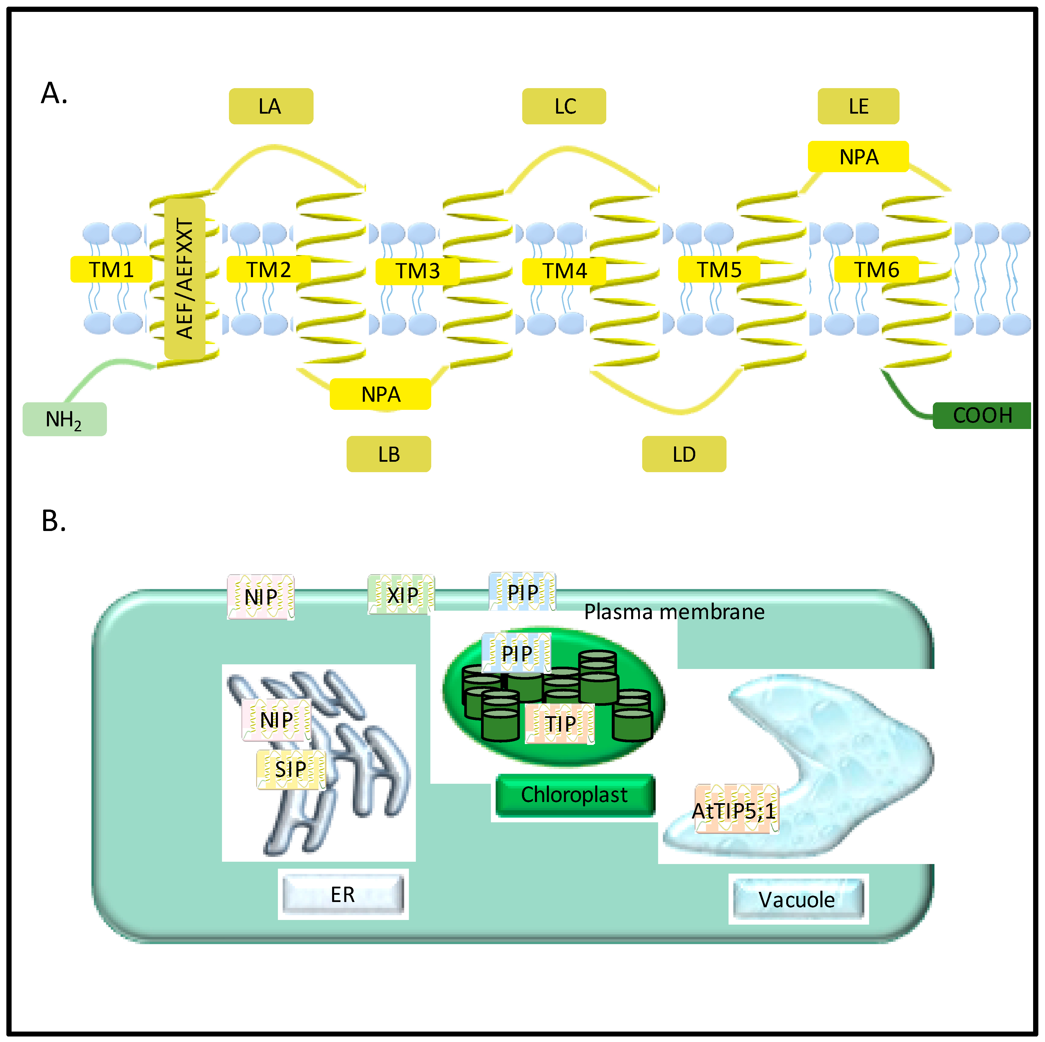
2. Diversity of Aquaporins in Plants

Presently, more than 800 MIPs [20,21] have been identified in bacteria [22], yeast [23], protozoa [24], archaea [25], insects [26] mammals [5,27] and plants [6]. Based on substrate specificity and protein sequence similarities, these 800 MIPs have been classified into three main subfamilies: (1) AQPs (aquaporins) involved in water and ion transport; (2) GLPs (glycerol-facilitators) permeable to glycerol and neutral molecules; and (3) GLAs (aquaglyceroporins) permeable to both water and glycerol. [20]. Unicellular organisms such as bacteria or yeast present the least MIP diversity, typically possessing only one or two aquaporin encoding genes. Plants do not have GLP orthologs [28], but they do exhibit the greatest diversity of ubiquitously localized AQPs [29,30] in comparison to mammals with 13 aquaporin isoforms (AQP0-12). Mammalian orthologs are primarily restricted to secretory glands [31] and fluid-conducting organs. Owing to the abundance and diversity of aquaporins in plants, MIPs are usually assimilated in a broader sense as aquaporins in literature (Figure 1).
Aquaporins include seven subfamilies categorized according to their intracellular locations and sequence similarities: the plasma membrane intrinsic proteins (PIPs), tonoplast intrinsic proteins (TIPs), NOD26-like intrinsic proteins (NIPs) and small, basic intrinsic proteins (SIPs), the GlpF-like intrinsic proteins (GIPs), hybrid intrinsic protein (HIP), and the uncategorized X intrinsic protein (XIP) [32] (Figure 1). Each subfamily could be further divided into different subgroups. For example, PIPs have been divided into two subgroups, PIP1 and PIP2. Each subgroup is then again divided to different isoforms, such as PIP1;1 and PIP1;2, which have specialized locations and functions (Figure 1).
The common characteristics that all MIPs share include six alpha-helical transmembrane helices (TM-1-TM-6), five inter-helical loops (LA-LE) [33] an AEF(Ala-Glu-Phe) or AEFXXT motif in the N-termial domain, and two highly conserved NPA (Asp-Pro-Ala) motifs (the “NPA box”) (Figure 1). Transport substrates, expression pattern, different levels of modification, regulation, and intracellular localizations contribute to the differences between the subfamilies of plant aquaporins. PIPs, NIPs, and XIPs are generally localized to the plasma membrane and expressed on the entire cell surface, while TIPs are localized to the tonoplast, the membrane of the vacuole. For most plant aquaporins, localization on endoplasmic reticulum (ER) can be observed during the processes of post-transcription, translation, and modification (Figure 1). However, SIPs and some NIPs have been found to localize to the ER [9], although the mechanism of targeting and their cellular functions are still not clear. SIPs tagged with green fluorescent protein were transiently expressed in Arabidopsis cells, showing the ER subcellular localization [34]. SIP1;1 and SIP1;2 may function as water channels in the ER, while SIP2;1 might act as an ER channel for other small molecules or ions [34]. Based on proteomic analyses in Arabidopsis, some PIPs and TIPs have been predicted to localized to the inner envelop and thylakoids [9], while it has also been found that AtTIP5;1 is located to tonoplast [35] (Figure 1).
Another difference between various MIP subfamilies is their substrate selectivity. Besides their function as water transporters, MIPs have been reported to transport atypical substrates, including ammonia, antimony, arsenite, boron, carbon dioxide, formamide, glycerol, hydrogen peroxide (H2O2), lactic acid, silicon, and urea. Two factors that contribute to their substrate selectivity are the conserved NPA motifs and amino acid residues including the ar/R (aromatic/arginine) region. The conserved NPA motifs contribute to the selectivity for water molecules [32], which are highly conserved in plant PIPs and TIPs, while alternative motifs have been found only in the NIP or SIP groups [36,37]. PIPs function as the transporters of water, glycerol, H2O2, carbon dioxide, and urea. PIP aquaporins are also involved in abiotic stress responses. However, several studies demonstrated altered responses of PIPs and TIPs family members upon salt, drought, or cold stresses [32]. The main role of TIPs has been described in the permeability of water. The water permeability function of plant aquaporins was first demonstrated in a TIP from Arabidopsis. Further studies have shown that TIPs may control water exchange between cytosolic and vacuolar compartments, which implies that they have a role in regulating cell turgor. Besides its water permeability function, TIPs also play roles in glycerol, urea, and ammonia transport and abiotic stress response. Specific TIP isoforms of rice and maize or Arabidopsis also show differential responses to water stress, salt, and cold stress. NIPs are able to transport water, glycerol, ammonia, silicic acid, and other solutes between plant and bacterial symbionts. Compared to PIPs and TIPs, NIPs have less water transport activity, but higher permeability to small organic molecules and mineral nutrients. The functions of NIPs include transporting beneficial and toxic metal molecules [38,39]. For example, OsNIP2;1 not only functions as a silicon influx transporter but is also involved in selenite uptake in rice [40]. SIPs have moderate water transport activity and may also function in original pore conformation. XIPs work as multifunctional channels permeable to water, metalloids and ROS. For example, PtXIP2;1 exhibited a differential expression in leaves and stems under the stress of drought, salicylic acid, or wounding [41].

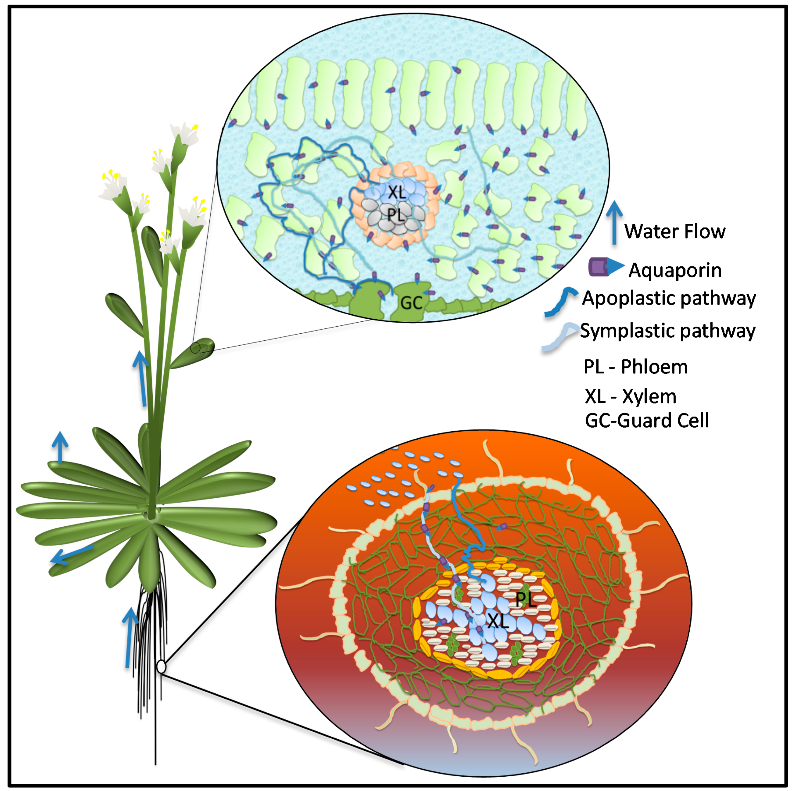
3. Roles of Aquaporins in Plant–Water Relations

Due to their autotrophic nature, plants require light and CO2 from their aerial environment culminated with water and nutrients from soil to carry out photosynthesis. A constant water flow starting from the absorption of water from soil to its distribution throughout the plant body and evaporation in the atmosphere is crucial for carrying out all the physiological activities of the plant and managing stress imposed by fluctuating environment. Plant–water relations result in establishing a soil-plant-atmosphere continuum [42]. Although water is a key element of all physiological processes, the plant, for its growth and metabolism, uses only a small fraction. The remaining 99.5% is lost during transpiration [43]. In order to fix one kilogram of carbon during photosynthesis, plants transport several hundred kilograms of water [44]. This bulk flow of water through plants can take three different routes: the apoplastic route along cell wall structure, the symplastic route from cell to cell through the plasmodesmata, and the transcellular path across the cellular membranes [45] (Figure 2).
Long-distance bulk flow of water during transpiration and sugar transport takes the apoplastic route through vascular bundles and is generally not limited by membrane barriers [46]. Water ascends through the xylem and phloem by capillary action driven by the gradient of water potential (ΔΨ) in which water moves from a region higher Ψ to a region of lower Ψ. In the light phase, stomatal opening leads to evaporation that causes a decreases in leaf Ψ that pulls water by creating tension in the xylem vessels resulting in the movement of water from the soil through the roots [42,47] (Figure 2).
Short-distance non-vascular water transport across cellular membranes is crucial to maintaining intracellular hydrostatic pressure (turgor) and cell water homeostasis at each level of organization. This includes (1) at the cell level: cellular/subcellular metabolic reactions, cell division, differentiation and elongation; (2) at the tissue level: stomatal movement; and (3) at the organ level: leaf petiole movement and maintenance of upright status of whole plant all processes depend upon cell water relations. Aquaporins as transmembrane water and solute transporter channels could be speculated as potential regulators of plant cell water relations that reflect their key roles in plant cell osmoregulation [48], root hydraulic conductivity (Lpr), leaf hydraulic conductivity [49], transpiration [50], and cell elongation [51] (Figure 2).
Maintenance of the cell’s osmotic potential under stress conditions such as pathogen infection, drought, flooding, salinity, high or low temperatures, and biotic stresses is a major challenge for plant growth and development. The majority of abiotic stress conditions directly impact plant water relations and stimulate an array of complicated cellular and physiological responses that lead to turning on plant water-saving strategies such as stomatal closure to cut off water loss during transpiration. As a tradeoff, water-saving photosynthetic activity is decreased due to the unavailability of CO2, ultimately leading to decreased plant biomass production. Consequently, a dire need exists to understand the underlying mechanisms which control plant–water relations in relation to photosynthesis and their response to biotic and abiotic stresses [52]. Aquaporins, as vital regulators of plant–water relations, are potential targets in developing stress resistant crop plants. Their significance in all facets of plant growth and development is well-established, but the mechanistic pathways behind their roles under plant defense responses remains to be elucidated [9,32]. Numerous comparative transcriptome studies under various abiotic stress conditions have shown a differential response of different aquaporin homologs in diverse plant tissues.
Figure 2. Water flow in plant from roots to aerial parts. Leaf cross-section shows flow of water through apoplastic and symplastic water pathways. In apoplastic pathway, water flows across the cells through cell wall. In symplastic pathway, water moves through cytoplasm and cellular membranes. Aquaporins are involved in symplastic path facilitating transmembrane water flow across cells and subcellular compartments.
Jdb 04 00009 g002
Reverse genetic approaches have also been extensively used to fine-tune the role of different aquaporin encoding genes in response to diverse environmental stresses. However, the presence of a large number of diverse aquaporins, integrated complex expression patterns, and technique limitations for measuring accurate solute and water movement across transmembrane aquaporin channels are major hurdles in establishing their conclusive roles in plant growth and survival under abiotic and biotic stresses [46].

4. Plant Aquaporins in Water Stress

Based on conclusive evidence, it is widely accepted that, in most plant species, water uptake and transcellular water flow in roots are largely mediated by PIPs and TIPs. These are the most abundant aquaporins in the plasma membrane and tonoplast of the plant cells, respectively [53]. Comparative transcriptome studies revealed differential expression of multiple aquaporin homologs in response to drought stress suggesting definite roles in stress responses. Induced expression of AtPIP2;3 under drought stress conditions is one of the earliest evidence of a drought responsive aquaporin [54]. In 2005, Alexandersson et al. monitored the expression of all 35 aquaporin homologs in Arabidopsis in response to drought stress alone and found that most PIP and some TIP genes have high levels of expression, while NIP genes have very low expression [55]. The authors also showed that all PIP genes are down-regulated in drought stress response in leaves except AtPIP1;4 and AtPIP2;5, which are up-regulated. Moreover, AtPIP2;6 and AtSIP1;1 are constitutively expressed and are not significantly affected by the drought stress [55]. Expression of AtPIP2;5 was also significantly up-regulated in leaves in response to a combination of drought and heat stresses [56]. Consistent with these results, several other studies in Arabidopsis have shown that among all subfamilies of aquaporins PIPs are most responsive to drought stress and most of them undergo a transcriptional down-regulation. Only a few genes were found to be up-regulated [57,58]. All of these PIP genes that are down-regulated in response to drought are highly expressed in the roots. Strong down-regulation of PIP genes transcription under drought stress was also observed in the roots and twigs of olive plants [59], in the roots of tobacco [60], and in the fruits of peach [61]. Moreover, most of these drought responsive transcriptional changes are conserved among different Arabidopsis accessions [57]. Distinctly, in many other plant species, differential responses by the same aquaporin homologs have been seen among different cultivars of the same plant species. For example, in the grapevines the expression of VvPIP1;1 in the root was up-regulated by drought stress in an anisohydric cultivar but not in an isohydric cultivar [62]. In another experiment, expression of the VvPIP2;1 gene was down-regulated under drought conditions [63]. Therefore, contrasting results have been reported between similar aquaporin homologs within the same species under drought stress. One transcriptome comparison of a field-grown cotton (Gossypium. hirsutum) under normal and drought stress conditions revealed that two highly homologous genes, PIP1;3 and PIP1;1 have contrasting expression patterns in leaves and roots [18] indicating that their roles in drought stress response differs despite their structural similarity. In addition to water deficiency dehydration caused by other environmental stimuli exhibited differential responses in some species. For example, osmotic stress induced by 10% polyethylene glycol (PEG) in rice revealed no effect on OsPIP1;3, but expression of OsPIP1;1 and OsPIP1;2 was up-regulated [64]. Contrastingly, expression of OsPIP1;1 was down-regulated in osmotic stress administrated by manitol [65] and also by drought stress [66]. Similar alterations were seen in reddish aquaporins in response to salt, PEG, and mannitol induced osmotic stress [67]. Recently, characterization of GoPIP1 from a legume forage Galega orientalis showed its association with drought tolerance. In contrast to rice and reddish, the transcripts levels of GoPIP1 increased significantly in roots upon exposure to the osmotic stress imposed by both high NaCl concentration and PEG. Overexpression of this gene in transgenic Arabidopsis made the plants more vulnerable to drought stress but not to salinity stress [68]. Stress levels also lead to differential responses, for example, in grapevine leaves, moderate drought stress led to a significant down regulation of the five PIP genes investigated, but prolonged stress increased their expression levels [69]. Water transport by aquaporins is also coupled with diurnal rhythms. Diurnal expression of PIPs in response to different intensities of drought was investigated in Fragaria vesca. Researchers found that most of the PIPs are down-regulated in roots and the expression of FvPIP1;1 and FvPIP2;1 was strongly correlated to the decrease in substrate moisture contents. In leaves, the amplitude of diurnal aquaporin expression was down-regulated in response to drought [70]. Responses of aquaporins to drought stress also vary based on plant symbiosis. In a study of Phaseolus vulgaris, it was observed that PIPs’ responses to drought stress vary depending on whether the plants is inoculated with arbuscular mycorrhizal fungi or not [71]. Arbuscular mycorrhizal symbiosis with maize plants has been shown to regulate aquaporin expression differentially in short term and prolonged drought stress conditions. Furthermore, various ZmPIPs, specifically the expression of ZmTIP1;2, ZmTIP2;3, and ZmNIP2;1 also varies in response to drought and symbiosis [72].
In transcriptome studies, the roles of PIPs in drought response is well pronounced in most of the cases but some reports specifically point out the role of TIPs in water deficient conditions. In rice, the expression of OsTIP1;1 was up-regulated in roots and shoot in response to water stress [73]. Root transcriptome analysis of chickpea plants under drought stressed showed both up and down-regulation of different PIPs, TIPs, and NIPs homologs suggesting their complex integrated roles in regulation of water balance under water deficit [74]. Reports about involvement of NIPs, SIPs, and XIPs in drought stress responses are very few. Detailed transcriptome studies of all aquaporin homologs in citrus plants under drought stress conditions revealed that all CsTIPs (11) and CsXIPS (3) were up-regulated in leaves but down-regulated in roots, whereas in leaves only two CsPIPs (CsPIP1;1 and CsPIP2;4), one CsNIP (CsNIP1;1), and one CsSIP (CsSIP1;2) were differentially up-regulated [13].
On the basis of transcriptome data, it is difficult to identify a concerted pattern of aquaporin expression in response to drought stress. Reverse genetics approaches based on one or a few genes provide a better snapshot than the whole transcriptome studies. Crucial involvement of PIPs in modulating Lpr in response to various environmental stresses has been widely observed [11,49,75]. Increased susceptibility of plants to water stress due to reduction in Lpr by silencing PIPs has been reported extensively in various plant species. For example, in Arabidopsis knockout mutants of AtPIP1;2 and AtPIP2;2 genes, there was a noteworthy reduction in water permeability of protoplasts [76] and a 14% decrease in Lpr, respectively making these mutants more vulnerable to drought stress [77]. Double antisense lines with reduced expression of AtPIP1 and AtPIP2 in Arabidopsis showed up to a 30-fold decrease in Lpr. In tobacco a 55% reduction in Lpr and increased sensitivity to drought was observed by targeting NtPIP1 gene using antisense technology. In addition to decreased Lpr, reduced expression of NtPIP1 also showed a significant decrease in transpiration rate [49]. In moss, Physcomitrella patens, knockout mutants of PpPIP2;1 and PpPIP2;2 exhibited severe stress phenotypes when grown under water-limited conditions. Based on these observations, it is plausible that these targeted aquaporins play a cumulative role as water transporters, and their decreased expressions make plants vulnerable to water stress due to a decreased Lpr. Further, it is also possible that decreased transpiration rates cause a reduction in photosynthesis, in turn affecting the overall survivability of the plant [78].
Consistently, transgenic plants overexpressing aquaporins showed enhanced drought tolerance. For example, the overexpression of a BnPIP1 from Brassica napus in transgenic tobacco plants resulted in increased tolerance to drought stress [79]. Similarly, transgenic tobacco plants overexpressing the wheat aquaporin gene TaAQP7 (PIP2) were more drought tolerant in comparison to non-transgenic tobacco plants due to enhanced water retention capabilities of transgenic plants [80]. Transgenic Arabidopsis plants expressing a Vicia faba PIP1 (VfPIP1) showed improved drought resistance by preventing water loss through transpiration due to the induction of stomatal closure [81]. In Arabidopsis, the overexpression of a banana PIP gene MaPIP1;1 showed increased root growth and enhanced survival rates of transgenic plants under drought stress, when compared to wild type plants [82]. Similarly, transgenic banana plants expressing banana PIP1;2 driven through two diverse promoters conferred enhanced tolerance to drought stress [17]. Overexpression of a tomato SlTIP2;2 gene in transgenic tomato plants resulted in increased drought-tolerance due to ability of plant to regulate its transpiration rate under drought stress conditions [83]. The above experimental evidence suggests that the overexpression of aquaporins make plants more resistant to drought stress. However, some contrasting results have also been observed because rapid water loss due to increased leaf and root hydraulic conductivity makes some plants even more vulnerable to drought stress conditions. For example, the overexpression of AtPIP1;4 or AtPIP2;5 in transgenic Arabidopsis plants showed no advantage under normal conditions but presented a rapid water loss under drought stress conditions. Similarly, overexpression of an Arabidopsis aquaporin gene AtPIP1b, in tobacco showed drastic water loss under drought stress [84].
In order to understand the role of aquaporins in drought tolerance many studies have been conducted in naturally drought tolerant plant species. In a drought tolerant plant Eragrostis nindensis (resurrection grass), TIP3;1 was found in small vacuoles of desiccation tolerant leaves, suggesting its importance in enhanced mobilization of water and solutes from these vacuoles upon rehydration [85]. Functional characterization of PIPs in two differential drought responsive poplar clones Populus balsamifera and P. simonii × P. balsamifera revealed a clear correlation between differential expressions of PIP genes in drought avoiding and drought tolerating strategies adopted by these two clones. The rapid reduction of stomatal conductance by reduction in PIPs activities was observed in leaves of drought avoiding P. simonii × P. balsamifera [86]. Water deficit exposure to a drought tolerant rice cultivar revealed up-regulation of the OsPIP1;3 gene suggesting its role in drought avoidance [87]. Strong up-regulation of some PIPs in a dehydration tolerant succulent plant, Craterostigma plantagineum, was observed under water stress [88]. Characterization of a FaPIP2;1 gene isolated from a drought-tolerant perennial grass species Festuca arundinacea showed its involvement in leaf dehydration status during water stress by overexpressing this gene in Arabidopsis. Transgenic plants showed enhanced resistance and better growth and development under drought stress [89]. In a drought tolerant Vitis hybrid, Richter-110 (Vitis berlandieri × Vitis rupestris) expression of five PIPs and two TIPs was monitored at different levels of water stress, and it was found that the aquaporin genes in leaves show differential regulation in response to moderate and drastic water stress. A moderate decrease in water availability results in down-regulation of the aquaporins. However, in roots, aquaporin expression showed complex patterns, with no generality among different aquaporins [69].
It can be concluded that drought stress response of aquaporins is highly variable depending on stress levels, aquaporin isoform, tissue, species, presence of symbionts, and the nature of stimuli causing dehydration similar to drought stress. However, a general down-regulation of most of the PIP genes is thought to reduce water loss and to help prevent backflow of water to drying soil. Although TIPs are found to play a key role in controlling cell water homeostasis by rapid water transport between the vacuole and cytoplasm of plant cells, experimental evidence on their roles in response to drought stress are limited in comparison to PIPs.

5. Aquaporins in CO2 Homeostasis in Water Deficit Conditions

CO2 conductivity and transpiration are intrinsically associated with stomatal movement. In order to cut off the loss of water through transpiration, stomatal closure is the paramount water-saving strategy of plants under water-limiting conditions, which result in a reduced rate of photosynthesis due to the unavailability of CO2. In addition to their pivotal role in the transmembrane, water transport, and regulation of stomatal aperture, reports have also shown the involvement of PIPs in CO2 conductivity relevant to the photosynthetic capacity of plant under water-limiting conditions. In tobacco, the involvement of NtAQP1 has been shown in leaf CO2 transport [90]. An AtPIP1;2 T-DNA insertion line facilitates the diffusion of CO2 suggesting a relevance of CO2 transport through aquaporins [91,92]. The Mesembryanthemum crystallinum aquaporin McMIPB, which is classified as a PIP1 that interacts with PIP2 by forming heterotetramers, showed enhanced mesophyll and stomatal conductance, CO2 diffusion, photosynthesis, and overall plant growth. This suggests that the co-expression of PIP2 and PIP1 may increase the activity of PIP1 not only for water flow but also for CO2 diffusion [93]. In Eucalyptus trees, the expression of EgPIP1 and EgPIP2 was co-suppressed by using RsPIP1;1 (Raphanus sativus aquaporin) and a low rate of CO2 assimilation was observed [94]. In rice, when HvPIP2;1 is overexpressed, enhanced CO2 conductance and CO2 assimilation is observed, but there is also greater sensitivity to salt stress [95]. In tomato (Solanum lycopersicum), overexpression of SlTIP2;2 regulates transpiration rates under stress conditions resulting in improved CO2 uptake and a more balanced water and nutrient supply [83].
Subsequently, under water stress conditions aquaporins are not only responsible for minimizing water loss from plant tissues, but they also serve a prized function in facilitating CO2 homeostasis.

6. Roles of Aquaporins in Salt Stress

Soil salinity is the combination of water and ionic stress that exerts noxious effects on plant growth and development by disturbing the cell’s osmotic balance. The primary responses of plants to salt stress are the inhibition of root-water uptake and a resultant decrease in Lpr [96], which is also manifested in cases of drought stress responses. Thus, most of the experimental evidence of aquaporin responses in salinity is in consistent with drought stress. For example, in Arabidopsis a significant reduction in Lpr, coupled with a 60% to 75% decrease in PIP and TIP aquaporin transcripts abundance, was observed after exposure to salt stress. Transgenic banana plants overexpressing MusaPIP1;2 and MusaPIP1;2 displayed enhanced tolerance to both salt and drought stresses [17]. Transcript and protein levels of the barley HvPIP2;1 gene were found to be down-regulated in roots but up-regulated in the shoots of plants under salt stress [97]. Additionally, the overexpression in transgenic barely revealed increased salt sensitivity, furthering suggesting a relevant role in salt tolerance [98]. A salinity-tolerant ice plant (M. crystallinum) also showed down-regulation of many PIP genes [99] and a TIP gene (MIPF) in roots and leaves, respectively under salt stress [100]. In Maize plants, an ABA mediated, salt-induced down regulation of most of the members of ZmPIP1 and ZmPIP2 were observed, but a transiently enhanced expression of ZmPIP1;1, ZmPIP1;5, and ZmPIP2;4 was also seen preferentially in the outer parts of the roots. No change in expression of ZmTIPs was observed in these experiments [101]. Subsequent studies showed involvement of some TIPs, therefore it can be anticipated that in addition to their role in increased water transport across the tonoplast, TIPs are also involved in the accumulation of ions in vacuoles in response to salt stress. For example, the overexpression of aAtTIP5;1 in Arabidopsis resulted in the tolerance of transgenic plants to high levels of borate, suggesting its involvement in vacuolar compartmentation of borate [102]. In rice, the expression of OsTIP1;1 was down-regulated in response to cold stress [12] but up-regulated during response to water and salinity stress [73]. The overexpression of the Panax ginseng aquaporin, PgTIP1, in Arabidopsis showed increased plant growth under optimal conditions and also enhanced tolerance to salt and drought stress [103]. Several studies involving the localization of aquaporins during salt stress have revealed relocalization or redistribution of aquaporins in response to high salt concentrations. For example, salt stress induced re-localization of AtTIP1;1 into intravacuolar invaginations is also shown in Arabidopsis [96]. In addition to redistribution, an alternative mechanism regulating PIP abundance in the plasma membrane is endocytosis of PIPs either through the clathrin-dependent pathway [104] or a salt- stimulated membrane raft–associated pathway [105]. Relocalization of a TIP1;1-GFP into intracellular spherical structures and internalization of PIP2;1-GFP in response to salt stress was also observed in Arabidopsis [96]. Both endocytosis and exocytosis caused by the cycling of AtPIP2;1 to and from the plasma membrane is also observed under salt stress [106].
Similar trends of PIPs and TIPs regulation in response to salt stress have been observed with only a few exceptions. For example, in drought-tolerant, salinity-sensitive grapevine the expression of the PIP2;1 gene is up-regulated under salt stress but down-regulated under drought [63]. In addition to PIPs and TIPs, being small solute and ion transporters, NIPs have also been found to be involved in salinity stress. The overexpression of wheat NIP, TaNIP, in Arabidopsis plants showed increased tolerance to salt stress. Expression of this TaNIP gene is up-regulated after ABA treatment, suggesting its association with other ABA regulated pathways [107]. Transcriptome analyses of citrus roots and leaves under salt stress revealed that in addition to CsPIPs and CsTIPs, most of the CsNIPs, CsXIPs, and CsSIPs are also up-regulated in roots, whereas in leaves both up and down regulation patterns were observed for some homologs of each aquaporin family except CsSIPs [13]. Similar to drought stress, most of the experimental evidence shows involvement of PIPs and TIPs, while only a few experiments point towards the involvement NIPs. Interestingly, only one account exists of XIPs’ involvement in salt stress.

7. Cold Stress and Aquaporins

Similar to water and salt stresses, cold stress is an important abiotic stress factor that significantly limits plant growth and development. The early response of Aquaporins to cold stress has been frequently reported in literature. For example, transgenic banana plants over expressing MusaPIP1;2 and MusaPIP1;2 displayed enhanced tolerance to both cold and drought stress [17]. Similarly, transgenic tobacco plants overexpressing a wheat aquaporin TaAQP7 (PIP2) gene exhibited enhanced cold-tolerance as well as drought tolerant in comparison to non-transgenic tobacco plants [80,108]. Distinctly, other experiments reveal opposite patterns. For example, transgenic Arabidopsis plants overexpressing AtPIP1;4 or AtPIP2;5 showed enhanced tolerance to cold stress but are more susceptible to drought due to rapid water loss. In cold sensitive plants like rice, prolonged exposure to cold stress cause an increase in Lpr which should be regulated through root aquaporins, as most, particularly OsPIP2;5, are found to be up-regulated [109]. In another experiment in rice, the mRNA levels of ten genes including OsTIP1;1 and OsTIP2;2 were significantly down-regulated but the expression of OsPIP1;3 increased up to 60% in roots on exposure to chilling treatment [12]. OsPIP1;3 is also significantly up-regulated in response to drought stress in drought-tolerant rice cultivar [87]. In contrast, OsTIP1;1 was up-regulated in response to water and salinity stress [73]. While some homologs present similarities in all dehydration inducing stresses like drought, salinity, and cold, others like OsTIP1;1, in the following example, are counter-regulated in cold in comparison to drought and salt. OsPIP1;1 presents different patterns of expression upon exposure to diverse osmotic stresses. Moreover, its involvement in all salt, drought and chilling tolerance has been suggested [64,65].
In Arabidopsis, only PIP2;5 and PIP2;6, which are normally among the low- expressed PIPs, are significantly up regulated by cold stress in both the roots and aerial parts of the plant. All other PIP genes were found to be down regulated by cold stress and their pattern of expression vary with the application of salt or drought stress. Most abiotic stresses, including chilling, induce the production of ABA. Plant responses to stress are generally ABA mediated. However, in an experiment, responses of each aquaporin to ABA treatment were different, suggesting that the regulation of aquaporins might follow both ABA-dependent and ABA-independent signaling pathways [58,67]. Aroca et al. in 2005 investigated the effect of long and short term chilling stress on the expression of aquaporins in cold-tolerant and cold-susceptible maize cultivars. They reported that long and short term chilling differentially induce aquaporin responses in similar patterns in both genotypes. These responses lead to recovery in tolerant genotypes, but not in sensitive genotypes including those that are unable to cope with the oxidative stress caused by chilling. Down regulation of PIPs in both cultivars is consistent with previous findings in rice and sugarcane [65,110,111].
The response of aquaporins to cold stress is linked with an increase in root hydraulic conductivity that may be regulated through increased aquaporin expression levels in plant roots upon exposure to low temperatures. Differential responses have been observed between short term and long term exposure to cold stress. Moreover, recovery from cold stress has been linked with aquaporin expression, particularly in PIPs.

8. Aquaporins in Micronutrient Homeostasis and Heavy Metal Stress

Although the water transport capacity of NIPs is very limited, they play a fundamental role in transporting other substrates involved in various cellular processes. Most NIP homologs appear to transport nutrients in plants. Arabidopsis knockout mutant of the NIP5;1 gene displayed striking growth retardation in response to low supply of boron (B). B is an essential element for plant growth, development, and reproduction, and its deficiency in arable areas has drastically effected crop production worldwide [112]. In the event of high B supply, a feedback inhibition of the AtNIP5;1 gene is observed, thus providing a strong evidence of the involvement of NIP5;1 in B homeostasis the adaptation of plants to B toxicity in soil. These results were further confirmed in maize, where a loss of function mutation in ZmNIP3;1, a maize ortholog of AtNIP5;1, was shown to be responsible for an abnormal phenotype caused by B deficiency. In addition, AtNIP6;1 and AtNIP7;1 homologs serve in B transport in Arabidopsis, facilitating its distribution in shoots and anthers, respectively [113,114]. Tolerance to high B was observed in barley by a reduced expression of the HvNIP2;1 gene. [115]. In addition to NIPs, an overexpression of TIP5;1 in Arabidopsis suggested its involvement in vacuolar compartmentation of borate [102]. Silicon (Si) plays an important role in plant defense against biotic and abiotic stresses. Si-mediated alleviation of plant stress is a well-known phenomenon [116]. The Si transporter OsNIP2;1 (Lsi1) was identified by quantitative trait loci (QTL) mapping in a rice mutant defective in Si uptake [117]. Furthermore, the Si transporter NIP2;2 (Lsi6) was also identified from rice [116]. Therefore, OsNIPs seems to play a significant role in Si transport in rice plants.
In addition to nutrient transport, NIPs are also found to be involved in both the transport and responses to heavy metal stress. A T-DNA knockout mutant of Arabidopsis NIP1;1 showed arsenite (As) tolerance suggesting its role as an As transporter [118]. Recently, AtNIP3;1 has shown to be involved in As transport. Double knockout mutants for both AtNIP1;1 and AtNIP3;1 show more pronounced tolerance against As stress. An additional four other isoforms, NIP5;1, NIP6;1, NIP7;1, and NIP1;2, are reported to be capable of As(III) transport on the basis of their expressions in yeast and oocytes [119]. The transport of antimony (Sb) by AtNIP1;1 has also been established, and its loss of function mutant showed an increased tolerance to high Sb stress [120]. A lactic acid transporter AtNIP2;1 was shown to be associated with water logging, leading to oxygen deprivation. Thus it can be anticipated that AtNIP2;1 is an anaerobic-induced lactic acid transporter that may play a role in plant adaptation to lactic fermentation under anaerobic stress conditions [121].

9. Aquaporins in Plant Symbiotic and Pathogenic Relations

Plant symbiotic relationships are known to affect plant aquaporin responses under different abiotic stress conditions [71,72]. It is hypothesized that in plant symbiotic relations, the re-distribution of water and nutrients takes place between the host and symbionts further shedding light on a relationship with aquaporins. The roles of NIPs in plant interactions with symbionts and pathogens are obvious but experimental evidence is scarce. The first identified aquaporin, nodulin-26, was found in soybean root nodules and is speculated to have formed as a result of a symbiotic interaction between plant and nitrogen fixing rhizobium. While the roles of NIPs in various forms of nitrogen transport (urea, ammonia) are well known, the mechanistic involvement of NIPs in driving these nutrient exchanges between plants and symbionts or plants and pathogens needs further investigation. The roles of plant aquaporin-mediated solute transport during plant symbiosis with arbuscular mycorrhizae have been examined by gene profiling. Such results have indicated that the transport of glycerol from plant to the microbe, in addition toNH4/NH3 from microbe to plant, is mediated by aquaporin genes including NIPs [72].
Like symbionts, plant pathogens rely on plant nutrients for their growth and survival on their plant hosts. Dehydration, as a result of pathogen infection, is a common observation responsible for regulating plant water relations and nutrient homeostasis. The production of reactive oxygen species is an integral component of plant immune system. The transport of H2O2 by aquaporin homologs also highlights functions in plant defense, despite not having a full understanding of these pathways [122,123]. Expression profiling of soybean leaves upon Pseudomonas syringae infection revealed down-regulation in 24 of 32 soybean aquaporin genes [124]. In citrus plants, six CsMIPs (CsPIP1;2, CsPIP2;2, CsNIP2;2, CsNIP5;2, CsNIP6;1, and CsSIP1;1) were found to be differentially expressed exclusively under the biotic stress imposed by the citrus infecting proteobacterium, Candidatus Liberibacter. Comparisons of CsPIP2;2, CsTIP1;2, CsTIP2;1, CsTIP2;2, and CsNIP5;1 expression patterns of susceptible sweet orange and tolerant rough lemon cultivars revealed that most CsMIPs are down regulated. Thus, they could be correlated with the disease development [13,125]. As a result, the roles of aquaporins in response to pathogen infection are highly anticipated. Evidence of NIPs’ involvement in biotic stress responses in maize was reported [126]. The induction of a tobacco root specific TIP1 gene expression upon nematode infection was observed [127]. In grasses (Festuca spp.), Si accumulation offers an effective defense against herbivore attacks [128]. Thus, the regulation of Si uptake through NIPs can be correlated with plant defense against herbivory. The precise modes of aquaporin regulation during infection and their response to pathogen signals are still unknown. Some interactions of aquaporins with bacterial and oomycete effectors were observed in yeast two hybrid systems [129]. TIP1 and TIP2 were found to interact with a cucumber mosaic virus (CMV) replication protein, CMV1a, in the SOS recruitment system suggesting that this TIPs-CMV1a interaction potentially affects CMV replication in the host plant’s tonoplasts [130]. Interactome and transcriptome data provide important clues about the involvement of aquaporins in host-pathogen interactions. Extensive reverse genetic-based characterization of individual or multiple putative aquaporin homologs needs to be undertaken in relation to pathogen infection to reach a definite conclusion about role of aquaporins in plant diseases.

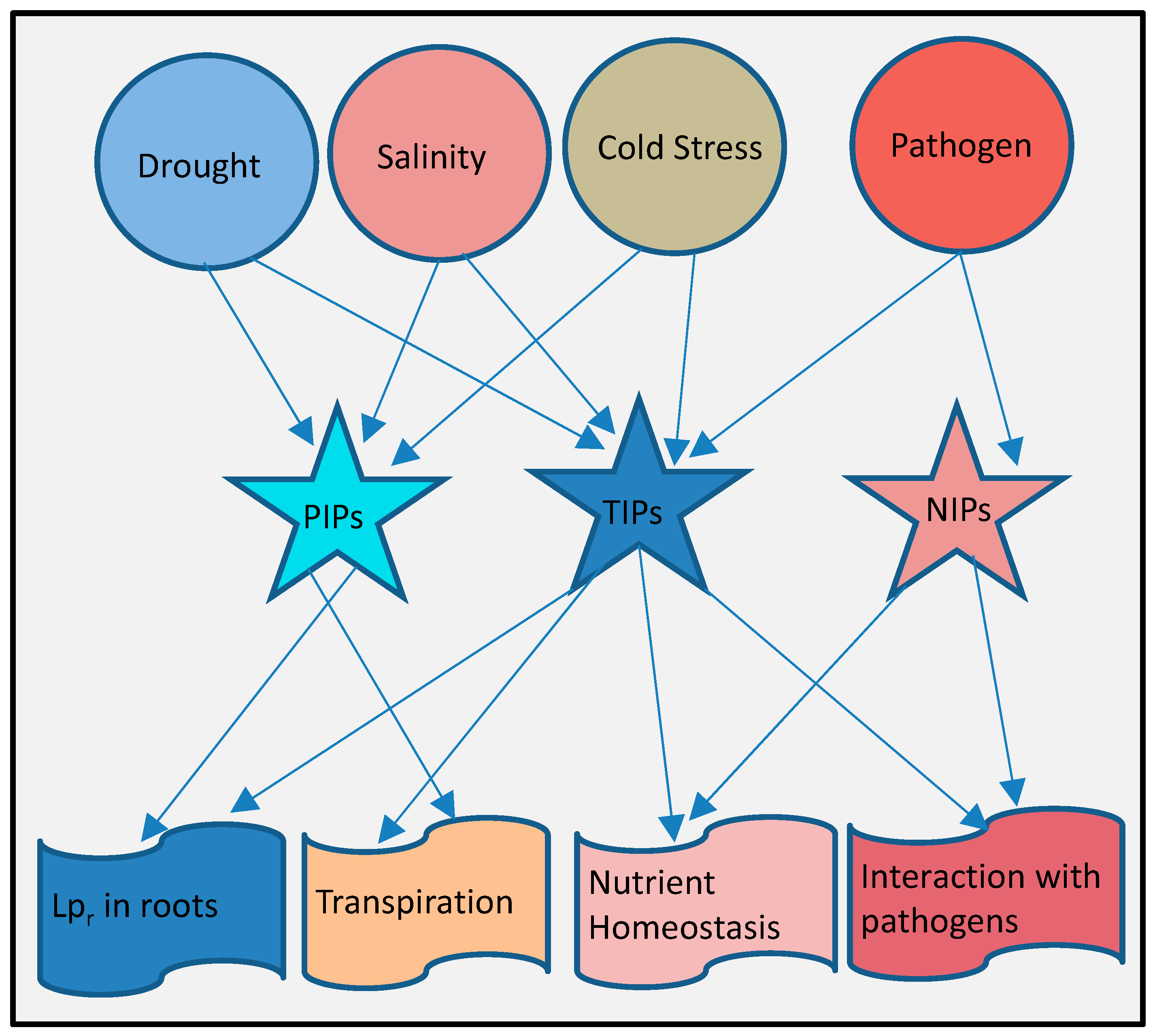
10. Complex Integrated Roles of Aquaporins in Plant Stresses

Despite enormous experimental evidence about the involvement of aquaporins genes in various stress responses (Figure 3), precise roles of individual genes or particular subfamilies are still hard to define because of their highly complex and integrated roles in the response to different environmental stimuli and involvement in other plant growth and developmental processes. These integrated contributions in multiple physiological facets can be more pronounced in transgenic plants containing foreign aquaporin genes. In transgenic plants expressing an exogenous aquaporin gene, tolerance or susceptibility is collectively effects by interactions with other plant genes. Transgenic Arabidopsis plants expressing a Vicia faba PIP1 (VfPIP1) showed improved drought resistance due to induction of stomatal closure, thereby preventing water loss in water- deficient conditions [81]. Hence, the interaction of VfPIP1 with other genes and gene products involved in stomatal movement contributes to overall drought tolerance. The silencing of endogenous genes by expressing exogenous aquaporins has also been observed when the Raphanus sativus aquaporin RsPIP1;1 gene is expressed in eucalyptus causing the silencing of endogenous EgPIP1 and EgPIP2 PIP aquaporins [94]. The overexpression of AtPIP1b in tobacco results in an increased stomatal density and increased growth rates under favorable conditions but drastic water loss under drought stress [84]. Transgenic tobacco plants overexpressing a wheat aquaporin TaAQP7 (PIP2) gene showed increased activity of detoxification enzymes resulting in an improved antioxidant defense and a reduction in H2O2 levels under osmotic stress [80]. Similarly, the overexpression of a wheat PIP, TdPIP1;1, in tobacco showed an enhanced stress tolerance coupled with increased leaf area and increased root size [131]. The overexpression of two foreign aquaporin isoforms, CfPIP2;1 (Cucurbita ficifolia) and CsPIP1;1 (C. sativus) in Arabidopsis, noticeably perturb the natural expression of other aquaporins, resulting in altered stress responses of the plants under various stress conditions [132]. In addition, the overexpression of a BnPIP1 from Brassica napus in transgenic tobacco plants results in an increased tolerance to drought stress, increased plant growth, and higher seed germination rates, whereas antisense plants showed developmental deformities and an increased susceptibility to drought stress [79].
In addition to these outcomes of over-expressing aquaporins in different transgenic backgrounds, advances in interactomics has shown physical interactions among different aquaporin homologs [133,134,135] as well as aquaporin interactions with several other proteins involved in various diverse functions in plants, alluding to their complex, integrated role in different physiological and stress responses [130,136,137,138,139,140,141].
Figure 3. A generalized summary of plant aquaporins in response to environmental stimuli. Both PIPs and TIPs are more responsive to drought, salt and cold stress that disturb cell osmotic balance, they regulate root hydraulic conductivity (Lpr) and transpiration rates. TIPs along with NIPs are involved in biotic stress responses and involved in regulating nutrient homeostasis between host and its pathogen. NIPs and TIPs are also found to interact with some pathogen proteins.
Jdb 04 00009 g003

11. Conclusions

Current data suggest that drought and salt stress induces more dramatic changes in aquaporin expression than any other abiotic stress [19]. A profound role of PIPs in response to osmotic stress stimuli like drought and salinity can be established (Figure 3). Knockout mutants of PIPs and their overexpressing transgenic plants revealed that PIPs respond to osmotic stresses in an integrated, complex way by regulating root water uptake and increasing transpiration rates as well as affecting the overall plant growth and development processes. Like PIPs, TIPs are involved in regulating osmotic stress responses, but experimental evidence is far less in comparison to PIPs. TIPs are also found to be involved in the uptake of some micronutrients facilitating responses to higher micronutrient concentrations. NIPs’ roles in nutrient homeostasis and heavy metal stress responses are more pronounced than any other aquaporin subfamily. Information on the involvement of aquaporins in biotic stress responses is still scarce. However, current analyses on the involvement of TIPs and NIPs in nutrient uptake points to a pronounced involvement of NIPs in plant pathogen interactions (Figure 3).
Since the discovery of aquaporins two decades ago, more information has been unveiled regarding structure, substrate specificity, gating properties, subcellular localization, and roles in various physiological processes in plant growth and development. Forward and reverse genetic approaches have also been used extensively to define their roles in plant defense mechanisms against environmental stresses. However, a clear cut mechanistic role of individual aquaporin homologs in response to a particular environmental stress has not yet been established because of the complex, integrated responses that vary among different plant species. Numerous studies of plant aquaporins under osmotic stress conditions have revealed their importance in regulating plant stress responses. Rapid changes in the expression levels of aquaporins, in response to diverse stresses, have been documented often, but the molecular and cellular mechanisms underlying these responses are still unknown. The localization patterns in specific membranes and subcellular compartments combined with their redistribution with exposure to diverse osmotic stresses could be crucial for efficacy of aquaporins [142,143]. Moreover, the responses of aquaporins to various stress induced hormones indicate their involvement in stress induced hormonal pathways. The interactions of aquaporins with other plant proteins also provide an insight into the complex mechanisms involved in aquaporin regulations and diverse stress responses. In order to arrive at a definite conclusion, more detailed studies are needed to map different pathways of aquaporin activities in plant physiology and stress responses. Differential responses of aquaporin homologs to stress induced hormones need thorough investigation to uncover their mechanistic involvement in plant stress responses.

Acknowledgments

We acknowledge funding from Fulbright graduate fellowship to Z.A., U.A.B., College of Arts and Science graduate fellowship to Y.S. and N.S.F. (IOS-1557796) to M.S.M. We greatly appreciate Jessica Lopez for critical reading the manuscript.

Author Contributions

M.S.M. conceived the ideas and designed the layout the article. Z.A., T.C.H. and Y.S. wrote the paper and contributed the figures

Conflicts of Interest

The authors declare no conflict of interest.

Abbreviations

The following abbreviations are used in this manuscript:
Lpr
Root hydraulic Conductivity
MIPs
Major intrinsic proteins
MIPs
Major intrinsic protein genes
PIPs
Plasma membrane intrinsic proteins
PIPs
Plasma membrane intrinsic protein genes
TIPs
Tonoplast intrinsic proteins
TIPs
Tonoplast intrinsic protein genes
NIPs
Nodulin-26 like intrinsic proteins
NIPs
Nodulin-26 like intrinsic protein genes
SIPs
Small basic intrinsic proteins
SIPs
Small basic intrinsic protein genes
GIPs
GlpF-like intrinsic proteins
HIPs
Hybrid intrinsic proteins
AQPs
Aquaporins
XIPs
Uncategorized X intrinsic proteins
XIPs
Uncategorized X intrinsic protein genes
ROS
Reactive oxygen species
GLPs
Glycerol-facilitators
ER
Endoplasmic reticulum
ΔΨ
Water potential
CMV
Cucumber mosaic virus

References

  1. Maurel, C.; Tacnet, F.; Guclu, J.; Guern, J.; Ripoche, P. Purified vesicles of tobacco cell vacuolar and plasma membranes exhibit dramatically different water permeability and water channel activity. Proc. Natl. Acad. Sci. USA 1997, 94, 7103–7108. [Google Scholar] [CrossRef] [PubMed]
  2. Koefoed-Johnsen, V. The contributions of diffusion and flow to the passage of D2O through living membranes: Effect of neurohypophysenl hormone 011 isolated anuran skin. Acta Physiol. Scand. 1953, 28, 60–76. [Google Scholar] [CrossRef] [PubMed]
  3. Macey, R.L.; Farmer, R.E. Inhibition of water and solute permeability in human red cells. Biochim. Biophys. Acta Biomembr. 1970, 211, 104–106. [Google Scholar] [CrossRef]
  4. Denker, B.M.; Smith, B.L.; Kuhajda, F.P.; Agre, P. Identification, purification, and partial characterization of a novel MR 28,000 integral membrane protein from erythrocytes and renal tubules. J. Biol. Chem. 1988, 263, 15634–15642. [Google Scholar] [PubMed]
  5. Preston, G.M.; Carroll, T.P.; Guggino, W.B.; Agre, P. Appearance of water channels in xenopus oocytes expressing red cell chip28 protein. Science 1992, 256, 385–387. [Google Scholar] [CrossRef] [PubMed]
  6. Fortin, M.G.; Morrison, N.A.; Verma, D.P.S. Nodulin-26, a peribacteroid membrane nodulin is expressed independently of the development of the peribacteroid compartment. Nucleic Acids Res. 1987, 15, 813–824. [Google Scholar] [CrossRef] [PubMed]
  7. Maurel, C.; Reizer, J.; Schroeder, J.I.; Chrispeels, M.J. The vacuolar membrane protein gamma-tip creates water specific channels in xenopus oocytes. EMBO J. 1993, 12, 2241–2247. [Google Scholar] [PubMed]
  8. Abascal, F.; Irisarri, I.; Zardoya, R. Diversity and evolution of membrane intrinsic proteins. Biochim. Biophys. Acta Gen. Subj. 2014, 1840, 1468–1481. [Google Scholar] [CrossRef] [PubMed]
  9. Maurel, C.; Boursiac, Y.; Luu, D.T.; Santoni, V.R.; Shahzad, Z.; Verdoucq, L. Aquaporins in plants. Physiol. Rev. 2015, 95, 1321–1358. [Google Scholar] [CrossRef] [PubMed]
  10. Johanson, U.; Karlsson, M.; Johansson, I.; Gustavsson, S.; Sjovall, S.; Fraysse, L.; Weig, A.R.; Kjellbom, P. The complete set of genes encoding major intrinsic proteins in arabidopsis provides a framework for a new nomenclature for major intrinsic proteins in plants. Plant Physiol. 2001, 126, 1358–1369. [Google Scholar] [CrossRef] [PubMed]
  11. Chaumont, F.O.; Barrieu, F.O.; Wojcik, E.; Chrispeels, M.J.; Jung, R. Aquaporins constitute a large and highly divergent protein family in maize. Plant Physiol. 2001, 125, 1206–1215. [Google Scholar] [CrossRef] [PubMed]
  12. Sakurai, J.; Ishikawa, F.; Yamaguchi, T.; Uemura, M.; Maeshima, M. Identification of 33 rice aquaporin genes and analysis of their expression and function. Plant Cell Physiol. 2005, 46, 1568–1577. [Google Scholar] [CrossRef] [PubMed]
  13. Martins, C.D.P.S.; Pedrosa, A.M.; Du, D.; Goncalves, L.P.; Yu, Q.; Gmitter, F.G., Jr.; Costa, M.G.C. Genome-wide characterization and expression analysis of major intrinsic proteins during abiotic and biotic stresses in sweet orange (Citrus sinensis L. Osb.). PLoS ONE 2015, 10, e0138786. [Google Scholar]
  14. Reuscher, S.; Akiyama, M.; Mori, C.; Aoki, K.; Shibata, D.; Shiratake, K. Genome-wide identification and expression analysis of aquaporins in tomato. PLoS ONE 2013, 8, e79052. [Google Scholar] [CrossRef] [PubMed]
  15. Gupta, A.B.; Sankararamakrishnan, R. Genome-wide analysis of major intrinsic proteins in the tree plant populus trichocarpa: Characterization of XIP subfamily of aquaporins from evolutionary perspective. BMC Plant Biol. 2009, 9. [Google Scholar] [CrossRef] [PubMed]
  16. Ali, Z.; Wang, C.B.; Xu, L.; Yi, J.X.; Xu, Z.L.; Liu, X.Q.; He, X.L.; Huang, Y.H.; Khan, I.A.; Trethowan, R. Genome-wide sequence characterization and expression analysis of major intrinsic proteins in soybean (Glycine max L.). PLoS ONE 2013, 8, e56312. [Google Scholar]
  17. Shekhawat, U.K.; Ganapathi, T.R. Overexpression of a native plasma membrane aquaporin for development of abiotic stress tolerance in banana. Plant Biotechnol. J. 2013, 11, 942–952. [Google Scholar]
  18. Park, W.; Scheffler, B.E.; Bauer, P.J.; Campbell, B.T. Genome-wide identification of differentially expressed genes under water deficit stress in upland cotton (Gossypium hirsutum L.). BMC Plant Biol. 2012, 12. [Google Scholar] [CrossRef] [PubMed]
  19. Reddy, P.S.; Rao, T.S.R.B.; Sharma, K.K.; Vadez, V. Genome-wide identification and characterization of the aquaporin gene family in Sorghum bicolor (L.). Plant Gene 2015, 1, 18–28. [Google Scholar] [CrossRef] [Green Version]
  20. Karkouri, K.; Gueune, H.; Delamarche, C. MIPDB: A relational database dedicated to MIP family proteins. Biol. Cell 2005, 97, 535–543. [Google Scholar] [CrossRef] [PubMed]
  21. Zardoya, R. Phylogeny and evolution of the major intrinsic protein family. Biol. Cell 2005, 97, 397–414. [Google Scholar] [CrossRef] [PubMed]
  22. Calamita, G.; Bishai, W.R.; Preston, G.M.; Guggino, W.B.; Agre, P. Molecular cloning and characterization of AQPZ, a water channel from escherichia coli. J. Biol. Chem. 1995, 270, 29063–29066. [Google Scholar] [PubMed]
  23. Carbrey, J.M.; Bonhivers, M.l.; Boeke, J.D.; Agre, P. Aquaporins in saccharomyces: Characterization of a second functional water channel protein. Proc. Natl. Acad. Sci. USA 2001, 98, 1000–1005. [Google Scholar] [CrossRef] [PubMed]
  24. Mitra, B.N.; Yoshino, R.; Morio, T.; Yokoyama, M.; Maeda, M.; Urushihara, H.; Tanaka, Y. Loss of a member of the aquaporin gene family, AQPA affects spore dormancy in dictyostelium. Gene 2000, 251, 131–139. [Google Scholar] [CrossRef]
  25. Kozono, D.; Ding, X.; Iwasaki, I.; Meng, X.; Kamagata, Y.; Agre, P.; Kitagawa, Y. Functional expression and characterization of an archaeal aquaporin aqpm from methanothermobacter marburgensis. J. Biol. Chem. 2003, 278, 10649–10656. [Google Scholar] [CrossRef] [PubMed]
  26. Beuron, F.; le Caherec, F.; Guillam, M.T.; Cavalier, A.; Garret, A.; Tassan, J.P.; Delamarche, C.; Schultz, P.; Mallouh, V.; Rolland, J.P. Structural analysis of a mip family protein from the digestive tract of cicadella viridis. J. Biol. Chem. 1995, 270, 17414–17422. [Google Scholar] [CrossRef] [PubMed]
  27. Agre, P.; Preston, G.M.; Smith, B.L.; Jung, J.; Raina, S.; Moon, C.; Guggino, W.B.; Nielsen, S. Aquaporin chip: The archetypal molecular water channel. Am. J. Physiol. Renal Physiol. 1993, 265, F463–F476. [Google Scholar]
  28. Zardoya, R.; Ding, X.; Kitagawa, Y.; Chrispeels, M.J. Origin of plant glycerol transporters by horizontal gene transfer and functional recruitment. Proc. Natl. Acad. Sci. USA 2002, 99, 14893–14896. [Google Scholar] [CrossRef] [PubMed]
  29. Johansson, I.; Karlsson, M.; Johanson, U.; Larsson, C.; Kjellbom, P. The role of aquaporins in cellular and whole plant water balance. Biochim. Biophys. Acta Biomembr. 2000, 1465, 324–342. [Google Scholar] [CrossRef]
  30. Maurel, C.; Javot, H.; Lauvergeat, V.; Gerbeau, P.; Tournaire, C.; Santoni, V.; Heyes, J. Molecular physiology of aquaporins in plants. Int. Rev. Cytol. 2002, 215, 105–148. [Google Scholar] [PubMed]
  31. Nielsen, S.R.; King, L.S.; Christensen, B.M.N.; Agre, P. Aquaporins in complex tissues. II. Subcellular distribution in respiratory and glandular tissues of rat. Am. J. Physiol. Cell Physiol. 1997, 273, C1549–C1561. [Google Scholar]
  32. Forrest, K.L.; Bhave, M. Major intrinsic proteins (MIPs) in plants: A complex gene family with major impacts on plant phenotype. Funct. Integr. Genomics 2007, 7, 263–289. [Google Scholar] [CrossRef] [PubMed]
  33. Hove, R.M.; Bhave, M. Plant aquaporins with non-aqua functions: Deciphering the signature sequences. Plant Mol. Biol. 2011, 75, 413–430. [Google Scholar] [CrossRef] [PubMed]
  34. Ishikawa, F.; Suga, S.; Uemura, T.; Sato, M.H.; Maeshima, M. Novel type aquaporin SIPs are mainly localized to the ER membrane and show cell-specific expression in arabidopsis thaliana. FEBS Lett. 2005, 579, 5814–5820. [Google Scholar] [CrossRef] [PubMed]
  35. Wudick, M.M.; Luu, D.T.; Tournaire-Roux, C.; Sakamoto, W.; Maurel, C. Vegetative and sperm cell-specific aquaporins of arabidopsis highlight the vacuolar equipment of pollen and contribute to plant reproduction. Plant Physiol. 2014, 164, 1697–1706. [Google Scholar] [CrossRef] [PubMed]
  36. Ishibashi, K. Aquaporin subfamily with unusual NPA boxes. Biochim. Biophys. Acta Biomembr. 2006, 1758, 989–993. [Google Scholar] [CrossRef] [PubMed]
  37. Ishibashi, K.; Kuwahara, M.; Kageyama, Y.; Sasaki, S.; Suzuki, M.; Imai, M. Molecular cloning of a new aquaporin superfamily in mammals. In Molecular Biology and Physiology of Water and Solute Transport; Springer: NewYork, NY, USA, 2000; pp. 123–126. [Google Scholar]
  38. Ma, J.F.; Yamaji, N.; Mitani, N.; Xu, X.Y.; Su, Y.H.; McGrath, S.P.; Zhao, F.J. Transporters of arsenite in rice and their role in arsenic accumulation in rice grain. Proc. Natl. Acad. Sci. USA 2008, 105, 9931–9935. [Google Scholar] [CrossRef] [PubMed]
  39. Zangi, R.; Filella, M. Transport routes of metalloids into and out of the cell: A review of the current knowledge. Chemico Biol. Interact. 2012, 197, 47–57. [Google Scholar] [CrossRef] [PubMed]
  40. Zhao, X.Q.; Mitani, N.; Yamaji, N.; Shen, R.F.; Ma, J.F. Involvement of silicon influx transporter OsNIP2;1 in selenite uptake in rice. Plant Physiol. 2010, 153, 1871–1877. [Google Scholar] [CrossRef] [PubMed]
  41. Lopez, D.; Bronner, G.L.; Brunel, N.; Auguin, D.; Bourgerie, S.; Brignolas, F.; Carpin, S.; Tournaire-Roux, C.; Maurel, C.; Fumanal, B. Insights into populus XIP aquaporins: Evolutionary expansion, protein functionality, and environmental regulation. J. Exp. Bot. 2012, 63, 2217–2230. [Google Scholar] [CrossRef] [PubMed] [Green Version]
  42. Steudle, E. The cohesion-tension mechanism and the acquisition of water by plant roots. Annu. Rev. Plant Biol. 2001, 52, 847–875. [Google Scholar] [CrossRef] [PubMed]
  43. Freeman, S. Biological Science, 3rd ed.; Benjamin Cummins: Upper Saddle River, NJ, USA, 2007. [Google Scholar]
  44. Tyerman, S.; Niemietz, C.; Bramley, H. Plant aquaporins: Multifunctional water and solute channels with expanding roles. Plant Cell Environ. 2002, 25, 173–194. [Google Scholar] [CrossRef] [PubMed]
  45. Steudle, E.; Peterson, C.A. How does water get through roots? J. Exp. Bot. 1998, 49, 775–788. [Google Scholar] [CrossRef]
  46. Kaldenhoff, R.; Ribas-Carbo, M.; Sans, J.F.; Lovisolo, C.; Heckwolf, M.; Uehlein, N. Aquaporins and plant water balance. Plant Cell Environ. 2008, 31, 658–666. [Google Scholar] [CrossRef] [PubMed]
  47. Lopez, D.; Venisse, J.S.P.; Fumanal, B.; Chaumont, F.O.; Guillot, E.; Daniels, M.J.; Cochard, H.; Julien, J.L.; Gousset-Dupont, A.L. Aquaporins and leaf hydraulics, poplar sheds new light. Plant Cell Physiol. 2013, 54, 1963–1975. [Google Scholar] [CrossRef] [PubMed]
  48. Wallace, I.S.; Choi, W.G.; Roberts, D.M. The structure, function and regulation of the nodulin 26-like intrinsic protein family of plant aquaglyceroporins. Biochim. Biophys. Acta Biomembr. 2006, 1758, 1165–1175. [Google Scholar] [CrossRef] [PubMed]
  49. Siefritz, F.; Tyree, M.T.; Lovisolo, C.; Schubert, A.; Kaldenhoff, R. PIP1 plasma membrane aquaporins in tobacco from cellular effects to function in plants. Plant Cell 2002, 14, 869–876. [Google Scholar] [CrossRef] [PubMed]
  50. Sade, N.; Gebretsadik, M.; Seligmann, R.; Schwartz, A.; Wallach, R.; Moshelion, M. The role of tobacco aquaporin1 in improving water use efficiency, hydraulic conductivity, and yield production under salt stress. Plant Physiol. 2010, 152, 245–254. [Google Scholar] [CrossRef] [PubMed]
  51. Hukin, D.; Doering-Saad, C.; Thomas, C.; Pritchard, J. Sensitivity of cell hydraulic conductivity to mercury is coincident with symplasmic isolation and expression of plasmalemma aquaporin genes in growing maize roots. Planta 2002, 215, 1047–1056. [Google Scholar] [PubMed]
  52. Park, W.J.; Campbell, B.T. Aquaporins as targets for stress tolerance in plants: Genomic complexity and perspectives. Turk. J. Bot. 2015, 39, 879–886. [Google Scholar] [CrossRef]
  53. Boursiac, Y.; Boudet, J.; Postaire, O.; Doan, T.L.; Colette, T.R.; Maurel, C. Stimulus induced downregulation of root water transport involves reactive oxygen species activated cell signalling and plasma membrane intrinsic protein internalization. Plant J. 2008, 56, 207–218. [Google Scholar] [CrossRef] [PubMed]
  54. Yamaguchi-Shinozaki, K.; Koizumi, M.; Urao, S.; Shinozaki, K. Molecular cloning and characterization of 9 cDNAs for genes that are responsive to desiccation in Arabidopsis thaliana: Sequenceanalysis of one cDNA clone that encodes a putative transmembrane channel protein. Plant Cell Physiol. 1992, 33, 217–224. [Google Scholar]
  55. Alexandersson, E.; Fraysse, L.; Sjovall-Larsen, S.; Gustavsson, S.; Fellert, M.; Karlsson, M.; Johanson, U.; Kjellbom, P. Whole gene family expression and drought stress regulation of aquaporins. Plant Mol. Biol. 2005, 59, 469–484. [Google Scholar] [CrossRef] [PubMed]
  56. Rizhsky, L.; Liang, H.; Shuman, J.; Shulaev, V.; Davletova, S.; Mittler, R. When defense pathways collide. The response of arabidopsis to a combination of drought and heat stress. Plant Physiol. 2004, 134, 1683–1696. [Google Scholar] [CrossRef] [PubMed]
  57. Alexandersson, E.; Danielson, J.A.; Rade, J.; Moparthi, V.K.; Fontes, M.; Kjellbom, P.; Johanson, U. Transcriptional regulation of aquaporins in accessions of arabidopsis in response to drought stress. Plant J. 2010, 61, 650–660. [Google Scholar] [CrossRef] [PubMed]
  58. Jang, J.Y.; Kim, D.G.; Kim, Y.O.; Kim, J.S.; Kang, H. An expression analysis of a gene family encoding plasma membrane aquaporins in response to abiotic stresses in arabidopsis thaliana. Plant Mol. Biol. 2004, 54, 713–725. [Google Scholar] [CrossRef] [PubMed]
  59. Secchi, F.; Lovisolo, C.; Schubert, A. Expression of OePIP2;1 aquaporin gene and water relations of OLEA europaea twigs during drought stress and recovery. Ann. Appl. Biol. 2007, 150, 163–167. [Google Scholar] [CrossRef]
  60. Mahdieh, M.; Mostajeran, A.; Horie, T.; Katsuhara, M. Drought stress alters water relations and expression of PIP-type aquaporin genes in Nicotiana tabacum plants. Plant Cell Physiol. 2008, 49, 801–813. [Google Scholar] [CrossRef] [PubMed]
  61. Sugaya, S.; Ohshima, I.; Gemma, H.; Iwahori, S. Expression analysis of genes encoding aquaporins during the development of peach fruit. In Proceedings of the XXVI International Horticultural Congress: Environmental Stress and Horticulture Crops 618, Toronto, AB, Canada, 11–17 August 2002; pp. 363–370.
  62. Vandeleur, R.K.; Mayo, G.; Shelden, M.C.; Gilliham, M.; Kaiser, B.N.; Tyerman, S.D. The role of plasma membrane intrinsic protein aquaporins in water transport through roots: Diurnal and drought stress responses reveal different strategies between isohydric and anisohydric cultivars of grapevine. Plant Physiol. 2009, 149, 445–460. [Google Scholar] [CrossRef] [PubMed]
  63. Cramer, G.R.; Ergul, A.; Grimplet, J.; Tillett, R.L.; Tattersall, E.A.; Bohlman, M.C.; Vincent, D.; Sonderegger, J.; Evans, J.; Osborne, C. Water and salinity stress in grapevines: Early and late changes in transcript and metabolite profiles. Funct. Integr. Genomics 2007, 7, 111–134. [Google Scholar] [CrossRef] [PubMed]
  64. Guo, L.; Wang, Z.Y.; Lin, H.; Cui, W.E.; Chen, J.; Liu, M.; Chen, Z.L.; Qu, L.J.; Gu, H. Expression and functional analysis of the rice plasma-membrane intrinsic protein gene family. Cell Res. 2006, 16, 277–286. [Google Scholar] [CrossRef] [PubMed]
  65. Li, L.G.; Li, S.F.; Tao, Y.; Kitagawa, Y. Molecular cloning of a novel water channel from rice: Its products expression in xenopus oocytes and involvement in chilling tolerance. Plant Sci. 2000, 154, 43–51. [Google Scholar] [CrossRef]
  66. Malz, S.; Sauter, M. Expression of two pip genes in rapidly growing internodes of rice is not primarily controlled by meristem activity or cell expansion. Plant Mol. Biol. 1999, 40, 985–995. [Google Scholar] [CrossRef] [PubMed]
  67. Suga, S.; Komatsu, S.; Maeshima, M. Aquaporin isoforms responsive to salt and water stresses and phytohormones in radish seedlings. Plant Cell Physiol. 2002, 43, 1229–1237. [Google Scholar] [CrossRef] [PubMed]
  68. Li, J.; Ban, L.; Wen, H.; Wang, Z.; Dzyubenko, N.; Chapurin, V.; Gao, H.; Wang, X. An aquaporin protein is associated with drought stress tolerance. Biochem. Biophys. Res. Commun. 2015, 459, 208–213. [Google Scholar] [CrossRef] [PubMed]
  69. Galmes, J.; Pou, A.; Alsina, M.M.; Tomas, M.; Medrano, H.l.; Flexas, J. Aquaporin expression in response to different water stress intensities and recovery in Richter-110 (Vitis sp.): Relationship with ecophysiological status. Planta 2007, 226, 671–681. [Google Scholar] [CrossRef] [PubMed]
  70. Surbanovski, N.; Sargent, D.J.; Else, M.A.; Simpson, D.W.; Zhang, H.; Grant, O.M. Expression of fragaria vesca PIP aquaporins in response to drought stress: PIP down-regulation correlates with the decline in substrate moisture content. PLoS ONE 2013, 8, e74945. [Google Scholar]
  71. Aroca, R.; Porcel, R.; Ruiz-Lozano, J.M. How does arbuscular mycorrhizal symbiosis regulate root hydraulic properties and plasma membrane aquaporins in phaseolus vulgaris under drought, cold or salinity stresses? New Phytol. 2007, 173, 808–816. [Google Scholar] [CrossRef] [PubMed]
  72. Barzana, G.; Aroca, R.; Bienert, G.P.; Chaumont, F.O.; Ruiz-Lozano, J.M. New insights into the regulation of aquaporins by the arbuscular mycorrhizal symbiosis in maize plants under drought stress and possible implications for plant performance. Mol. Plant Microb. Interact. 2014, 27, 349–363. [Google Scholar] [CrossRef] [PubMed]
  73. Liu, Q.; Umeda, M.; Uchimiya, H. Isolation and expression analysis of two rice genes encoding the major intrinsic protein. Plant Mol. Biol. 1994, 26, 2003–2007. [Google Scholar] [CrossRef] [PubMed]
  74. Molina, C.; Rotter, B.R.; Horres, R.; Udupa, S.M.; Besser, B.; Bellarmino, L.; Baum, M.; Matsumura, H.; Terauchi, R.; Kahl, G.N. Supersage: The drought stress-responsive transcriptome of chickpea roots. BMC Genomics 2008, 9. [Google Scholar] [CrossRef] [PubMed]
  75. Martre, P.; Morillon, R.L.; Barrieu, F.O.; North, G.B.; Nobel, P.S.; Chrispeels, M.J. Plasma membrane aquaporins play a significant role during recovery from water deficit. Plant Physiol. 2002, 130, 2101–2110. [Google Scholar] [CrossRef] [PubMed]
  76. Kaldenhoff, R.; Grote, K.; Zhu, J.J.; Zimmermann, U. Significance of plasmalemma aquaporins for water transport in arabidopsis thaliana. Plant J. 1998, 14, 121–128. [Google Scholar] [CrossRef] [PubMed]
  77. Javot, H.L.N.; Lauvergeat, V.; Santoni, V.R.; Martin-Laurent, F.; Guclu, J.; Vinh, J.L.; Heyes, J.; Franck, K.I.; Schaffner, A.R.; Bouchez, D. Role of a single aquaporin isoform in root water uptake. Plant Cell 2003, 15, 509–522. [Google Scholar] [CrossRef] [PubMed]
  78. Lienard, D.; Durambur, G.; Kiefer-Meyer, M.C.; Nogue, F.; Menu-Bouaouiche, L.; Charlot, F.; Gomord, V.R.; Lassalles, J.P. Water transport by aquaporins in the extant plant physcomitrella patens. Plant Physiol. 2008, 146, 1207–1218. [Google Scholar] [CrossRef] [PubMed]
  79. Yu, Q.; Hu, Y.; Li, J.; Wu, Q.; Lin, Z. Sense and antisense expression of plasma membrane aquaporin bnpip1 from brassica napus in tobacco and its effects on plant drought resistance. Plant Sci. 2005, 169, 647–656. [Google Scholar] [CrossRef]
  80. Zhou, S.; Hu, W.; Deng, X.; Ma, Z.; Chen, L.; Huang, C.; Wang, C.; Wang, J.; He, Y.; Yang, G. Overexpression of the wheat aquaporin gene, TaAQP7, enhances drought tolerance in transgenic tobacco. PLoS ONE 2012, 7, e52439. [Google Scholar] [CrossRef] [PubMed]
  81. Cui, X.H.; Hao, F.S.; Chen, H.; Chen, J.; Wang, X.C. Expression of the vicia faba VFPIP1 gene in arabidopsis thaliana plants improves their drought resistance. J. Plant Res. 2008, 121, 207–214. [Google Scholar] [CrossRef] [PubMed]
  82. Xu, Y.; Hu, W.; Liu, J.; Zhang, J.; Jia, C.; Miao, H.; Xu, B.; Jin, Z. A banana aquaporin gene, MaPIP1;1, is involved in tolerance to drought and salt stresses. BMC Plant Biol. 2014, 14. [Google Scholar] [CrossRef] [PubMed]
  83. Sade, N.; Vinocur, B.J.; Diber, A.; Shatil, A.; Ronen, G.; Nissan, H.; Wallach, R.; Karchi, H.; Moshelion, M. Improving plant stress tolerance and yield production: Is the tonoplast aquaporin SlTIP2;2 a key to isohydric to anisohydric conversion? New Phytol. 2009, 181, 651–661. [Google Scholar] [CrossRef] [PubMed]
  84. Aharon, R.; Shahak, Y.; Wininger, S.; Bendov, R.; Kapulnik, Y.; Galili, G. Overexpression of a plasma membrane aquaporin in transgenic tobacco improves plant vigor under favorable growth conditions but not under drought or salt stress. Plant Cell 2003, 15, 439–447. [Google Scholar] [CrossRef] [PubMed]
  85. Vander Willigen, C.; Pammenter, N.; Mundree, S.G.; Farrant, J.M. Mechanical stabilization of desiccated vegetative tissues of the resurrection grass eragrostis nindensis: Does a TIP3;1 and/or compartmentalization of subcellular components and metabolites play a role? J. Exp. Bot. 2004, 55, 651–661. [Google Scholar] [CrossRef] [PubMed]
  86. Almeida-Rodriguez, A.M.; Cooke, J.E.; Yeh, F.; Zwiazek, J.J. Functional characterization of drought responsive aquaporins in populus balsamifera and populus simonii X balsamifera clones with different drought resistance strategies. Physiol. Plant. 2010, 140, 321–333. [Google Scholar] [CrossRef] [PubMed]
  87. Lian, H.L.; Yu, X.; Ye, Q.; Ding, X.S.; Kitagawa, Y.; Kwak, S.S.; Su, W.A.; Tang, Z.C. The role of aquaporin RWC3 in drought avoidance in rice. Plant Cell Physiol. 2004, 45, 481–489. [Google Scholar] [CrossRef] [PubMed]
  88. Smith-Espinoza, C.; Richter, A.; Salamini, F.; Bartels, D. Dissecting the response to dehydration and salt (NACL) in the resurrection plant craterostigma plantagineum. Plant Cell Environ. 2003, 26, 1307–1315. [Google Scholar] [CrossRef]
  89. Zhuang, L.; Liu, M.; Yuan, X.; Yang, Z.; Huang, B. Physiological effects of aquaporin in regulating drought tolerance through overexpressing of festuca arundinacea aquaporin gene FaPIP2;1. J. Am. Soc. Hortic. Sci. 2015, 140, 404–412. [Google Scholar]
  90. Flexas, J.; Miquel, R.C.; Hanson, D.T.; Bota, J.; Otto, B.; Cifre, J.; McDowell, N.; Medrano, H.; Kaldenhoff, R. Tobacco aquaporin NTAQP1 is involved in mesophyll conductance to CO2 in vivo. Plant J. 2006, 48, 427–439. [Google Scholar] [CrossRef] [PubMed]
  91. Boudichevskaia, A.; Heckwolf, M.; Kaldenhoff, R. T-DNA insertion in aquaporin gene AtPIP1;2 generates transcription profiles reminiscent of a low CO2 response. Plant Cell Environ. 2015, 38, 2286–2298. [Google Scholar] [CrossRef] [PubMed]
  92. Uehlein, N.; Sperling, H.; Heckwolf, M.; Kaldenhoff, R. The arabidopsis aquaporin PIP1;2 rules cellular CO2 uptake. Plant Cell Environ. 2012, 35, 1077–1083. [Google Scholar] [CrossRef] [PubMed]
  93. Flexas, J.; Barbour, M.M.; Brendel, O.; Cabrera, H.N.M.; CarriquÃ, M.; DÃaz-Espejo, A.; Douthe, C.; Dreyer, E.; Ferrio, J.P.; Gago, J. Mesophyll diffusion conductance to CO2: An unappreciated central player in photosynthesis. Plant Sci. 2012, 193, 70–84. [Google Scholar] [CrossRef] [PubMed] [Green Version]
  94. Tsuchihira, A.; Hanba, Y.T.; Kato, N.; Doi, T.; Kawazu, T.; Maeshima, M. Effect of overexpression of radish plasma membrane aquaporins on water-use efficiency, photosynthesis and growth of eucalyptus trees. Tree Physiol. 2010, 30, 417–430. [Google Scholar] [CrossRef] [PubMed]
  95. Hanba, Y.T.; Shibasaka, M.; Hayashi, Y.; Hayakawa, T.; Kasamo, K.; Terashima, I.; Katsuhara, M. Overexpression of the barley aquaporin HvPIP2;1 increases internal CO2 conductance and CO2 assimilation in the leaves of transgenic rice plants. Plant Cell Physiol. 2004, 45, 521–529. [Google Scholar] [CrossRef] [PubMed]
  96. Boursiac, Y.; Chen, S.; Luu, D.T.; Sorieul, M.; van den Dries, N.; Maurel, C. Early effects of salinity on water transport in arabidopsis roots. Molecular and cellular features of aquaporin expression. Plant Physiol. 2005, 139, 790–805. [Google Scholar] [CrossRef] [PubMed]
  97. Katsuhara, M.; Akiyama, Y.; Koshio, K.; Shibasaka, M.; Kasamo, K. Functional analysis of water channels in barley roots. Plant Cell Physiol. 2002, 43, 885–893. [Google Scholar] [CrossRef] [PubMed]
  98. Katsuhara, M.; Koshio, K.; Shibasaka, M.; Hayashi, Y.; Hayakawa, T.; Kasamo, K. Over-expression of a barley aquaporin increased the shoot/root ratio and raised salt sensitivity in transgenic rice plants. Plant Cell Physiol. 2003, 44, 1378–1383. [Google Scholar] [CrossRef] [PubMed]
  99. Yamada, S.; Katsuhara, M.; Kelly, W.B.; Michalowski, C.B.; Bohnert, H.J. A family of transcripts encoding water channel proteins: Tissue-specific expression in the common ice plant. Plant Cell 1995, 7, 1129–1142. [Google Scholar] [CrossRef] [PubMed]
  100. Kirch, H.H.; Vera-Estrella, R.; Golldack, D.; Quigley, F.; Michalowski, C.B.; Barkla, B.J.; Bohnert, H.J. Expression of water channel proteins in mesembryanthemum crystallinum. Plant Physiol. 2000, 123, 111–124. [Google Scholar] [CrossRef] [PubMed]
  101. Zhu, C.; Schraut, D.; Hartung, W.; SchÃffner, A.R. Differential responses of maize MIP genes to salt stress and ABA. J. Exp. Bot. 2005, 56, 2971–2981. [Google Scholar] [CrossRef] [PubMed]
  102. Pang, Y.; Li, L.; Ren, F.; Lu, P.; Wei, P.; Cai, J.; Xin, L.; Zhang, J.; Chen, J.; Wang, X. Overexpression of the tonoplast aquaporin AtTIP5;1 conferred tolerance to boron toxicity in arabidopsis. J. Genet. Genomics 2010, 37, 389–397. [Google Scholar] [CrossRef]
  103. Peng, Y.; Lin, W.; Cai, W.; Arora, R. Overexpression of a panax ginseng tonoplast aquaporin alters salt tolerance, drought tolerance and cold acclimation ability in transgenic arabidopsis plants. Planta 2007, 226, 729–740. [Google Scholar] [CrossRef] [PubMed]
  104. Dhonukshe, P.; Aniento, F.; Hwang, I.; Robinson, D.G.; Mravec, J.; Stierhof, Y.D.; Friml, J.Ã. Clathrin-mediated constitutive endocytosis of PIN auxin efflux carriers in arabidopsis. Curr. Biol. 2007, 17, 520–527. [Google Scholar] [CrossRef] [PubMed]
  105. Li, X.; Wang, X.; Yang, Y.; Li, R.; He, Q.; Fang, X.; Luu, D.T.; Maurel, C.; Lin, J. Single-molecule analysis of PIP2;1 dynamics and partitioning reveals multiple modes of arabidopsis plasma membrane aquaporin regulation. Plant Cell 2011, 23, 3780–3797. [Google Scholar] [CrossRef] [PubMed]
  106. Luu, D.T.; Martiniere, A.; Sorieul, M.; Runions, J.; Maurel, C. Fluorescence recovery after photobleaching reveals high cycling dynamics of plasma membrane aquaporins in arabidopsis roots under salt stress. Plant J. 2012, 69, 894–905. [Google Scholar] [CrossRef] [PubMed]
  107. Gao, Z.; He, X.; Zhao, B.; Zhou, C.; Liang, Y.; Ge, R.; Shen, Y.; Huang, Z. Overexpressing a putative aquaporin gene from wheat, TaNIP, enhances salt tolerance in transgenic arabidopsis. Plant Cell Physiol. 2010, 51, 767–775. [Google Scholar] [CrossRef] [PubMed]
  108. Huang, C.; Zhou, S.; Hu, W.; Deng, X.; Wei, S.; Yang, G.; He, G. The wheat aquaporin gene TAAQP7 confers tolerance to cold stress in transgenic tobacco. Z. Naturforschung. C 2014, 69, 142–148. [Google Scholar] [CrossRef]
  109. Ahamed, A.; Murai-Hatano, M.; Ishikawa-Sakurai, J.; Hayashi, H.; Kawamura, Y.; Uemura, M. Cold stress-induced acclimation in rice is mediated by root-specific aquaporins. Plant Cell Physiol. 2012, 53, 1445–1456. [Google Scholar] [CrossRef] [PubMed]
  110. Aroca, R.; Amodeo, G.; Fernandez-Illescas, S.; Herman, E.M.; Chaumont, F.; Chrispeels, M.J. The role of aquaporins and membrane damage in chilling and hydrogen peroxide induced changes in the hydraulic conductance of maize roots. Plant Physiol. 2005, 137, 341–353. [Google Scholar] [CrossRef] [PubMed]
  111. Nogueira, F.B.T.; de Rosa, V.E.; Menossi, M.; Ulian, E.N.C.; Arruda, P. RNA expression profiles and data mining of sugarcane response to low temperature. Plant Physiol. 2003, 132, 1811–1824. [Google Scholar] [CrossRef] [PubMed]
  112. Takano, J.; Wada, M.; Ludewig, U.; Schaaf, G.; von Wiren, N.; Fujiwara, T. The arabidopsis major intrinsic protein NIP5;1 is essential for efficient boron uptake and plant development under boron limitation. Plant Cell 2006, 18, 1498–1509. [Google Scholar] [CrossRef] [PubMed]
  113. Li, T.; Choi, W.G.; Wallace, I.S.; Baudry, J.; Roberts, D.M. Arabidopsis thaliana NIP7;1: An anther-specific boric acid transporter of the aquaporin superfamily regulated by an unusual tyrosine in HELIX 2 of the transport pore. Biochemistry 2011, 50, 6633–6641. [Google Scholar] [CrossRef] [PubMed]
  114. Tanaka, M.; Wallace, I.S.; Takano, J.; Roberts, D.M.; Fujiwara, T. NIP6;1 is a boric acid channel for preferential transport of boron to growing shoot tissues in arabidopsis. Plant Cell 2008, 20, 2860–2875. [Google Scholar] [CrossRef] [PubMed]
  115. Schnurbusch, T.; Hayes, J.; Hrmova, M.; Baumann, U.; Ramesh, S.A.; Tyerman, S.D.; Langridge, P.; Sutton, T. Boron toxicity tolerance in barley through reduced expression of the multifunctional aquaporin HvNIP2;1. Plant Physiol. 2010, 153, 1706–1715. [Google Scholar] [CrossRef] [PubMed]
  116. Yamaji, N.; Mitatni, N.; Ma, J.F. A transporter regulating silicon distribution in rice shoots. Plant Cell 2008, 20, 1381–1389. [Google Scholar] [CrossRef] [PubMed]
  117. Ma, J.F.; Tamai, K.; Yamaji, N.; Mitani, N.; Konishi, S.; Katsuhara, M.; Ishiguro, M.; Murata, Y.; Yano, M. A silicon transporter in rice. Nature 2006, 440, 688–691. [Google Scholar] [CrossRef] [PubMed]
  118. Kamiya, T.; Tanaka, M.; Mitani, N.; Ma, J.F.; Maeshima, M.; Fujiwara, T. NIP1;1, an aquaporin homolog, determines the arsenite sensitivity of arabidopsis thaliana. J. Biol. Chem. 2009, 284, 2114–2120. [Google Scholar] [CrossRef] [PubMed]
  119. Bienert, G.P.; Thorsen, M.; Schussler, M.D.; Nilsson, H.R.; Wagner, A.; Tamas, M.J.; Jahn, T.P. A subgroup of plant aquaporins facilitate the bi-directional diffusion of As(OH)3 and Sb(OH)3 across membranes. BMC Biol. 2008, 6. [Google Scholar] [CrossRef] [PubMed]
  120. Kamiya, T.; Fujiwara, T. Arabidopsis NIP1;1 transports antimonite and determines antimonite sensitivity. Plant Cell Physiol. 2009, 50, 1977–1981. [Google Scholar] [CrossRef] [PubMed]
  121. Choi, W.G.; Roberts, D.M. Arabidopsis NIP2;1, a major intrinsic protein transporter of lactic acid induced by anoxic stress. J. Biol. Chem. 2007, 282, 24209–24218. [Google Scholar] [CrossRef] [PubMed]
  122. Dynowski, M.; Schaaf, G.; Loque, D.; Moran, O.; Ludewig, U. Plant plasma membrane water channels conduct the signalling molecule H2O2. Biochem. J. 2008, 414, 53–61. [Google Scholar] [CrossRef] [PubMed]
  123. Hooijmaijers, C.; Rhee, J.Y.; Kwak, K.J.; Chung, G.C.; Horie, T.; Katsuhara, M.; Kang, H. Hydrogen peroxide permeability of plasma membrane aquaporins of arabidopsis thaliana. J. Plant Res. 2012, 125, 147–153. [Google Scholar] [CrossRef] [PubMed]
  124. Zou, J.; Rodriguez-Zas, S.; Aldea, M.; Li, M.; Zhu, J.; Gonzalez, D.O.; Vodkin, L.O.; DeLucia, E.; Clough, S.J. Expression profiling soybean response to pseudomonas syringae reveals new defense-related genes and rapid HR-specific downregulation of photosynthesis. Mol. Plant Microb. Interact. 2005, 18, 1161–1174. [Google Scholar] [CrossRef] [PubMed]
  125. Aritua, V.; Achor, D.; Gmitter, F.G.; Albrigo, G.; Wang, N. Transcriptional and microscopic analyses of citrus stem and root responses to Candidatus Liberibacter asiaticus infection. PLoS ONE 2013, 8, e73742. [Google Scholar]
  126. Lawrence, S.; Novak, N.; Xu, H.; Cooke, J. Herbivory of maize by southern corn rootworm induces expression of the major intrinsic protein ZmNIP1;1 and leads to the discovery of a novel aquaporin ZmPIP2;8. Plant Signal. Behav. 2013, 8, e24937. [Google Scholar] [CrossRef] [PubMed]
  127. Opperman, C.H.; Taylor, C.G.; Conkling, M.A. Root-knot nematode-directed expression of a plant root-specific gene. Science 1994, 263, 221–223. [Google Scholar] [CrossRef] [PubMed]
  128. Hartley, S.E.; Fitt, R.N.; McLarnon, E.L.; Wade, R.N. Defending the leaf surface: Intra-and inter-specific differences in silicon deposition in grasses in response to damage and silicon supply. Front. Plant Sci. 2015, 6, 35. [Google Scholar] [CrossRef] [PubMed]
  129. Mukhtar, M.S.; Carvunis, A.R.; Dreze, M.; Epple, P.; Steinbrenner, J.; Moore, J.; Tasan, M.; Galli, M.; Hao, T.; Nishimura, M.T. Independently evolved virulence effectors converge onto hubs in a plant immune system network. Science 2011, 333, 596–601. [Google Scholar] [CrossRef] [PubMed]
  130. Kim, M.J.; Kim, H.R.; Paek, K.H. Arabidopsis tonoplast proteins Tip1 and Tip2 interact with the cucumber mosaic virus 1A replication protein. J. Gen. Virol. 2006, 87, 3425–3431. [Google Scholar] [CrossRef] [PubMed]
  131. Ayadi, M.; Cavez, D.; Miled, N.; Chaumont, F.O.; Masmoudi, K. Identification and characterization of two plasma membrane aquaporins in durum wheat (Triticum turgidum L. Subsp. Durum) and their role in abiotic stress tolerance. Plant Physiol. Biochem. 2011, 49, 1029–1039. [Google Scholar] [CrossRef] [PubMed]
  132. Jang, J.Y.; Lee, S.H.; Rhee, J.Y.; Chung, G.C.; Ahn, S.J.; Kang, H. Transgenic arabidopsis and tobacco plants overexpressing an aquaporin respond differently to various abiotic stresses. Plant Mol. Biol. 2007, 64, 621–632. [Google Scholar] [CrossRef] [PubMed]
  133. Zelazny, E.; Borst, J.W.; Muylaert, M.L.; Batoko, H.; Hemminga, M.A.; Chaumont, F.O. FRET imaging in living maize cells reveals that plasma membrane aquaporins interact to regulate their subcellular localization. Proc. Natl. Acad. Sci. USA 2007, 104, 12359–12364. [Google Scholar] [CrossRef] [PubMed]
  134. Chen, W.; Yin, X.; Wang, L.; Tian, J.; Yang, R.; Liu, D.; Yu, Z.; Ma, N.; Gao, J. Involvement of rose aquaporin RhPIP1;1 in ethylene-regulated petal expansion through interaction with RhPIP2;1. Plant Mol. Biol. 2013, 83, 219–233. [Google Scholar] [CrossRef] [PubMed]
  135. Li, D.D.; Ruan, X.M.; Zhang, J.; Wu, Y.J.; Wang, X.L.; Li, X.B. Cotton plasma membrane intrinsic protein 2s (PIP2s) selectively interact to regulate their water channel activities and are required for fibre development. New Phytol. 2013, 199, 695–707. [Google Scholar] [CrossRef] [PubMed]
  136. Besserer, A.; Burnotte, E.; Bienert, G.P.; Chevalier, A.S.; Errachid, A.; Grefen, C.; Blatt, M.R.; Chaumont, F.O. Selective regulation of maize plasma membrane aquaporin trafficking and activity by the snare SYP121. Plant Cell 2012, 24, 3463–3481. [Google Scholar] [CrossRef] [PubMed]
  137. Hachez, C.; Laloux, T.E.; Reinhardt, H.; Cavez, D.; Degand, H.; Grefen, C.; de Rycke, R.; InzÃ, D.; Blatt, M.R.; Russinova, E. Arabidopsis snares syp61 and SYP121 coordinate the trafficking of plasma membrane aquaporin PIP2;7 to modulate the cell membrane water permeability. Plant Cell 2014, 26, 3132–3147. [Google Scholar] [CrossRef] [PubMed]
  138. Hachez, C.; Veljanovski, V.; Reinhardt, H.; Guillaumot, D.; Vanhee, C.; Chaumont, F.O.; Batoko, H. The arabidopsis abiotic stress-induced tspo-related protein reduces cell-surface expression of the aquaporin PIP2;7 through protein-protein interactions and autophagic degradation. Plant Cell 2014, 26, 4974–4990. [Google Scholar] [CrossRef] [PubMed]
  139. Lee, H.K.; Cho, S.K.; Son, O.; Xu, Z.; Hwang, I.; Kim, W.T. Drought stress-induced RMA1H1, a ring membrane-anchor E3 ubiquitin ligase homolog, regulates aquaporin levels via ubiquitination in transgenic arabidopsis plants. Plant Cell 2009, 21, 622–641. [Google Scholar] [CrossRef] [PubMed]
  140. Masalkar, P.; Wallace, I.S.; Hwang, J.H.; Roberts, D.M. Interaction of cytosolic glutamine synthetase of soybean root nodules with the C-terminal domain of the symbiosome membrane nodulin 26 aquaglyceroporin. J. Biol. Chem. 2010, 285, 23880–23888. [Google Scholar] [CrossRef] [PubMed]
  141. Wu, X.N.; Rodriguez, C.S.; Pertl-Obermeyer, H.; Obermeyer, G.; Schulze, W.X. Sucrose-induced receptor kinase SIRK1 regulates a plasma membrane aquaporin in arabidopsis. Mol. Cell. Proteomics 2013, 12, 2856–2873. [Google Scholar] [CrossRef] [PubMed]
  142. Chevalier, A.S.; Chaumont, F.O. Trafficking of plant plasma membrane aquaporins: Multiple regulation levels and complex sorting signals. Plant Cell Physiol. 2015, 56, 819–829. [Google Scholar] [CrossRef] [PubMed]
  143. Vera-Estrella, R.; Barkla, B.J.; Bohnert, H.J.; Pantoja, O. Novel regulation of aquaporins during osmotic stress. Plant Physiol. 2004, 135, 2318–2329. [Google Scholar] [CrossRef] [PubMed]

