Article

The Smk1 MAPK and Its Activator, Ssp2, Are Required for Late Prospore Membrane Development in Sporulating Saccharomyces cerevisiae

Department of Biology, University of Massachusetts Boston, 100 Morrisey Boulevard, Boston, MA 02125, USA
*
Author to whom correspondence should be addressed.
These authors contributed equally to this work.
Present Affiliation: Merck, 33 Avenue Louis Pasteur, Boston, MA 02115, USA.
§
Present Affiliation: Generation Bio, 301 Binney Street, Cambridge, MA 02124, USA.
Present Affiliation: Department of Biology, Boston College, 140 Commonwealth Avenue, Chestnut Hill, MA 02467, USA.
Submission received: 13 November 2020 / Revised: 29 December 2020 / Accepted: 9 January 2021 / Published: 14 January 2021
(This article belongs to the Special Issue Formation and Function of Fungal Ascospores)

Abstract

:
During sporulation in the budding yeast Saccharomyces cerevisiae, proper development of the prospore membrane is necessary for the formation of viable spores. The prospore membrane will eventually become the plasma membrane of the newly formed haploid spore and also serves as the template for the deposition of the spore wall. The prospore membrane is generated de novo during meiosis II and the growing edge of the prospore membrane is associated with the Leading Edge Protein (LEP) complex. We find that the Smk1 MAP kinase, along with its activator Ssp2, transiently localizes with the LEP during late meiosis II. SSP2 is required for the leading edge localization of Smk1; this localization is independent of the activation state of Smk1. Like other LEP components, the localization of Smk1 at the leading edge also depends on Ady3. Although prospore membrane development begins normally in smk1 and ssp2 mutants, late prospore membrane formation is disrupted, with the formation of ectopic membrane compartments. Thus, MAP kinase signaling plays an important role in the formation of the prospore membrane.

1. Introduction

Diploid Saccharomyces cerevisiae cells will form spores when starved of carbon and nitrogen (reviewed in [1]). The sporulation process involves meiosis followed by spore morphogenesis, resulting in the formation of four haploid spores from a diploid mother cell. The formation of spores requires a new membrane that begins growth during meiosis II; this membrane is called the prospore membrane. Cellularization of the haploid cells occurs when the prospore membrane grows to surround the haploid meiotic products.
The prospore membrane is critical for the development of the spore. The prospore membrane starts as a double lipid bilayer and serves as the template for spore wall deposition. Ultimately, the inner lipid bilayer becomes the plasma membrane of the new haploid cells. The outer bilayer is degraded during spore wall formation. The spore wall differs from the vegetative cell wall and is made up of four layers: the inner two layers are made of mannan and glucan, while the outer two layers are made up of chitosan and dityrosine.
Growth of the prospore membrane requires post-Golgi vesicles and begins at the spindle pole body [2,3,4]. The prospore membrane takes on characteristic shapes as it grows, starting as cups in early development, transitioning to elongated tube-like structures, and eventually rounding up and closing [5]. As the prospore membrane grows, a protein complex called the Leading Edge Protein (LEP) complex associates with the growing edge of the membrane. Members of the LEP include Ssp1, Don1, Ady3, and Irc10 [6,7,8,9,10]. The LEP localizes to the ring-shaped opening that forms as the prospore membrane grows around the nucleus and is thought to be necessary for the shape of the growing membrane and for keeping the lip of the membrane open as it grows [8,10,11]. The removal of this complex at the end of meiosis II is needed for the closure of the prospore membrane [5,6,12].
The Smk1 mitogen-activated protein kinase (MAPK) is required for spore wall formation [13,14]. SMK1 negatively regulates the glucan synthase activity of Gsc2, an enzyme involved in the synthesis of the glucan layer of the spore wall [15]. smk1∆ cells lack a chitosan layer [14,15], leading to the model that the chitosan layer may only form when glucan synthesis is properly shut off [15]. SMK1 likely has other roles beyond the regulation of Gsc2, as smk1 mutant cells do not form refractile spores [13,14,15], which differs from the gsc2∆ mutants, which can form refractile compartments, even though spore wall formation is disrupted [15].
Conventional MAP kinases are activated by dual-specificity MAPK kinases (MAPKKs) that act as both a tyrosine and serine/threonine kinase. Smk1 activation does not require a typical MAPKK, even though Smk1 does have the conserved activation loop threonine (T) and tyrosine (Y). These conserved residues are important, as an in vitro kinase assay demonstrated that Smk1 requires phosphorylation at T207 and Y209 for its activity [15]. Instead of activation by a dual-specificity MAPKK, Smk1 activation requires both Ssp2 (which does not contain a protein kinase domain) and Cak1 (a serine/threonine protein kinase) [14,16,17]. Cak1 phosphorylates Smk1 at T207 while Ssp2 binding to Smk1 leads to autophosphorylation of Y209 [16,17].
ssp2 mutants share some phenotypes with smk1 mutants, suggesting these genes may play similar roles. SSP2 is needed for spore wall formation [18,19] and ssp2 mutants do not make the chitosan layer of the spore wall [19]. Smk1 and Ssp2 both localize to the prospore membrane during meiosis II [16,20], although defects in prospore membrane development have not been previously reported in smk1 or ssp2 mutants.
In this study, we show that Smk1 and Ssp2 have a transient localization at the leading edge of the growing prospore membrane late in meiosis II; this localization to the leading edge of the prospore membrane depends on SSP2 and ADY3 but not the activation state of Smk1. We demonstrate a role for SMK1 and SSP2 in late prospore membrane development, suggesting a role for MAP kinase signaling in this process.

2. Materials and Methods

2.1. Yeast Strains and Plasmids

All strains used in this study are in the SK1 background [21] and are described in Supplementary Table S1. All strains are derived from LH177 [15]. Standard genetic methods were used to create and propagate strains unless otherwise noted [22]. Epitope-tagged strains and knockout alleles were generated by PCR-mediated recombination techniques, as previously described [23,24,25].
The pTEF1-SPO2051–91-mKate2::HIS3 allele used in this study was created using plasmid mod_pRS303-pTEF1-mKate2-SPO2051–91 [26], which was cut with PstI (New England Biolabs, Ipswich, MA, USA) and integrated at the HIS3 locus. The pTEF1-ENVY-SPO2051–91-tCYC1::URA3 allele used to examine prospore membranes was constructed by linearizing pRS426-E20 [27] with StuI and integrated at the URA3 locus. The pTEF1-ENVY-SPO2051–91::HIS3 allele was created by replacing mKATE2 with GFPENVY in mod_pRS303-pTEF1-mKate2-SPO2051–91 [26], to generate the resulting plasmid BSp56, which was verified by sequencing before cutting with Pst1 and integrated into the HIS3 locus. The pRS426-pTEF1-SPO2051–91-mTagBFP plasmid used to label prospore membranes was previously described [28].
The plasmids containing the smk1 activation loop mutations (CSp211, CSp212, CSp213) were constructed in pRS316 [29], a centromere-based low-copy plasmid. The activation loop mutations of smk1 are expressed under the control of the SMK1 promoter and have GFPENVY at the C-terminus of the protein. Primers used to construct these mutations are listed in Table 1. The Smk1 promoter and part of SMK1 was amplified using OLH1756 and OLH1757, while the remainder of the SMK1 gene was amplified using OLH1773 and OLH1774 while also providing overlap with the linker sequence for the insertion of GFPENVY, which was amplified using OLH1758 and OLH1759. OLH1756 created an SacI site at the 5′ end, while OLH1759 created a XhoI site at the 3′ end of the construct, allowing for the assembly of the SMK1 locus in pRS316. DNA fragments containing the T207A, Y209F, and T207 Y209F changes were obtained from plasmids carrying these mutations [15] using the naturally occurring BglII and HindIII site within the SMK1 locus. Plasmids were sequenced to verify appropriate construction.
Cells were grown in standard yeast media and sporulated in a synchronous manner as previously described [15]. Briefly, yeast cells were grown in YPD (yeast extract—peptone—dextrose, Thermofisher, Waltham, MA, USA) overnight at 30 °C and then transferred to YPAcetate (yeast extract—peptone–potassium acetate, Thermofisher, Waltham, MA, USA) and grown to ~1.5 O.D600/mL overnight. These cells were collected, washed in ddH2O and resuspended in 1% potassium acetate at 2.0 O.D600/mL.

2.2. Immunoblotting

Cells were collected after 6 h in sporulation media and prepared using the trichloroacetic acid (TCA) precipitation method [30], which involves the addition of lysis buffer (1.85N NaOH, 10% v/v β-mercaptoethanol, Sigma-Aldrich, St. Louis, MO, USA), followed by precipitation of proteins with 50% (v/v) TCA. Precipitated protein lysates are then pelleted, washed with ice-cold acetone, and resuspended in 2× sample buffer (20% glycerol (w:v), 2% SDS (w:v), 2% β-mercaptoethanol (v:v), 0.05% bromophenol blue (w:v), 62.5 mM Tris-HCl pH 6.8, Thermofisher, Waltham, MA, USA) neutralized with 5 µL 1 M Tris base; samples were boiled before loading. Protein lysates were separated on an 8% SDS-PAGE gel.
Separated protein extracts were then transferred onto Immobilon low-fluorescence PVDF membrane, blocked with TBS blocking buffer (LI-COR, Lincoln, NE, USA), and incubated with appropriate primary antibodies. HA was detected using anti-HA.11 16B12 antibody (901501, Biolegend, San Diego, CA, USA) at a 1:2000 dilution. Pgk1 was detected using a rabbit polyclonal anti-Pgk1 antibody (PA5-28612, ThermoFisher, Waltham, MA, USA) at a 1:5000 dilution. Membranes were imaged using a LI-COR Odyssey Infrared Imaging System, using IRDye 800CW anti-mouse secondary antibodies and IRDye680RD anti-rabbit secondary antibodies at a 1:10,000 dilution (LI-COR, Lincoln, NE, USA).

2.3. Microscopy

Widefield microscopy was performed using a 100× (NA 1.45) objective on an Axioskop Mot2 (Zeiss, White Plains, NY, USA). Images were taken using an Orca-ER cooled CCD camera (Hamamatsu) using iVision (BioVision Technologies, Exton, PA, USA) software for image acquisition. Cells were optically sectioned in the Z-dimension in each channel. Z-sections were then merged using max intensity projection, adjusted for equivalent brightness and contrast, pseudocolored, merged, and cropped using FIJI [31].

3. Results

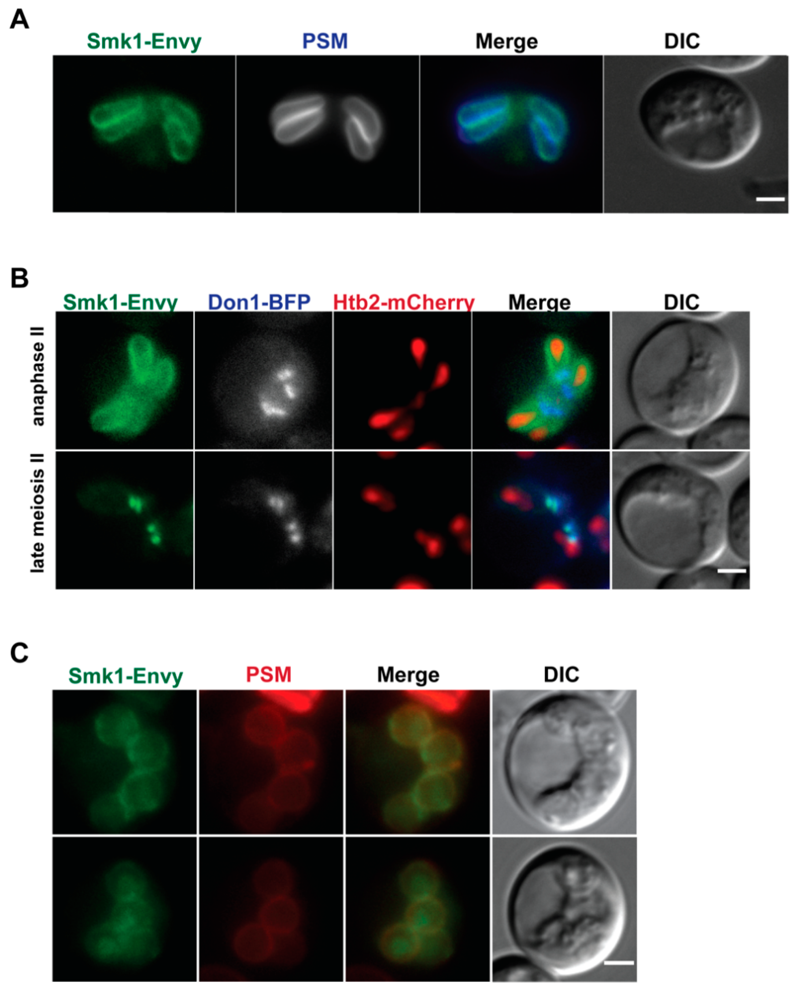
3.1. Smk1 Localizes to the Leading Edge of Elongated Prospore Membranes

Previous studies that report Smk1 localization to the prospore membrane utilized an overexpressed version of Smk1-GFP, due to difficulties in imaging endogenous levels of Smk1 [16]. Because GFPENVY is brighter and more photostable [25], we made a genomically integrated SMK1-GFPENVYallele and examined Smk1 localization during sporulation. As previously reported, we see localization at the prospore membrane during meiosis II (Figure 1A and 1B top) and co-localization of the Smk1-GFPENVY signal with a marker for the prospore membrane that fuses a fluorescent protein to a lipid-binding domain from the Spo20 protein (residues 51–91) [32].
During late meiosis II, as assayed by examining the prospore membrane morphology and by following meiosis using the nuclear marker Htb2-mCherry, we see transient concentrated localization of Smk1 (Figure 1B bottom). These regions of concentrated Smk1 co-localize with Don1, a member of the LEP (Figure 1B bottom), showing that Smk1 localizes to the leading edge of the prospore membrane during late meiosis II. Once PSMs round up and close, Smk1 becomes more diffuse and can be seen more broadly in the cell, where it can be found in the cytosol, along the prospore membrane, as well as in the nucleus (Figure 1C). Thus, Smk1 localization is dynamic, starting all along the prospore membrane, becoming concentrated at the leading edge after anaphase II, and becoming more broadly localized after prospore membranes round and close.

3.2. Smk1 Localization at the Leading Edge of the Prospore Membrane Requires SSP2

Previous studies that demonstrated Ssp2 localization on the prospore membrane also utilized an overexpressed version of Ssp2 [16]. Because Ssp2 complexes with Smk1, we examined whether Ssp2 was also localized at the leading edge of the prospore membrane. We created a genomically integrated SSP2-GFPENVY allele and see that in addition to the previously reported localization along the prospore membrane, Ssp2 can also localize to the leading edge of the prospore membrane late in meiosis II, when prospore membranes are elongated (Figure 2A). Thus, the localization of Smk1 and Ssp2 are similar during meiosis II.
We tested whether the localization of Smk1 at the leading edge required SSP2. We compared the localization of Smk1-GFPENVY in wild type cells and ssp2∆ cells. We only counted cells in late meiosis II, as assayed by using Htb2-mCherry and Don1-BFP to identify cells with four distinct nuclei that still had Don1-BFP localized as a tight ring or dot (like that seen in Figure 1B bottom). We found that ~30% of these cells had Smk1 concentrated along the leading edge of the prospore membranes (Figure 2B). Cells that did not have Smk1 at the leading edge had Smk1 localized along the prospore membrane. We hypothesize that the reason we do not see 100% of wild type cells having Smk1 at the leading edge is due to the transient nature of the leading edge localization coupled with the imperfect synchrony seen in sporulating SK1 cells; when we have used genetic methods to synchronize sporulation, the best we have seen is 50% of cells having Smk1 along the leading edge in the otherwise wild type culture (unpublished observations).
In contrast, we do not see Smk1 localizing at the leading edge of the growing prospore membrane in ssp2∆ cells in late meiosis II (as assayed using Htb2-mCherry and Don1-BFP) (Figure 2B). Instead, 100% of the ssp2∆ cells in late meiosis II had Smk1 localized along the prospore membrane. Thus, although the localization of Smk1 on the prospore membrane is independent of SSP2 (as previously reported [16]), its localization at the leading edge requires SSP2.

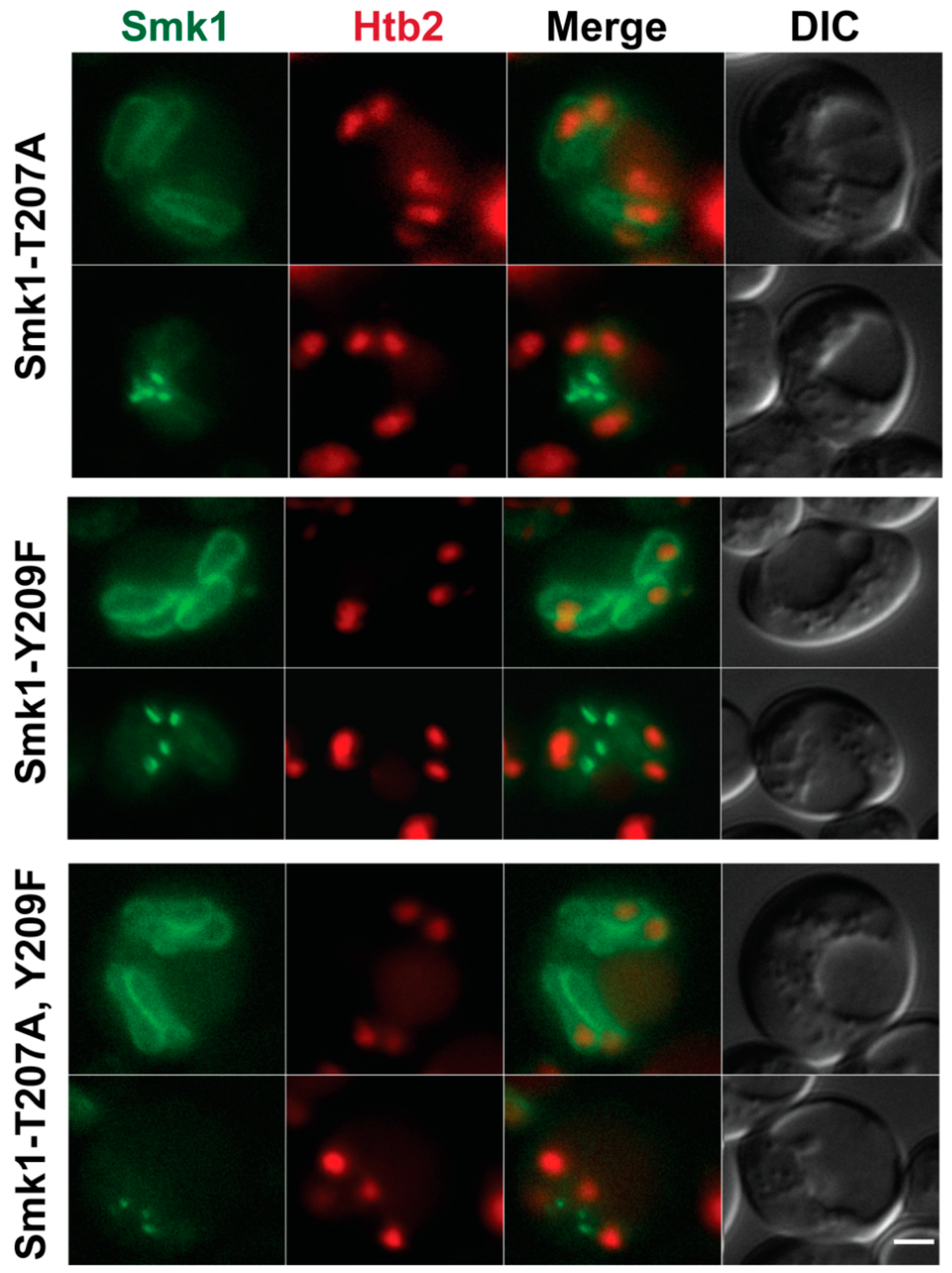
3.3. Smk1 Localization Is Independent of Activation State

Because Ssp2 activates Smk1, we asked whether localization of Smk1 at the leading edge of the prospore membrane depended on its activation state. We created GFPENVY-tagged versions of smk1-T207A, smk1-Y209F, and smk1-T207A Y209F, mutating the conserved threonine (T) and tyrosine (Y) in the conserved activation loop to the non-phosphorylateable amino acids alanine (A) and phenylalanine (F).
We examined the localization of the Smk1-T207A, Smk1-Y209F, and Smk1-T207A Y209F proteins during meiosis II and found that all three of these inactivation mutants of Smk1 can still localize along the prospore membrane during most of meiosis II and also concentrates at the leading edge towards the end of meiosis II (Figure 3). These results suggest that the ability of Smk1 to localize at the prospore membrane and the leading edge is independent of the phosphorylation state of its activation loop and also of its activity, as these mutations inactivate the Smk1 kinase [15].

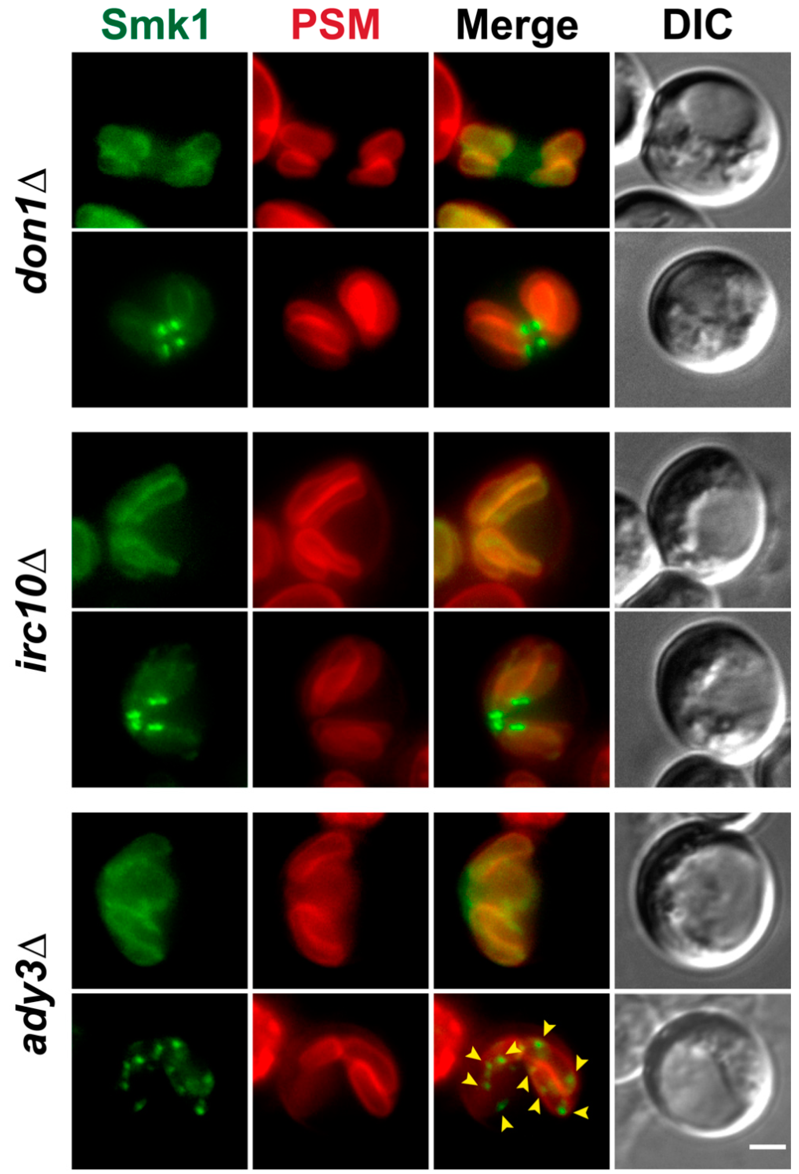
3.4. Localization of Smk1 Requires the LEP Member ADY3, But Not IRC10 or DON1

Because Smk1 co-localizes with the LEP, we tested whether Smk1 localization at the leading edge depended on IRC10 or DON1, two members of the LEP. Like in wild type cells, in irc10∆ or don1∆ cells, Smk1 can be seen on the prospore membrane during most of meiosis II, including at the elongated tubular stage of prospore membrane development (Figure 4, top panel of each genotype). Late in meiosis, after anaphase II, Smk1 can be seen concentrated at the leading edge in irc10∆ and don1∆ cells (Figure 4, bottom panel of each genotype). These results show that the localization of Smk1 along the prospore membrane and at the leading edge does not require IRC10 or DON1.
ADY3 is a member of the LEP and is important for the localization of other LEP components [8,9,10]. When we examine Smk1 localization in cells lacking ADY3, we see that during most of meiosis II, Smk1 localizes to the prospore membrane, as in wild type cells (Figure 4). However, during late meiosis II (when prospore membranes are elongated), at the time when Smk1 would co-localize with the leading edge protein Don1 (Figure 1B), we instead see Smk1 localizing to puncta along the prospore membrane in ady3∆ mutants (Figure 4). Thus, although ADY3 is dispensable for the localization of Smk1 during most of meiosis II, ADY3 is required for appropriate Smk1 localization during late prospore membrane development.

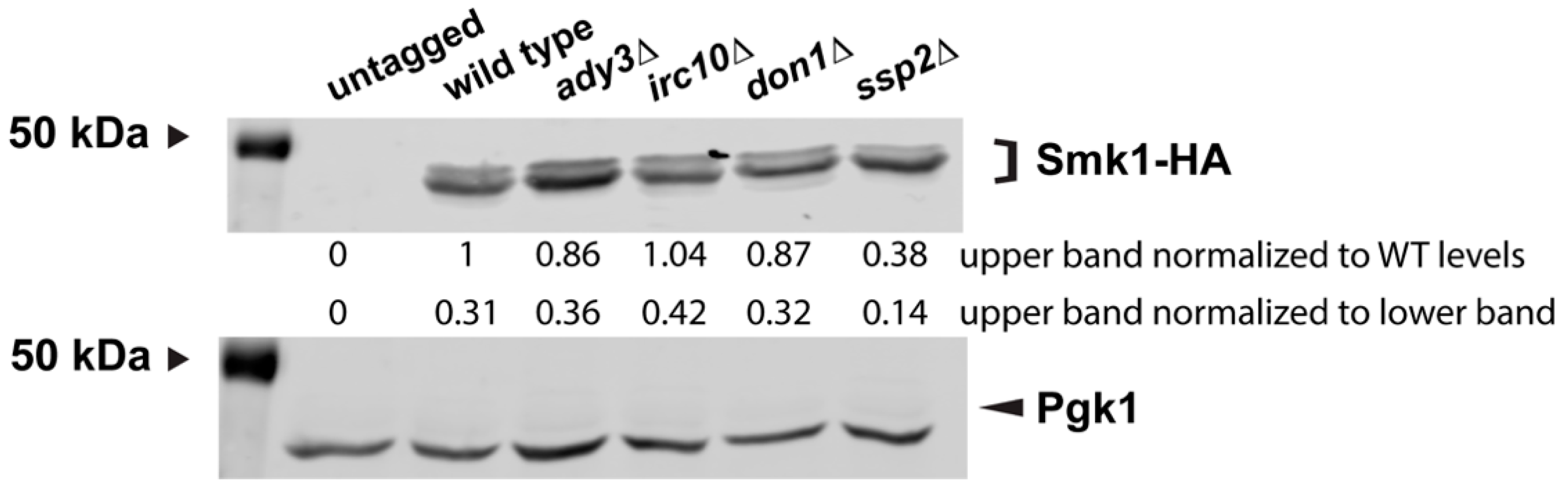
3.5. Smk1 Phosphorylation Does Not Require the LEP Complex

Smk1 is phosphorylated around 6 h during sporulation; this phosphorylation can be seen on immunoblots as a more slowly migrating band. This more slowly migrating band is missing when Smk1 first appears during sporulation (~5 h) and appears around the time when cell are in anaphase II (~6 h) [15].
To see whether the LEP is required for the post-translational modification of Smk1, we examined cells 6 h into sporulation when the characteristic doublet can be easily seen, using the SMK1-HA allele. We do not see significant differences in Smk1 post-translational modification in irc10∆, don1∆, ssp2∆, or ady3∆ mutants (Figure 5). As expected, we do see a slight decrease in the more slowly migrating Smk1 band in ssp2∆ mutants, consistent with SSP2 being required for the full phosphorylation and activation of Smk1 [16].

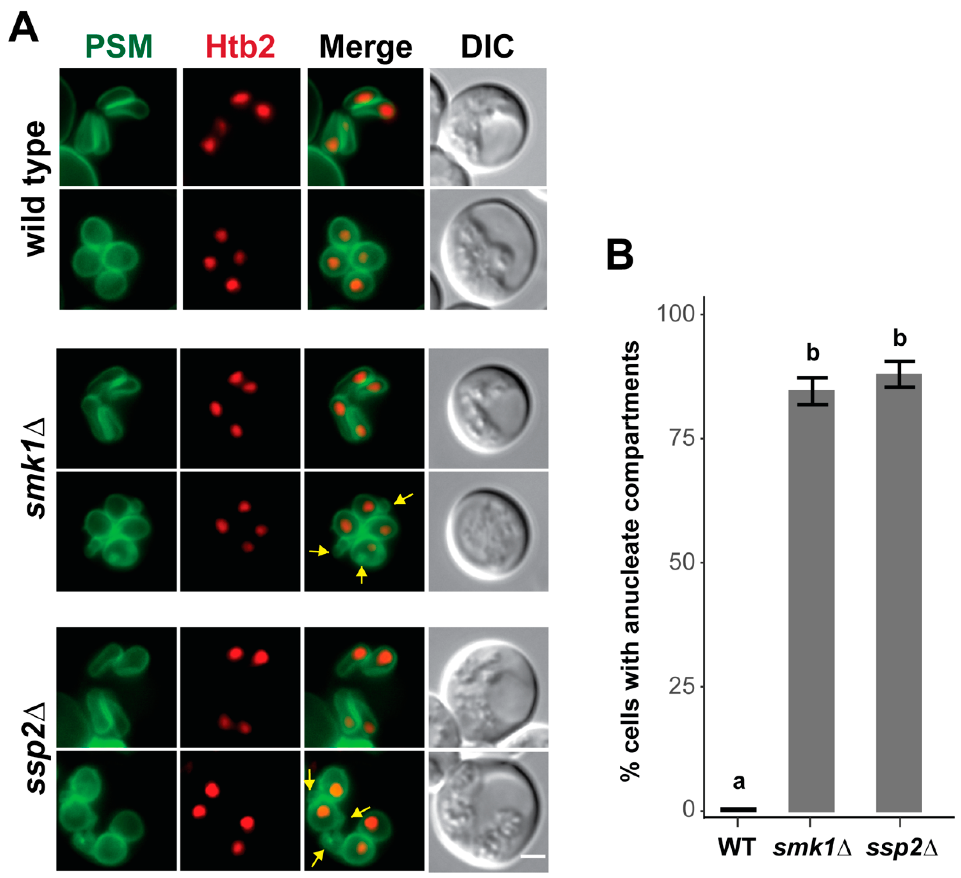
3.6. SMK1 and SSP2 Are Required for Late Prospore Membrane Development

Given the localization of Smk1 and Ssp2 to the leading edge of the prospore membrane in late meiosis II, we asked whether prospore membrane development was disrupted in smk1∆ or ssp2∆ cells. As previously reported, we do not see obvious prospore membrane defects in the early stages of prospore membrane during meiosis up to anaphase II, when prospore membrane form elongated tube-like structures (Figure 6).
However, at the end of meiosis II, when prospore membranes round up and close, we see additional membrane compartments in the smk1∆ and ssp2∆ mutant cells (Figure 6A). These extra compartments are anucleate and are only seen late in prospore membrane development after anaphase II, when the four meiotic nuclei are distinctly separated. These anucleate compartments are seen in >85% of smk1∆ and ssp2∆ cells with rounded prospore membranes, and are not found in wild type cells (Figure 6B). Thus, although SMK1 and SSP2 are dispensable during most of meiosis II, late prospore membrane development does not occur properly in the absence of these genes.

4. Discussion

Here, we find a role for the Smk1 MAPK and its activator, Ssp2, in late prospore membrane development. Using an integrated GFPENVY-tagged Smk1, we see the transient localization of Smk1 and Ssp2 at the leading edge of the prospore membrane after anaphase II, before cellularization when the prospore membrane rounds up and closes. This leading edge localization is transient and can only be seen in cells at the very end of meiosis II.
Previous studies that had examined Smk1 and Ssp2 only reported the earlier occurring prospore membrane localization [16,20]. These studies used an overexpression plasmid due to difficulties in detecting endogenous levels of Smk1. GFPENVY is brighter and more photostable than the more widely used eGFP [25], which allowed for the visualization of Smk1 expressed from the genomic locus under its own promoter at two copies per cell. It is possible that the leading edge localization was previously missed, either because the overproduction of Smk1 led to its localization at both the prospore membrane and the leading edge in late meiosis II, which would obscure the leading edge localization, or possibly because the leading edge localization is transient and can only be caught at a brief window at the end of meiosis II (meiosis II occurs relatively rapidly within about 40–50 min and anaphase II only lasts 10–20 min [33].)
Our current results do not allow us to distinguish between models of where the Smk1 and Ssp2 at the leading edge originates. Neither Smk1 nor Ssp2 are predicted to be transmembrane proteins, and thus, are likely peripheral membrane proteins; a membrane binding domain has been identified at the N-terminus of Ssp2 [16]. It is possible that the Smk1 and Ssp2 that is concentrated at the leading edge comes from newly recruited pools from the cytoplasm. Alternatively, it is possible that Smk1 and/or Ssp2 is relocalized from the pool of Smk1 and Ssp2 already on the prospore membrane.
We find that the LEP component ADY3 is required for the proper recruitment of Smk1 to the leading edge of the prospore membrane, even though it is not required for the initial localization of Smk1 to the prospore membrane. Interestingly, the punctate localization of Smk1 on the prospore membrane in ady3∆ mutants is reminiscent of the mislocalization of the LEP component Ssp1 seen in ady3∆ and ady3∆ irc10∆ mutants [8,10]. The consequence of these mislocalized proteins is unclear, as ady3∆ mutants can form mature spores (unlike ssp1∆ or smk1∆), although ady3∆ mutants have a tendency to form asci with only two spores [8,9].
We also see that SSP2, which encodes the activator of Smk1, is required for the proper recruitment of Smk1 to the leading edge of the prospore membrane. The earlier Smk1 localization to the prospore membrane occurs independently of SSP2. Ssp2 complexes with Smk1 [16] and shares the same localization, suggesting that Smk1 activation could occur in a localized fashion. However, a mutant that lacks the Ssp2 membrane binding domain at its N-terminus (ssp2-∆N137) sporulates normally [16], suggesting that Smk1 activation cannot only occur at the prospore membrane. We see that Smk1 localization to the prospore membrane and the leading edge is independent of its activation state, as mutations that prevent activation do not perturb the localization of Smk1. Thus, SSP2 may play a role in the movement and/or recruitment of inactive Smk1 to the leading edge, in addition to its role in mediating Smk1 autophosphorylation on Y209. It is possible that Smk1 and Ssp2 are recruited to the prospore membrane independently. Whether the mechanism that relocalizes Ssp2 to the leading edge also brings along Smk1 as a member of the complex, or whether Ssp2 is directly responsible for the movement/recruitment of Smk1 once Ssp2 arrives at the leading edge and does not complex with Smk1 until they are both at the leading edge, still needs to be determined.
Because the removal of the LEP at the end of meiosis II is important for prospore membrane closure, we initially hypothesized a role in closure for SMK1 and SSP2. However, the smk1∆ and ssp2∆ prospore membrane phenotype is distinct from the defect seen in mutants such as ama1∆, cdc15∆, spo77∆, and sps1∆, which play a role in prospore membrane closure [5,12,27]. These mutants make hyperelongated prospore membranes that round up and close much later than wild type cells.
Instead, in smk1∆ and ssp2∆ mutants, we observed the formation of ectopic anucleate membrane compartments in post-anaphase II cells. These extra prospore membrane compartments may correspond to the ectopic membrane compartments previously seen by electron microscopy in the ssp2∆ mutants [20]. Whether the prospore membrane phenotype is related to the localization of Smk1 and Ssp2 at the leading edge of the prospore membrane remains to be determined. How these extra membrane compartments form is also unclear: are cells closing the prospore membrane in the wrong place, creating the extra compartments, or are these extra compartments the result of inappropriate membrane synthesis after prospore membrane closure? A further examination of how SMK1 and SSP2 affect prospore membrane development can be a starting point for learning more about how MAP kinase signaling can affect membrane development.

Supplementary Materials

The following are available online at https://0-www-mdpi-com.brum.beds.ac.uk/2309-608X/7/1/53/s1, Table S1: Yeast Strains.

Author Contributions

L.S.H., J.M.R., M.D., C.J.S. and X.M. conceived and designed the experiments; M.D., J.M.R., C.J.S., E.K., B.C.S. and Z.W. constructed and created reagents, strains, and materials important for the experiments; M.D., J.M.R. and X.M. performed the experiments; L.S.H., J.M.R., M.D. and X.M. analyzed the data and made the figures; L.S.H., M.D. and J.M.R. wrote the manuscript. All authors have read and agreed to the published version of the manuscript.

Funding

Z.W. was supported by a Research Experience for Undergraduates grant from the National Science Foundation (DBI1659347). B.C.S. was supported by a College of Science and Mathematics Dean’s Doctoral Research Fellowship. This work was funded by a grant from the National Institutes of health to L.S.H (GM086805).

Institutional Review Board Statement

Not applicable.

Informed Consent Statement

Not applicable.

Data Availability Statement

Not applicable.

Conflicts of Interest

The authors declare no conflict of interest. The funders had no role in the design of the study; in the collection, analyses, or interpretation of data; in the writing of the manuscript, or in the decision to publish the results.

References

  1. Neiman, A.M. Sporulation in the budding yeast Saccharomyces cerevisiae. Genetics 2011, 189, 737–765. [Google Scholar] [CrossRef] [Green Version]
  2. Neiman, A.M. Prospore Membrane Formation Defines a Developmentally Regulated Branch of the Secretory Pathway in Yeast. J. Cell Biol. 1998, 140, 29–37. [Google Scholar] [CrossRef] [Green Version]
  3. Mathieson, E.M.; Suda, Y.; Nickas, M.; Snydsman, B.; Davis, T.N.; Muller, E.G.D.; Neiman, A.M.; Brennwald, P.J. Vesicle Docking to the Spindle Pole Body Is Necessary to Recruit the Exocyst During Membrane Formation in Saccharomyces cerevisiae. Mol. Biol. Cell 2010, 21, 3693–3707. [Google Scholar] [CrossRef] [PubMed] [Green Version]
  4. Suda, Y.; Tachikawa, H.; Inoue, I.; Kurita, T.; Saito, C.; Kurokawa, K.; Nakano, A.; Irie, K. Activation of Rab GTPase Sec4 by its GEF Sec2 is required for prospore membrane formation during sporulation in yeast Saccharomyces cerevisiae. FEMS Yeast Res. 2018, 18, fox095. [Google Scholar] [CrossRef] [PubMed]
  5. Diamond, A.E.; Park, J.-S.; Inoue, I.; Tachikawa, H.; Neiman, A.M. The Anaphase Promoting Complex Targeting Subunit Ama1 Links Meiotic Exit to Cytokinesis during Sporulation in Saccharomyces cerevisiae. Mol. Biol. Cell 2009, 20, 134–145. [Google Scholar] [CrossRef] [PubMed] [Green Version]
  6. Maier, P.; Rathfelder, N.; Finkbeiner, M.G.; Taxis, C.; Mazza, M.; Panse, S.L.; Haguenauer-Tsapis, R.; Knop, M. Cytokinesis in yeast meiosis depends on the regulated removal of Ssp1p from the prospore membrane. EMBO J. 2007, 26, 1843–1852. [Google Scholar] [CrossRef]
  7. Knop, M.; Strasser, K. Role of the spindle pole body of yeast in mediating assembly of the prospore membrane during meiosis. EMBO J. 2000, 19, 3657–3667. [Google Scholar] [CrossRef] [PubMed]
  8. Moreno-Borchart, A.C.; Strasser, K.; Finkbeiner, M.G.; Shevchenko, A.; Shevchenko, A.; Knop, M. Prospore membrane formation linked to the leading edge protein (LEP) coat assembly. EMBO J. 2001, 20, 6846–6857. [Google Scholar] [CrossRef] [PubMed] [Green Version]
  9. Nickas, M.E.; Neiman, A.M. Ady3p Links Spindle Pole Body Function to Spore Wall Synthesis in Saccharomyces cerevisiae. Genetics 2002, 160, 1439–1450. [Google Scholar] [PubMed]
  10. Lam, C.; Santore, E.; Lavoie, E.; Needleman, L.; Fiacco, N.; Kim, C.; Neiman, A.M. A visual screen of protein localization during sporulation identifies new components of prospore membrane-associated complexes in budding yeast. Eukaryot. Cell 2014, 13, 383–391. [Google Scholar] [CrossRef] [Green Version]
  11. Maier, P.; Rathfelder, N.; Maeder, C.I.; Colombelli, J.; Stelzer, E.H.K.; Knop, M. The SpoMBe pathway drives membrane bending necessary for cytokinesis and spore formation in yeast meiosis. EMBO J. 2008, 27, 2363–2374. [Google Scholar] [CrossRef] [Green Version]
  12. Paulissen, S.M.; Slubowski, C.J.; Roesner, J.M.; Huang, L.S. Timely closure of the prospore membrane requires SPS1 and SPO77 in Saccharomyces cerevisiae. Genetics 2016, 203, 1203–1216. [Google Scholar] [CrossRef] [PubMed] [Green Version]
  13. Krisak, L.; Strich, R.; Winters, R.S.; Hall, J.P.; Mallory, M.J.; Kreitzer, D.; Tuan, R.S.; Winter, E. SMK1, a developmentally regulated MAP kinase, is required for spore wall assembly in Saccharomyces cerevisiae. Genes Dev. 1994, 8, 2151–2161. [Google Scholar] [CrossRef] [PubMed] [Green Version]
  14. Wagner, M.; Briza, P.; Pierce, M.; Winter, E. Distinct Steps in Yeast Spore Morphogenesis Require Distinct SMK1 MAP Kinase Thresholds. Genetics 1999, 15, 1327–1340. [Google Scholar]
  15. Huang, L.S.; Doherty, H.K.; Herskowitz, I. The Smk1p MAP kinase negatively regulates Gsc2p, a 1,3-glucan synthase, during spore wall morphogenesis in Saccharomyces cerevisiae. Proc. Natl. Acad. Sci. USA 2005, 102, 12431–12436. [Google Scholar] [CrossRef] [Green Version]
  16. Tio, C.W.; Omerza, G.; Sunder, S.; Winter, E. Autophosphorylation of the Smk1 MAPK is spatially and temporally regulated by Ssp2 during meiotic development in yeast. Mol. Biol. Cell 2015, 26, 3546–3555. [Google Scholar] [CrossRef]
  17. Tio, C.W.; Omerza, G.; Phillips, T.; Lou, H.J.; Turk, B.E.; Winter, E. Ssp2 Binding Activates the Smk1 Mitogen-Activated Protein Kinase. Mol. Cell Biol. 2017, 37, e00607-16. [Google Scholar] [CrossRef] [Green Version]
  18. Sarkar, P.; Florczyk, M.; McDonough, K.; Nag, D. SSP2, a sporulation-specific gene necessary for outer spore wall assembly in the yeast Saccharomyces cerevisiae. Mol. Genet. Genom. 2002, 267, 348–358. [Google Scholar] [CrossRef]
  19. Coluccio, A.; Bogengruber, E.; Conrad, M.N.; Dresser, M.E.; Briza, P.; Neiman, A.M. Morphogenetic Pathway of Spore Wall Assembly in Saccharomyces cerevisiae. Eukaryot. Cell 2004, 3, 1464–1475. [Google Scholar] [CrossRef] [PubMed] [Green Version]
  20. Li, J.; Agarwal, S.; Roeder, G.S. SSP2 and OSW1, two sporulation-specific genes involved in spore morphogenesis in Saccharomyces cerevisiae. Genetics 2007, 175, 143–154. [Google Scholar] [CrossRef] [Green Version]
  21. Kane, S.M.; Roth, R. Carbohydrate Metabolism during Ascospore Development in Yeast. J. Bacteriol. 1974, 118, 8–14. [Google Scholar] [CrossRef] [PubMed] [Green Version]
  22. Rose, M.D.; Fink, G.F. Methods in Yeast Genetics; Cold Spring Harbor Laboratory Press: Cold Spring Harbor, NY, USA, 1990. [Google Scholar]
  23. Longtine, M.S.; Mckenzie III, A.; Demarini, D.J.; Shah, N.G.; Wach, A.; Brachat, A.; Philippsen, P.; Pringle, J.R. Additional modules for versatile and economical PCR-based gene deletion and modification in Saccharomyces cerevisiae. Yeast 1998, 14, 953–961. [Google Scholar] [CrossRef]
  24. Lee, S.; Lim, W.A.; Thorn, K.S. Improved Blue, Green, and Red Fluorescent Protein Tagging Vectors for S. cerevisiae. PLoS ONE 2013, 8, e67902. [Google Scholar] [CrossRef] [PubMed]
  25. Slubowski, C.J.; Funk, A.D.; Roesner, J.M.; Paulissen, S.M.; Huang, L.S. Plasmids for C-terminal tagging in Saccharomyces cerevisiae that contain improved GFP proteins, Envy and Ivy. Yeast 2015, 32, 379–387. [Google Scholar] [CrossRef] [PubMed] [Green Version]
  26. Nakamura, T.S.; Numajiri, Y.; Okumura, Y.; Hidaka, J.; Tanaka, T.; Inoue, I.; Suda, Y.; Takahashi, T.; Nakanishi, H.; Gao, X.D.; et al. Dynamic localization of a yeast development-specific PP1 complex during prospore membrane formation is dependent on multiple localization signals and complex formation. Mol. Biol. Cell 2017, 28, 3881–3895. [Google Scholar] [CrossRef] [Green Version]
  27. Paulissen, S.M.; Hunt, C.A.; Seitz, B.C.; Slubowski, C.J.; Yu, Y.; Mucelli, X.; Truong, D.; Wallis, Z.; Nguyen, H.T.; Newman-Toledo, S.; et al. A noncanonical hippo pathway regulates spindle disassembly and cytokinesis during meiosis in Saccharomyces cerevisiae. Genetics 2020, 216, 447–462. [Google Scholar] [CrossRef]
  28. Lin, C.P.C.; Kim, C.; Smith, S.O.; Neiman, A.M. A Highly Redundant Gene Network Controls Assembly of the Outer Spore Wall in S. cerevisiae. PLoS Genet. 2013, 9, e1003700. [Google Scholar] [CrossRef] [Green Version]
  29. Sikorski, R.S.; Hieter, P. A System of Shuttle Vectors and Yeast Host Strains Designed for Efficient Manipulation of DNA in Saccharomyces cerevisiae. Genetics 1989, 122, 19–27. [Google Scholar] [CrossRef]
  30. Philips, J.; Herskowitz, I. Identification of Kel1p, a Kelch Domain-containing Protein Involved in Cell Fusion and Morphology in Saccharomyces cerevisiae. J. Cell Biol. 1998, 143, 375–389. [Google Scholar] [CrossRef] [PubMed] [Green Version]
  31. Schindelin, J.; Arganda-Carreras, I.; Frise, E.; Kaynig, V.; Longair, M.; Pietzsch, T.; Preibisch, S.; Rueden, C.; Saalfeld, S.; Schmid, B.; et al. Fiji: An open-source platform for biological-image analysis. Nat. Methods 2012, 9, 676–682. [Google Scholar] [CrossRef] [Green Version]
  32. Nakanishi, H.; de los Santos, P.; Neiman, A.M. Positive and Negative Regulation of a SNARE Protein by Control of Intracellular Localization. Mol. Biol. Cell 2004, 15, 1802–1815. [Google Scholar] [CrossRef] [PubMed]
  33. Carpenter, K.; Bell, R.B.; Yunus, J.; Amon, A.; Berchowitz, L.E. Phosphorylation-Mediated Clearance of Amyloid-like Assemblies in Meiosis. Dev. Cell 2018, 45, 392–405. [Google Scholar] [CrossRef] [PubMed] [Green Version]
Figure 1. Smk1 exhibits two distinct localizations during prospore membrane development. (A) Smk1-GFPEnvy co-localizes with the prospore membrane (PSM) marker pRS426-Spo2051–91-mTagBFP along the PSM. (B) Smk1-GFPEnvy localizes to PSM during the elongation stage of membrane development, and is concentrated at the leading edge of the prospore membrane after anaphase II. Htb2-mCherry is used as a histone marker. The leading edge of the growing prospore membrane is marked using Don1-BFP. (C) Smk1-GFPEnvy becomes more broadly localized after prospore membranes round up and close. Prospore membranes are in red and are labeled using an integrated Spo2051–91-mKate2 marker. For all strains, the genomic copy of SMK1 is epitope tagged with GFPENVY at its C-terminus; strains are homozygous for SMK1-GFPENVY. Yeast strains used for each panel: (A) LH1116, (B), LH1117, and (C) LH1133. Scale bar = 2 µM.
Jof 07 00053 g001
Figure 2. SSP2 is required for Smk1 localization at the leading edge of the prospore membrane. (A) Ssp2 localizes to the leading edge of the prospore membrane. Ssp2-GFPEnvy is found at the leading edge of the elongated prospore membrane (LH1118). Prospore membranes (PSMs) are labeled with pRS426-Spo2051–91-mTagBFP. Yellow arrowheads indicate leading edge localization at the open ends of the tubular elongated prospore membranes. Scale bar = 2 µM. (B) Percentage of cells in late meiosis II that have Smk1 localized at the leading edge of the prospore membrane. Wild type (WT; LH1117), ssp2∆ (LH1134). Three biological replicates, 100 cells per replicate, were counted for each genotype. Error bars show standard error of the mean. p-value was calculated using Fisher’s exact test (two-tailed).
Jof 07 00053 g002
Figure 3. Smk1 localization is independent of its activation state. Localization of Smk1-T207A (LH1119), Smk1-Y209F (LH1120), and Smk1-T207A, Y209F (LH1121) to both elongated prospore membranes and the leading edge of the prospore membrane. smk1 activation site mutations are epitope tagged with GFPENVY. Htb2-mCherry is used as a histone marker. Scale bar = 2 µM.
Jof 07 00053 g003
Figure 4. Smk1 localization requires the Leading Edge Protein complex member ADY3, but not IRC10 or DON1. Smk1-GFPENVY is localized to the prospore membrane and the leading edge in don1∆ (LH1123) and irc10∆ (LH1124) mutants. Smk1-GFPENVY is localized to the prospore membrane (PSM) in ady3∆ (LH1125) mutants early in meiosis II, but becomes localized as puncta along the leading edge later in development. Prospore membranes are in red and are labeled using an integrated Spo2051–91-mKate2 marker. Yellow arrowheads indicate some of the ectopic Smk1 puncta seen along the prospore membrane in ady3∆ mutant cells. Scale bar = 2 µM.
Jof 07 00053 g004
Figure 5. Smk1 post-translational modification does not require the leading edge protein complex members ADY3, IRC10, and DON1. Immunoblot of protein extracts examining Smk1-HA in an untagged strain lacking Smk1-HA (LH177), Smk1-HA (LH178), ady3∆ (LH1126), irc10∆ (LH1127), don1∆ (LH1128), and ssp2∆ (LH1129). Pgk1 is used as a loading control. The 50 kDa band from the Precision Plus Protein Markers (BioRad) is indicated on the left of each blot. The upper quantification corresponds to densitometry, in which the slower migrating (upper) Smk1-HA band was first normalized to Pgk1 levels, followed by the normalization of the slower migrating Smk1-HA band in each mutant to wild-type levels of expression. The lower quantification corresponds to densitometry, in which the slower migrating Smk1-HA band was normalized to the faster migrating Smk1-HA band for each genotype.
Jof 07 00053 g005
Figure 6. SMK1 and SSP2 are required for late prospore membrane development. (A) Cells with elongated tubular prospore membranes and post-anaphase II rounded prospore membranes are shown in wild type (LH1130), smk1∆ (LH1131), and ssp2∆ (LH1132) cells. Prospore membranes (PSM) are in green and are labeled using an integrated Spo2051–91-GFPENVY marker. Htb2-mCherry was used as a histone marker in LH1131 and LH1132, while Htb2-mRuby2 was used as the histone marker in LH1130. Yellow arrows point to ectopic membrane compartments found in mutant cells with rounded prospore membranes. Scale bar = 2 µM. (B) Quantitation of anucleate compartments. Post-anaphase II cells that had four distinct nuclei and rounded prospore membranes were assayed for the presence of anucleate compartments in wild type (WT; LH1130), smk1∆ (LH1131), and ssp2∆ (LH1132). At least three biological replicates, 100 cells per replicate, were counted for each genotype. Error bars show the standard error of the mean. The wild type strain (a) is significantly different from the smk1∆ and ssp2∆ strains (b); one way ANOVA (F(2,8) = 549, p < 0.001) followed by Tukey’s HSD post hoc test (alpha = 0.01).
Jof 07 00053 g006
Table 1. Primers used to construct smk1 activation loop mutations.
Primer NameSequence (5′ to 3′)
OLH1756AATCGAGCTCAGTAATAAATACTGTGTTGTTTG
OLH1757GAAAATGTTGGACCATG
OLH1773GTACTCCTGATAAAGATATTCTG
OLH1774CAGCACCGTCACCTAAAGACGAGGAGGACAAATC
OLH1758TATAAAAAAATCCCATGGTC
OLH1759ATTGCTCGAGTTATTTGTACAATTCGTCCATTC
Publisher’s Note: MDPI stays neutral with regard to jurisdictional claims in published maps and institutional affiliations.

