
Fusarium as a Novel Fungus for the Synthesis of Nanoparticles: Mechanism and Applications

 , , ,
, , ,  ,
,  ,
,
Abstract
:1. Introduction
1.1. Diversity of Fusarium spp. for the Synthesis of Different Nanopartilces
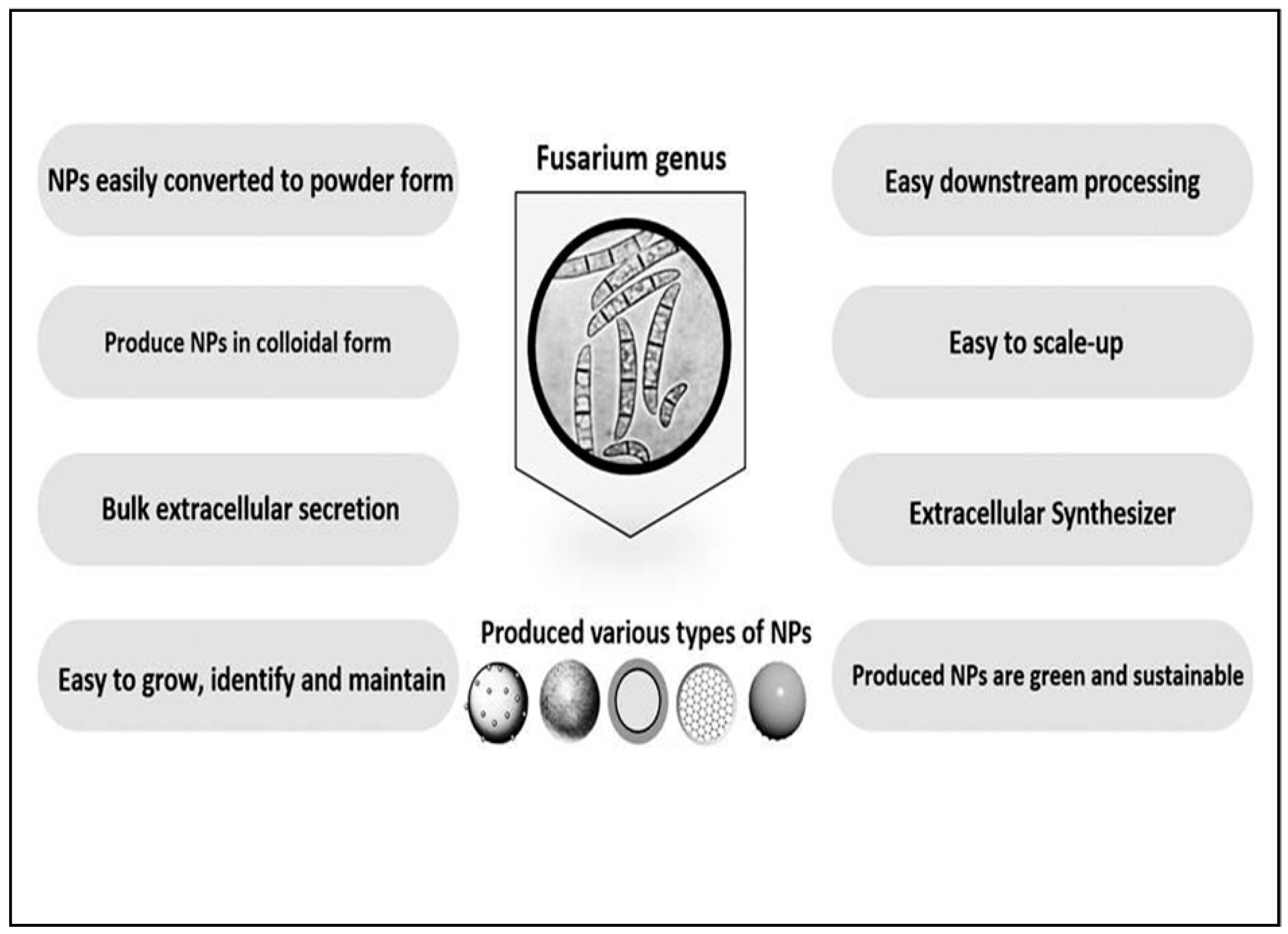
1.2. F. oxysporum as a Novel Organism for Synthesis of Nanoparticles
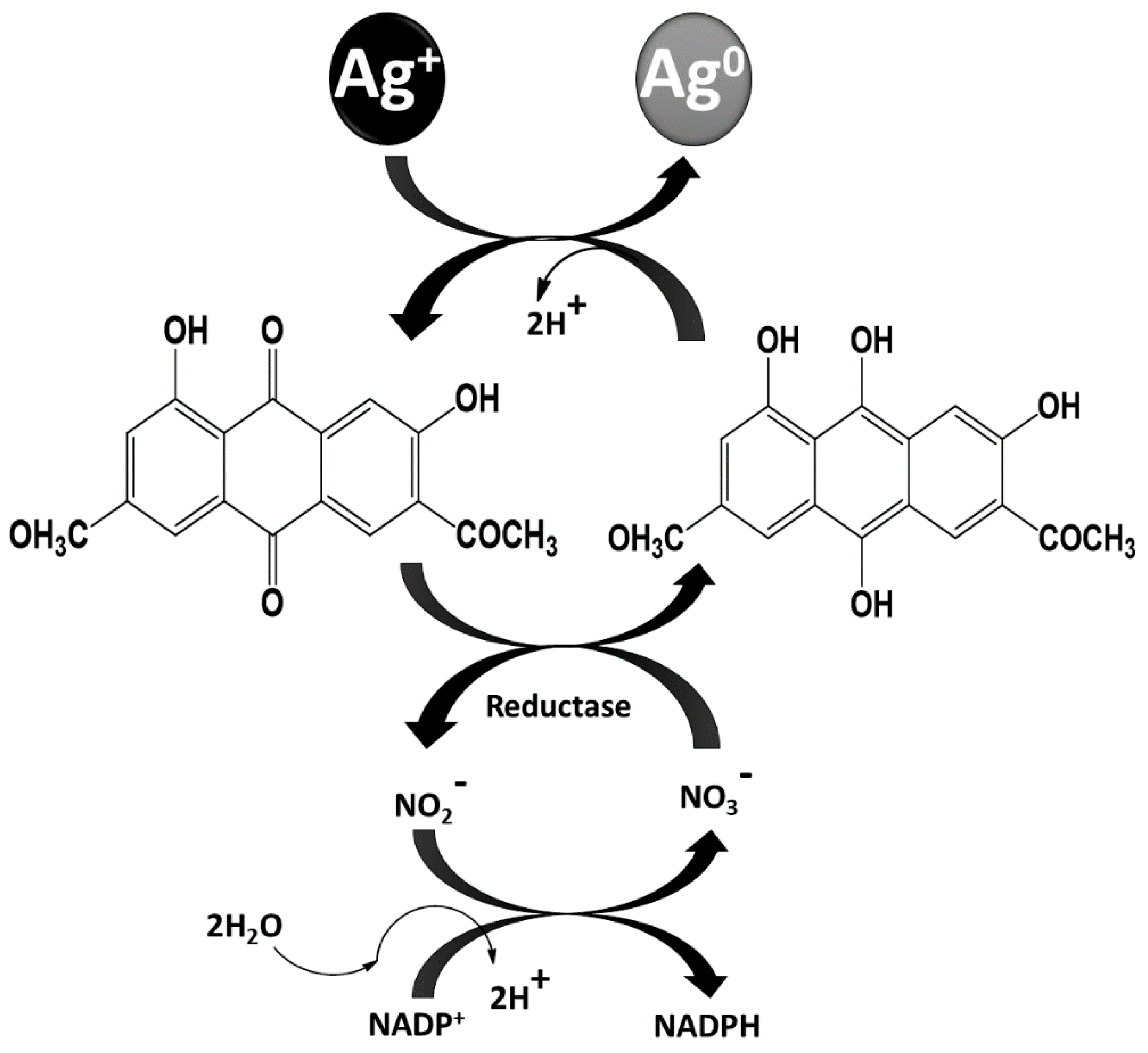
2. Mechanism of Nanoparticle Synthesis from Fusarium
2.1. Biomedical Applications of Nanoparticles Synthesized Using Fusarium Spp.
2.1.1. Antibacterial Activity of Nanoparticles
2.1.2. Antiviral Activity of Nanoparticles
2.1.3. Anticancer Activity of Nanoparticles
2.1.4. Antifungal Activity of NPs
2.1.5. Antiparasitic Activity Against Vectors
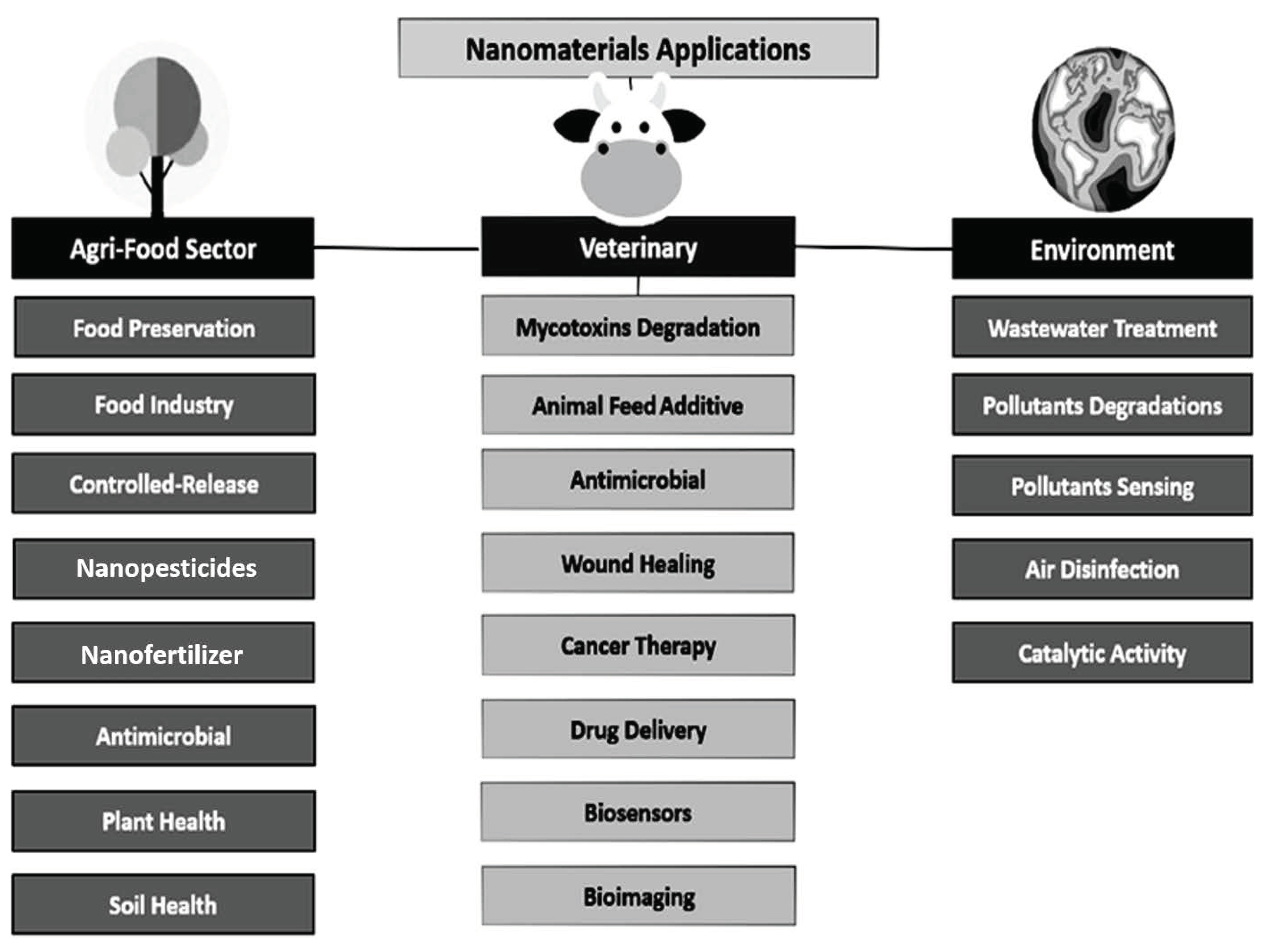
3. Applications in Agriculture
4. Toxicity of Fusarium Nanoparticles
4.1. Effect of Size and Shape of Nanoparticles on Cytotoxicity
4.2. Mechanisms of Nanoparticle Cytotoxicity
4.3. Effect of Nanoparticles on Cell Membranes
4.4. Effect of Nanoparticles on Mitochondria and/or Metabolic Activity
4.5. Effect of Nanoparticles on Cell Proteins
4.6. Effect of Nanoparticles on Cell Nucleic Acids
4.7. Other Nanoparticle Activity
5. Conclusions
Author Contributions
Funding
Institutional Review Board Statement
Informed Consent Statement
Data Availability Statement
Acknowledgments
Conflicts of Interest
References
- Ranjani, S.; Shariq, A.M.; Adnan, M.; Senthil-Kumar, N.; Ruckmani, K.; Hemalatha, S. Synthesis, characterization and applications of endophytic fungal nanoparticles. Inorg. Nano Metal Chem. 2020, 51, 280–287. [Google Scholar] [CrossRef]
- Upadhyay, P.K.; Jain, V.K.; Sharma, K.; Sharma, R. Synthesis and applications of ZnO nanoparticles in biomedicine. Res. J. Pharm. Technol. 2020, 13, 1636. [Google Scholar] [CrossRef]
- Usman, M.; Farooq, M.; Wakeel, A.; Nawaz, A.; Cheema, S.A.; Rehman, H.U.; Ashraf, I.; Sanaullah, M. Nanotechnology in agriculture: Current status, challenges and future opportunities. Sci. Total. Environ. 2020, 721, 137778. [Google Scholar] [CrossRef] [PubMed]
- Yaqoob, A.A.; Ahmad, H.; Parveen, T.; Ahmad, A.; Oves, M.; Ismail, I.M.I.; Qari, H.A.; Umar, K.; Ibrahim, M.N.M. Recent advances in metal decorated nanomaterials and their various biological applications: A Review. Front. Chem. 2020, 8, 341. [Google Scholar] [CrossRef]
- Bououdina, M.; Rashdan, S.; Bobet, J.L.; Ichiyanagi, Y. Nanomaterials for biomedical applications: Synthesis, Characterization, and applications. J. Nanomater. 2013, 2013, 1–4. [Google Scholar] [CrossRef]
- Paramo, L.A.; Feregrino-Pérez, A.A.; Guevara, R.; Mendoza, S.; Esquivel, K. Nanoparticles in agroindustry: Applications, toxicity, challenges, and trends. Nanomaterials 2020, 10, 1654. [Google Scholar] [CrossRef]
- Ramos, M.M.; Morais, E.D.S.; Sena, I.D.S.; Lima, A.L.; De Oliveira, F.R.; De Freitas, C.M.; Fernandes, C.P.; De Carvalho, J.C.T.; Ferreira, I.M. Silver nanoparticle from whole cells of the fungi Trichoderma spp. isolated from Brazilian Amazon. Biotechnol. Lett. 2020, 42, 833–843. [Google Scholar] [CrossRef]
- Ahmed, S.; Ahmad, M.; Swami, B.L.; Ikram, S. A review on plants extract mediated synthesis of silver nanoparticles for antimicrobial applications: A green expertise. J. Adv. Res. 2016, 7, 17–28. [Google Scholar] [CrossRef] [PubMed] [Green Version]
- Guilger-Casagrande, M.; De Lima, R. Synthesis of silver nanoparticles mediated by fungi: A Review. Front. Bioeng. Biotechnol. 2019, 7, 287. [Google Scholar] [CrossRef] [Green Version]
- Ingle, A.; Gade, A.; Pierrat, S.; Sonnichsen, C.; Rai, M. Mycosynthesis of silver nanoparticles using the fungus Fusarium acuminatum and its activity against some human pathogenic bacteria. Curr. Nanosci. 2008, 4, 141–144. [Google Scholar] [CrossRef]
- Ingle, A.; Rai, M.; Gade, A.; Bawaskar, M. Fusarium solani: A novel biological agent for the extracellular synthesis of silver nanoparticles. J. Nanopart. Res. 2009, 11, 2079–2085. [Google Scholar] [CrossRef]
- Rai, M.; Yadav, A.; Gade, A. Silver nanoparticles as a new generation of antimicrobials. Biotechnol. Adv. 2009, 27, 76–83. [Google Scholar] [CrossRef]
- Yadav, A.; Kon, K.; Kratosova, G.; Duran, N.; Ingle, A.P.; Rai, M. Fungi as an efficient mycosystem for the synthesis of metal nanoparticles: Progress and key aspects of research. Biotechnol. Lett. 2015, 37, 2099–2120. [Google Scholar] [CrossRef]
- Rai, M.; Yadav, A.; Bridge, P.; Gade, A. Myconanotechnology: A new and emerging science. In Applied Mycology; Rai, M.K., Bridge, P.D., Eds.; CABI: Wallingford, UK, 2009; pp. 267–285. [Google Scholar] [CrossRef]
- Gade, A.; Ingle, A.; Whiteley, C.; Rai, M. Mycogenic metal nanoparticles: Progress and applications. Biotechnol. Lett. 2010, 32, 593–600. [Google Scholar] [CrossRef] [PubMed]
- Ahmad, A.; Mukherjee, P.; Senapati, S.; Mandal, D.; Khan, M.I.; Kumar, R.; Sastry, M. Extracellular biosynthesis of silver nanoparticles using the fungus Fusarium oxysporum. Coll. Surf. B Biointerfaces 2003, 28, 313–318. [Google Scholar] [CrossRef]
- Bansal, V.; Rautaray, D.; Ahmad, A.; Sastry, M. Biosynthesis of zirconia nanoparticles using the fungus Fusarium oxysporum. J. Mater. Chem. 2004, 14, 3303–3305. [Google Scholar] [CrossRef]
- Reyes, L.R.; Gómez, I.; Garza, M.T. Biosynthesis of cadmium sulfide nanoparticles by the fungi Fusarium sp. Int. J. Green Nanotechnol. Biomed. 2009, 1, B90–B95. [Google Scholar] [CrossRef]
- Basavaraja, S.; Balaji, S.D.; Lagashetty, A.; Rajasab, A.H.; Venkataraman, A. Extracellular biosynthesis of silver nanoparticles using the fungus Fusarium semitectum. Mater. Res. Bull. 2008, 43, 1164–1170. [Google Scholar] [CrossRef]
- Siddiqi, K.S.; Husen, A. Fabrication of metal nanoparticles from fungi and metal salts: Scope and application. Nanoscale Res. Lett. 2016, 11, 98. [Google Scholar] [CrossRef] [Green Version]
- Khan, N.T.; Jameel, M.; Jameel, J. Silver nanoparticles biosynthesis by Fusarium oxysporum and determination of its antimicrobial potency. J. Nanomed. Biotherapeutic. Discov. 2017, 7, 1. [Google Scholar] [CrossRef] [Green Version]
- Mahmoud, M.A.; Al-Sohaibani, S.A.; Al-Othman, M.R.; El-Aziz, A.M.A.; Eifan, S.A. Synthesis of extracellular silver nanoparticles using Fusarium semitectum (KSU-4) isolated from Saudi Arabia. Dig. J. Nanomat. Biostruct. 2013, 8, 589–596. [Google Scholar] [CrossRef]
- Elamawi, R.M.; Al-Harbi, R.E.; Hendi, A.A. Biosynthesis and characterization of silver nanoparticles using Trichoderma longibrachiatum and their effect on phytopathogenic fungi. Egypt. J. Biol. Pest Control 2018, 28, 28. [Google Scholar] [CrossRef] [Green Version]
- Noor, S.; Shah, Z.; Javed, A.; Ali, A.; Hussain, S.B.; Zafar, S.; Ali, H.; Muhammad, S.A. A fungal based synthesis method for copper nanoparticles with the determination of anticancer, antidiabetic and antibacterial activities. J. Microbiol. Methods 2020, 174, 105966. [Google Scholar] [CrossRef]
- Lange, K.; Swift, P.G.F.; Pankowska, E.; Danne, T. Diabetes education in children and adolescents. Pediatric Diabetes 2014, 15, 77–85. [Google Scholar] [CrossRef]
- Solomon, L.; Tomii, V. Dick Importance of fungi in the petroleum, agro-allied, agriculture and pharmaceutical industries. N. Y. Sci. J. 2019, 12, 5. [Google Scholar] [CrossRef]
- Tidke, P.R.; Gupta, I.; Gade, A.K.; Rai, M. Fungus-mediated synthesis of gold nanoparticles and standardization of parameters for its biosynthesis. IEEE Trans. NanoBiosci. 2014, 13, 397–402. [Google Scholar] [CrossRef]
- Bawaskar, M.; Gaikwad, S.; Ingle, A.; Rathod, D.; Gade, A.; Durán, N.; Marcato, P.D.; Rai, M. A New Report on mycosynthesis of silver nanoparticles by Fusarium culmorum. Curr. Nanosci. 2010, 6, 376–380. [Google Scholar] [CrossRef]
- Khalil, N.M.; El-Ghany, M.N.A.; Rodríguez-Couto, S. Antifungal and anti-mycotoxin efficacy of biogenic silver nanoparticles produced by Fusarium chlamydosporum and Penicillium chrysogenum at non-cytotoxic doses. Chemosphere 2019, 218, 477–486. [Google Scholar] [CrossRef] [PubMed]
- Gaikwad, S.C.; Birla, S.S.; Ingle, A.P.; Gade, A.K.; Marcato, P.D.; Rai, M.K.; Durán, N. Screening of different Fusarium species to select potential species for the synthesis of silver nanoparticles. J. Braz. Chem. Soc. 2013, 24, 1974–1982. [Google Scholar] [CrossRef]
- Mohamed, A.A.; Fouda, A.; Abdel-Rahman, M.A.; Hassan, S.E.-D.; El-Gamal, M.S.; Salem, S.S.; Shaheen, T.I. Fungal strain impacts the shape, bioactivity and multifunctional properties of green synthesized zinc oxide nanoparticles. Biocatal. Agric. Biotechnol. 2019, 19, 101103. [Google Scholar] [CrossRef]
- Mohamed, A.A.; Fouda, A.; Elgamal, M.S.; Hassan, S.; Shaheen, T.I.; Salem, S.S. Enhancing of cotton fabric antibacterial properties by silver nanoparticles synthesized by new egyptian strain Fusarium keratoplasticum A1–3. In Egyptian Journal of Chemistry 60, Proceedings of the 8th International Conference of the Textile Research Division (ICTRD 2017), Cairo, Egypt, 25–27 September 2017; National Research Centre: Dokki, Egypt, 2017; pp. 63–71. [Google Scholar] [CrossRef] [Green Version]
- Birla, S.S.; Gaikwad, S.C.; Gade, A.K.; Rai, M.K. Rapid Synthesis of silver nanoparticles from Fusarium oxysporum by optimizing physicocultural conditions. Sci. World J. 2013, 2013, 1–12. [Google Scholar] [CrossRef] [Green Version]
- Bansod, S.; Bonde, S.; Tiwari, V.; Bawaskar, M.; Deshmukh, S.; Gaikwad, S.; Gade, A.; Rai, M. Bioconjugation of gold and silver nanoparticles synthesized by Fusarium oxysporum and their use in rapid identification of Candida species by using bioconjugate-nano-polymerase chain reaction. J. Biomed. Nanotechnol. 2013, 9, 1962–1971. [Google Scholar] [CrossRef]
- Sanyal, A.; Rautaray, D.; Bansal, V.; Ahmad, A.; Sastry, M. Heavy-metal remediation by a fungus as a means of production of lead and cadmium carbonate crystals. Langmuir 2005, 21, 7220–7224. [Google Scholar] [CrossRef]
- Rautaray, D.; Sanyal, A.; Adyanthaya, S.D.; Ahmad, A.; Sastry, M. Biological synthesis of strontium carbonate crystals using the fungus Fusarium oxysporum. Langmuir 2004, 20, 6827–6833. [Google Scholar] [CrossRef]
- Ahmad, A.; Mukherjee, P.; Mandal, D.; Senapati, S.; Khan, M.I.; Kumar, R.; Sastry, M. Enzyme mediated extracellular synthesis of CdS nanoparticles by the fungus, Fusarium oxysporum. J. Am. Chem. Soc. 2002, 124, 12108–12109. [Google Scholar] [CrossRef] [PubMed]
- Bansal, V.; Rautaray, D.; Bharde, A.; Ahire, K.; Sanyal, A.; Ahmad, A.; Sastry, M. Fungus-mediated biosynthesis of silica and titania particles. J. Mater. Chem. 2005, 15, 2583–2589. [Google Scholar] [CrossRef]
- Bansal, V.; Poddar, P.; Ahmad, A.A.; Sastry, M. Room-temperature biosynthesis of ferroelectric barium titanate nanoparticles. J. Am. Chem. Soc. 2006, 128, 11958–11963. [Google Scholar] [CrossRef]
- Bansal, V.; Sanyal, A.; Rautaray, D.; Ahmad, A.; Sastry, M. Bioleaching of sand by the fungus Fusarium oxysporum as a means of producing extracellular silica nanoparticles. Adv. Mater. 2005, 17, 889–892. [Google Scholar] [CrossRef]
- Gupta, K.; Chundawat, T.S. Bio-inspired synthesis of platinum nanoparticles from fungus Fusarium oxysporum: Its characteristics, potential antimicrobial, antioxidant and photocatalytic activities. Mater. Res. Express 2019, 6, 1–10. [Google Scholar] [CrossRef]
- Bharde, A.; Rautaray, D.; Bansal, V.; Ahmad, A.; Sarkar, I.; Yusuf, S.M.; Sanyal, M.; Sastry, M. Extracellular biosynthesis of magnetite using fungi. Small 2006, 2, 135–141. [Google Scholar] [CrossRef]
- Kumar, S.; Kumar, D.; Dilbaghi, N. Preparation, characterization, and bio-efficacy evaluation of controlled release carbendazim-loaded polymeric nanoparticles. Environ. Sci. Pollut. Res. 2017, 24, 926–937. [Google Scholar] [CrossRef]
- Senapati, S.; Syed, A.; Khan, S.; Pasricha, R.; Khan, M.I.; Kumar, R.; Ahmad, A. Extracellular biosynthesis of metal sulfide nanoparticles using the fungus Fusarium oxysporum. Curr. Nanosci. 2014, 10, 588–595. [Google Scholar] [CrossRef]
- Ganapathy, S.; Siva, K. Bio-synthesis, characterisation and application of titanium oxide nanoparticles by Fusarium oxysporum. Int. J. Life Sci. Res. 2016, 4, 69–75. [Google Scholar] [CrossRef] [Green Version]
- Boruah, S.; Dutta, P. Fungus mediated biogenic synthesis and characterization of chitosan nanoparticles and its combine effect with Trichoderma asperellum against Fusarium oxysporum, Sclerotium rolfsii and Rhizoctonia solani. Indian Phytopathol. 2020, 1–13. [Google Scholar] [CrossRef]
- Mirzadeh, S.; Darezereshki, E.; Bakhtiari, F.; Fazaelipoor, M.H.; Hosseini, M.R. Characterization of zinc sulfide (ZnS) nanoparticles biosynthesized by Fusarium oxysporum. Mater. Sci. Semiconduct. Process. 2013, 16, 374–378. [Google Scholar] [CrossRef]
- Thakker, J.N.; Dalwadi, P.; Dhandhukia, P.C. Biosynthesis of gold nanoparticles using Fusarium oxysporum f. sp. cubense JT1, a plant pathogenic fungus. ISRN Biotechnol. 2013, 2013, 1–5. [Google Scholar] [CrossRef]
- Rajput, S.; Werezuk, R.; Lange, R.M.; McDermott, M.T. Fungal isolate optimized for biogenesis of silver nanoparticles with enhanced colloidal stability. Langmuir 2016, 32, 8688–8697. [Google Scholar] [CrossRef]
- Riddin, T.L.; Gericke, M.; Whiteley, C.G. Analysis of the inter- and extracellular formation of platinum nanoparticles by Fusarium oxysporum f. sp. lycopersiciusing response surface methodology. Nanotechnology 2006, 17, 3482–3489. [Google Scholar] [CrossRef] [PubMed]
- Cárdenas, D.I.S.; Gomez-Ramirez, M.; Rojas-Avelizapa, N.G.; Vidales-Hurtado, M.A. Synthesis of cadmium sulfide nanoparticles by biomass of Fusarium oxysporum f. sp. lycopersici. J. Nano Res. 2017, 46, 179–191. [Google Scholar] [CrossRef]
- Karbasian, M.; Atyabi, S.M.; Siadat, S.D.; Momen, S.B.; Norouzian, D. Optimizing nanosilver formation by Fusarium oxysporum PTCC 5115 employing response surface methodology. AJABS 2008, 3, 433–437. [Google Scholar] [CrossRef] [Green Version]
- Ajah, A.H.; Hassan, A.S.; Aja, H.A. Extracellular biosynthesis of silver nanoparticles using Fusarium graminearum and their antimicrobial activity. J. Global Pharma Technol. 2018, 10, 683–689. [Google Scholar]
- Rodríguez-Serrano, C.; Guzmán-Moreno, J.; Ángeles-Chávez, C.; Rodríguez-González, V.; Ortega-Sigala, J.J.; Ramírez-Santoyo, R.M.; Vidales-Rodríguez, L.E. Biosynthesis of silver nanoparticles by Fusarium scirpi and its potential as antimicrobial agent against uropathogenic Escherichia coli biofilms. PLoS ONE 2020, 15, e0230275. [Google Scholar] [CrossRef] [PubMed] [Green Version]
- Sawle, B.D.; Salimath, B.; Deshpande, R.; Bedre, M.D.; Prabhakar, B.K.; Venkataraman, A. Biosynthesis and stabilization of Au and Au–Ag alloy nanoparticles by fungus, Fusarium semitectum. Sci. Technol. Adv. Mater. 2008, 9, 035012. [Google Scholar] [CrossRef]
- Madakka, M.; Jayaraju, N.; Rajesh, N. Mycosynthesis of silver nanoparticles and their characterization. MethodsX 2018, 5, 20–29. [Google Scholar] [CrossRef]
- Abbas, H.; Baker, D.A. Biological evaluation of selenium nanoparticles biosynthesized by Fusarium semitectum as antimicrobial and anticancer agents. Egypt. J. Chem. 2020, 4, 18–19. [Google Scholar] [CrossRef]
- Mohammed, A.E.; Baz, F.F.B.; Albrahim, J.S. Calligonum comosum and Fusarium sp. extracts as bio-mediator in silver nanoparticles formation: Characterization, antioxidant and antibacterial capability. 3 Biotech 2018, 68, 72. [Google Scholar] [CrossRef] [PubMed]
- Kavitha, N.S.; Venkatesh, K.S.; Palani, N.S.; Ilangovan, R. Synthesis and characterization of zirconium oxide nanoparticles using Fusarium solani extract. In Proceedings of the 64th DAE Solid State Physics Symposium, Mumbai, India, 18–22 December 2019; AIP Publishing LLC.: Melville, NY, USA, 2020; Volume 2265, p. 030057. [Google Scholar] [CrossRef]
- Clarance, P.; Luvankar, B.; Sales, J.; Khusro, A.; Agastian, P.; Tack, J.-C.; Al Khulaifi, M.M.; Al-Shwaiman, H.A.; Elgorban, A.M.; Syed, A.; et al. Green synthesis and characterization of gold nanoparticles using endophytic fungi Fusarium solani and its in-vitro anticancer and biomedical applications. Saudi J. Biol. Sci. 2020, 27, 706–712. [Google Scholar] [CrossRef]
- Mekkawy, A.I.; El-Mokhtar, M.A.; Nafady, N.A.; Yousef, N.; Hamad, M.A.; El-Shanawany, S.M.; Ibrahim, E.H.; Elsabahy, M. In vitro and in vivo evaluation of biologically synthesized silver nanoparticles for topical applications: Effect of surface coating and loading into hydrogels. Int. J. Nanomed. 2017, 12, 759–777. [Google Scholar] [CrossRef] [Green Version]
- Gade, A.; Bonde, P.; Ingle, A.P.; Marcato, P.D.; Duran, N.; Rai, M. Exploitation of Aspergillus niger for synthesis of silver nanoparticles. J. Biobased Mater. Bioenergy 2008, 2, 243–247. [Google Scholar] [CrossRef]
- Slavin, Y.N.; Asnis, J.; Häfeli, U.O.; Bach, H. Metal nanoparticles: Understanding the mechanisms behind antibacterial activity. J. Nanobiotechnol. 2017, 15, 65. [Google Scholar] [CrossRef]
- Naimi-Shamel, N.; Pourali, P.; Dolatabadi, S. Green synthesis of gold nanoparticles using Fusarium oxysporum and antibacterial activity of its tetracycline conjugant. J. Mycologie Médicale 2019, 29, 7–13. [Google Scholar] [CrossRef]
- Pourali, P.; Yahyaei, B.; Afsharnezhad, S. Bio-synthesis of gold nanoparticles by Fusarium oxysporum and assessment of their conjugation possibility with two types of β-lactam antibiotics without any additional linkers. Microbiology 2018, 87, 229–237. [Google Scholar] [CrossRef]
- El Sayed, M.T.; El-Sayed, A.S.A. Biocidal Activity of Metal Nanoparticles Synthesized by Fusarium solani against Multidrug-Resistant Bacteria and Mycotoxigenic Fungi. J. Microbiol. Biotechnol. 2020, 30, 226–236. [Google Scholar] [CrossRef]
- Srivastava, S.; Bhargava, A.; Pathak, N.; Srivastava, P. Production, characterization and antibacterial activity of silver nanoparticles produced by Fusarium oxysporum and monitoring of protein-ligand interaction through in-silico approaches. Microb. Pathog. 2019, 129, 136–145. [Google Scholar] [CrossRef]
- Durán, N.; Marcato, P.D.; Alves, O.L.; De Souza, G.I.H.; Esposito, E. Mechanistic aspects of biosynthesis of silver nanoparticles by several Fusarium oxysporum strains. J. Nanobiotechnol. 2005, 3, 8. [Google Scholar] [CrossRef] [Green Version]
- Kumar, S.A.; Abyaneh, M.K.; Gosavi, S.W.; Kulkarni, S.K.; Pasricha, R.; Ahmad, A.; Khan, M.I. Nitrate reductase-mediated synthesis of silver nanoparticles from AgNO3. Biotechnol. Lett. 2007, 29, 439–445. [Google Scholar] [CrossRef]
- Fadeel, B.; Garcia-Bennett, A.E. Better safe than sorry: Understanding the toxicological properties of inorganic nanoparticles manufactured for biomedical applications. Adv. Drug Deliv. Rev. 2009, 62, 362–374. [Google Scholar] [CrossRef] [PubMed]
- Tian, F.; Prina-Mello, A.; Estrada, G.; Beyerle, A.; Möller, W.; Schulz, H.; Kreyling, W.; Stoeger, T. A novel assay for the quantification of internalized nanoparticles in macrophages. Nanotoxicology 2008, 2, 232–242. [Google Scholar] [CrossRef]
- Cui, D.; Tian, F.; Coyer, S.R.; Wang, J.; Pan, B.; Gao, F.; He, R.; Zhang, Y. Effects of antisense-myc-conjugated single-walled carbon nanotubes on HL-60Cells. J. Nanosci. Nanotechnol. 2007, 7, 1639–1646. [Google Scholar] [CrossRef] [PubMed]
- Kowshik, M.; Ashtaputre, S.; Kharrazi, S.; Vogel, W.; Urban, J.; Kulkarni, S.K.; Paknikar, K.M. Extracellular synthesis of silver nanoparticles by a silver-tolerant yeast strain MKY3. Nanotechnol. 2003, 14, 95–100. [Google Scholar] [CrossRef]
- Loo, Y.Y.; Rukayadi, Y.; Nor-Khaizura, M.R.; Kuan, C.H.; Chieng, B.W.; Nishibuchi, M.; Radu, M. In vitro antimicrobial activity of green synthesized silver nanoparticles against selected gram-negative foodborne pathogens. Front. Microbiol. 2018, 9, 1555. [Google Scholar] [CrossRef]
- Al-Askar, A.; Hafez, E.E.; Kabeil, S.A.; Meghad, A. Bioproduction of silver-nano particles by Fusarium oxysporum and their antimicrobial activity against some plant pathogenic bacteria and fungi. Life Sci. J. 2013, 10, 2470–2475. [Google Scholar]
- Ishida, K.; Cipriano, T.F.; Rocha, G.M.; Weissmüller, G.; Gomes, F.; Miranda, K.; Rozental, S.; Cruz, M.; de Janeiro, R. Silver nanoparticle production by the fungus Fusarium oxysporum: Nanoparticle characterisation and analysis of antifungal activity against pathogenic yeasts. Mem. Inst. Oswaldo Cruz 2014, 109, 220–228. [Google Scholar] [CrossRef] [PubMed]
- Elechiguerra, J.L.; Burt, J.L.; Morones, J.R.; Camacho-Bragado, A.; Gao, X.; Lara, H.H.; Yacaman, M.J. Interaction of silver nanoparticles with HIV-1. J. Nanobiotechnol. 2005, 3, 1–8. [Google Scholar] [CrossRef] [Green Version]
- Akbarzadeh, A.; Kafshdooz, L.; Razban, Z.; Tbrizi, A.D.; Rasoulpour, S.; Khalilov, R.; Kavetskyy, T.; Saghfi, S.; Nasibova, A.N.; Kaamyabi, S.; et al. An overview application of silver nanoparticles in inhibition of herpes simplex virus. Artif. Cells Nanomed. Biotechnol. 2018, 46, 263–267. [Google Scholar] [CrossRef] [PubMed]
- Husseiny, S.M.; Salah, T.A.; Anter, H.A. Biosynthesis of size controlled silver nanoparticles by Fusarium oxysporum, their antibacterial and antitumor activities. Beni Suef Univ. J. Basic Appl. Sci. 2015, 4, 225–231. [Google Scholar] [CrossRef] [Green Version]
- Horky, P.; Skalickova, S.; Baholet, D.; Skladanka, J. Nanoparticles as a solution for eliminating the risk of mycotoxins. Nanomater. 2018, 8, 727. [Google Scholar] [CrossRef] [Green Version]
- Sayed, A.M.M.; Kim, S.; Behle, R.W. Characterisation of silver nanoparticles synthesised by Bacillus thuringiensis as a nanobiopesticide for insect pest control. Biocontrol Sci. Technol. 2017, 27, 1308–1326. [Google Scholar] [CrossRef]
- Dhanasekaran, D.; Thangaraj, R. Evaluation of larvicidal activity of biogenic nanoparticles against filariasis causing Culex mosquito vector. Asian Pac. J. Trop. Dis. 2013, 3, 174–179. [Google Scholar] [CrossRef]
- Jo, Y.K.; Kim, B.H.; Jung, G. Antifungal activity of silver ions and nanoparticles on phytopathogenic fungi. Plant Dis. 2009, 93, 1037–1043. [Google Scholar] [CrossRef] [Green Version]
- U.N. Department of Economic and Social Affairs. Population Division 2019, World Population Prospects; United Nations: New York, NY, USA, 2019. [Google Scholar]
- Hietzschold, S.; Walter, A.; Davis, C.; Taylor, A.A.; Sepunaru, L. Does nitrate reductase play a role in silver nanoparticle synthesis? evidence for NADPH as the sole reducing agent. ACS Sustain. Chem. Eng. 2019, 7, 8070–8076. [Google Scholar] [CrossRef]
- Partila, A.M. Bioproduction of silver nanoparticles and its potential applications in agriculture. In Nanotechnology for Agriculture; Panpatte, D.G., Jhala, Y.K., Eds.; Springer: Singapore, 2019. [Google Scholar] [CrossRef]
- He, X.; Deng, H.; Hwang, H.-M. The current application of nanotechnology in food and agriculture. J. Food Drug Anal. 2019, 27, 1–21. [Google Scholar] [CrossRef] [PubMed] [Green Version]
- Oliveira, H.C.; Stolf-Moreira, R.; Martinez, C.B.R.; Grillo, R.; De Jesus, M.B.; Fraceto, L.F.U. Nanoencapsulation enhances the post-emergence herbicidal activity of atrazine against mustard plants. PLoS ONE 2015, 10, e0132971. [Google Scholar] [CrossRef]
- Cao, L.; Zhou, Z.; Niu, S.; Cao, C.; Li, X.; Shan, Y.; Huang, Q. Positive-charge functionalized mesoporous silica nanoparticles as nanocarriers for controlled 2,4-dichlorophenoxy acetic acid sodium salt release. J. Agric. Food Chem. 2018, 66, 6594–6603. [Google Scholar] [CrossRef]
- Duhan, J.S.; Kumar, R.; Kumar, N.; Kaur, P.; Nehra, K.; Duhan, S. Nanotechnology: The new perspective in precision agriculture. Biotechnol. Rep. 2017, 15, 11–23. [Google Scholar] [CrossRef] [PubMed]
- Khot, L.R.; Sankaran, S.; Maja, J.M.; Ehsani, R.; Schuster, E.W. Applications of nanomaterials in agricultural production and crop protection: A review. Crop. Prot. 2012, 35, 64–70. [Google Scholar] [CrossRef]
- Sekhon, B.S. Nanotechnology in agri-food production: An overview. Nanotechnol. Sci. Appl. 2014, 7, 31–53. [Google Scholar] [CrossRef] [PubMed] [Green Version]
- Dimkpa, C.O.; McLean, J.E.; Britt, D.W.; Anderson, A.J. Antifungal activity of ZnO nanoparticles and their interactive effect with a biocontrol bacterium on growth antagonism of the plant pathogen Fusarium graminearum. BioMetals 2013, 26, 913–924. [Google Scholar] [CrossRef]
- Rajiv, P.; Rajeshwari, S.; Venckatesh, R. Bio-Fabrication of zinc oxide nanoparticles using leaf extract of Parthenium hysterophorus L. and its size-dependent antifungal activity against plant fungal pathogens. Spectrochim. Acta Part A 2013, 112, 384–387. [Google Scholar] [CrossRef] [PubMed]
- Bramhanwade, K.; Shende, S.; Bonde, S.; Gade, A.; Rai, M. Fungicidal activity of Cu nanoparticles against Fusarium causing crop diseases. Environ. Chem. Lett. 2016, 14, 229–235. [Google Scholar] [CrossRef]
- Dimkpa, C.O.; Bindraban, P.S. Nanofertilizers: New Products for the industry? J. Agric. Food Chem. 2018, 66, 6462–6473. [Google Scholar] [CrossRef]
- Tripathi, K.M.; Bhati, A.; Singh, A.; Sonker, A.K.; Sarkar, S.; Sonkar, S.K. Sustainable changes in the contents of metallic micronutrients in first generation gram seeds imposed by carbon nano-onions: Life cycle seed to seed study. ACS Sustain. Chem. Eng. 2017, 5, 2906–2916. [Google Scholar] [CrossRef]
- Khalifa, N.S.; Hasaneen, M.N. The effect of chitosan-PMAA-NPK nanofertilizer on Pisum sativum plants. 3 Biotech 2018, 8, 193. [Google Scholar] [CrossRef]
- Abdel-Aziz, H.M.M.; Hasaneen, M.N.A.; Omer, A.M. Nano chitosan-NPK fertilizer enhances the growth and productivity of wheat plants grown in sandy soil. Spanish J. Agri. Res. 2016, 14, e0902. [Google Scholar] [CrossRef]
- Shende, S.; Rathod, D.; Gade, A.; Rai, M. Biogenic copper nanoparticles promote the growth of pigeon pea (Cajanus cajan L.). IET Nanobiotechnol. 2017, 11, 773–781. [Google Scholar] [CrossRef]
- Abbacia, A.; Azzouz, N.; Bouznit, Y. A new copper doped montmorillonite modified carbon paste electrode for propineb detection. Appl. Clay Sci. 2014, 90, 130–134. [Google Scholar] [CrossRef]
- Wibowo, K.; Sahdan, M.; Ramli, N.; Muslihati, A.; Rosni, N.; Tsen, V.; Saïm, H.; Ahmad, S.; Sari, Y.; Mansor, Z. Detection of Escherichia coli bacteria in wastewater by using graphene as a sensing material. In Journal of Physics: Conference Series, Proceedings of the 2017 International Seminar on Mathematics and Physics in Sciences and Technology (ISMAP 2017), Batu Pahat, Malaysia, 28–29 October 2017; IOP Publishing Ltd.: Bristol, UK, 2018; Volume 995, p. 012063. [Google Scholar] [CrossRef]
- Deng, H.; Gao, Y.; Dasari, T.P.S.; Ray, P.C.; Yu, H. A facile 3D construct of graphene oxide embedded with silver nanoparticles and its potential application as water filter. J. Miss. Acad. Sci. 2016, 61, 190–197. Available online: https://www.researchgate.net/publication/312115792 (accessed on 13 October 2020).
- Esser, B.; Schnorr, J.M.; Swager, T.M. Selective detection of ethylene gas using carbon nanotube-based devices: Utility in determination of fruit ripeness. Angew. Chem. Int. Ed. 2012, 51, 5752–5756. [Google Scholar] [CrossRef]
- Geszke-Moritz, M.; Clavier, G.; Lulek, J.; Schneider, R. Copper- or manganese-doped ZnS quantum dots as fluorescent probes for detecting folic acid in aqueous media. J. Lumin. 2012, 132, 987–991. [Google Scholar] [CrossRef]
- Jokar, M.; Safaralizadeh, M.H.; Hadizadeh, F.; Rahmani, F.; Kalani, M.R. Design and evaluation of an apta-nano-sensor to detect acetamiprid in vitro and in silico. J. Biomol. Struct. Dyn. 2016, 34, 2505–2517. [Google Scholar] [CrossRef]
- Lin, Y.W.; Huang, C.C.; Chang, H.T. Gold nanoparticle probes for the detection of mercury, lead and copper ions. Analyst 2011, 136, 863–871. [Google Scholar] [CrossRef] [PubMed]
- Korbekandi, H.; Ashari, Z.; Iravani, S.; Abbasi, S. Optimization of biological synthesis of silver nanoparticles using Fusarium oxysporum. Iran. J. Pharm. Res. 2013, 12, 289–298. [Google Scholar] [CrossRef]
- Hamedi, S.; Ghaseminezhad, M.; Shokrollahzadeh, S.; Shojaosadati, S.A. Controlled biosynthesis of silver nanoparticles using nitrate reductase enzyme induction of filamentous fungus and their antibacterial evaluation. Artif. Cells Nanomed. Biotechnol. 2017, 45, 1588–1596. [Google Scholar] [CrossRef]
- Zhang, X.; He, X.; Wang, K.; Yang, X. Different active biomolecules involved in biosynthesis of gold nanoparticles by three fungus species. J. Biomed. Nanotechnol. 2011, 7, 245–254. [Google Scholar] [CrossRef]
- Senapati, S.; Ahmad, A.; Khan, M.I.; Sastry, M.; Kumar, R. Extracellular biosynthesis of bimetallic Au-Ag alloy nanoparticles. Small 2005, 1, 517–520. [Google Scholar] [CrossRef]
- Govender, Y.; Riddin, T.; Gericke, M.; Whiteley, C.G. Bioreduction of platinum salts into nanoparticles: A mechanistic perspective. Biotechnol. Lett. 2009, 31, 95–100. [Google Scholar] [CrossRef]
- Velmurugan, P.; Shim, J.; You, Y.; Choi, S.; Kamala-Kannan, S.; Lee, K.J.; Kim, H.J.; Oh, B.T. Removal of zinc by live, dead, and dried biomass of Fusarium spp. isolated from the abandoned-metal mine in South Korea and its perspective of producing nanocrystals. J. Hazard. Mater. 2010, 182, 317–324. [Google Scholar] [CrossRef]
- Camargo, P.H.C.; Satyanarayana, K.G.; Wypych, F. Nanocomposites: Synthesis, structure, properties and new application opportunities. Mater. Res. 2009, 12, 1–39. [Google Scholar] [CrossRef] [Green Version]
- Hamzeh, M.; Sunahara, G.I. In vitro cytotoxicity and genotoxicity studies of titanium dioxide (TiO2) nanoparticles in Chinese hamster lung fibroblast cells. Toxicol. Vitro 2013, 27, 864–873. [Google Scholar] [CrossRef] [PubMed]
- Yu, S.J.; Yin, Y.G.; Liu, J.F. Silver nanoparticles in the environment. Environ. Sci. Process. Impacts 2013, 15, 78–92. [Google Scholar] [CrossRef] [PubMed]
- Naidu, K.S.B.; Adam, J.K.; Govender, P. Biomedical applications and toxicity of nanosilver: A review. Med Technol. SA 2015, 29, 13–19. [Google Scholar] [CrossRef]
- Akter, M.; Sikder, T.; Rahman, M.; Ullah, A.A.; Hossain, K.F.B.; Banik, S.; Hosokawa, T.; Saito, T.; Kurasaki, M. A systematic review on silver nanoparticles-induced cytotoxicity: Physicochemical properties and perspectives. J. Adv. Res. 2018, 9, 1–16. [Google Scholar] [CrossRef]
- Salaheldin, T.A.; Husseiny, S.M.; Al-Enizi, A.M.; Elzatahry, A.; Cowley, A.H. Evaluation of the Cytotoxic Behavior of Fungal extracellular synthesized Ag nanoparticles using confocal laser scanning microscope. Int. J. Mol. Sci. 2016, 17, 329. [Google Scholar] [CrossRef] [Green Version]
- Akhtar, M.S.; Panwar, J.; Yun, Y.-S. Biogenic synthesis of metallic nanoparticles by plant extracts. ACS Sustain. Chem. Eng. 2013, 1, 591–602. [Google Scholar] [CrossRef]
- Karatoprak, G.Ş.; Aydin, G.; Altinsoy, B.; Altinkaynak, C.; Koşar, M.; Ocsoy, I. The Effect of Pelargonium endlicherianum Fenzl. root extracts on formation of nanoparticles and their antimicrobial activities. Enzyme Microb. Technol. 2017, 97, 21–26. [Google Scholar] [CrossRef]
- Siddiqi, K.S.; Husen, A.; Rao, R.A.K. A review on biosynthesis of silver nanoparticles and their biocidal properties. J. Nanobiotechnol. 2018, 16, 14. [Google Scholar] [CrossRef] [PubMed]
- Barbalinardo, M.; Caicci, F.; Cavallini, M.; Gentili, D. Protein corona mediated uptake and cytotoxicity of silver nanoparticles in mouse embryonic fibroblast. Small 2018, 14, e1801219. [Google Scholar] [CrossRef]
- Ritz, S.; Schöttler, S.; Kotman, N.; Baier, G.; Musyanovych, A.; Kuharev, J.; Landfester, K.; Schild, H.; Jahn, O.; Tenzer, S.; et al. Protein corona of nanoparticles: Distinct proteins regulate the cellular uptake. Biomacromolecules 2015, 16, 1311–1321. [Google Scholar] [CrossRef]
- Nguyen, V.H.; Lee, B.J. Protein corona: A new approach for nanomedicine design. Int. J. Nanomed. 2017, 12, 3137–3151. [Google Scholar] [CrossRef] [PubMed] [Green Version]
- Abbaszadegan, A.; Ghahramani, Y.; Gholami, A.; Hemmateenejad, B.; Dorostkar, S.; Nabavizadeh, M.; Sharghi, H. The effect of charge at the surface of silver nanoparticles on antimicrobial activity against Gram-positive and Gram-negative bacteria: A preliminary study. J. Nanomater. 2015, 2015, 1–8. [Google Scholar] [CrossRef] [Green Version]
- Asharani, P.V.; Hande, M.P.; Valiyaveettil, S. Anti-proliferative activity of silver nanoparticles. BMC Cell Biol. 2009, 10, 65. [Google Scholar] [CrossRef] [PubMed] [Green Version]
- Walczyk, D.; Bombelli, F.B.; Monopoli, M.P.; Lynch, I.; Dawson, K.A. What the Cell “Sees” in Bionanoscience. J. Am. Chem. Soc. 2010, 132, 5761–5768. [Google Scholar] [CrossRef] [PubMed]
- Monopoli, M.P.; Åberg, C.; Salvati, A.; Dawson, K.A. Biomolecular coronas provide the biological identity of nanosized materials. Nat. Nanotechnol. 2012, 7, 779–786. [Google Scholar] [CrossRef]
- Lee, Y.K.; Choi, E.-J.; Webster, T.J.; Kim, S.-H.; Khang, D. Effect of the protein corona on nanoparticles for modulating cytotoxicity and immunotoxicity. Int. J. Nanomed. 2014, 10, 97–113. [Google Scholar] [CrossRef] [Green Version]
- Lesniak, A.; Fenaroli, F.; Monopoli, M.P.; Åberg, C.; Dawson, K.A.; Salvati, A. Effects of the presence or absence of a protein corona on silica nanoparticle uptake and impact on cells. ACS Nano 2012, 6, 5845–5857. [Google Scholar] [CrossRef] [PubMed]
- Yilma, A.N.; Singh, S.R.; Dixit, S.; Dennis, V.A. Anti-inflammatory effects of silver-polyvinyl pyrrolidone (Ag-PVP) nanoparticles in mouse macrophages infected with live Chlamydia trachomatis. Int. J. Nanomed. 2013, 8, 2421–2432. [Google Scholar] [CrossRef] [Green Version]
- Yin, N.; Liu, Q.; Liu, J.; He, B.; Cui, L.; Li, Z.; Yun, Z.; Qu, G.; Liu, S.; Zhou, Q.; et al. Silver nanoparticle exposure attenuates the viability of rat cerebellum granule cells through apoptosis coupled to oxidative stress. Small 2013, 9, 1831–1841. [Google Scholar] [CrossRef] [PubMed]
- Braydich-Stolle, L.; Hussain, S.; Schlager, J.J.; Hofmann, M.C. In vitro cytotoxicity of nanoparticles in mammalian germline stem cells. Toxicol. Sci. 2005, 88, 412–419. [Google Scholar] [CrossRef] [Green Version]
- Navya, P.N.; Daima, H.K. Rational engineering of physicochemical properties of nanomaterials for biomedical applications with nanotoxicological perspectives. Nano Converg. 2016, 3, 1–14. [Google Scholar] [CrossRef] [Green Version]
- Soleimani, F.F.; Saleh, T.; Shojaosadati, S.A.; Poursalehi, R. Green synthesis of different shapes of silver nanostructures and evaluation of their antibacterial and cytotoxic activity. BioNanoScience 2018, 8, 72–80. [Google Scholar] [CrossRef]
- Aueviriyavit, S.; Phummiratch, D.; Maniratanachote, R. Mechanistic study on the biological effects of silver and gold nanoparticles in Caco-2 cells—Induction of the Nrf2/HO-1 pathway by high concentrations of silver nanoparticles. Toxicol. Lett. 2014, 224, 73–83. [Google Scholar] [CrossRef]
- Benn, T.; Cavanagh, B.; Hristovski, K.; Posner, J.D.; Westerhoff, P. The release of nanosilver from consumer products used in the home. J. Environ. Qual. 2010, 39, 1875–1882. [Google Scholar] [CrossRef] [Green Version]
- Nel, A.; Xia, T.; Mädler, L.; Li, N. Toxic potential of materials at the nanolevel. Science 2006, 311, 622–627. [Google Scholar] [CrossRef] [Green Version]
- Mishra, A.R.; Zheng, J.; Tang, X.; Goering, P.L. Silver nanoparticle-induced autophagic-lysosomal disruption and NLRP3-inflammasome activation in HepG2 cells is size-dependent. Toxicol. Sci. 2016, 150, 473–487. [Google Scholar] [CrossRef] [PubMed]
- Chen, X.; Schluesener, H.J. Nanosilver: A nanoproduct in medical application. Toxicol. Lett. 2008, 176, 1–12. [Google Scholar] [CrossRef] [PubMed]
- Simon-Deckers, A.; Gouget, B.; Mayne-L’Hermite, M.; Herlin-Boime, N.; Reynaud, C.; Carrière, M. In vitro investigation of oxide nanoparticle and carbon nanotube toxicity and intracellular accumulation in A549 human pneumocytes. Toxicol. 2008, 253, 137–146. [Google Scholar] [CrossRef]
- Xiu, Z.M.; Zhang, Q.B.; Puppala, H.L.; Colvin, V.L.; Alvarez, P.J. Negligible particle-specific antibacterial activity of silver nanoparticles. Nano Lett. 2012, 12, 4271–4275. [Google Scholar] [CrossRef]
- Chernousova, S.; Epple, M. Silver as antibacterial agent: Ion, nanoparticle, and metal. Angew. Chem. Int. Ed. 2013, 52, 1636–1653. [Google Scholar] [CrossRef] [PubMed]
- Cho, J.G.; Kim, K.T.; Ryu, T.K.; Lee, J.W.; Kim, J.E.; Kim, J.; Lee, B.C.; Jo, E.H.; Yoon, J.; Eom, I.C.; et al. Stepwise embryonic toxicity of silver nanoparticles on Oryzias latipes. BioMed Res. Int. 2013, 2013, 1–7. [Google Scholar] [CrossRef] [Green Version]
- Gorth, D.J.; Rand, D.M.; Webster, T.J. Silver nanoparticle toxicity in Drosophila: Size does matter. Int. J. Nanomed. 2011, 6, 343–350. [Google Scholar] [CrossRef] [Green Version]
- Kruszewski, M.; Brzoska, K.; Brunborg, G.; Asare, N.; Dobrzyńska, M.; Dušinská, M.; Fjellsbø, L.M.; Georgantzopoulou, A.; Gromadzka-Ostrowska, J.; Gutleb, A.C.; et al. Toxicity of silver nanomaterials in higher eukaryotes. In Advances in Molecular Toxicology; Fishbein, J.C., Ed.; Elsevier: Amsterdam, The Netherlands, 2011; Volume 5, pp. 179–218. [Google Scholar]
- Van Der Zande, M.; Vandebriel, R.J.; Van Doren, E.; Kramer, E.; Rivera, Z.H.; Serrano-Rojero, C.S.; Gremmer, E.R.; Mast, J.; Peters, R.J.B.; Hollman, P.C.H.; et al. Distribution, elimination, and toxicity of silver nanoparticles and silver ions in rats after 28-Day oral exposure. ACS Nano 2012, 6, 7427–7442. [Google Scholar] [CrossRef] [PubMed]
- Sambale, F.; Wagner, S.; Stahl, F.; Khaydarov, R.R.; Scheper, T.; Bahnemann, D.W. Investigations of the toxic effect of silver nanoparticles on mammalian cell lines. J. Nanomater. 2015, 2015, 1–9. [Google Scholar] [CrossRef]
- El Badawy, A.M.; Silva, R.G.; Morris, B.; Scheckel, K.G.; Suidan, M.T.; Tolaymat, T.M. Surface charge-dependent toxicity of silver nanoparticles. Environ. Sci. Technol. 2011, 45, 283–287. [Google Scholar] [CrossRef]
- Joshi, N.; Ngwenya, B.T.; French, C.E. Enhanced resistance to nanoparticle toxicity is conferred by overproduction of extracellular polymeric substances. J. Hazard. Mater. 2012, 241, 363–370. [Google Scholar] [CrossRef] [PubMed]
- Khan, S.S.; Mukherjee, A.; Chandrasekaran, N. Studies on interaction of colloidal silver nanoparticles (SNPs) with five different bacterial species. Coll. Surf. B Biointerfaces 2011, 87, 129–138. [Google Scholar] [CrossRef]
- Khan, S.S.; Srivatsan, P.; Vaishnavi, N.; Mukherjee, A.; Chandrasekaran, N. Interaction of silver nanoparticles (SNPs) with bacterial extracellular proteins (ECPs) and its adsorption isotherms and kinetics. J. Hazard. Mater. 2011, 192, 299–306. [Google Scholar] [CrossRef]
- Baruwati, B.; Simmons, S.O.; Varma, R.S.; Veronesi, B. “Green” synthesized and coated nanosilver alters the membrane permeability of barrier (intestinal, brain endothelial) cells and stimulates oxidative stress pathways in neurons. ACS Sustain. Chem. Eng. 2013, 1, 753–759. [Google Scholar] [CrossRef]
- Ferreira, L.A.; Dos Reis, S.B.; da Silva, E.D.N.; Cadore, S.; da Silva Bernardes, J.; Durán, N.; de Jesus, M.B. Thiol-antioxidants interfere with assessing silver nanoparticle cytotoxicity. Nanomed. Nanotechnol. Biol. Med. 2020, 24, 102130. [Google Scholar] [CrossRef] [PubMed]
- Marcato, P.D.; Nakasato, G.; Brocchi, M.; Melo, P.S.; Huber, S.C.; Ferreira, I.R.; Alves, O.L.; Durán, N. Biogenic Silver Nanoparticles: Antibacterial and cytotoxicity applied to textile fabrics. J. Nano Res. 2012, 20, 69–76. [Google Scholar] [CrossRef]
- Vijayan, S.; Divya, K.; George, T.K.; Jisha, M.S. Biogenic synthesis of silver nanoparticles using endophytic fungi Fusarium oxysporum isolated from Withania somnifera (L.), its antibacterial and cytotoxic activity. J. Bionanosci. 2016, 10, 369–376. [Google Scholar] [CrossRef]
- Asharani, P.V.; Mun, G.L.K.; Hande, M.P.; Valiyaveettil, S. Cytotoxicity and genotoxicity of silver nanoparticles in human cells. ACS Nano 2009, 3, 279–290. [Google Scholar] [CrossRef] [PubMed]
- Gurunathan, S.; Park, J.H.; Han, J.W.; Kim, J.H. Comparative assessment of the apoptotic potential of silver nanoparticles synthesized by Bacillus tequilensis and Calocybe indica in MDA-MB-231 human breast cancer cells: Targeting p53 for anticancer therapy. Int. J. Nanomed. 2015, 10, 4203–4223. [Google Scholar] [CrossRef] [Green Version]
- Supino, R. MTT Assays: In Vitro Toxicity Testing Protocols. In Methods in Molecular Biology; O’Hare, S., Atterwill, C.K., Eds.; Humana Press: Totowa, NJ, USA, 1995; Volume 43. [Google Scholar]
- Pourali, P.; Badiee, S.H.; Manafi, S.; Noorani, T.; Rezaei, A.; Yahyaei, B. Biosynthesis of gold nanoparticles by two bacterial and fungal strains, Bacillus cereus and Fusarium oxysporum, and assessment and comparison of their nanotoxicity in vitro by direct and indirect assays. Electron. J. Biotechnol. 2017, 29, 86–93. [Google Scholar] [CrossRef]
- Andersson, L.O. Study of some silver-thiol complexes and polymers: Stoichiometry and optical effects. J. Polym. Sci. Part A-1 Polym. Chem. 1972, 10, 1963–1973. [Google Scholar] [CrossRef]
- Li, X.; Lenhart, J.J. Aggregation and dissolution of silver nanoparticles in natural surface water. Environ. Sci. Technol. 2012, 46, 5378–5386. [Google Scholar] [CrossRef]
- Almofti, M.R.; Ichikawa, T.; Yamashita, K.; Terada, H.; Shinohara, Y. Silver ion induces a cyclosporine a-insensitive permeability transition in rat liver mitochondria and release of apoptogenic cytochrome c. J. Biochem. 2003, 134, 43–49. [Google Scholar] [CrossRef]
- Manivasagan, P.; Oh, J. Production of a novel fucoidanase for the green synthesis of gold nanoparticles by Streptomyces sp. and its cytotoxic effect on HeLa Cells. Mar. Drugs 2015, 13, 6818–6837. [Google Scholar] [CrossRef] [Green Version]
- Park, H.Y.; Park, S.H.; Kim, G.Y.; Choi, I.W.; Kim, S.; Kim, H.S.; Cha, H.J.; Choi, Y.H.; Jeong, J.W.; Yoon, D.; et al. Induction of p53-independent apoptosis and G1 cell cycle arrest by fucoidan in HCT116 human colorectal carcinoma cells. Mar. Drugs 2017, 15, 154–159. [Google Scholar] [CrossRef] [PubMed] [Green Version]
- Hamzah, H.M.; Salah, R.F.; Maroof, M.N. Fusarium mangiferae as new cell factories for producing silver nanoparticles. J. Microbiol. Biotechnol. 2018, 28, 1654–1663. [Google Scholar] [CrossRef] [Green Version]
- Al-Sharqi, S.A.H. Histological and biometric study of the effects of Fusarium graminarum silver nanoparticles on the kidney in male albino mice. Med. Leg. Update 2020, 20, 1028–1035. [Google Scholar] [CrossRef] [Green Version]


| Fusarium spp. | Type of Nanoparticles | Reference |
|---|---|---|
| Fusarium acuminatum | Silver, | Ingle et al. [10] |
| Fusarium solani | Silver | Ingle et al. [11] |
| Fusarium semitectum | Silver | Basavaraja et al. [19] |
| Fusarium acuminatum | Gold | Tidke et al. [27] |
| Fusarium culmorum | Silver | Bawaskar et al. [28] |
| Fusarium chlamydosporum NG30 | Silver | Khalil et al. [29] |
| Fusarium equiseti, Fusarium tricinctum | Silver | Gaikwad et al. [30] |
| Fusarium proliferatum | Silver | Gaikwad et al. [30] |
| Fusarium keratoplasticum A1-3 | Zinc oxide Silver | Mohamed et al. [31] Mohamed et al. [32] |
| Fusarium monoliforme | Silver | Gaikwad et al. [30] |
| Fusarium oxysporum | Silver, Gold, Lead and Cadmium Carbonate, Strontium Carbonate, Cadmium Sulfide, Silica and Titania, Silica, Barium Titanate, Zirconia Platinum Magnetite CdSe Quantum dot CdTe Quantum dot Titanium oxide Chitosan Zinc Sulfide | Birla et al. [33] Bansod et al. [34] Sanyal et al. [35] Rautaray et al. [36] Ahmad et al. [37] Bansal et al. [38] Bansal et al. [39] Bansal et al. [40] Gupta and Chundawat [41] Bharde et al. [42] Kumar et al. [43] Senapati et al. [44] Ganpathy and Siva [45] Boruah and Dutta [46] Mirzadeh et al. [47] |
| Fusarium oxysporum f. sp. cubense JT1 | Gold | Thakker et al. [48] |
| Fusarium oxysporum 405 | Silver | Rajput et al. [49] |
| Fusarium oxysporum f. sp. lycopersici | Platinum Cadmium Sulphide | Riddin et al. [50] Cardenas et al. [51] |
| Fusarium oxysporum PTCC 5115 | Silver | Karbasian et al. [52] |
| Fusarium graminearum | Silver | Ajah et al. [53] |
| Fusarium scirpi | Silver | Rodríguez-Serrano et al. [54] |
| Fusarium semitectum | Gold and Gold-silver alloy Silver Selenium | Sawle et al. [55] Madakka et al. [56] Abbas and Baker [57] |
| Fusarium semitectum (KSU-4) | Silver | Mahmoud et al. [58] |
| Fusarium solani | Zirconium Oxide | Kavitha et al. [59] |
| Fusarium solani ATLOY–8 | Gold | Clarance et al. [60] |
| Fusarium verticillioides | Silver | Mekkawy Mekkawy et al. [61] |
| Name of Fungi | Synthesised NPs | Localization | Size (in nm) and Shape | References |
|---|---|---|---|---|
| Fusarium oxysporum | Ag | Extracellular | 10–20 and Spherical | Birla et al. [33] |
| 25–50 and almost spherical | Korbekandi et al. [108] | |||
| - | Ishida et al. [76] | |||
| 5–13 and Spherical | Husseiny et al. [79] | |||
| 23 and Spherical | Hamedi et al. [109] | |||
| Fusarium verticillioides | Ag | Extracellular | - | Mekkawy et al. [61] |
| Fusarium semitectum | Au | - | 25 and Spherical | Sawle et al. [54] |
| Fusarium oxysporum | Au | - | 2–50 and Spherical, monodispered | Zhang et al. [110] |
| Fusarium oxysporum | Au-Ag bimetallic | Extracellular | 8–14 and Quasi-spherical | Senapati et al. [111] |
| Fusarium semitectum | Au-Ag alloy | - | 25 and Spherical | Sawle et al. [55] |
| Fusarium oxysporum | Fe3O4 | Extracellular | 20–50 and Irregular, quasi-spherical | Bharde et al. [42] |
| Fusarium oxysporum | Pt | - | 70–180 and Rectangular, triangular, spherical and aggregates | Govender et al. [112] |
| Fusarium oxysporum f. sp. lycopersici | Pt | Extra-and intracellular | 10–100 and Hexagonal, pentagonal, circular, squares, rectangles | Riddin et al. [50] |
| Fusarium spp. | Zn | Intracellular | 100–200 and Irregular, some spherical | Velmurugan et al. [113] |
Publisher’s Note: MDPI stays neutral with regard to jurisdictional claims in published maps and institutional affiliations. |
© 2021 by the authors. Licensee MDPI, Basel, Switzerland. This article is an open access article distributed under the terms and conditions of the Creative Commons Attribution (CC BY) license (http://creativecommons.org/licenses/by/4.0/).
Share and Cite
Rai, M.; Bonde, S.; Golinska, P.; Trzcińska-Wencel, J.; Gade, A.; Abd-Elsalam, K.A.; Shende, S.; Gaikwad, S.; Ingle, A.P. Fusarium as a Novel Fungus for the Synthesis of Nanoparticles: Mechanism and Applications. J. Fungi 2021, 7, 139. https://0-doi-org.brum.beds.ac.uk/10.3390/jof7020139
Rai M, Bonde S, Golinska P, Trzcińska-Wencel J, Gade A, Abd-Elsalam KA, Shende S, Gaikwad S, Ingle AP. Fusarium as a Novel Fungus for the Synthesis of Nanoparticles: Mechanism and Applications. Journal of Fungi. 2021; 7(2):139. https://0-doi-org.brum.beds.ac.uk/10.3390/jof7020139
Chicago/Turabian StyleRai, Mahendra, Shital Bonde, Patrycja Golinska, Joanna Trzcińska-Wencel, Aniket Gade, Kamel A. Abd-Elsalam, Sudhir Shende, Swapnil Gaikwad, and Avinash P. Ingle. 2021. "Fusarium as a Novel Fungus for the Synthesis of Nanoparticles: Mechanism and Applications" Journal of Fungi 7, no. 2: 139. https://0-doi-org.brum.beds.ac.uk/10.3390/jof7020139