Conductive Electrospun Nanofiber Mats


1
Institute of Physics—Centre for Science and Education, Silesian University of Technology, 44-100 Gliwice, Poland
2
Faculty of Engineering and Mathematics, Bielefeld University of Applied Sciences, 33619 Bielefeld, Germany
*
Author to whom correspondence should be addressed.
Materials 2020, 13(1), 152; https://0-doi-org.brum.beds.ac.uk/10.3390/ma13010152
Submission received: 29 November 2019
/
Revised: 23 December 2019
/
Accepted: 30 December 2019
/
Published: 31 December 2019
(This article belongs to the Special Issue Electrospinning: Nanofabrication and Application)
Abstract
:Conductive nanofiber mats can be used in a broad variety of applications, such as electromagnetic shielding, sensors, multifunctional textile surfaces, organic photovoltaics, or biomedicine. While nanofibers or nanofiber from pure or blended polymers can in many cases unambiguously be prepared by electrospinning, creating conductive nanofibers is often more challenging. Integration of conductive nano-fillers often needs a calcination step to evaporate the non-conductive polymer matrix which is necessary for the electrospinning process, while conductive polymers have often relatively low molecular weights and are hard to dissolve in common solvents, both factors impeding spinning them solely and making a spinning agent necessary. On the other hand, conductive coatings may disturb the desired porous structure and possibly cause problems with biocompatibility or other necessary properties of the original nanofiber mats. Here we give an overview of the most recent developments in the growing field of conductive electrospun nanofiber mats, based on electrospinning blends of spinning agents with conductive polymers or nanoparticles, alternatively applying conductive coatings, and the possible applications of such conductive electrospun nanofiber mats.
1. Introduction
Electrospinning is a relatively simple method to produce nanofibers from diverse polymers or polymer blends [1,2]. Embedding nanoparticles to modify their physical or chemical properties is often reported in the literature [3,4,5]. These composite fibers can in many cases be calcinated afterward to create pure metallic, semiconducting, or other non-polymeric nanofibers [6,7,8].
Generally, electrospinning is performed by pressing a polymer solution or a melt through a needle [9,10] or by coating wires, cylinders, and other objects by the polymer melt or solution [11,12,13,14]. In both cases, a strong electric field is generated by a high voltage which drags the polymer to a substrate, in this way stretching and thinning the polymer drops to create long, fine nanofibers. This means, however, that while most polymers and other materials can unambiguously be embedded in a spinning agent like polyacrylonitrile (PAN) or polyethylene oxide (PEO) [15,16], electrospinning becomes dangerous or even impossible if conductive solutions or melts are used which may form undesired connections between both high voltage electrodes along which the high voltage may discharge, resulting in flashovers.
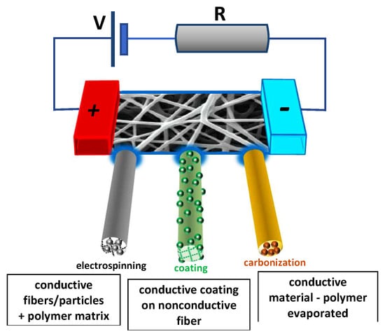
One of the possibilities to nevertheless prepare conductive nanofiber mats is based on the carbonization of polyacrylonitrile, the most often used precursor for carbon nanofibers [17], lignin [18] or other nanofiber mats. These approaches are not discussed in detail here. Instead, we give an overview of different electrospinning techniques that can be applied in the case of conductive polymers as well as diverse after-treatment steps, enabling the preparation of originally non-conductive nanofiber mats which are made conductive afterward (Figure 1), followed by possible applications of such conductive nanofiber mats.
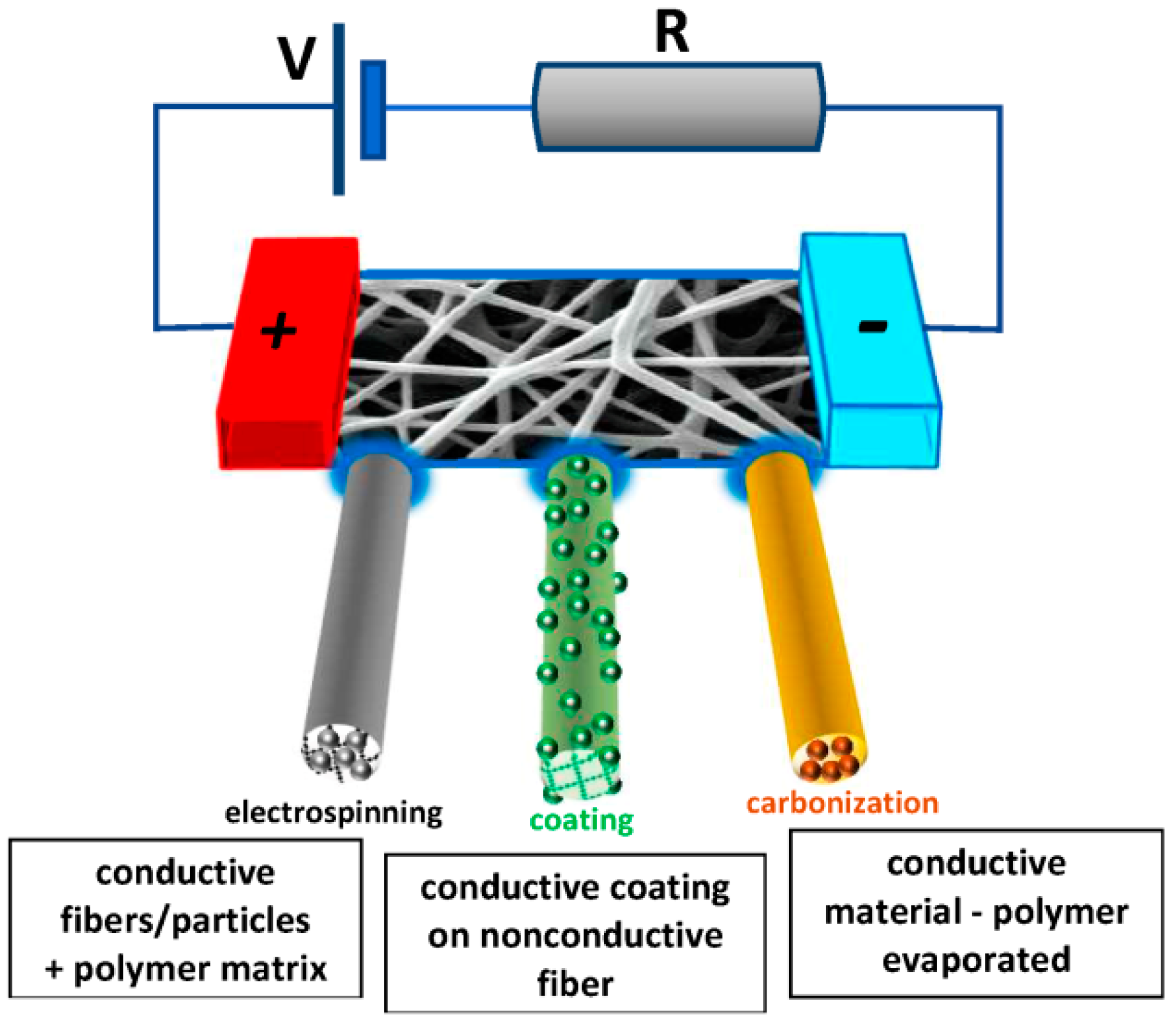
The number of studies on electrospun conductive nanofiber mats has strongly increased during recent years due to their large field of applications, as visible in Figure 2. It must be mentioned, however, that due to the broad spectrum of applications, “conductive” nanofibers do not always have conductivities of comparable orders of magnitude in these studies, but also span a wide range of conductivities or sheet resistances, respectively.
2. Electrospinning from Conductive Solutions or Melts
One possibility to create conductive nanofibers by electrospinning is based on including conducting nanoparticles or nano-sheets and sintering after electrospinning to remove the non-conductive polymeric matrix (Figure 2). Li et al. describe the process of creating a hydrophobic and conductive composite nanofiber mat in this way. In their needle-based electrospinning setup, two needles on a positive voltage share a common cylinder substrate on negative voltage. By co-electrospinning trimethylethoxysilane (MTES) and a conductive polyvinylpyrrolidone/graphene solution, they removed the PVP by sintering at 500 °C. The resulting MTES/graphene nanofiber mats showed sheet resistance up to nearly 2000 S/m [19].
Wang et al. prepared conductive core-shell nanofiber mats by needle-based electrospinning a solution from multi-wall carbon nanotubes (CNTs) in polycaprolactone (PCL) and silk fibroin. While the conductivity of these nanofiber mats is not reported in their paper, the nanofiber mats were found to allow for neurite extension and cell migration along the nanofibers [20].
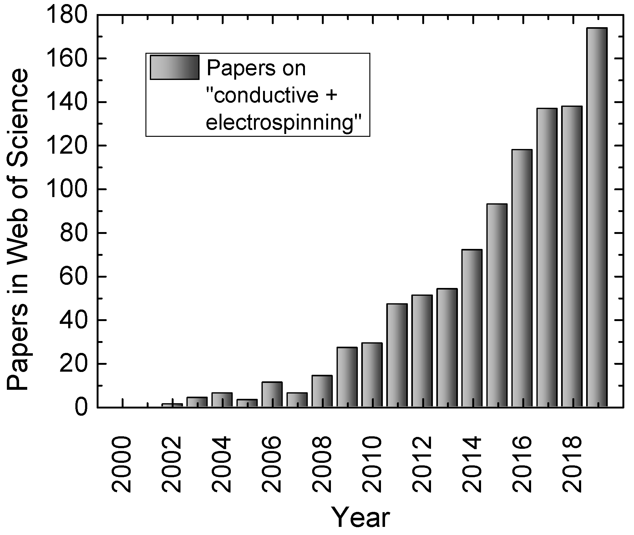
Generally, however, embedding such special carbon modifications does not necessarily result in very high conductivities. With functionalized single-wall carbon nanotubes (CNTs), a conductivity of 1 S/m was reached [21], while even high amounts of multi-wall CNTs resulted in only 10 nS/cm [22]. Using graphite nanoplatelets as fillers in electrospun polystyrene nanofibers which were cold- and hot-pressed after spinning, Guo et al. reached a higher value of approximately 1 S/cm for the highest graphite loading, as depicted in Figure 3 [23]. Shrestha et al. included functionalized multi-wall CNTs in a polyurethane/silk spinning solution and found conductivities of nearly 60 µS/cm for the resulting nanofiber mats, as compared to values below 1 µS/cm for pure polyurethane (PU) or PU/silk nanofiber mats [24]. Combining electrospraying of polyurethane (PU) with simultaneous electrospraying of multi-wall CNTs, Shokraei et al. reached conductivities between 10−5 and 10−2 S/cm, as compared to the conductivity of the pure isolating PU of 10−10 S/cm [25].
Abedi et al. report on conductive chitosan/PEDOT:PSS nanofiber mats [26]. The complicated electrospinning process of chitosan solutions is often attributed to their low conductivity [27]; a problem that can be solved by using different spinning agents [28,29]. They showed that adding up to 1% PEDOT:PSS was sufficient to increase the conductivity of the resulting nanofiber mat by two orders of magnitude, in this way significantly increasing cell proliferation in cardiac tissue engineering.
Adding conductive MoS2 nano-sheets to a nonconductive nylon spinning solution resulted in s significantly increased conductivity of the solution of approximately 20 µS/cm; however, this value is still far below a critical value for electrospinning [30]. Nevertheless, the resulting increase of conductivity of the nanofiber mats was sufficient to increase cellular attachment and cell proliferation, and even to induce cardiogenic differentiation of mouse embryonic cardiac cells.
Conductive polymers are usually hard to electrospin solely because they often show a low solubility in most solvents and usually have a low molecular weight which impedes fiber formation. Blending them with spinning agents is one possibility to prepare conductive nanofiber mats from such conductive polymers [31]. Bittencourt et al., e.g., used electrospinning from different PVA/polyaniline (PAni) solutions with de-doped PAni, resulting in nanofiber mats with conductivities around 35 nS/m which was sufficient for the use as ammonia gas sensor [32].
Akcoren et al. prepared nanofiber mats from blends of polypyrrole and poly(butyl acrylate-co-methyl methacrylate), in this way, increasing the alternating current (AC) conductivity to a range of 0.4–0.5 µS/cm [33]. Poly(caprolactone)(PCL)/PAni nanofibers were electrospun using the camphorsulfonic acid doped green form of PAni by Garrudo et al., resulting in much higher conductivities in the range of 10−4–10−1 S/cm [34]. Liu et al. used a side-by-side spinneret to spin camphoric acid doped PAni together with PEO and found an increased spinnability combined with conductivities between 10−6 and 10−4 S/cm [35].
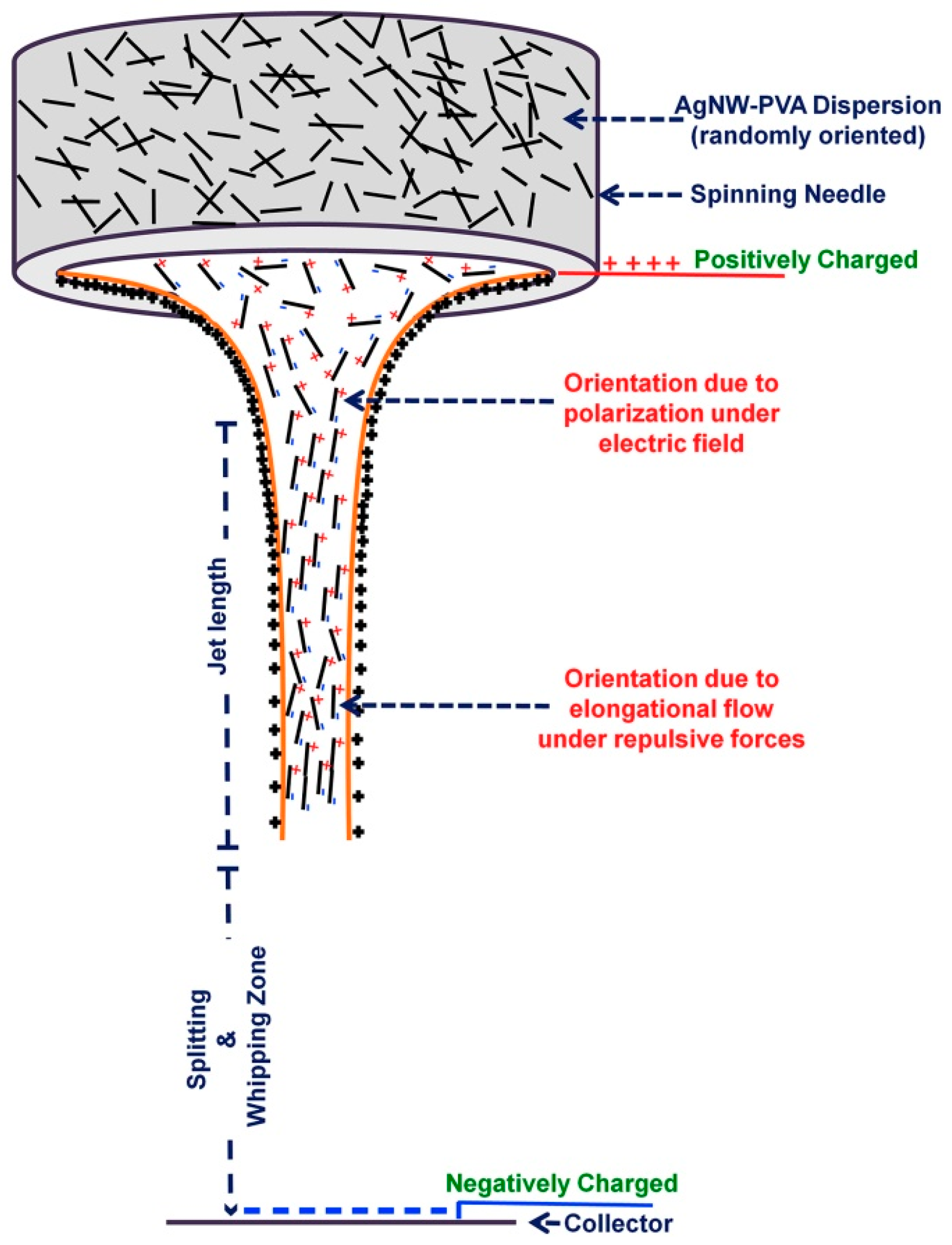
These conductivities, however, are still low as compared to values that can be achieved by adding conductive nanowires, etc. Yadav et al. recently reported on polyvinyl alcohol (PVA) electrospun nanofiber mats which included approximately 1/3 weight percent of silver nanowires [36]. They found that this approach resulted in conductivity of more than 650 S/cm, i.e., a much higher value than reached by the aforementioned conductive polymer blends. This high conductivity was attributed to the orientation in the jet during electrospinning, as depicted in Figure 4.
3. Electrospinning and Subsequent Calcination of a Polymer
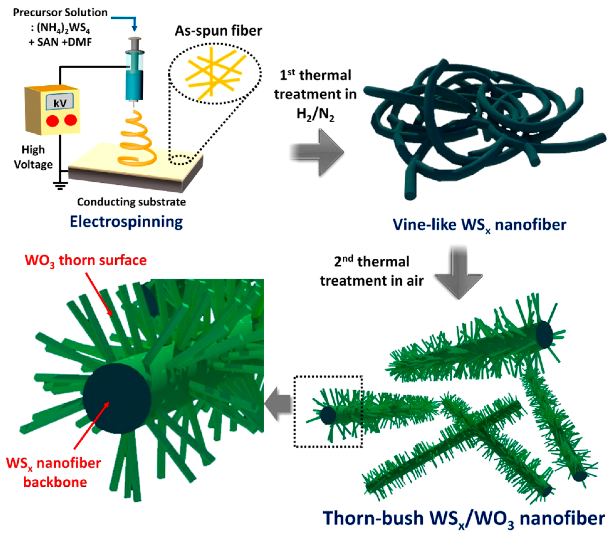
Completely metallic nanofibers were produced by electrospinning copper(II) acetate and PVA in a needle-based setup with a rotating collector, followed by calcination (Figure 2) to maintain pure CuO nanofibers which were used as translucent conductive layers with sheet resistances around 0.4–5.4 MΩ [37]. Silver-electroplated electrospun nickel microfibers showed much lower sheet resistance of less than 0.2 Ω [38]. For the application as anode materials in Na-ion batteries, Ryu et al. prepared hierarchically structured WSx/WO3 thorn-bush nanofibers by electrospinning (NH4)2WS4/styrene acrylonitrile solutions in dimethylformamide (DMF) and two subsequent thermal treatments, resulting in the growth of thorns vertical to the nanofiber surface which further increased the fiber surface [39] (Figure 5).
4. Conductive Coatings
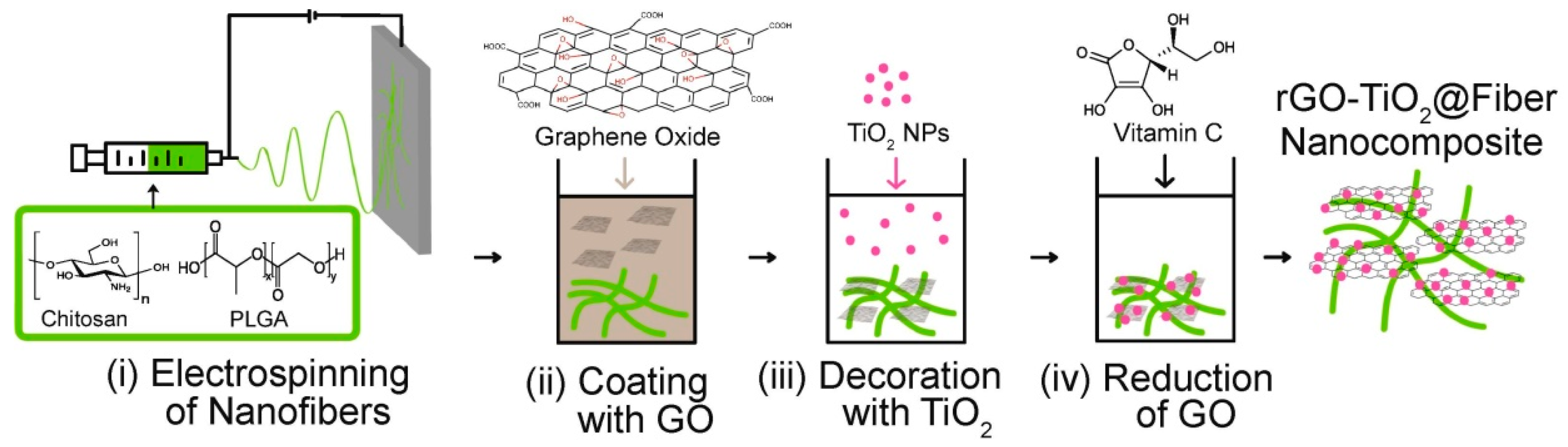
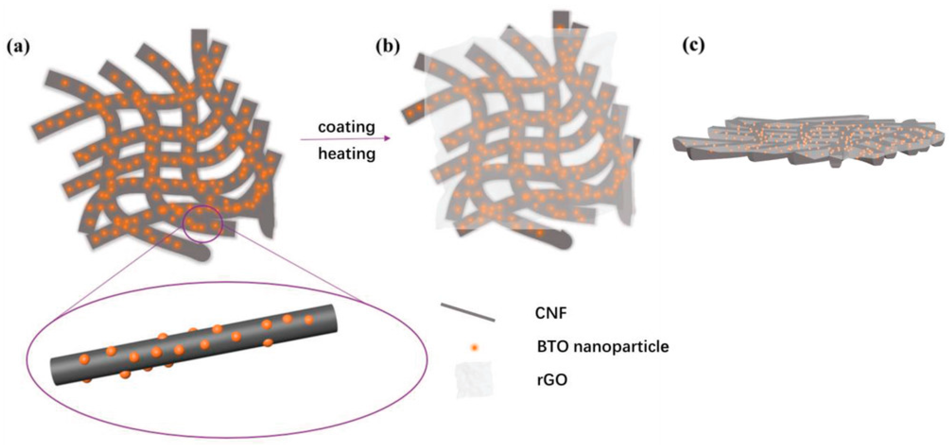
Another possibility to create conductive nanofibers, also based on a two-step process, is performed by applying a conductive coating on non-conductive or weakly-conductive nanofibers (Figure 2). Fausey et al., e.g., prepared a nanofiber mat from a chitosan/poly(lactic-co-glycolic) acid polymer blend by needle-based electrospinning and afterward dip-coated this substrate by conductive graphene oxide, followed by dip-coating in TiO2 and afterward reduction of the graphene oxide with vitamin C to increase its conductivity (Figure 6), resulting in increased arsenic oxidation due to the faster shuttling of electrons from the valence band of the TiO2 and thus reduction of electron-hole recombination [40].
Similarly, Ahmed et al. prepared a poly(vinylidene fluoride-co-trifluoro ethylene) (PVDF-TrFE) nanofiber mat by electrospinning and spray-coated it with a mixture of multi-wall CNTs and reduced graphene oxide five times, before this nanofiber mat was finally coated with PEDOT. This resulted in a conductivity of nearly 4000 S/cm, depending on the exact material combination, and allowed for using the nanofiber mat as a conductive electrode in a piezoelectric pressure sensor [41]. Conductivities of approximately 0.3 S/m were found by Li et al. who coated electrospun PAN yarn with multi-wall CNTs [42].
Polypyrrole (PPy) is often used for the preparation of artificial muscles. Ebadi et al. produced polyurethane (PU) nanofibers by needle-based electrospinning and afterward coated PPy onto these PU nanofibers. For this, pyrrole monomer with LiTFSI was dissolved in water, and an oxidizing agent was gradually added to polymerize PPY on the PU nanofibers, a process in which bonding occurred by radical cations [43]. Similarly, the same group coated PU nanofibers with a p-toluenesulfonate doped PPy layer, resulting in electrical conductivity of approximately 276 S/cm. These nanofiber mats could be used as bending actuators [44]. To prepare scaffolds for neural cell growth, Xu et al. prepared poly(l-lactide acid)-PCL fibers and coated them electrochemically with chitosan and PPy, resulting in conductivities of approximately 1 S/m [45]. In-situ polymerization of pyrrole on Fe3O4/polylactic acid-glycolic acid resulted in magnetic nanofibers with a conductivity of up to 0.58 S/cm [46].
Dognani et al. coated an electrospun polyvinylidenefluoride-co-hexafluoropropilene (VDF-HFP) nanofiber mat with PAni, resulting in modified pore sizes, and water contact angles from the clearly hydrophobic surface of pure PVDF-HFP nanofiber mats to hydrophilic surfaces of PVDF-HFP/PAni nanofiber mats [47]. Pure PEDOT nanofibers were prepared by Laforgue and Robitaille by applying an EDOT coating on an electrospun PVP nanofiber mat, polymerizing it to PEDOT and afterward calcinating the PVP core, in this way creating nanofibers with a conductivity of approximately 60 S/cm [48]. Similarly, pure PAni nanofibers were prepared by coaxial electrospinning of a PAni shell around a poly(methyl methacrylate (PMMA) core, resulting in nearly identical conductivities [49], while PVDF nanofibers with an aniline coating polymerized on them resulted in one order of magnitude lower conductivity [50].
Coating PU nanofibers with silver nanowires, Kim et al. found sheet resistances between 0.7 Ω and 510 Ω, depending on the areal weight of the coating [51].
5. Applications of Conductive Electrospun Nanofiber Mats
5.1. Electromagnetic Shielding
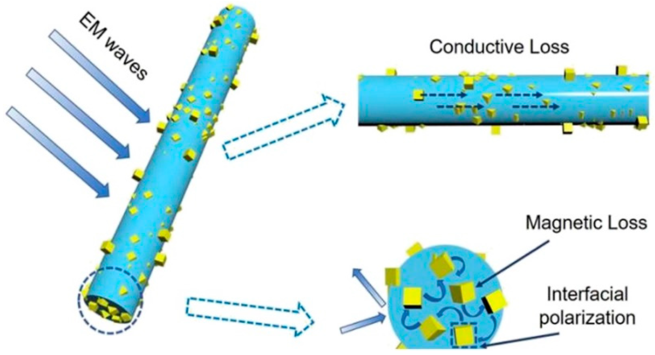
One of the large areas in which electrospun nanofiber mats are used is electromagnetic shielding. Typically, lightweight electromagnetic (EM) wave absorbers are prepared as heterogeneous structures from magnetic and dielectric loss materials, with the heterogeneous structure supporting the interaction between an electromagnetic wave and absorber [52]. This results in a strong use of combinations of magnetic loss materials like magnetic metals with dielectric loss materials like carbon in different modifications for the preparation of lightweight EM composite absorbers, as depicted in Figure 7 [53,54,55,56]. Nanofiber mats electrospun from other combinations such as ZnO/C are also reported to show good microwave absorption [57].
5.2. Energy Storage
Another application of conductive nanofiber mats are electrodes of lithium-ion batteries. Here again, metallic and carbon-based materials are often combined to gain a sufficient conductivity. Typically, the anode is prepared from MgFe2O4 in combination with graphene [58], carbon nanotubes [59] or graphene aerogel [60,61]. MoS2/carbon nanofiber membranes were prepared by needle-based electrospinning and carbonization of the PAN-based precursor and used as binder-free anodes for sodium-ion batteries [62].
Interlayers for Li-S batteries were prepared by Zhang et al., combining a reduced graphene oxide layer with BaTiO3 decorated carbon nanofibers prepared by electrospinning and subsequent calcination (Figure 8), resulting in low resistances around 30 Ω in the fresh state and around 6 Ω after cycling, resulting in a high rate performance and cycling performance [63].
Supercapacitors, on the other hand, can be created by firstly electrospinning TiO2 nanofibers from a solution of Ti(OC4O9)4 and poly(vinyl pyrrolidone) (PVP), followed by calcination to remove the polymer and retain the pure semiconductive nanofibers. Next, nitridization via ammonia annealing resulted in highly conductive TiN nanofibers. These nanofibers were afterward coated with MnO2 nanosheets, resulting in increased specific capacitance and cycle stability [64].
5.3. Electronic Components
Even memristors were produced by conductive nanofiber mats. Lapkin et al. used electrospinning to produce polyamide-6 nanofiber mats on which PAni was polymerized, resulting in a conductivity around 1 S/cm. Combined with a solid polymer electrolyte and a silver counter electrode, a memristor could be realized which showed resistive switching due to a voltage-controlled change in the PAni redox state [65]. Döpke et al. suggested producing conductive magnetic nanofiber mats for data storage and transfer [4].
5.4. Tissue Engineering and Cell Growth
Tissue engineering generally is often based on electrospun nanofiber mats. In order to engineer cardiac tissue, it is not only necessary to create porous nanofiber scaffolds, but these scaffolds should also mimic the extra-cellular matrix of the target tissue, i.e., should be conductive in case of growing cardiac muscle tissue on them with undisturbed intracellular signaling [66,67]. In general, scaffolds with embedded conductive materials often show advances against non-conductive nanofiber mats, whether prepared with PAni, PPy or CNTs [68,69,70].
Nekouian et al. report on conductive electrospun nanofiber mats, prepared from PCL/PPy/multi-wall CNTs which were used to examine the influence of electrical stimulation on the photoreceptor differentiation of mesenchymal stem cells, showing that rhodopsin and peripherin gene expressions could significantly be increased by the electrical stimulation [71]. Rahmani et al. used silk fibroin nanofibers filled with conductive reduced graphene oxide, resulting in electrochemical series resistances around 20–30 Ω, to grow conjunctiva mesenchymal stem cells under electrical stimulation and found formation of neuron-like cell morphology and alignment along the electrical field [72]. PCL/PAni scaffolds with conductivities up to approximately 80 µS/cm were used by Garrudo et al. for the cultivation of neural stem cells, showing that the typical cell morphology was retained, and the nanofiber mats were biocompatible [34]. Even lower values of approximately 1 µS/cm were reported by Ghasemi et al. who doped electrospun polyethylene terephthalate (PET) nanofibers with graphene oxide to prepare cardiac patches for cardiac regeneration after myocardial infarcts [73]. For the same purpose, Walker et al. suggested using electrospun gelatin methacryloyl with bio-ionic liquid to combine adhesive and conductive properties [74].
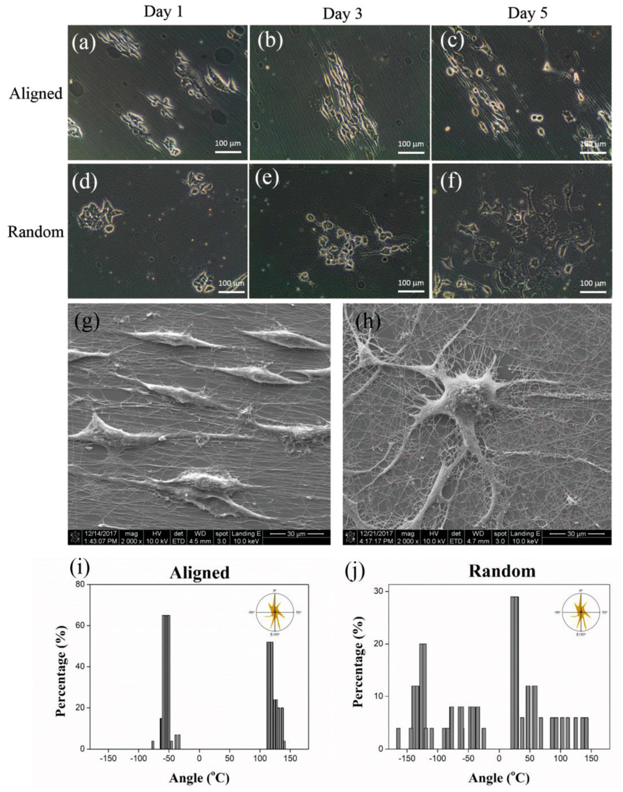
Cell proliferation and gene expression could also be optimized by doping PAni scaffolds with graphene oxide and plasma treatment to hydrophilize the fiber surface [75]. Attachment, spreading and proliferation of fibroblasts and endothelial cells was optimized by tailoring the concentration of multilayer graphene flakes in electrospun polyurethane nanofiber mats [76]. Embedding reduced graphene oxide in electrospun poly(ester amide) (PEA) and PEA/chitosan scaffolds increased cardiac differentiation [77]. Similarly, electrospinning PEO/PEDOT:PSS nanofibers showed a positive effect on neurite outgrowth, i.e., neural differentiation of neuron-like model cells, which is especially interesting since a spin-coated PEO/PEDOT:PSS film showed contact repulsion limiting cell attachment and proliferation (Figure 9) [78].
Osteoblast cells were found to grow and proliferate well on electrospun poly(l-lactic acid)/PAni/p-toluene sulfonic acid nanofiber mats [79]. Keratinocytes were shown to grow on electrospun PAN/PPy and PAN/PPy/CNT nanofiber mats [80]. Coating electrospun polyurethane nanofibers with PAni reduced the water contact angle significantly, resulted in a certain anticoagulant effect and was found supportive for cell adhesion, proliferation, and extension [81].
5.5. Dye-Sensitized Solar Cells
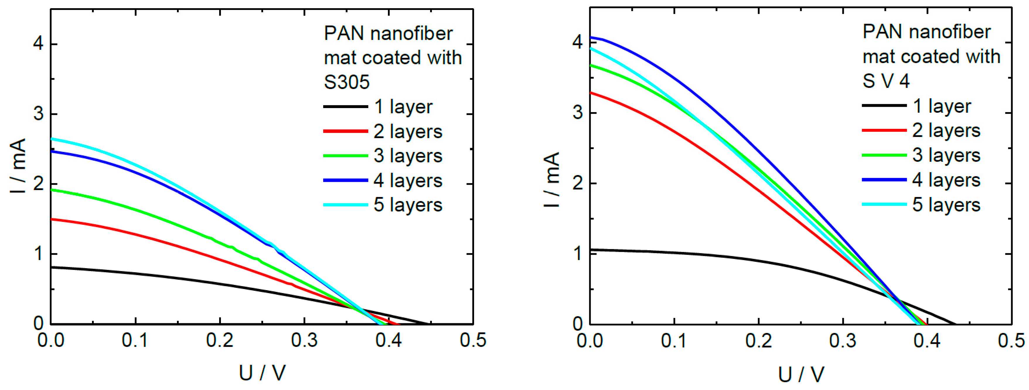
Counter electrodes of dye-sensitized solar cells (DSSCs) were prepared by coating an electrospun nanofiber mat with PEDOT:PSS. Juhász Junger et al. used several dip-coating steps to optimize the electrode conductivity while partly retaining the nanostructured surface and thus the large contact area with the neighboring layers (Figure 10) [82]. The optimum number of layers resulted in a sheet resistance around 150 Ω, reduced from approximately 550 Ω for a single coating layer [82,83]. A similar approach was recently suggested by Kohn et al. who prepared fully electrospun DSSCs with both electrodes prepared by separately dip-coating them in PEDOT:PSS [84].
Eslah and Nouri, on the other hand, used spin-coating of WO3 nanoparticles on electrospun PAN/PAni nanofibers to prepare counter electrodes of DSSCs [85]. For the possible use in LEDs and solar cells, Jiang et al. developed transparent conductive electrodes by electrospinning copper nanofibers and immersing them in silver ink as a protective layer, resulting in sheet resistances below 10 Ω [86].
5.6. Hydrogen Evolution
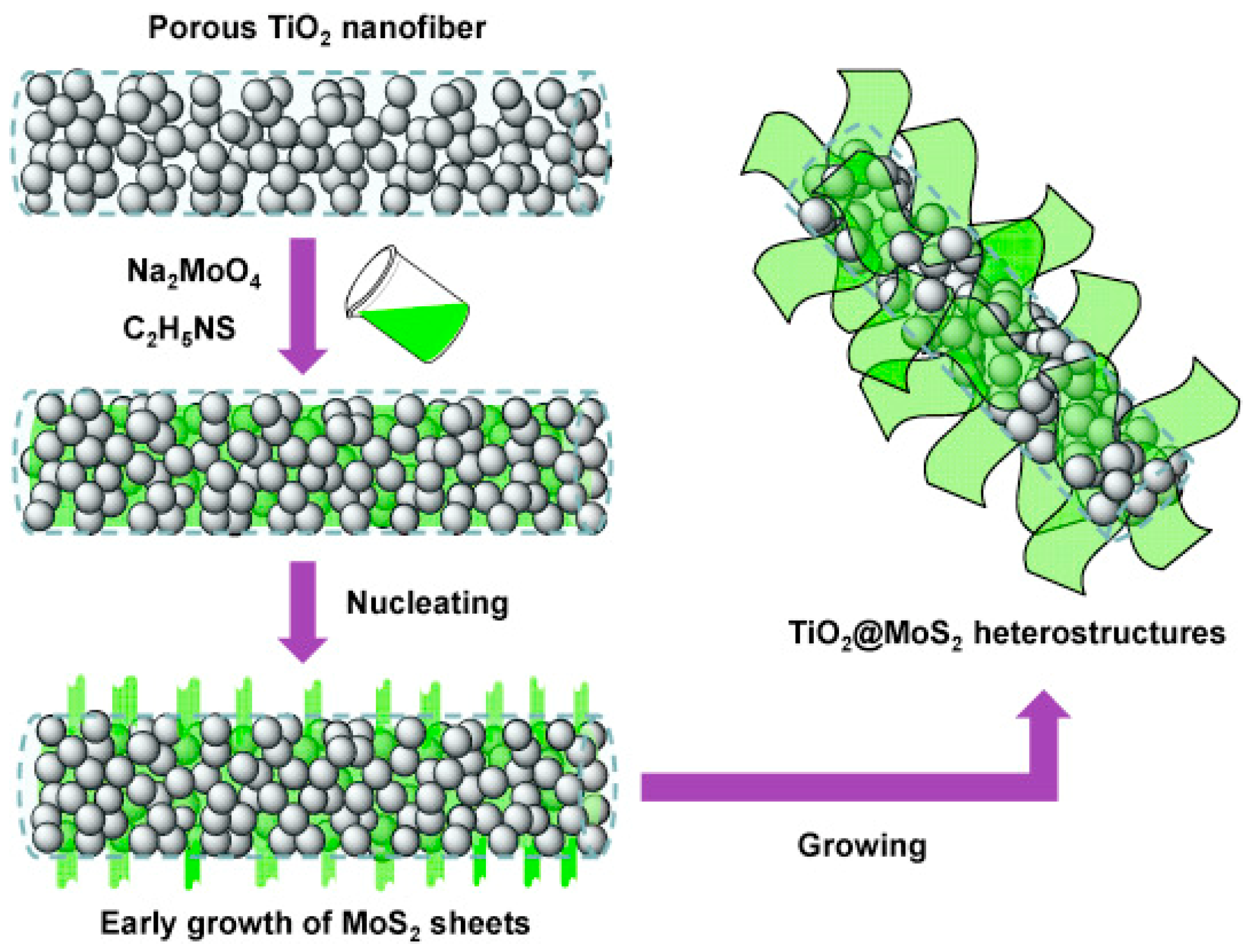
Another interesting application is hydrogen evolution. Sun et al. most recently prepared electrospun carbon/Ni/Mo2C nanofibers which were used as electrocatalysts in hydrogen evolution reaction in an alkaline electrolyte [87]. Li et al. used nitrogen-doped carbon/Ni nanofibers decorated with Pt for hydrogen evolution, resulting in a high electrochemical activity combined with reduced usage of Pt [88]. Zhang et al. prepared binder-free MoS2/carbon nanofiber electrodes by electrospinning and carbonization of the resulting nanofibers, allowing them to tailor the porosity chemically, which could be used for electrocatalytic hydrogen production [89]. Rheem et al. used a hierarchical structure of MoS2 nanosheets on conductive MoO2 nanofibers, gained by electrospinning, calcination, and sulfurization, to increase the hydrogen evolution reaction [90]. A similar hierarchical structure was prepared earlier by Liu et al. who used porous electrospun TiO2 nanofibers as a substrate for growing MoS2 nanosheets perpendicular to the nanofiber surfaces, resulting in high photocatalytic hydrogen production (Figure 11) [91].
5.7. Sensors
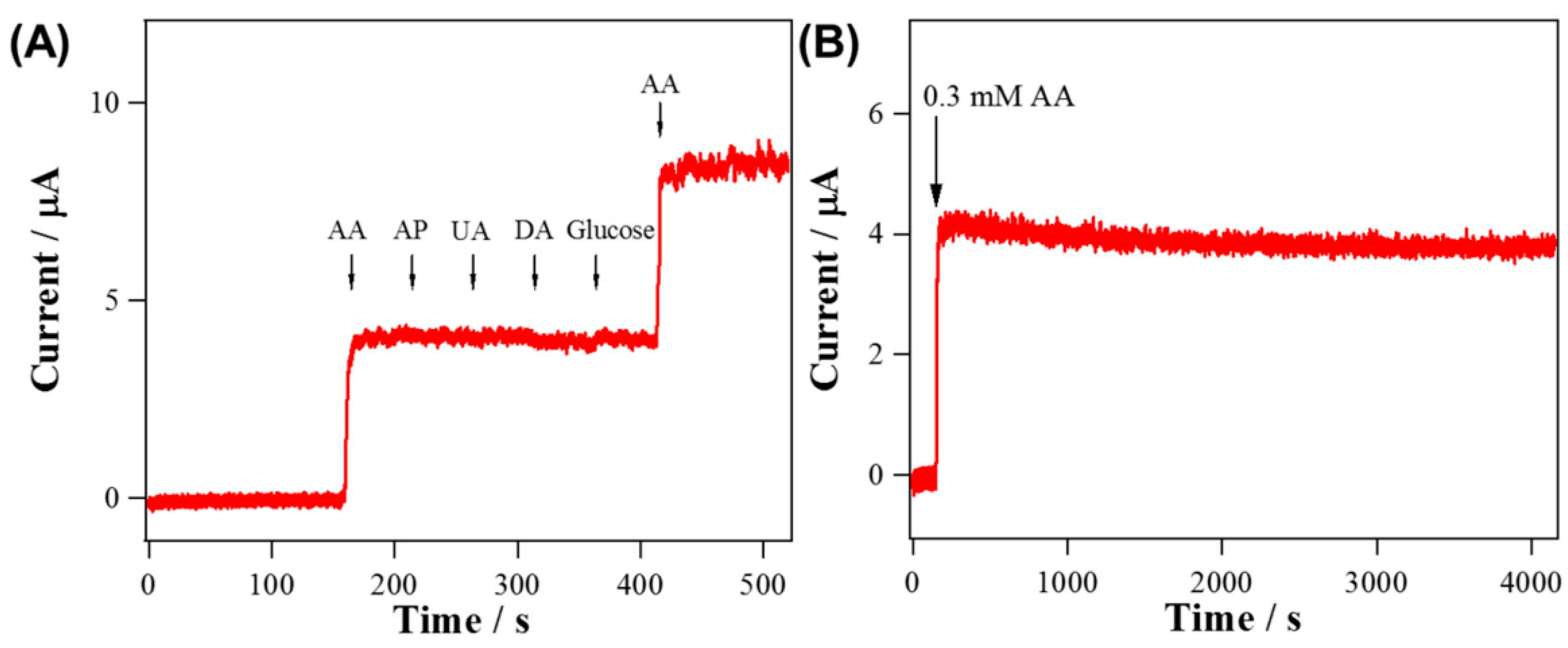
Lee et al. used electrospun WO3 nanofibers coated with RuO2 nanorods as a sensor for H2O2 and L-ascorbic acid. They could show that by the addition of the RuO2 nanorods, the electrocatalytic activity was increased, and the sensing abilities were significantly improved in comparison with pure WO3 nanofibers, as shown in Figure 12 [92].
To sense dopamine, Ozoemena et al. used electrospun PAN/onion-like carbon nanofibers and found a high conductivity and sensitivity of the resulting nanofibers [93]. By electrospinning polystyrene/polyhydroxibutyrate filled with graphitized carbon and partly doped with porphyrin on an interdigitated electrode, Avossa et al. prepared gas sensors for volatile organic compounds [94].
Shaker et al. developed a polyurethane/PEDOT:PSS electrospun nanofiber mat which exhibited a resistance of approximately 3 kΩ and could be used as a reliable strain gauge sensor [95]. Yang et al. coated highly conductive MXene sheets on electrospun PU nanofibers mats to produce highly sensitive strain sensors [96]. Flexible strain sensors with up to 1000% elongation were prepared from conductively coated electrospun styrenebutadiene-styrene copolymer [97]. A similar stretchability was reached by Ren et al., electrospinning a thermoplastic polyurethane nanofiber mat with a wavelike structure, followed by wrapping CNTs around the nanofibers [98]. Wrapping conductive nanofiber yarn produced from graphene oxide-doped PAN nanofibers with in-situ polymerized PPy around elastic yarns results in high sensitivity and repeatability, in this way enabling detection or breathing or human motion [99].
Harjo et al. developed conductive fiber scaffolds by coating electrospun glucose-gelatin nanofiber mats with polypyrrole and investigated their electro-chemo-mechanical response, showing stable actuation for more than 100 cycles as well as reasonable sensor properties [100]. They found conductivities of approximately 3 µS/cm in the unstretched state and approximately half this value when stretched in aqueous or organic electrolyte solutions.
Finally, Table 1 gives an overview of the conductivities mentioned in this article, reached with different methods, again showing the broad range of conductivities reached by different methods and sufficient for various applications.
6. Conclusions
In this review, we report we give an overview of the most recent developments in the research area of conductive electrospun nanofiber mats. As well as the possible applications, varying from biomedicine to sensors to batteries to hydrogen evolution, the range of conductivities achievable with different methods is wide. While conductivities in the range of some 10 µS/cm are sufficient for some biotechnological applications, some techniques such as embedding silver nanowires into the electrospinning solution or coating nanofiber mats with diverse conductive materials result in high conductivities of some 100 to some 1000 S/cm. In many cases, the authors of the cited studies report on additional advantageous findings, such as enhanced biocompatibility or improved fiber diameters.
While this review can only give a short overview of the most recent development, it aims at supporting the growing number of researchers working in this highly interesting field of conductive nanofiber mats to find the best solutions for their own applications.
Author Contributions
Conceptualization, T.B. and A.E.; visualization, T.B. and A.E.; writing—original draft preparation, A.E. and T.B. All authors have read and agreed to the published version of the manuscript.
Funding
This research was funded by Volkswagen Foundation grant “Adaptive Computing with Electrospun Nanofiber Networks” no. 93679.
Conflicts of Interest
The authors declare no conflict of interest. The funders had no role in the design of the study; in the collection, analyses, or interpretation of data; in the writing of the manuscript, or in the decision to publish the results.
References
- Greiner, A.; Wendorff, J.H. Electrospinning: A fascinating method for the preparation of ultrathin fibers. Angew. Chem. Int. Ed. 2007, 46, 5670–5703. [Google Scholar] [CrossRef] [PubMed]
- Yalcinkaya, F. A review on advanced nanofiber technology for membrane distillation. J. Eng. Fibers Fabr. 2019, 14. [Google Scholar] [CrossRef]
- Banitaba, S.N.; Semnani, D.; Rezaei, B.; Ensafi, A.A. Evaluating the electrochemical properties of PEO-based nanofibrous electrolytes incorporated with TiO2 nanofiller applicable in lithium-ion batteries. Polym. Adv. Technol. 2019, 30, 1234–1242. [Google Scholar] [CrossRef]
- Döpke, C.; Grothe, T.; Steblinski, P.; Klöcker, M.; Sabantina, L.; Kosmalska, D.; Blachowicz, T.; Ehrmann, A. Magnetic Nanofiber Mats for Data Storage and Transfer. Nanomaterials 2019, 9, 92. [Google Scholar] [CrossRef] [PubMed] [Green Version]
- Andre, R.S.; Mercante, L.A.; Facure, M.H.M.; Mattoso, L.H.C.; Correa, D.S. Enhanced and selective ammonia detection using In2O3/reduced graphene oxide hybrid nanofibers. Appl. Surf. Sci. 2019, 473, 133–140. [Google Scholar] [CrossRef]
- Mahmoodi, N.M.; Keshavarzi, S.; Oveisi, M.; Rahimi, S.; Hayati, B. Metal-organic framework (ZIF-8)/inorganic nanofiber (Fe2O3) nanocomposite: Green synthesis and photocatalytic degradation using LED irradiation. J. Mol. Liq. 2019, 291, 111333. [Google Scholar] [CrossRef]
- Sabzehmeidani, M.M.; Karimi, H.; Ghaedi, M. Visible light-induced photo-degradation of methylene blue by n-p heterojunction CeO2/CuS composite based on ribbon-like CeO2 nanofibers via electrospinning. Polyhedron 2019, 170, 160–171. [Google Scholar] [CrossRef]
- Rodriguez, A.V.; Sabino, N.L. Synthesis of photoluminescent β-Ga2O3 nanostructures using electrospinning method, and control of length-diameter ratio by calcination heating rates. J. Mater. Sci. Mater. Electron. 2019, 30, 16910–16916. [Google Scholar] [CrossRef]
- Amand, F.K.; Esmaeli, A. Investigating the properties of electrospun nanofibers made of hybride polymer containing anticoagulant drugs. Carbohydr. Polym. 2020, 228, 115397. [Google Scholar] [CrossRef]
- Jahan, I.; Jadhav, A.; Wang, L.J.; Wang, X. Electrospinning from a convex needle with multiple jet toward better controlling and enhanced production rate. J. Appl. Polym. Sci. 2019, 136, 48014. [Google Scholar] [CrossRef]
- Hwang, M.; Karenson, M.O.; Elabd, Y.A. High Production Rate of High Purity, High Fidelity Nafion Nanofibers via Needleless Electrospinning. ACS Appl. Polym. Mater. 2019, 1, 2731–2740. [Google Scholar] [CrossRef]
- Grothe, T.; Wehlage, D.; Böhm, T.; Remche, A.; Ehrmann, A. Needleless Electrospinning of PAN nanofibre Mats. Tekstilec 2017, 60, 290–295. [Google Scholar] [CrossRef]
- Rosenthal, T.; Weller, J.M.; Chan, C.K. Needleless Electrospinning for High Throughput Production of Li7La3Zr2O12 Solid Electrolyte Nanofibers. Ind. Eng. Chem. Res. 2019, 58, 17399–17405. [Google Scholar] [CrossRef]
- Yalcinkaya, F.; Komarek, M. Polyvinyl Butyral (PVB) Nanofiber/Nanoparticle-Covered Yarns for Antibacterial Textile Surfaces. Int. J. Mol. Sci. 2019, 20, 4317. [Google Scholar] [CrossRef] [PubMed] [Green Version]
- Wehlage, D.; Blattner, H.; Sabantina, L.; Böttjer, R.; Grothe, T.; Rattenholl, A.; Gudermann, F.; Lütkemeyer, D.; Ehrmann, A. Sterilization of PAN/gelatin nanofibrous mats for cell growth. Tekstilec 2019, 62, 78–88. [Google Scholar] [CrossRef]
- Grothe, T.; Brikmann, J.; Meissner, H.; Ehrmann, A. Influence of solution and spinning parameters on nanofiber mat creation of poly(ethylene oxide) by needleless electrospinning. Mater. Sci. Medzg. 2017, 23, 342–349. [Google Scholar] [CrossRef] [Green Version]
- García-Mateos, F.J.; Ruiz-Rosas, R.; Rosas, J.J.; Rodríguez-Mirasol, J.; Cordero, T. Controlling the Composition, Morphology, Porosity, and Surface Chemistry of Lignin-Based Electrospun Carbon Materials. Front. Mater. 2019, 6, 114. [Google Scholar] [CrossRef] [Green Version]
- Sabantina, L.; Rodriguez-Cano, M.A.; Klöcker, M.; Garcia-Mateos, F.J.; Ternero-Hidalgo, J.J.; Mamun, A.; Beermann, F.; Schwakenberg, M.; Voigt, A.L.; Rodriguez-Mirasol, J.; et al. Fixing PAN Nanofiber Mats during Stabilization for Carbonization and Creating Novel Metal/Carbon Composites. Polymers 2018, 10, 735. [Google Scholar] [CrossRef] [Green Version]
- Li, T.Y.; Xu, Y.L.; Wang, K.J.; Song, J.H.; Hu, H.W.; Liu, H.; Liu, Y.Q.; Liu, Y.; Wu, J.; Pi, H.H.; et al. Preparation and performance of hydrophobic and conductive silica composite fiber membrane. J. Mater. Sci. 2020, 55, 191–202. [Google Scholar] [CrossRef]
- Wang, L.; Wu, Y.B.; Hu, T.L.; Ma, P.X.; Guo, B.L. Aligned conductive core-shell biomimetic scaffolds based on nanofiber yarns/hydrogel for enhanced 3D neurite outgrowth alignment and elongation. Acta Biomater. 2019, 96, 175–187. [Google Scholar] [CrossRef]
- Naeem, F.; Prestayko, R.; Saem, S.; Nowicki, L.; Imit, M.; Adronov, A.; Moran-Mirabal, J.M. Fabrication of conductive polymer nanofibers through SWNT supramolecular functionalization and aqueous solution processing. Nanotechnology 2015, 26, 395301. [Google Scholar] [CrossRef] [PubMed]
- Wang, J.; Naguib, H.E.; Bazylak, A. Electrospun porous conductive polymer membranes. In Proceedings of the SPIE—The International Society for Optical Engineering, San Diego, CA, USA, 11–15 March 2012. [Google Scholar]
- Guo, Y.Q.; Pan, L.L.; Yang, X.T.; Ruan, K.P.; Han, Y.X.; Kong, J.; Gu, J.W. Simultaneous improvement of thermal conductivities and electromagnetic interference shielding performances in polystyrene composites via constructing interconnection oriented networks based on electrospinning technology. Compos. Part A Appl. Sci. Manuf. 2019, 124, 105484. [Google Scholar] [CrossRef]
- Shrestha, S.; Shrestha, B.K.; Lee, J.; Joong, O.K.; Kim, B.S.; Park, C.H.; Kim, C.S. A conducting neural interface of polyurethane/silk-functionalized multiwall carbon nanotubes with enhanced mechanical strength for neuroregeneration. Mater. Sci. Eng. C Mater. Biol. Appl. 2019, 102, 511–523. [Google Scholar] [CrossRef] [PubMed]
- Shokraei, N.; Asadpour, S.; Shokraei, S.; Sabet, M.N.; Faridi-Majidi, R.; Ghanbari, H. Development of electrically conductive hybrid nanofibers based on CNT-polyurethane nanocomposite for cardiac tissue engineering. Microsc. Res. Tech. 2019, 82, 1316–1325. [Google Scholar] [CrossRef] [PubMed]
- Abedi, A.; Zasanzadeh, M.; Tayebi, L. Conductive nanofibrous chitosan/PEDOT: PSS tissue engineering scaffolds. Mater. Chem. Phys. 2019, 237, 121882. [Google Scholar] [CrossRef]
- Costa-Júnior, E.S.; Barbosa-Stancioli, E.F.; Mansur, A.A.P.; Vasconcelos, W.L.; Mansur, H.S. Preparation and characterization of chitosan/poly(vinyl alcohol) chemically crosslinked blends for biomedical applications. Carbohydr. Polym. 2009, 76, 472–481. [Google Scholar] [CrossRef]
- Grimmelsmann, N.; Homburg, S.V.; Ehrmann, A. Needleless electrospinning of pure and blended chitosan. IOP Conf. Ser. Mater. Sci. Eng. 2017, 225, 012098. [Google Scholar] [CrossRef] [Green Version]
- Grimmelsmann, N.; Homburg, S.V.; Ehrmann, A. Electrospinning chitosan blends for nonwovens with morphologies between nanofiber mat and membrane. IOP Conf. Ser. Mater. Sci. Eng. 2017, 213, 012007. [Google Scholar] [CrossRef] [Green Version]
- Nazari, H.; Heirani-Tabasi, A.; Alavijeh, M.S.; Jeshvaghani, Z.S.; Esmaeili, E.; Hossenzadeh, S.; Mohabatpour, F.; Taheri, B.; Tafti, S.H.A.; Seleimani, M. Nanofibrous Composites Reinforced by MoS2 Nanosheets as a Conductive Scaffold for Cardiac Tissue Engineering. Chemistryselect 2019, 4, 11557–11563. [Google Scholar] [CrossRef]
- Merlini, C.; Barra, G.; Araujo, T.M.; Pegoretti, A. Electrically pressure sensitive poly (vinylidene fluoride)/polypyrrole electrospun mats. RSC Adv. 2014, 4, 15749–15758. [Google Scholar] [CrossRef]
- Bittencourt, J.C.; de Santana Gois, B.H.; Rodrigues de Oliveira, V.J.; da Silva Agostini, D.L.; de Almeida Olivati, C. Gas sensor for ammonia detection based on poly (vinyl alcohol) and polyaniline electrospun. J. Appl. Polym. Sci. 2019, 136, 47288. [Google Scholar] [CrossRef]
- Akcoren, D.; Avci, M.Z.; Gokce, Z.G.; Balkan, T.; Sarac, A.S. Fabrication and characterization of poly(butyl acrylate-co-methyl methacrylate)-polypyrrole nanofibers. Polym. Bull. 2018, 75, 1607–1617. [Google Scholar] [CrossRef]
- Garrudo, F.F.F.; Chapman, C.A.; Hoffman, P.R.; Udangawa, R.W.; Silva, J.C.; Mikael, P.E.; Rodrigues, C.A.V.; Cabral, J.M.S.; Morgado, J.M.F.; Ferreira, F.C.; et al. Polyaniline-polycaprolactone blended nanofibers for neural cell culture. Eur. Polym. J. 2019, 117, 28–37. [Google Scholar] [CrossRef]
- Liu, W.C.; Zhang, J.W.; Liu, H. Conductive bicomponent fibers containing polyaniline produced via side-by-side electrospinning. Polymers 2019, 11, 954. [Google Scholar] [CrossRef] [PubMed] [Green Version]
- Yadav, K.; Nain, R.; Jassal, M.; Agrawal, A.K. Free standing flexible conductive PVA nanoweb with aligned silver nanowires. Compos. Sci. Technol. 2019, 182, 107766. [Google Scholar] [CrossRef]
- Saveh-Shemshaki, N.; Bagherzadeh, R.; Latifi, M. Electrospun metal oxide nanofibrous mat as a transparent conductive layer. Org. Electron. 2019, 70, 131–139. [Google Scholar] [CrossRef]
- Il Kim, Y.; An, S.; Kim, M.W.; Jo, H.S.; Kim, T.G.; Swihart, M.T.; Yarin, A.L.; Yoon, S.S. Highly transparent, conducting, body-attachable metallized fibers as a flexible and stretchable film. J. Alloys Compd. 2019, 790, 1127–1136. [Google Scholar] [CrossRef]
- Ryu, W.H.; Wilson, H.; Sohn, S.W.; Li, J.Y.; Tong, X.; Shaulsky, E.; Schroers, J.; Elimelech, M.; Taylor, A.D. Heterogeneous WSx/WO3 thorn-bush nanofiber electrodes for sodium-ion batteries. ACS Nano 2016, 10, 3257–3266. [Google Scholar] [CrossRef]
- Fausey, C.L.; Zucker, I.; Shaulsky, E.; Zimmerman, J.B.; Elimelech, M. Removal of arsenic with reduced graphene oxide-TiO2-enabled nanofibrous mats. Chem. Eng. J. 2019, 375, 122040. [Google Scholar] [CrossRef]
- Ahmed, A.; Jia, Y.M.; Huang, Y.; Khoso, N.A.; Deb, H.; Fan, Q.G.; Shao, J.Z. Preparation of PVDF-TrFE based electrospun nanofibers decorated with PEDOT-CNT/rGO composites for piezo-electric pressure sensor. J. Mater. Sci. Mater. Electron. 2019, 30, 14007–14021. [Google Scholar] [CrossRef]
- Li, Y.; Gora, A.; Anariba, F.; Baji, A. Enhanced tensile strength and electrical conductivity of electrospun polyacrylonitrile yarns via post-treatment. Polym. Compos. 2019, 40, 1702–1707. [Google Scholar] [CrossRef]
- Ebadi, S.V.; Semnani, D.; Fashandi, H.; Rezaei, B. Highly conductive Faradaic artificial muscle based on nanostructured polypyrrole-bis(trifluoromethylsulfonyl)imide synthesized onto electrospun polyurethane nanofibers. Sens. Actuators B Chem. 2019, 297, 126736. [Google Scholar] [CrossRef]
- Ebadi, S.V.; Semnani, D.; Fashandi, H.; Rezaei, B. Synthesis and characterization of a novel polyurethane/polypyrrole-p-toluenesulfonate (PU/PPy-pTS) electroactive nanofibrous bending actuator. Polym. Adv. Technol. 2019, 30, 2261–2274. [Google Scholar] [CrossRef]
- Xu, Y.X.; Huang, Z.B.; Pu, X.M.; Yin, G.F.; Zhang, J.K. Fabrication of Chitosan/Polypyrrole-coated poly(L-lactic acid)/Polycaprolactone aligned fibre films for enhancement of neural cell compatibility and neurite growth. Cell Prolif. 2019, 52, e12588. [Google Scholar] [CrossRef]
- Li, K.; Zhang, S.P.; Wang, S.Y.; Zhu, F.N.; Liu, M.L.; Gu, X.N.; Li, P.; Fan, Y.B. Positive Effect of Magnetic-Conductive Bifunctional Fibrous Scaffolds on Guiding Double Electrical and Magnetic Stimulations to Pre-Osteoblasts. J. Biomed. Nanotechnol. 2019, 15, 477–486. [Google Scholar] [CrossRef] [PubMed]
- Dognani, G.; Hadi, P.; Ma, H.Y.; Cabrera, F.C.; Job, A.E.; Agostini, D.L.S.; Hsiao, B.S. Effective chromium removal from water by polyaniline-coated electrospun adsorbent membrane. Chem. Eng. J. 2019, 372, 341–351. [Google Scholar] [CrossRef]
- Laforgue, A.; Robitaille, L. Production of conductive PEDOT nanofibers by the combination of electrospinning and vapor-phase polymerization. Macromolecules 2010, 43, 4194–4200. [Google Scholar] [CrossRef] [Green Version]
- Zhang, Y.; Rutledge, G.C. Electrical conductivity of electrospun polyaniline and polyaniline-blend fibers and mats. Macromolecules 2012, 45, 4238–4246. [Google Scholar] [CrossRef] [Green Version]
- Merlini, C.; Barra, G.M.O.; Daniela, S.; Ramoa, A.S.; Contri, G.; dos Santos Almeida, R.; Akira d’Ávila, M.; Soares, B.G. Electrically conductive polyaniline-coated electrospun poly(vinylidene fluoride) mats. Front. Mater. 2015, 2, 14. [Google Scholar] [CrossRef]
- Kim, I.; Lee, E.G.; Jang, E.; Cho, G. Characteristics of polyurethane nanowebs treated with silver nanowire solutions as strain sensors. Text. Res. J. 2018, 88, 1215–1225. [Google Scholar] [CrossRef]
- Liu, X.F.; Hao, C.C.; Jiang, H.; Zeng, M.; Yu, R.H. Hierarchical NiCo2O4/Co3O4/NiO porous composite: A lightweight electromagnetic wave absorber with tunable absorbing performance. J. Mater. Chem. C. 2017, 5, 3770–3778. [Google Scholar] [CrossRef]
- Meng, X.F.; Dong, S.H. Design and construction of lightweight C/Co heterojunction nanofibers for enhanced microwave absorption performance. J. Alloys Compd. 2019, 810, 151806. [Google Scholar] [CrossRef]
- Li, W.X.; Qi, H.X.; Guo, F.; Du, Y.E.; Song, N.J.; Liu, Y.Y.; Chen, Y.Q. Co nanoparticles supported on cotton-based carbon fibers: A novel broadband microwave absorbent. J. Alloys Compd. 2019, 772, 760–769. [Google Scholar] [CrossRef]
- Wang, F.Y.; Sun, Y.Q.; Li, D.R.; Zhong, B.; Wu, Z.G.; Zuo, S.Y.; Yan, D.; Zhuo, R.F.; Feng, J.J.; Yan, P.X. Microwave absorption properties of 3D cross-linked Fe/C porous nanofibers prepared by electrospinning. Carbon 2018, 134, 264–273. [Google Scholar] [CrossRef]
- Liu, H.H.; Li, Y.J.; Yuan, M.W.; Sun, G.B.; Liao, Q.L.; Zhang, Y. Solid and macroporous Fe3C/N-C nanofibers with enhanced electromagnetic wave absorbability. Sci. Rep. 2018, 8, 16832. [Google Scholar] [CrossRef] [PubMed]
- Gu, W.H.; Lv, J.; Quan, B.; Liang, X.H.; Zhang, B.S.; Ji, G.B. Achieving MOF-derived one-dimensional porous ZnO/C nanofiber with lightweight and enhanced microwave response by an electrospinning method. J. Alloys Compd. 2019, 806, 983–991. [Google Scholar] [CrossRef]
- Yin, Y.H.; Liu, W.F.; Huo, N.N.; Yang, S.T. Synthesis of vesicle-like MgFe2O4/graphene 3D network anode materials with enhanced lithium storage performance. ACS Sustain. Chem. Eng. 2017, 5, 563–570. [Google Scholar] [CrossRef]
- Pereira, C.; Costa, R.S.; Lopes, L.; Bachiller-Baeza, B.; Rodriguez-Ramos, I.; Guerrero-Ruiz, A.; Tavares, P.B.; Freire, C.; Pereira, A.M. Multifunctional mixed valence N-doped CNT@MFe2O4 hybrid nanomaterials: From engineered one-pot coprecipitation to application in energy storage paper supercapacitors. Nanoscale 2018, 10, 12820–12840. [Google Scholar] [CrossRef]
- Luo, L.; Chen, Z.; Ke, H.Z.; Sha, S.; Cai, G.M.; Li, D.W.; Yang, H.J.; Yang, X.W.; Zhang, R.Q.; Li, J.Q.; et al. Facile synthesis of three-dimensional MgFe2O4/graphene aerogel composites for high lithium storage performance and its application in full cell. Mater. Des. 2019, 182, 108043. [Google Scholar] [CrossRef]
- Brown, E.; Yan, P.L.; Tekik, H.; Elangovan, A.; Wang, J.; Lin, D.; Li, J. 3D printing of hybrid MoS2-graphene aerogels as highly porous electrode materials for sodium ion battery anodes. Mater. Des. 2019, 170, 107689. [Google Scholar] [CrossRef]
- Xiong, X.Q.; Luo, W.; Hu, X.L.; Chen, C.J.; Qie, L.; Hou, D.F.; Huang, Y.H. Flexible Membranes of MoS2/C Nanofibers by Electrospinning as Binder-Free Anodes for High-Performance Sodium-Ion Batteries. Sci. Rep. 2015, 5, 9254. [Google Scholar] [CrossRef] [PubMed]
- Zhang, S.Q.; Qin, X.Y.; Liu, Y.M.; Zhang, L.H.; Liu, D.Q.; Xia, Y.; Zhu, H.; Li, B.H.; Kang, F.Y. A Conductive/Ferroelectric Hybrid Interlayer for Highly Improved Trapping of Polysulfides in Lithium-Sulfur Batteries. Adv. Mater. Interfaces 2019, 6, 1900984. [Google Scholar] [CrossRef]
- Xu, K.B.; Shen, Y.N.; Zhang, K.; Yang, F.; Li, S.J.; Hu, J.Q. Hierarchical assembly of manganese dioxide nanosheets on one-dimensional titanium nitride nanofibers for high-performance supercapacitors. J. Colloid Interface Sci. 2019, 552, 712–718. [Google Scholar] [CrossRef] [PubMed]
- Lapkin, D.A.; Malakhov, S.N.; Demin, V.A.; Chvalun, S.N.; Feigin, L.A. Hybrid polyaniline/polyamide-6 fibers and nonwoven materials for assembling organic memristive elements. Synth. Met. 2019, 254, 63–67. [Google Scholar] [CrossRef]
- Qazi, T.H.; Rai, R.; Dippold, D.; Roether, J.E.; Schubert, D.W.; Rosellini, E.; Barbani, N.; Baccaccini, A.R. Development and characterization of novel electrically conductive PANI–PGS composites for cardiac tissue engineering applications. Acta Biomater. 2014, 10, 2434–2445. [Google Scholar] [CrossRef] [PubMed]
- Martins, A.M.; Eng, G.; Caridade, G.; Mano, F.; Reis, R.L. Electrically conductive chitosan/carbon scaffolds for cardiac tissue engineering. Biomacromolecules 2014, 15, 635–643. [Google Scholar] [CrossRef] [PubMed]
- Guo, B.; Ma, P.X. Conducting polymers for tissue engineering. Biomacromolecules 2018, 19, 1764–1782. [Google Scholar] [CrossRef]
- Prabhakaran, M.P.; Ghasemi-Mobarakeh, L.; Jin, G.; Ramakrishna, S. Electrospun conducting polymer nanofibers and electrical stimulation of nerve stem cells. J. Biosci. Bioeng. 2011, 112, 501–507. [Google Scholar] [CrossRef]
- Wu, Y.; Wang, L.; Guo, B.; Ma, P.X. Interwoven aligned conductive nanofiber yarn/hydrogel composite scaffolds for engineered 3D cardiac anisotropy. ACS Nano 2017, 11, 5646–5659. [Google Scholar] [CrossRef]
- Nekouian, S.; Sojoodi, M.; Nadri, S. Fabrication of conductive fibrous scaffold for photoreceptor differentiation of mesenchymal stem cell. J. Cell. Physiol. 2019, 234, 15800–15808. [Google Scholar] [CrossRef]
- Rahmani, A.; Nadri, S.; Kazemi, H.S.; Mortazavi, Y.; Sojoodi, M. Conductive electrospun scaffolds with electrical stimulation for neural differentiation of conjunctiva mesenchymal stem cells. Artif. Organs 2019, 43, 780–790. [Google Scholar] [CrossRef] [PubMed]
- Ghasemi, A.; Imani, R.; Yousefzadeh, M.; Bonakdar, S.; Solouk, A.; Fakhrzadeh, H. Studying the Potential Application of Electrospun Polyethylene Terephthalate/Graphene Oxide Nanofibers as Electroconductive Cardiac Patch. Macromol. Mater. Eng. 2019, 304, 1900187. [Google Scholar] [CrossRef]
- Walker, B.W.; Lara, R.P.; Yu, C.H.; Sani, E.S.; Kimball, W.; Joyce, S.; Annabi, N. Engineering a naturally-derived adhesive and conductive cardiopatch. Biomaterials 2019, 207, 89–101. [Google Scholar] [CrossRef] [PubMed] [Green Version]
- Almasi, N.; Hosseinzadeh, S.; Hatamie, S.; Sangsari, G.T. Stable conductive and biocompatible scaffold development using graphene oxide (GO) doped polyaniline (PANi). Int. J. Polym. Mater. Polym. Biomater. 2019. [Google Scholar] [CrossRef]
- Bahrami, S.; Solouk, A.; Mirzadeh, H.; Seifalian, A.M. Electroconductive polyurethane/graphene nanocomposite for biomedical applications. Compos. Part B Eng. 2019, 168, 421–431. [Google Scholar] [CrossRef]
- Stone, H.; Lin, S.G.; Mequanint, K. Preparation and characterization of electrospun rGO-poly(ester amide) conductive scaffolds. Mater. Sci. Eng. C Mater. Biol. Appl. 2019, 98, 324–332. [Google Scholar] [CrossRef]
- Tsai, N.C.; She, J.W.; Wu, J.G.; Chen, P.L.; Hsiao, Y.S.; Yu, J.S. Poly(3,4-ethylenedioxythiophene) Polymer Composite Bioelectrodes with Designed Chemical and Topographical Cues to Manipulate the Behavior of PC12 Neuronal Cells. Adv. Mater. Interfaces 2019, 6, 1801576. [Google Scholar] [CrossRef]
- Yao, J.Y.; Chen, Y.F.; Li, W.D.; Chen, X.; Fan, X.D. Fabrication and characterization of electrospun PLLA/PANI/TSA fibers. RSC Adv. 2019, 9, 5610–5619. [Google Scholar] [CrossRef] [Green Version]
- Yardimci, A.I.; Aypek, H.; Ozturk, O.; Yilmaz, S.; Ozcivici, E.; Mese, G.; Selamet, Y. CNT Incorporated Polyacrilonitrile/Polypyrrole Nanofibers as Keratinocytes Scaffold. J. Biomim. Biomater. Biomed. Eng. 2019, 41, 69–81. [Google Scholar] [CrossRef]
- Li, Y.M.; Zhao, R.; Li, X.; Wang, C.Y.; Bao, H.W.; Wang, S.D.; Fang, J.; Huang, J.Q.; Wang, C. Blood-compatible Polyaniline Coated Electrospun Polyurethane Fiber Scaffolds for Enhanced Adhesion and Proliferation of Human Umbilical Vein Endothelial Cells. Fibers Polym. 2019, 20, 250–260. [Google Scholar] [CrossRef]
- Juhász Junger, I.; Wehlage, D.; Böttjer, R.; Grothe, T.; Juhász, L.; Grassmann, C.; Blachowicz, T.; Ehrmann, A. Dye-sensitized solar cells with electrospun-nanofiber mat based counter electrodes. Materials 2018, 11, 1604. [Google Scholar] [CrossRef] [Green Version]
- Juhász, L.; Juhász Junger, I. Spectral analysis and parameter identification of textile-based dye-sensitized solar cells. Materials 2018, 11, 1623. [Google Scholar] [CrossRef] [Green Version]
- Kohn, S.; Wehlage, D.; Juhász Junger, I.; Ehrmann, A. Electrospinning a dye-sensitized solar cell. Catalysts 2019, 9, 975. [Google Scholar] [CrossRef] [Green Version]
- Eslah, S.; Nouri, M. Synthesis and characterization of tungsten trioxide/polyaniline/polyacrylonitrile composite nanofibers for application as a counter electrode of DSSCs. Russ. J. Electrochem. 2019, 55, 291–304. [Google Scholar] [CrossRef]
- Jiang, D.H.; Tsai, P.C.; Kuo, C.C.; Jhuang, F.C.; Guo, H.C.; Chen, S.P.; Liao, Y.C.; Satoh, T.; Tung, S.H. Facile Preparation of Cu/Ag Core/Shell Electrospun Nanofibers as Highly Stable and Flexible Transparent Conductive Electrodes for Optoelectronic Devices. ACS Appl. Mater. Interfaces 2019, 11, 10118–10127. [Google Scholar] [CrossRef]
- Sun, J.H.; Liu, J.N.; Chen, H.; Han, X.; Wu, Y.; He, J.; Han, C.; Yang, G.C.; Shan, Y.P. Strongly coupled Mo2C and Ni nanoparticles with in-situ formed interfaces encapsulated by porous carbon nanofibers for efficient hydrogen evolution reaction under alkaline conditions. J. Colloid Interface Sci. 2020, 558, 100–105. [Google Scholar] [CrossRef]
- Li, M.X.; Zhu, Y.; Song, N.; Wang, C.; Lu, X.F. Fabrication of Pt nanoparticles on nitrogen-doped carbon/Ni nanofibers for improved hydrogen evolution activity. J. Colloids Interface Sci. 2018, 514, 199–207. [Google Scholar] [CrossRef]
- Zhang, Z.X.; Wang, Y.X.; Leng, X.X.; Crespi, V.H.; Kang, F.Y.; Lv, R.T. Controllable edge exposure of MoS2 for efficient hydrogen evolution with high current density. ACS Appl. Energy Mater. 2018, 1, 1268–1275. [Google Scholar] [CrossRef]
- Rheem, Y.; Han, Y.; Lee, K.H.; Choi, S.M.; Myung, N.V. Synthesis of hierarchical MoO2MoS2 nanofibers for electrocatalytic hydrogen evolution. Nanotechnology 2017, 28, 105605. [Google Scholar] [CrossRef]
- Liu, C.B.; Wang, L.L.; Tang, Y.H.; Luo, S.L.; Liu, Y.T.; Zhang, S.Q.; Zeng, Y.X.; Xu, Y.Z. Vertical single or few-layer MoS2 nanosheets rooting into TiO2 nanofibers for highly efficient photocatalytic hydrogen evolution. Appl. Catal. B Environ. 2015, 164, 1–9. [Google Scholar] [CrossRef]
- Lee, H.; Kim, Y.; Yu, A.; Jin, D.; Jo, A.; Lee, Y.; Kim, M.H.; Lee, C. An Efficient Electrochemical Sensor Driven by Hierarchical Hetero-Nanostructures Consisting of RuO2 Nanorods on WO3 Nanofibers for Detecting Biologically Relevant Molecules. Sensors 2019, 19, 3295. [Google Scholar] [CrossRef] [Green Version]
- Ozoemena, O.C.; Shai, L.J.; Maphumulo, T.; Ozoemena, K.I. Electrochemical Sensing of Dopamine Using Onion-like Carbons and Their Carbon Nanofiber Composites. Electrocatalysts 2019, 10, 381–391. [Google Scholar] [CrossRef]
- Avossa, J.; Paolesse, R.; Di Natale, C.; Zampetti, E.; Bertoni, G.; De Cesare, F.; Scarascia-Mugnozza, G.; Macagnano, A. Electrospinning of Polystyrene/Polyhydroxybutyrate Nanofibers Doped with Porphyrin and Graphene for Chemiresistor Gas Sensors. Nanomaterials 2019, 9, 280. [Google Scholar] [CrossRef] [Green Version]
- Shaker, A.; Hassanin, A.H.; Shaalan, N.M.; Hassan, M.A.; Abd El-Moneim, A. Micropatterned flexible strain gauge sensor based on wet electrospun polyurethane/PEDOT: PSS nanofibers. Smart Mater. Struct. 2019, 28, 075029. [Google Scholar] [CrossRef]
- Yang, K.; Yin, F.X.; Xia, D.; Peng, H.F.; Yang, J.Z.; Yuan, W.J. A highly flexible and multifunctional strain sensor based on a network-structured MXene/polyurethane mat with ultra-high sensitivity and a broad sensing range. Nanoscale 2019, 11, 9949–9957. [Google Scholar] [CrossRef]
- Khalili, N.; Chu, M.; Naguib, H.E. Solvent-assisted electrospun fibers with ultrahigh stretchability and strain sensing capabilities. Smart Mater. Struct. 2019, 28, 055018. [Google Scholar] [CrossRef]
- Ren, M.N.; Zhou, Y.J.; Wang, Y.; Zheng, G.Q.; Dai, K.; Liu, C.T.; Shen, C.Y. Highly stretchable and durable strain sensor based on carbon nanotubes decorated thermoplastic polyurethane fibrous network with aligned wavelike structure. Chem. Eng. J. 2019, 360, 762–777. [Google Scholar] [CrossRef]
- Nan, N.; He, J.X.; You, X.L.; Sun, X.Q.; Zhou, Y.M.; Qi, K.; Shao, W.L.; Liu, F.; Chu, Y.Y.; Ding, B. A Stretchable, Highly Sensitive, and Multimodal Mechanical Fabric Sensor Based on Electrospun Conductive Nanofiber Yarn for Wearable Electronics. Adv. Mater. Technol. 2019, 4, 1088338. [Google Scholar] [CrossRef]
- Harjo, M.; Zondaka, Z.; Leemets, K.; Järvekülg, M.; Tamm, T.; Kiefer, R. Polypyrrole-coated fiber-scaffolds: Concurrent linear actuation and sensing. J. Appl. Polym. Sci. 2019. [Google Scholar] [CrossRef]
Figure 1.
Common techniques to create conductive nanofibers, described in chapters 2–4.

Figure 2.
Number of papers on “conductive” and “electrospinning”, listed in the Web of Science (analyzed on 23 December 2019).
Figure 2.
Number of papers on “conductive” and “electrospinning”, listed in the Web of Science (analyzed on 23 December 2019).

Figure 3.
Comparing the oriented distribution of graphite nanoplatelets in composites with polystyrene, prepared by electrospinning, with random dispersion. Reprinted from [23], with permission from Elsevier.
Figure 3.
Comparing the oriented distribution of graphite nanoplatelets in composites with polystyrene, prepared by electrospinning, with random dispersion. Reprinted from [23], with permission from Elsevier.

Figure 4.
Alignment of silver nanowires inside nanofibers during the electrospinning process. Reprinted from [36], with permission from Elsevier.
Figure 4.
Alignment of silver nanowires inside nanofibers during the electrospinning process. Reprinted from [36], with permission from Elsevier.

Figure 5.
Electrospinning (NH4)2WS4/styrene acrylonitrile and double thermal treatment to prepare WSx/WO3 thorn-bush nanofibers. Reprinted with permission from [39]. Copyright (2016) American Chemical Society.
Figure 5.
Electrospinning (NH4)2WS4/styrene acrylonitrile and double thermal treatment to prepare WSx/WO3 thorn-bush nanofibers. Reprinted with permission from [39]. Copyright (2016) American Chemical Society.

Figure 6.
Preparation of reduced graphene oxide/TiO2 enabled nanofibers. Reprinted from [40], with permission from Elsevier.
Figure 6.
Preparation of reduced graphene oxide/TiO2 enabled nanofibers. Reprinted from [40], with permission from Elsevier.

Figure 7.
Electromagnetic wave absorption due to magnetic and dielectric losses. Reprinted from [53], with permission from Elsevier.
Figure 7.
Electromagnetic wave absorption due to magnetic and dielectric losses. Reprinted from [53], with permission from Elsevier.

Figure 8.
Pure BaTiO3@CNF interlayer and interlayer loaded with rGO. Reprinted from [63], with permission from WILEY-VCH Verlag GmbH & Co. KGaA. (a) BaTiO3@CNF interlayer; (b) rGO/BaTiO3@CNF interlayer in top view; (c) rGO/BaTiO3@CNF interlayer in side view.
Figure 8.
Pure BaTiO3@CNF interlayer and interlayer loaded with rGO. Reprinted from [63], with permission from WILEY-VCH Verlag GmbH & Co. KGaA. (a) BaTiO3@CNF interlayer; (b) rGO/BaTiO3@CNF interlayer in top view; (c) rGO/BaTiO3@CNF interlayer in side view.

Figure 9.
PC12 cells grown on aligned (a–c,g) and random nanofibers (d–f,h), resulting in oriented or random neurites (i,j). Reprinted from [78], with permission from WILEY-VCH Verlag GmbH & Co. KGaA.
Figure 9.
PC12 cells grown on aligned (a–c,g) and random nanofibers (d–f,h), resulting in oriented or random neurites (i,j). Reprinted from [78], with permission from WILEY-VCH Verlag GmbH & Co. KGaA.

Figure 10.
Current-voltage curves of DSSCs, prepared with different PEDOT:PSS counter electrodes. Reprinted from [82], originally published under a CC BY license.
Figure 10.
Current-voltage curves of DSSCs, prepared with different PEDOT:PSS counter electrodes. Reprinted from [82], originally published under a CC BY license.

Figure 11.
Nucleation and growth of MoS2 nanosheets on porous TiO2 nanofibers. Reprinted from [91], with permission from Elsevier.
Figure 11.
Nucleation and growth of MoS2 nanosheets on porous TiO2 nanofibers. Reprinted from [91], with permission from Elsevier.

Figure 12.
The amperometric response of the WO3 nanofiber with RuO2 nanorods, showing the stability for detection of L-ascorbic acid (AA) against additions of diverse chemicals (A) and against time (B). Reprinted from [92], originally published under a CC BY license.
Figure 12.
The amperometric response of the WO3 nanofiber with RuO2 nanorods, showing the stability for detection of L-ascorbic acid (AA) against additions of diverse chemicals (A) and against time (B). Reprinted from [92], originally published under a CC BY license.


Table 1.
Examples of electrical conductivities of nanofiber mats mentioned in this paper, sorted by conductivity.
Table 1.
Examples of electrical conductivities of nanofiber mats mentioned in this paper, sorted by conductivity.
| Nanofiber Materials | Conductivity/(S/cm) | Ref. |
|---|---|---|
| PVA/polyaniline | 3.5 × 10⁻¹⁰ | [32] |
| Multi-wall CNTs in polystyrene | 10−8 | [22] |
| Polypyrrole/poly(butyl acrylate-co-methyl methacrylate) | 5 × 10⁻⁷ | [33] |
| Polyethylene terephthalate/graphene oxide | 10−6 | [73] |
| Glucose-gelatin coated with polypyrrole | 3 × 10⁻⁶ | [81] |
| Multi-wall CNTs in a polyurethane/silk | 6 × 10⁻⁵ | [24] |
| PCL/PAni | 8 × 10⁻⁵ | [34] |
| Camphoric acid doped PAni/poly(ethylene oxide) | 10−6–10−4 | [35] |
| PAN coated with multi-wall CNTs | 3 × 10⁻³ | [42] |
| Multi-wall CNTs/polyurethane | 10−5–10−2 | [25] |
| Poly(caprolactone)/PAni | 10−4–10−1 | [34] |
| Poly(l-lactide acid) coated with chitosan/ polypyrrole | 10−2 | [45] |
| Fe3O4/polylactic acid-glycolic acid coated with pyrrole | 0.58 | [45] |
| Graphite in polystyrene | 1 | [23] |
| Polyamide-6 nanofiber mats coated with PAni | 1 | [65] |
| Trimethylethoxysilane/graphene | 20 | [19] |
| PEDOT | 60 | [48] |
| PU coated with p-toluenesulfonate doped PPy | 276 | [44] |
| Silver nanowires in polyvinyl alcohol | 650 | [36] |
| Poly(vinylidene fluoride-co-trifluoro ethylene) coated with multi-wall CNTs and reduced graphene oxide | 4000 | [41] |
© 2019 by the authors. Licensee MDPI, Basel, Switzerland. This article is an open access article distributed under the terms and conditions of the Creative Commons Attribution (CC BY) license (http://creativecommons.org/licenses/by/4.0/).
Share and Cite
MDPI and ACS Style
Blachowicz, T.; Ehrmann, A. Conductive Electrospun Nanofiber Mats. Materials 2020, 13, 152. https://0-doi-org.brum.beds.ac.uk/10.3390/ma13010152
AMA Style
Blachowicz T, Ehrmann A. Conductive Electrospun Nanofiber Mats. Materials. 2020; 13(1):152. https://0-doi-org.brum.beds.ac.uk/10.3390/ma13010152
Chicago/Turabian StyleBlachowicz, Tomasz, and Andrea Ehrmann. 2020. "Conductive Electrospun Nanofiber Mats" Materials 13, no. 1: 152. https://0-doi-org.brum.beds.ac.uk/10.3390/ma13010152
Note that from the first issue of 2016, this journal uses article numbers instead of page numbers. See further details here.