Article

Fibrin Biopolymer Incorporated with Antimicrobial Agents: A Proposal for Coating Denture Bases

by
Helena Sandrini Venante
1,
Ana Paula Chappuis-Chocano
1,
Oscar Oswaldo Marcillo-Toala
2,
Rafaela Alves da Silva
3,
Rodrigo Moreira Bringel da Costa
1,
Mariana Domingues Pordeus
1,
Benedito Barraviera
4,5,
Rui Seabra Ferreira Junior
4,5,
Vanessa Soares Lara
3,
Karin Hermana Neppelenbroek
1,*,
Heitor Marques Honório
6 and
Vinicius Carvalho Porto
1
1
Department of Periodontics and Prosthodontics, Dental School of Bauru, FOB-USP—University of São Paulo, Al. Octávio Pinheiro Brisola, 9-75, Bauru 17012-901, Brazil
2
School of Dentistry, Universidad Espíritu Santo, Samborondon, Guayas 09-01-952, Ecuador
3
Department of Surgery, Stomatology, Pathology and Radiology, Dental School of Bauru, FOB-USP—University of São Paulo, Al. Octávio Pinheiro Brisola, 9-75, Bauru 17012-901, Brazil
4
Department of Infectology, Dermatology, Imaging Diagnosis and Radiotherapy, Botucatu Medical School, FMB, São Paulo State University, UNESP, Av. Prof. Mário Rubens Guimarães Montenegro, Botucatu 18610-307, Brazil
5
Center for the Studies of Venoms and Venomous Animals, CEVAP, São Paulo State University, UNESP, Av. Universitária, 3780, Fazenda Lageado, Botucatu 18610-307, Brazil
6
Department de Pediatric Dentistry, Orthodontics and Public Health, Dental School of Bauru, FOB-USP—University of São Paulo, Al. Octávio Pinheiro Brisola, 9-75, Bauru 17012-901, Brazil
*
Author to whom correspondence should be addressed.
Submission received: 21 February 2021 / Revised: 20 March 2021 / Accepted: 22 March 2021 / Published: 26 March 2021
(This article belongs to the Special Issue Current and Future Trends in Dental Materials)

Abstract

:
The characteristics of the denture base surface, in combination with the oral environment, promote the colonization and development of Candida albicans biofilm, which is the main cause of denture stomatitis. This study evaluated the effectiveness of fibrin biopolymer with digluconate chlorhexidine or Punica granatum alcoholic extract to prevent C. albicans biofilm. Conventional heat polymerized and pre-polymerized poly(methyl methacrylate) (PMMA) circular specimens (10 × 2 mm) were fabricated (n = 504) and randomly divided into groups: no treatment (control—CT), fibrin biopolymer coating (FB), fibrin biopolymer with P. granatum (FBPg), or digluconate of chlorhexidine (FBCh) coating. The specimens were inoculated with C. albicans SC5314 (1 × 10⁷ cells/mL) and incubated for 24, 48, and 72 h. Crystal violet and colony-forming unit assays were used to quantify the total biofilm biomass and biofilm-living cells. A qualitative analysis was performed using confocal laser scanning microscopy. Data obtained are expressed as means and standard deviations and were statistically analyzed using a three-way analysis of variance (α = 0.05). The FBPg and FBCh groups inhibited the growth of C. albicans biofilm in both PMMA materials analyzed, with FBCh performing better in all periods evaluated (p < 0.0001). The colony forming unit (CFU) assay showed that the FB group favored the C. albicans biofilm growth at 24 h and 48 h (p < 0.0001), with no differences with CT group at 72 h (p = 0.790). All groups showed an enhancement in biofilm development up to 72 h (p < 0.0001), except the FBCh group (p = 0.100). No statistical differences were found between the PMMA base materials (p > 0.050), except in the FB group (p < 0.0001). Fibrin biopolymer, albeit a scaffold for the growth of C. albicans, when combined with chlorhexidine digluconate or P. granatum, demonstrated excellent performance as a drug delivery system, preventing and controlling the formation of denture biofilm.

1. Introduction

Denture stomatitis (DS) is a chronic inflammatory condition that affects 15% to 70% of denture wearers and is mainly related to inflammation caused by Candida albicans of the palatal mucosa supporting the denture [1,2,3]. C. albicans can colonize and develop a biofilm on the inner surface of the dentures, a process influenced by the hydrophobicity, surface free energy, and surface roughness of the poly(methyl methacrylate) (PMMA) resins [4,5].
The adhesion of C. albicans to the denture base is a critical factor in the development of DS [6,7]. This adhesion occurs through the colonization of microorganisms to the denture surface. These act as protective reservoirs that inhibit Candida from being removed by the self-cleaning effect of saliva, mechanical cleaning, or dislodgment forces [8]. This yeast has significant proliferation capacity, especially for immunocompromised patients [9].
Conventional heat-cured PMMA resin is the most widely used material for denture bases, and it has been proven that the processing method used increases the porosity of the acrylic surface [10]. In addition to conventional PMMA, newer processing systems for obtaining denture bases have been reported, such as computer-aided design and computer-aided manufacturing (CAD-CAM) PMMA-based polymers [11,12,13]. CAD-CAM-fabricated complete dentures have several promising advantages, including a decrease in porosity as the polymer base is formed from a pre-polymerized block of acrylic resin industrially polymerized under protocol conditions at high heat and pressure [13,14,15]. Moreover, denture bases fabricated using CAD-CAM release a small amount of monomer, which may affect microbial adhesion [16]. The internal surface of the denture bases in both PMMA processing methods are not polished, which may affect the roughness threshold, favoring Candida biofilm adhesion [17,18].
Besides reinforcing oral hygiene, DS treatment usually involves the administration of local and systemic antifungals [2,19,20]. However, these approaches may have certain setbacks such as local and systemic side effects, and antifungal resistance, which can lead to a disease recurrence [2,20]. Denture cleansers and oral mouth rinses are also used as antimicrobial agents; for the management and control of DS, however, their effectiveness depends directly on their continuous and proper use, following the manufacturer’s recommendations on the preparation and immersion period [21,22,23]. In addition, their use has been associated with increased surface roughness and the color changes of the base of the denture, which consequently favors the biofilm development [22,23]. Thus, it is necessary to develop new alternatives to promote anti-adherent and antimicrobial activity on PMMA substrates.
In this context, a growing interest in medicinal plants has been recently observed, and their therapeutic and preventive effects have been reported, including for DS [2,24,25]. The pomegranate (Punica granatum) is a fruit that is commonly consumed fresh or in beverages and has high antioxidant activity [25]. Furthermore, its alcoholic extract has already been proven to possess antifungal activity against Candida species [2,19,25,26,27]. Almeida et al. [2] incorporated in vitro anti-C. albicans, an alcoholic extract of P. granatum, into a denture adhesive. Furthermore, a study by Esawy et al. [28] showed that mouthwashes with fractions of P. granatum extract were effective as anti-calculus and anti-hemorrhagic agents.
In addition, coating materials have also been suggested as an alternative to promote changes in the topography and physicochemical characteristics of the PMMA, resulting in it being less prone to the adhesion and growth of C. albicans, such as synthetic tissue adhesives, specifically cyanoacrylates (CA) [1,4,29,30]. However, the high cost and controversial biocompatibility of CA continue to be issues [31].
More recently, a new heterologous fibrin sealant composed of fibrinogen-rich cryoprecipitate, extracted from Bubalus bubalis buffalo blood, and a thrombin-like enzyme, purified from the venom of Crotalus durissus terrificus, has been considered as a promising material in medicine and dentistry [32,33,34,35,36]. This biopolymer has biocompatibility (nontoxic), low-cost to produce in addition to hemostatic, sealant, adhesive, scaffold, and drug delivery properties [32,33,34,35,36,37,38,39]. In the presence of calcium, the thrombin-like enzyme acts on fibrinogen molecules, turning them into fibrin monomers, resulting in a stable clot [33,34,35]. In dentistry, Barbosa et al. [35,40] and Chiquito et al. [41] reported that using FB (fibrin biopolymer) to immobilize free gingival grafts is as effective as conventional sutures. In addition, it promoted advanced healing and reduced inflammatory cell density. However, its application as a coating material for complete dentures such as a drug delivery system has never been used.
According to the literature, the incorporation of antimicrobial agents into coating materials has demonstrated promising results, providing clinical advantages as a useful strategy to avoid microbial colonization [42,43,44]. Redding et al. [42] evaluated the effectiveness of incorporating chlorhexidine diacetate, amphotericin B, and nystatin into a thin-film polymer. Although all antifungals promoted satisfactory biofilm inhibition, the chlorhexidine diacetate group presented significantly better results. Therefore, considering the lack of studies and the necessity of new alternatives to treat and prevent DS, the aim of this study was to evaluate for the first time the effectiveness of a new heterologous fibrin sealant incorporated with digluconate chlorhexidine or P. granatum alcoholic extract against C. albicans biofilm using two different PMMA base materials. The null hypothesis was that the different coating conditions would not affect the formation of C. albicans biofilm.

2. Materials and Methods

2.1. Plant Material and Extract Preparation

P. granatum fruit were purchased at the “Boa Fruta” fruit and seedling distributor supermarket. The fruit was cultivated in Petrolina, Pernambuco, Brazil (9°4630′ S, 24°2130′ W). For this study, crude powder extract produced from the peel of the P. granatum fruit was used. The peels were dried in an air circulation oven at 45 °C (Drying Kiln with Renovation/Air Circulation Marconi N480 Novus; Marconi Ltda, Piracicaba, Brazil) and crushed in a knife mill (MA340—Macro Moinho de Willey Knives; Marconi Ltda, Piracicaba, Brazil) [2]. For the extraction of its compounds, percolation was performed at room temperature. This produced a hydroalcoholic extract [2]. The concentrated solution was subjected to a rotary evaporator (Bath Model Hei Vap Precision G3; Heidolph, Schwabach, Germany) under reduced pressure and at temperatures below 50 °C to remove the organic solvent. Finally, the crude extract was lyophilized in its powder form [25,45].

2.2. Specimen Preparation

A total of 504 circular specimens (10 mm diameter × 2 mm thick) using a pre-polymerized resin (Vipi block gum; Vipi, Pirassununga, Brazil) and heat polymerized acrylic resin (Vipi Cril Plus; Vipi, Pirassununga, Brazil) were prepared in accordance with the manufacturer’s instructions. To obtain a highly rough surface compatible with the growth of microorganisms (2–3 µm range), each specimen had one surface sanded using a polishing machine (PFL; Fortel, São Paulo, Brazil) with 120-grit sandpaper (Norton Abrasivos, Guarulhos, Brasil) for 15 s [30,46]. Surface roughness was measured using a roughness tester (Hommel Tester T 1000 basic; Hommelwerke GmbH, Vs-Schwenningen, Germany), and four readings were taken at different positions on the roughened surface of each specimen [47]. Subsequently, the specimens were immersed in 2 mL of distilled water at 37 °C for 48 h to allow for the release of residual monomer (ISO 10139-2, International Organization for Standardization, 1999) and were sterilized using ethylene oxide (Acecil Ltda, Campinas, Brazil) [47,48]. Finally, the specimens were randomly distributed into four groups: without coating or control group (CT), coated with FB only, coated with biopolymer fibrin incorporated with chlorhexidine (FBCh) or P. granatum (FBPg).

2.3. Surface Treatment of the Specimens

The fibrin biopolymer was discovered, developed and supplied by the Center for the Study of Venoms and Venomous Animals from São Paulo State University (CEVAP), Botucatu, São Paulo, Brazil. CEVAP maintains a serpentarium with authorization for the management of wild fauna (no. 3507.7263/2012-SP) and registration as a scientific breeder for research purposes at the Brazilian Institute of the Environment and Natural Resources (IBAMA) (protocol number 02001.005670/90–77). The extraction of the venom from Crotalus durissus terrificus snakes is carried out following the strictest standards of good laboratory practices and standard operating protocols to guarantee the quality and purity required in the production of biopharmaceuticals [49]. First, the venom is extracted and filtered, then the processing is carried out by evaluating its protein dosage. The venom undergoes a lyophilization process and then fractionated through high-performance liquid chromatography (HPLC), where the purity of the thrombin-like enzyme is evaluated by sequencing techniques and mass spectrometry [50,51]. Next, the fraction is stored at −20 °C. More information about the fibrin biopolymer is available on YouTube at https://youtu.be/y6ho6M0amA8 (accessed on 22 March 2021).
Bubalus bubalis buffaloes are used for the large-scale production of cryoprecipitate [37]. To guarantee that the cryoprecipitate is safe and free of any foreign or detrimental substance to the human organism, donor animals are selected and their health is evaluated. The animals are submitted to frequent sanitary management (vaccines and deworming, diagnostic serological tests, and tuberculinization tests, besides clinical examinations). CEVAP researchers extract the cryoprecipitate, which is then analyzed by two-dimensional electrophoresis, to isolate and identify proteins. The research bases for processing and traceability were reported by Pontes et al., 2017 [52] and Ferreira Jr. et al., 2019 [53]. The specifications of formulation are protected by patents no. BR 10 2014 011432 7 and BR 10 2014 011436-0 [54,55].
The concentrations of the tested antimicrobials that were incorporated into the fibrin biopolymer were established according to a pilot study and corresponded to 20 mg/mL of P. granatum and 4 mg/mL of chlorhexidine (data not shown). The biopolymer is available in three solutions: fraction 1 (0.400 mL)—Crotalus durissus terrificus thrombin-like enzyme, fraction 2 (1 mL)—buffalo cryoprecipitate solution, and diluent (0.600 mL)—containing calcium chloride. These compounds were kept frozen and thawed prior to use. The amounts used were 0.400 mL of fraction 1, 0.400 mL of diluent, and finally, 0.200 mL of fraction 2, which should be the last to be homogenized, since it is the responsible for polymerization, totaling 1 mL. Then, the powdered medicines were incorporated into the biopolymer until a homogeneous mixture was formed.
The experimental specimens were coated with 50 µL of each experimental product, which was equally distributed across the surface using a disposable brush tip (Disposable Brush Tips/60; 3M ESPE, Sumaré, Brazil). Afterward, the specimens were dried at room temperature for 40 min.

2.4. Yeast Strain, Growth Conditions and Biofilm Development

C. albicans (strain SC5413) frozen culture stocks (−80 °C) were incubated in tryptic soy broth (Accumidia manufactures Inc, Lansing, MI, USA) with 1% chloramphenicol (Quemicetina Succinato, Pfizer, Guarulhos, Brazil) at 30 °C for 24 h under aerobic conditions. Subsequently, the suspension was centrifuged at 5000 rpm for 10 min at 22 °C and the cells were harvested and washed with phosphate-buffered saline (PBS, pH 7.2) and standardized to 1 × 10⁷ cell/mL−1 in PBS using a hemocytometer [46,56].
To form the biofilm, all acrylic resin specimens were carefully washed with PBS and immersed in 1 mL of the previously standardized cell suspension and incubated for 90 min at 37 °C, at 75 rpm. Then, the specimens were washed in 1 mL of PBS to remove non-adherent organisms and immersed in 1 mL of Roswell Park Memorial Institute solution (RPMI-1640; Gibco, New York, NY, USA) for 24, 48, and 72 h at 37 °C (75 rpm). The medium was changed at 24-h intervals [2,46].

2.5. Viable Cell Count (Colony-Forming Units (CFU)/mL)

For this experiment, 108 specimens of each acrylic resin was fabricated. Each group included 3 specimens, and the experiment was repeated 3 times per period. After each evaluation period, the specimens were gently washed in 1 mL of PBS. The biofilm was removed from the specimen surface with the aid of a cell scraper (Costar® 3010; Corning, New York, NY, USA) and stored in 1 mL of PBS [2,57]. These suspensions were serially diluted (10−1 to 10−4), and aliquots (50 µL) of each dilution were plated in triplicate on Sabouraud Dextrose agar (Accumedia Manufacturers, Lansing, MI, USA) and incubated for 48 h at 37 °C [58]. After this period, the colonies were counted and expressed as mean CFU/mL values.

2.6. Total Biomass of the C. albicans Biofilm

For this assay, 108 specimens of each acrylic resin were produced. Each group included 3 specimens, and the experiment was repeated 3 times per period. After the formation of the biofilm, the non-adherent cells were removed by washing the specimens in 1 mL of PBS in each well. Then, the biofilm formed on the rough surface was fixed with 1 mL of 99% methanol (Merck Millipore, Burlington, VT, USA) for 15 min, dried at room temperature, and immersed in 2 mL of 0.1% violet crystal (VC) solution (Sigma–Aldrich, St. Louis, MO, USA) for 20 min. The specimens were then washed with distilled water to remove the excess VC [59]. To dissolve the stain, the specimens were immersed in 2 mL of 95% ethanol (Synth, Diadema, Brazil) [60], and an aliquot of 100 µL was transferred to a 96 wells microtiter plate for spectrophotometer reading, programmed with a wavelength of 570 nm [46,61].

2.7. Confocal Laser Scanning Microscopy

After 24, 48, and 72 h of incubation, 36 specimens of each resin type were transferred to a sterile 24-well plate and carefully washed with PBS. In sequence, in the absence of light, the specimens were stained with LIVE/DEAD® BaclightTM L7007 Kit (Molecular Probes; Sigma–Aldrich, St. Louis, MO, USA) at 1% for 20 min at 37 °C. The stained C. albicans biofilm remaining on the acrylic resin surface was qualitatively analyzed using confocal laser scanning microscopy (CLSM) (TCS-SPE; Leica Mycrosystems, Wetzlar, Germany) [1].

2.8. Statistical Analysis

Data obtained from the CFU and VC assays are presented as means and standard deviations and were statistically analyzed using a 3-way analysis of variance (α = 0.05).

3. Results

3.1. CFU Assay

Data from the CFU assay is shown in Table 1 and Table 2. The chlorhexidine group incorporated in the fibrin biopolymer (FBCh) exhibited high inhibitory values in all of the materials studied, showing statistically significant differences when compared with the CT, FB, and FBPg groups (p < 0.0001). In addition, it can be confirmed that this material has an inhibitory capacity of up to 72 h of exposure in microbial biofilm, with no significant differences between the periods evaluated (p = 0.100) in all of the materials studied (Table 1 and Table 2).
At 24 h, it was evident that the FBPg group was not statistically different (p = 0.060) than the control group in all of the materials studied. However, at 48 and 72 h, this group showed inhibitory capacity when compared to the control group (p < 0.0001) in all of the materials studied. FB favored C. albicans biofilm growth compared to the control group at 24 and 48 h in all of the materials evaluated (p < 0.0001). However, at 72 h, these differences were not significant (p = 0.790; Table 1 and Table 2).
Comparison among materials in the evaluation periods revealed significant differences between heat polymerized and CAD-CAM resins when FB coating was applied (p = 0.0100).

3.2. Metabolic Activity Test (VC)

Cellular accounting, expressed in absorbance values, is described in Table 3 and Table 4. Significant differences were observed for both the factors “groups” and “periods” (p < 0.0001). However, no statistical significance was found for the factor “material” (p = 0.830). Inhibitory effects were observed for the FBPg and FBCh groups when compared to the control group (p < 0.0001), with FBCh having the lowest values. However, there were no significant differences between the CT and FB groups (p = 0.590).
Regarding the factor “period”, a significant increase in the biofilm adhered to the surface was observed at 72 h in all the groups evaluated (p < 0.0001).

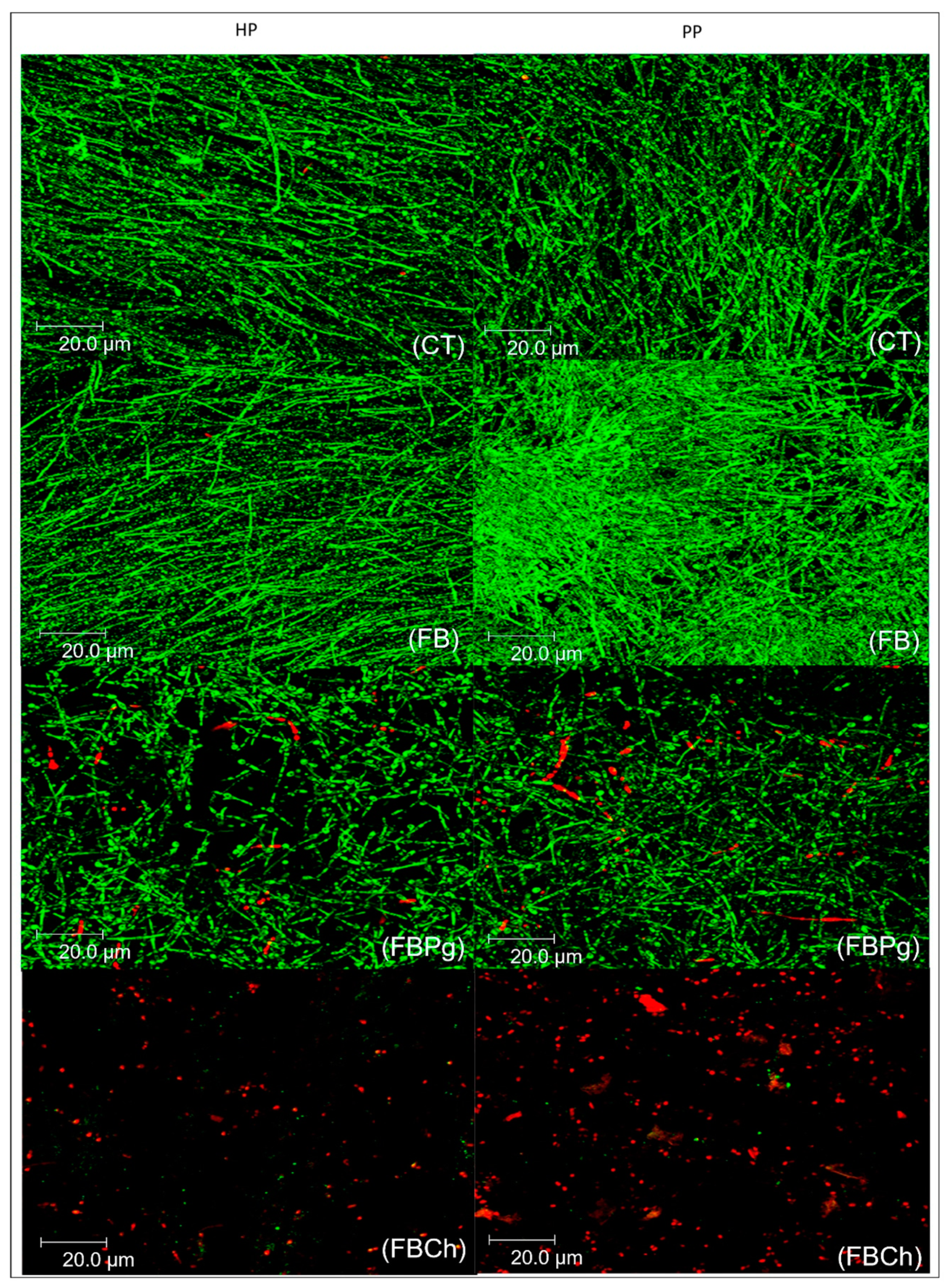
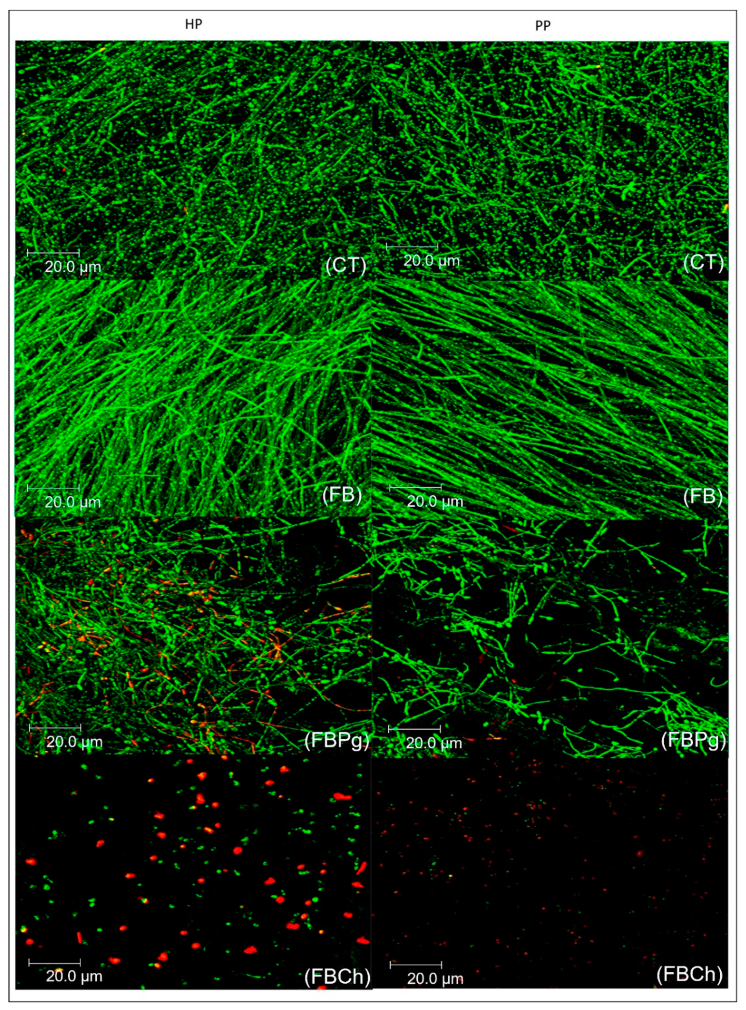
3.3. Qualitative Confocal Analysis

After 24 h of incubation, the control counterpart showed a higher proportion of uniform and elongated C. albicans yeast arranged in clusters (Figure 1). Similarly, at the same time point, the FB group was densely populated with uniform yeast (Figure 1). In contrast, growth suppression of C. albicans by FBCh (Figure 1) and FBPg (Figure 1) was seen with LIVE/DEAD staining, confirming the CFU and VC assay data.
After 48 h of incubation, in the control (Figure 2) and FB groups (Figure 2), a large amount of yeast and hyphae were evident, ratifying the quantitative CFU and VC findings. Some C. albicans blastopores and yeasts were noted in biofilms treated with FBPg (Figure 2), while sparsely distributed C. albicans blastopores and dead yeast were detected in the FBCh group (Figure 2).
After 72 h of incubation, the FB (Figure 3) and control groups (Figure 3) were highly densely populated with uniform yeast, pseudohyphae, and hyphae, similar to the results found in the CFU and VC assay. At the same time point, sparsely distributed C. albicans blastopores and a considerable amount of dead yeast were visible among scattered C. albicans in the FBCh group (Figure 3). Yeast and some hyphae of diffuse formats were evident in the group treated with FBPg (Figure 3).

4. Discussion

DS, the most common oral condition involving denture wearers, is mainly caused by the adherence of C. albicans to the porous and rough surfaces of denture base materials [7,8]. Coating the denture surface may alter these rough characteristics and prevent the colonization of this yeast. Strategies such as the incorporation of antimicrobials into the composition of coating materials can provide additional benefits in preventing the development of biofilms on the inner surface of removable dentures [42,43]. Thus, in the present study, we assessed whether the coating of fibrin biopolymer adhesive with P. granatum and digluconate chlorhexidine could affect the adhesion of C. albicans on the surface of two different PMMA resins.
To the best of our knowledge, no study has investigated fibrin biopolymer combined with antimicrobial agents nor its effectiveness against fungus as an alternative therapy for DS. The results showed that heat-treated and pre-polymerized resin specimens coated with fibrin biopolymer incorporated with chlorhexidine (FBCh) significantly reduced C. albicans biofilm formation and growth in all of the evaluated assays and periods. These findings are supported by Redding et al. [42] who evaluated chlorhexidine incorporated thin-film polymer on acrylic resin specimens after 24 h of incubation. In addition, Garaicoa et al. [44] showed the antifungal capacity of chlorhexidine incorporated in denture adhesives. Ellepola and Samaranayake [62] found that the chlorhexidine digluconate compound provoked rupture in the cell membrane yeast, even at low concentrations, and was a potent antifungal. Moreover, the antimicrobial efficacy of chlorhexidine is also associated with its substantivity, assuring its gradual release, and the promotion of its efficacy over a long period [63].
Although not as efficient as chlorhexidine, the incorporation of P. granatum in fibrin biopolymer (FBPg) also showed an inhibitory capacity on C. albicans biofilm at all periods and in both evaluated PMMA materials. Almeida et al. [2] also reported reduced biofilm development up to 12 h after biofilm induction in specimens treated with prosthetic adhesive with P. granatum incorporation. The antifungal effect of crude hydroalcoholic extract from P. granatum peel has been attributed to several structural compounds of the peel, specifically the punicalagin and ellagitannin derivatives [25,26,64]. These components are found in abundance in the crude hydroalcoholic extract of P. granatum peels, causing serious damage to the cellular structure of C. albicans yeast [26,27], possibly related to their molecular structure and toxicity, and astringent properties of tannins [45].
Surprisingly, and observed for the first time, FB favored biofilm development for at least 72 h in both quantitative tests. As far as we know, none of the studies before found these C. albicans-scaffold properties. Therefore, only indirect comparisons can be worthy of the previous discussion. Biofilm overgrowth could be related to the presence of abundant fibrin net—a special biological material—acting as a nutrient reservoir for C. albicans development on the resin surface [65]. However, it is noteworthy that this study proved the wet tolerance of FB and its efficiency as a drug delivery system for antimicrobials or antifungals. In this way, FBs incorporated with antimicrobials could be a sustainable alternative for the local prevention and management of DS. Moreover, previous studies have demonstrated the biocompatibility or absence of cytotoxicity of FB in human and animal cells [32,33,34]. It should be noted that for the first time, a biodegradable biological material (fibrin biopolymer) was innovatively applied together with antimicrobial-yeast candidate agents to prevent DS.
Among the two different PMMA materials, the CAD-CAM pre-polymerized resin presented the best inhibitory effect to the development of C. albicans biofilm. Recent studies have demonstrated that the surface of CAD-CAM specimens exhibit significantly less adherence to C. albicans than heat-cured specimens [11,14]. These results could be attributed to the surface roughness of the materials once the pre-polymerized PMMA presents a smoother surface compared to heat-cured resin [14,15].
Considering the time exposure evaluated in this investigation, all groups presented a significant overgrowth of C. albicans biofilm after 72 h, as observed in other Candida species-related studies [44]. The development of C. albicans biofilm proceeds in three developmental phases: early (0–11 h), intermediate (12–30 h), and maturation (38–72 h). In the present study, the overgrowth of C. albicans biofilm was detected in the mature phase of biofilm development, which is probably associated with its highly heterogeneous architecture and extracellular material [49], besides greater drug tolerance since mature biofilm starts to express resistance genes [3,59]. In addition, “persister” necrotic fungal cells in mature biofilm subjected to antifungal agents, which are subpopulations of cells highly tolerant of stress conditions, protected by the cell-matrix can repopulate the biofilm and interact and co-aggregate with other microorganisms present in the oral environment. This represents an important factor in its virulence [66,67].
In summary, the results of the present study suggested that fibrin biopolymer, a natural biological material, facilitates the growth of C. albicans, possibly due to the robust fibrin network. On the other hand, when this biological medicine was applied as a drug delivery system, associated with molecules that are candidates for antifungal agents, such as chlorhexidine or P. granatum used in this investigation, there was an important inhibition in dentures biofilm production. These outcomes suggest that the incorporation of P. granatum or digluconate chlorhexidine in a vehicle tolerant to a wet environment has the potential to prevent and treat DS by acting on the main etiological factor, C. albicans. However, among the limitations of this study, it comprised in vitro rather than in vivo tests, thus, these findings must be carefully applied to clinical conditions, since the adhesive will be subjected to routine hygiene and disinfection protocols performed by denture users, in addition to thermal and pH variations that may affect the effect and durability of this experimental product. Thus, further in vivo investigations on the anti-adherent potential against biofilm-associated with the denture base are needed to prove its effectiveness against DS, determine its longevity, and whether it causes any damage to the structure of dentures.

5. Conclusions

Within the limitations of this in vitro study and according to the data obtained, it was possible to conclude that, although fibrin biopolymer facilitated C. albicans growth, it proved to be effective when used as a drug delivery-system in combination with digluconate chlorhexidine or P. granatum, resulting in a reduction of fungal biofilm growth on the surface of two different PMMA base materials up to 72 h.

Author Contributions

Below is the description of credit roles of each author. H.S.V.: conceptualization, methodology, data curation, investigation, writing—original draft, writing—review and editing. A.P.C.-C.: data curation, investigation, formal analysis, software, writing—original draft, writing—review and editing. O.O.M.-T.: validation, methodology, data curation, writing—original draft. R.A.d.S.: conceptualization, methodology, data curation, investigation, writing—review and editing. R.M.B.d.C.: validation, formal analysis, software, writing—original draft. M.D.P.: data curation, investigation, writing—review and editing. B.B.: validation, methodology, resources, writing—review and editing. R.S.F.J.: conceptualization, methodology, visualization, resources writing—review and editing. V.S.L.: investigation, resources, formal analysis, software, writing—review and editing. H.M.H.: data curation (statistical analysis), formal analysis, review and editing. K.H.N.: validation, methodology, formal analysis, resources, writing—review and editing. V.C.P.: conceptualization, visualization, supervision, project administration, writing—review and editing. All authors have read and agreed to the published version of the manuscript.

Funding

This study was supported by São Paulo Research Foundation-FAPESP (Grant-2009/14152-1), by Coordination for the Improvement of Higher Education Personnel—CAPES (Finance Code 001) and National Council for Scientific and Technological Development—CNPq (H.S.V. and V.S.L. are fellow researchers—process number 88882.182729/2018-01 and 307603/2018-0, respectively). R.S.F.Jr. is a CNPq PQ1C fellow researcher [303224/2018-5]. B.B. is a CNPq PQ-2 fellow researcher [306339/2020-0].

Institutional Review Board Statement

Not applicable.

Informed Consent Statement

Not applicable.

Data Availability Statement

The data presented in this study are available on request from the corresponding author.

Conflicts of Interest

The authors declare no conflict of interest. The funders had no role in the design of the study; in the collection, analyses, or interpretation of data; in the writing of the manuscript; nor in the decision to publish the result.

References

  1. Távora, F.F.F.; Chocano, A.P.C.; Oliveira, D.G.; Pereira, J.R.; Almeida, R.S.; Neppelenborek, K.H.; Porto, V.C. Effects of Ethyl-Cyanoacrylate Coating Against Candida Albicans Biofilm Formation. Braz. Dent. J. 2019, 30, 266–271. [Google Scholar]
  2. Almeida, N.L.M.; Saldanha, L.L.; Da Silva, R.A.; Pinke, K.H.; Costa, E.F.; Porto, V.C.; Dokkedal, A.L.; Lara, V.S. Antimicrobial activity of denture adhesive associated with Equisetum giganteum- and Punica granatum-enriched fractions against Candida albicans biofilms on acrylic resin surfaces. Biofouling 2018, 34, 62–73. [Google Scholar] [CrossRef] [Green Version]
  3. Silva, S.; Negri, M.; Henriques, M.; Oliveira, R.; Williams, D.W.; Azeredo, J. Candida glabrata, Candida parapsilosis and Candida tropicalis: Biology, epidemiology, pathogenicity and antifungal resistance. FEMS Microbiol. Rev. 2012, 36, 288–305. [Google Scholar] [CrossRef] [Green Version]
  4. Acosta, L.D.; Pérez-Camacho, O.; Acosta, R.; Escobar, D.M.; Gallardo, C.A.; Sánchez-Vargas, L.O. Reduction of Candida albicans biofilm formation by coating polymethyl methacrylate denture bases with a photopolymerized film. J. Prosthet. Dent. 2019, 124, 605–613. [Google Scholar] [CrossRef]
  5. AlBin-Ameer, M.A.; Alsrheed, M.Y.; Aldukhi, I.A.; Matin, A.; Khan, S.Q.; Abualsaud, R.; Gad, M.M. Effect of Protective Coating on Surface Properties and Candida albicans Adhesion to Denture Base Materials. J. Prosthodont. 2020, 29, 80–86. [Google Scholar] [CrossRef]
  6. Spratt, D.A.; Pratten, J. Biofilms and the oral cavity. Rev. Environ. Sci. Biotechnol. 2003, 2, 109–120. [Google Scholar] [CrossRef]
  7. Ramage, G.; Martínez, J.P.; López-Ribot, J.L. Candida biofilms on implanted biomaterials: A clinically significant problem. FEMS Yeast Res. 2006, 6, 979–986. [Google Scholar] [CrossRef] [Green Version]
  8. Ramage, G.; Tomsett, K.; Wickes, B.L.; López-Ribot, J.L.; Redding, S.W. Denture stomatitis: A role for Candida biofilms. Oral Surg. Oral Med. Oral Pathol. Oral Radiol. Endod. 2004, 98, 53–59. [Google Scholar] [CrossRef] [PubMed]
  9. Pellon, A.; Sadeghi Nasab, S.D.; Moyes, D.L. New Insights in Candida albicans Innate Immunity at the Mucosa: Toxins, Epithelium, Metabolism, and Beyond. Front. Cell Infect. Microbiol. 2020, 10, 81. [Google Scholar] [CrossRef] [PubMed] [Green Version]
  10. Singh, S.; Palaskar, J.N.; Mittal, S. Comparative evaluation of surface porosities in conventional heat polymerized acrylic resin cured by water bath and microwave energy with microwavable acrylic resin cured by microwave energy. Contemp. Clin. Dent. 2013, 4, 147–151. [Google Scholar] [PubMed]
  11. Al-Fouzan, A.F.; Al-Mejrad, L.A.; Albarrag, A.M. Adherence of Candida to complete denture surfaces in vitro: A comparison of conventional and CAD/CAM complete dentures. J. Adv. Prosthodont. 2017, 9, 402–408. [Google Scholar] [CrossRef] [PubMed] [Green Version]
  12. Yoon, S.N.; Oh, K.C.; Lee, S.J.; Han, J.S.; Yoon, H.I. Tissue surface adaptation of CAD-CAM maxillary and mandibular complete denture bases manufactured by digital light processing: A clinical study. J. Prosthet. Dent. 2020, 124, 682–689. [Google Scholar] [CrossRef]
  13. Bidra, A.S.; Taylor, T.D.; Agar, J.R. Computer-aided technology for fabricating complete dentures: Systematic review of historical background, current status, and future perspectives. J. Prosthet. Dent. 2013, 109, 361–366. [Google Scholar] [CrossRef]
  14. Murat, S.; Alp, G.; Alatalı, C.; Uzun, M. In vitro Evaluation of Adhesion of Candida albicans on CAD/CAM PMMA-Based Polymers. J. Prosthodont. 2019, 28, e873–e879. [Google Scholar] [CrossRef]
  15. Al-Dwairi, Z.N.; Tahboub, K.Y.; Baba, N.Z.; Goodacre, C.J.; Özcan, M. A Comparison of the Surface Properties of CAD/CAM and Conventional Polymethylmethacrylate (PMMA). J. Prosthodont. 2019, 28, 452–457. [Google Scholar] [CrossRef] [PubMed] [Green Version]
  16. Steinmassl, P.A.; Wiedemair, V.; Huck, C.; Klaunzer, F.; Steinmassl, O.; Grunert, I.; Dumfahrt, H. Do CAD/CAM dentures really release less monomer than conventional dentures? Clin. Oral Investig. 2017, 21, 1697–1705. [Google Scholar] [CrossRef] [Green Version]
  17. Bollen, C.M.; Lambrechts, P.; Quirynen, M. Comparison of surface roughness of oral hard materials to the threshold surface roughness for bacterial plaque retention: A review of the literature. Dent. Mater. 1997, 13, 258–269. [Google Scholar] [CrossRef]
  18. Radford, D.R.; Sweet, S.P.; Challacombe, S.J.; Walter, J.D. Adherence of Candida albicans to denture-base materials with different surface finishes. J. Dent. 1998, 26, 577–583. [Google Scholar] [CrossRef]
  19. Vasconcelos, L.C.; Sampaio, M.C.; Sampaio, F.C.; Higino, J.S. Use of Punica granatum as an antifungal agent against candidosis associated with denture stomatitis. Mycoses 2003, 46, 192–196. [Google Scholar] [CrossRef]
  20. Polychronakis, N.; Polyzois, G.; Lagouvardos, P.; Andreopoulos, A.; Ngo, H.C. Long-term microwaving of denture base materials: Effects on dimensional, color and translucency stability. J. Appl. Oral Sci. 2018, 26, e20170536. [Google Scholar] [CrossRef]
  21. Gad, M.M.; Fouda, S.M. Current perspectives and the future of Candida albicans-associated denture stomatitis treatment. Dent. Med. Probl. 2020, 57, 95–102. [Google Scholar]
  22. Al-Thobity, A.M.; Gad, M.A.A.; Alnassar, T.; Al-Khalifa, K.S. Impact of denture cleansing solution immersion on some properties of different denture base materials: An in vitro study. J. Prosthodont. 2019, 28, 913–919. [Google Scholar] [CrossRef]
  23. Porwal, A.; Khandelwal, M.; Punia, V.; Sharma, V. Effect of denture cleansers on color stability, surface roughness, and hardness of different denture base resins. J. Indian. Prosthodont. Soc. 2017, 17, 61–67. [Google Scholar] [PubMed]
  24. Mansourian, A.; Boojarpour, N.; Ashnagar, S.; Momen Beitollahi, J.; Shamshiri, A.R. The comparative study of antifungal activity of Syzygium aromaticum, Punica granatum and nystatin on Candida albicans; an in vitro study. J. Mycol. Med. 2014, 24, e163–e168. [Google Scholar] [CrossRef]
  25. Endo, E.H.; Ueda-Nakamura, T.; Nakamura, C.V.; Filho, B.P. Activity of spray-dried microparticles containing pomegranate peel extract against Candida albicans. Molecules 2012, 17, 10094–10107. [Google Scholar] [CrossRef] [PubMed]
  26. Anibal, P.C.; Peixoto, I.T.; Foglio, M.A.; Hoflihg, J.F. Antifungal activity of the ethanolic extracts of Punica granatum L. and evaluation of the morphological and structural modifications of its compounds upon the cells of Candida spp. Braz. J. Microbiol. 2013, 44, 839–848. [Google Scholar] [CrossRef] [PubMed] [Green Version]
  27. Bakkiyaraj, D.; Nandhini, J.R.; Malathy, B.; Pandian, S.K. The anti-biofilm potential of pomegranate (Punica granatum L.) extract against human bacterial and fungal pathogens. Biofouling 2013, 29, 929–937. [Google Scholar] [CrossRef] [PubMed]
  28. Esawy, M.A.; Ragab, T.I.; Basha, M.; Emam, M. Evaluated bioactive component extracted from Punica granatum peel and its Ag NPs forms as mouthwash against dental plaque. Biocatal. Agric. Biotechnol. 2019, 18, 101073. [Google Scholar] [CrossRef]
  29. Ali, A.A.; Alharbi, F.A.; Suresh, C.S. Effectiveness of coating acrylic resin dentures on preventing Candida adhesion. J. Prosthodont. 2013, 22, 445–450. [Google Scholar] [CrossRef]
  30. Silva, M.J.; De Oliveira, D.G.; Marcillo, O.O.; Neppelenbroek, K.H.; Lara, V.S.; Porto, V.C. Effect of denture-coating composite on Candida albicans biofilm and surface degradation after disinfection protocol. Int. Dent. J. 2016, 66, 86–92. [Google Scholar] [CrossRef]
  31. Borie, E.; Rosas, E.; Kuramochi, G.; Etcheberry, S.; Olate, S.; Weber, B. Oral Applications of Cyanoacrylate Adhesives: A Literature Review. Biomed. Res. Int. 2019, 2019, 8217602. [Google Scholar] [CrossRef] [PubMed]
  32. Buchaim, D.V.; Cassaro, C.V.; Shindo, J.V.T.C.; Coletta, B.B.D.; Pomini, K.T.; Rosso, M.P.O.; Campos, L.M.G.; Ferreira, R.S., Jr.; Barraviera, B.; Buchaim, R.L. Unique heterologous fibrin biopolymer with hemostatic, adhesive, sealant, scaffold and drug delivery properties: A systematic review. J. Venom. Anim. Toxins Incl. Trop. Dis. 2019, 25, e20190038. [Google Scholar]
  33. Creste, C.F.Z.; Orsi, P.R.; Landim-Alvarenga, F.C.; Justulin, L.A.; Golim, M.A.; Barraviera, B.; Ferreira, R.S., Jr. Highly effective fibrin biopolymer scaffold for stem cells upgrading bone regeneration. Materials 2020, 13, 2747. [Google Scholar] [CrossRef]
  34. Abbade, L.P.F.; Ferreira, R.S., Jr.; Dos Santos, L.D.; Barraviera, B. Chronic venous ulcers: A review on treatment with fibrin sealant and prognostic advances using proteomic strategies. J. Venom. Anim. Toxins Incl. Trop. Dis. 2020, 26, e20190101. [Google Scholar] [CrossRef] [PubMed]
  35. Barbosa, M.D.; Stipp, A.C.; Passanezi, E.; Greghi, S.L. Fibrin adhesive derived from snake venom in periodontal surgery: Histological analysis. J. Appl. Oral Sci. 2008, 16, 310–315. [Google Scholar] [CrossRef] [Green Version]
  36. Frauz, K.; Teodoro, L.F.R.; Carneiro, G.D.; Cristina da Veiga, F.; Lopes Ferrucci, D.; Luis Bombeiro, A.; Waleska Simões, P.; Elvira Álvares, L.; Leite R de Oliveira, A.; Pontes Vicente, C.; et al. Transected Tendon Treated with a New Fibrin Sealant Alone or Associated with Adipose-Derived Stem Cells. Cells 2019, 8, 56. [Google Scholar] [CrossRef] [Green Version]
  37. Barros, L.C.; Ferreira, R.S., Jr.; Barraviera, S.R.C.S.; Stolf, H.O.; Thomazini-Santos, I.A.; Mendes-Giannini, M.J.S. A new fibrin sealant from crotalus durissus terrificus venom: Applications in medicine. J. Toxicol. Environ. Health B Crit. Ver. 2009, 12, 553–571. [Google Scholar]
  38. Ferreira, R.S., Jr.; De Barros, L.C.; Abbade, L.P.F.; Barraviera, S.R.C.S.; Silvares, M.R.C.; de Pontes, L.G.; Dos Santos, L.D.; Barraviera, B. Heterologous fibrin sealant derived from snake venom: From bench to bedside—An overview. J. Venom. Anim. Toxins Incl. Trop. Dis. 2017, 23, 21. [Google Scholar] [CrossRef] [Green Version]
  39. Orsi, P.R.; Landim-Alvarenga, F.C.; Justulin, L.A.; Kaneno, R.; De Assis Golilm, M.; Dos Santos, D.C.; Creste, C.F.Z.; Oba, E.; Maia, L.; Barraviera, B.; et al. A unique heterologous fibrin sealant (HFS) as a candidate biological scaffold for mesenchymal stem cells in osteoporotic rats. Stem Cell Res. Ther. 2017, 8, 1–14. [Google Scholar]
  40. Barbosa, M.D.S.; Gregh, S.L.A.; Passanezi, E. Fibrin adhesive derived from snake venom in periodontal surgery. J. Periodontol. 2007, 78, 2026–2031. [Google Scholar] [CrossRef] [PubMed]
  41. Chiquito, G.C.M. Comparison between suture and fibrin adhesive derived from snake venom for fixation of connective tissue graft in correction of marginal tissue recession. Venom. Anim. Toxins Incl. Trop. Dis. 2007, 13, 559. [Google Scholar]
  42. Redding, S.; Bhatt, B.; Rawls, H.R.; Siegel, G.; Scott, K.; Lopez-Ribot, J. Inhibition of Candida albicans biofilm formation on denture material. Oral Surg. Oral Med. Oral Pathol. Oral Radiol. Endod. 2009, 107, 669–672. [Google Scholar] [CrossRef]
  43. Garner, S.J.; Nobbs, A.H.; McNally, L.M.; Barbour, M.E. An antifungal coating for dental silicones composed of chlorhexidine nanoparticles. J. Dent. 2015, 43, 362–372. [Google Scholar] [CrossRef]
  44. Garaicoa, J.L.; Fischer, C.L.; Bates, A.M.; Holloway, J.; Avila-Ortiz, G.; Guthmiller, J.M.; Johnson, G.K.; Stanford, C.; Brogden, K.A. Promise of Combining Antifungal Agents in Denture Adhesives to Fight Candida Species Infections. J. Prosthodont. 2018, 27, 755–762. [Google Scholar] [CrossRef] [PubMed]
  45. Bassiri-Jahromi, S.; Katiraee, F.; Hajimahmoodi, M.; Mostafavi, E.; Talebi, M.; Pourshafie, M.R. In vitro antifungal activity of various Persian cultivars of Punica granatum L. extracts against Candida species. Jundishapur J. Nat. Pharm. Prod. 2015, 10, e19754. [Google Scholar] [CrossRef]
  46. Da Silva, R.A.; Bernardo, L.P.; Moreno, J.M.L.; Lara, V.S.; Porto, V.C. Equisetum giganteum influences the ability of Candida albicans in forming biofilms over the denture acrylic resin surface. Pharm. Biol. 2017, 55, 1698–1702. [Google Scholar] [CrossRef] [Green Version]
  47. Rahal, J.S.; Mesquita, M.F.; Henriques, G.E.; Nobilo, M.A. Surface roughness of acrylic resins submitted to mechanical and chemical polishing. J. Oral Rehabil. 2004, 31, 1075–1079. [Google Scholar] [CrossRef]
  48. Procópio, A.L.F.; Da Silva, R.A.; Maciel, J.G.; Sugio, C.Y.C.; Soares, S.; Urban, V.M.; Neppelenbroek, K.H. Antimicrobial and cytotoxic effects of denture base acrylic resin impregnated with cleaning agents after long-term immersion. Toxicol. In Vitro 2018, 52, 8–13. [Google Scholar] [CrossRef] [PubMed]
  49. Santos, L.; Oliveira, C.; Vasconcelos, B.M.; Vilela, D.; Melo, L.; Ambrósio, L.; Da Silva, A.; Murback, L.; Kurissio, J.; Cavalcante, J.; et al. Good management practices of venomous snakes in captivity to produce biological venom-based medicines: Achieving replicability and contributing to pharmaceutical industry. J. Toxicol. Environ. Health B Crit. Rev. 2021, 24, 30–50. [Google Scholar] [CrossRef] [PubMed]
  50. Barros, L.; Soares, A.; Costa, F.; Rodrigues, V.; Fuly, A.; Giglio, J.; Gallacci, M.; Thomazini-Santos, I.A.; Barraviera, S.R.C.S.; Barraviera, B.; et al. Biochemical and biological evaluation of gyroxin isolated from Crotalus durissus terrificus venom. J. Venom. Anim. Toxins Incl. Trop. Dis. 2011, 17, 23–33. [Google Scholar] [CrossRef] [Green Version]
  51. Abbade, L.P.; Barraviera, S.R.C.S.; Silvares, M.R.; Lima, A.B.; Haddad, G.R.; Gatti, M.A.; Medolago, N.B.; Rigotto Carneiro, M.T.; Dos Santos, L.D.; Ferreira, R.S., Jr.; et al. Treatment of chronic venous ulcers with heterologous fibrin sealant: A phase I/II clinical trial. Front. Immunol. 2021, 12, 627541. [Google Scholar] [CrossRef] [PubMed]
  52. Pontes, L.G.; Cavassan, N.R.V.; De Barros, L.C.; Ferreira, R.S., Jr.; Barraviera, B.; Dos Santos, L.D. Plasma proteome of buffaloes. Proteom. Clin. Appl. 2017, 11, 1600138. [Google Scholar] [CrossRef]
  53. Ferreira, R.S., Jr.; Da Silva, D.A.F.; Biscola, N.P.; Sartori, M.M.P.; Denadai, J.C.; Jorge, A.M.; Dos Santos, L.D.; Barraviera, B. Traceability of animal protein byproducts in ruminants by multivariate analysis of isotope ratio mass spectrometry to prevent transmission of prion diseases. J. Venom. Anim. Toxins Incl. Trop. Dis. 2019, 25, e148718. [Google Scholar] [CrossRef] [PubMed] [Green Version]
  54. Ferreira, R.S., Jr.; Barraviera, B. Arcabouço Tridimensional Para Células Tronco, Processo de Obtenção do Mesmo e Seu Uso. Nacional-BR 10 2014 011432 7, 8 May 2015. [Google Scholar]
  55. Ferreira, R.S., Jr.; Barraviera, B.; Barraviera, S.R.S. Selante de Fibrina Para Uso Tópico, Método de Formação do Mesmo e seu uso. Nacional-BR 1020140114360, 8 May 2015. [Google Scholar]
  56. Chandra, J.; Kuhn, D.M.; Mukherjee, P.K.; Hoyer, L.L.; McCormick, T.; Ghannoum, M.A. Biofilm formation by the fungal pathogen Candida albicans: Development, architecture, and drug resistance. J. Bacteriol. 2001, 183, 5385–5394. [Google Scholar] [CrossRef] [Green Version]
  57. Tobudic, S.; Lassnigg, A.; Kratzer, C.; Graninger, W.; Presterl, E. Antifungal activity of amphotericin B, caspofungin and posaconazole on Candida albicans biofilms in intermediate and mature development phases. Mycoses 2010, 53, 208–214. [Google Scholar] [CrossRef]
  58. Sanitá, P.V.; Vergani, C.E.; Giampaolo, E.T.; Pavarina, A.C.; Machado, A.L. Growth of Candida species on complete dentures: Effect of microwave disinfection. Mycoses 2009, 52, 154–160. [Google Scholar] [CrossRef] [PubMed]
  59. Monteiro, D.R.; Silva, S.; Negri, M.; Gorup, L.F.; Camargo, E.R.; Oliveira, R.; Barbosa, D.B.; Henriques, M. Silver nanoparticles: Influence of stabilizing agent and diameter on antifungal activity against Candida albicans and Candida glabrata biofilms. Lett. Appl. Microbiol. 2012, 54, 383–391. [Google Scholar] [CrossRef] [Green Version]
  60. Li, X.; Yan, Z.; Xu, J. Quantitative variation of biofilms among strains in natural populations of Candida albicans. Microbiology 2003, 149, 353–362. [Google Scholar] [CrossRef] [Green Version]
  61. Peeters, E.; Nelis, H.J.; Coenye, T. Comparison of multiple methods for quantification of microbial biofilms grown in microtiter plates. J. Microbiol. Methods 2008, 72, 157–165. [Google Scholar] [CrossRef] [Green Version]
  62. Ellepola, A.N.; Samaranayake, L.P. Oral candidal infections and antimycotics. Crit. Rev. Oral Biol. Med. 2000, 11, 172–198. [Google Scholar] [CrossRef] [Green Version]
  63. Spiechowicz, E.; Santarpia, R.P., 3rd; Pollock, J.J.; Renner, R.P. In vitro study on the inhibiting effect of different agents on the growth of Candida albicans on acrylic resin surfaces. Quintessence Int. 1990, 21, 35–40. [Google Scholar]
  64. García-Villalba, R.; Espín, J.C.; Aaby, K.; Alasalvar, C.; Heinonen, M.; Jacobs, G.; Voorspoels, S.; Koivumäki, T.; Kroon, P.A.; Pelvan, E.; et al. Validated Method for the Characterization and Quantification of Extractable and Nonextractable Ellagitannins after Acid Hydrolysis in Pomegranate Fruits, Juices, and Extracts. J. Agric. Food Chem. 2015, 63, 6555–6566. [Google Scholar] [CrossRef] [PubMed]
  65. Waheed, H.F.; Moin, S.; Choudhary, M.I. Snake venom: From deadly toxins to life-saving therapeutics. Curr. Med. Chem. 2017, 24, 1874–1891. [Google Scholar]
  66. Pereira-Cenci, T.; Deng, D.M.; Kraneveld, E.A.; Manders, E.M.; Del Bel Cury, A.A.; Ten Cate, J.M.; Crielaard, W. The effect of Streptococcus mutans and Candida glabrata on Candida albicans biofilms formed on different surfaces. Arch. Oral Biol. 2008, 53, 755–764. [Google Scholar] [CrossRef] [PubMed]
  67. Lewis, K. Riddle of biofilm resistance. Antimicrob. Agents Chemother. 2001, 45, 999–1007. [Google Scholar] [CrossRef] [Green Version]
Figure 1. Confocal images of C. albicans biofilms developed on heat-polymerized (HP) and pre-pol ymerized (PP) surfaces after 24 h of incubation. CT: control group; FB: Fibrin biopolymer; FBPg: P. granatum incorporated in fibrin biopolymer; FBCh: Chlorhexidine incorporated in fibrin biopolymer.
Materials 14 01618 g001
Figure 2. Confocal images of C. albicans biofilms developed on heat-polymerized (HP) and pre-polymerized (PP) surfaces after 48 h of incubation. CT: control; FB: Fibrin biopolymer; FBPg: P. granatum incorporated in fibrin biopolymer; FBCh: Chlorhexidine incorporated in fibrin biopolymer.
Materials 14 01618 g002
Figure 3. Confocal images of C. albicans biofilms developed on heat-polymerized (HP) and pre-polymerized (PP) surfaces after 72 h of incubation. CT: control group; FB: Fibrin biopolymer; FBPg: P. granatum incorporated in fibrin biopolymer; FBCh: Chlorhexidine incorporated in fibrin biopolymer.
Materials 14 01618 g003
Table 1. Mean values of colony forming unit (CFU)/mL (103) ± standard deviations (SD) in heat polymerized resin.
GroupsExperimental Periods Mean ± SD/Group
24 h48 h72 h
CT1304.44 ± 500.07 Aa1726.44 ± 386.45 Aa3144.44 ± 474.11 Ba2058.44 ± 453.55
FB2297.78 ± 613.55 Ab2557.78 ± 758.19 Ab3220 ± 424.15 Ba2691.85 ± 598.63
FBPg891.11 ± 178.36 Aa1277.78 ± 196.58 Ac2186.67 ± 220.45 Bb1451.85 ± 198.46
FBCh0.0 ± 0.0 c0.07 ± 0.2 d0.02 ± 0.07 c0.03 ± 0.09
Mean ± SD/Period1123.33 ± 322.99 (103)1390.52 ± 335.35 (103)2137.78 ± 279.69 (103)
CT: Control group; FB: Fibrin biopolymer; FBPg: P. granatum incorporated in fibrin biopolymer; FBCh: chlorhexidine incorporated in fibrin biopolymer. Horizontally, different capital letters indicate a statistical difference between the experimental periods for the same group (p < 0.05). Vertically, different lowercase letters indicate a statistical difference between the groups for the same period (p < 0.05).
Table 2. Mean values of CFU/mL (103) ± standard deviations (SD) in computer-aided design and computer-aided manufacturing (CAD-CAM) resin.
GroupsExperimental PeriodsMean ± SD/Group
24 h48 h72 h
CT1271.11 ± 315.73 Aa1504.44 ± 458.89 Aa2664.44 ± 402.71 Ba1813.33 ± 392.44
FB1842.22 ± 410.18 Ab1753.33 ± 597.99 Ab3044,44 ± 434.14 Ba2213.33 ± 480.77
FBPg888.89 ± 251.62 Aa1053.33 ± 190.79 Ac1753.33 ± 410.97 Bb1231.85 ± 284.46
FBCh0.02 ± 0.07 c0.02 ± 0.07 d0.09 ± 0.2 c0.04 ± 0.11
Mean ± SD/Period1000.56 ± 244.39 (103)1077.78 ± 311.94 (103)1865.58 ± 312.01 (103)
CT: Control group; FB: Fibrin biopolymer; FBPg: P. granatum incorporated in fibrin biopolymer; FBCh: chlorhexidine incorporated in fibrin biopolymer. Horizontally, different capital letters indicate a statistical difference between the experimental periods for the same group (p < 0.05). Vertically, different lowercase letters indicate a statistical difference between the groups for the same period (p < 0.05).
Table 3. Mean absorbance values (nm) ± standard deviations (SD) of the metabolic activity of the C. albicans biofilm for the adhesives tested on specimens made with conventional heat polymerized resin.
GroupsExperimental PeriodsMean ± SD/Group
24 h48 h72 h
CT1576.56 ± 850. 561897.67 ± 702.622763.444 ± 387.192079.23 ± 646.79 a
FB1879.78 ± 702.882003.44 ± 7762102.333 ± 1179.41995.18 ± 886.09 a
FBPg1153.44 ± 647.591470.56 ± 591.571598.333 ± 672.351407.44 ± 637.17 b
FBCh110 ± 2.74120.11 ± 4.76127.889 ± 5.71119.33 ± 4.4 c
Mean ± SD/Period1179.95 ± 550.94 A1372.95 ± 518.74 A1647.99 ± 561.16 B
CT: Control group; FB: Fibrin biopolymer; FBPg: P. granatum incorporated in fibrin biopolymer; FBCh: chlorhexidine incorporated in fibrin biopolymer. Horizontally, different capital letters indicate a statistical difference between the experimental periods for the same group (p < 0.05). Vertically, different lowercase letters indicate a statistical difference between the groups for the same period (p < 0.05).
Table 4. Average absorbance values (nm) ± standard deviations (SD) of the metabolic activity of the C. albicans biofilm for the adhesives tested on specimens made with CAD-CAM resin.
GroupsExperimental PeriodsMean ± SD/Group
24 h48 h72 h
CT1591.778 ± 583.751475.667 ± 612.72126.556 ± 1204.091731.33 ± 800.18 a
FB1925.889 ± 438.851777.667 ± 685.532643.333 ± 3402115.63 ± 488.13 a
FBPg1500.778 ± 177.551330.222 ± 520.121807.111 ± 976.891546.04 ± 558.19 b
FBCh136.222 ± 2.28126 ± 14.16141.333 ± 12.37134.52 ± 9.6 c
Mean ± SD/Period1288.67 ± 300.61 A1177.39 ± 458.13 A1679.58 ± 633.34 B
CT: Control group; FB: Fibrin biopolymer; FBPg: P. granatum incorporated in fibrin biopolymer; FBCh: chlorhexidine incorporated in fibrin biopolymer. Horizontally, different capital letters indicate a statistical difference between the experimental periods for the same group (p < 0.05). Vertically, different lowercase letters indicate a statistical difference between the groups for the same period (p < 0.05).
Publisher’s Note: MDPI stays neutral with regard to jurisdictional claims in published maps and institutional affiliations.

