
Analysis and Comparison of the Corrosive Behavior of Nickel-Based and Cobalt-Based Dental Alloys

 ,
,
Abstract
:1. Introduction
2. Materials and Methods
2.1. Materials, Specimens Preparation
2.2. Microstructural Characterization
2.3. Electrochemical Measurements
2.3.1. Potentiostatic Polarization Studies—Chronoamperometry
2.3.2. EIS—Electrochemical Impedance Spectroscopy
3. Results and Discussions
4. Conclusions
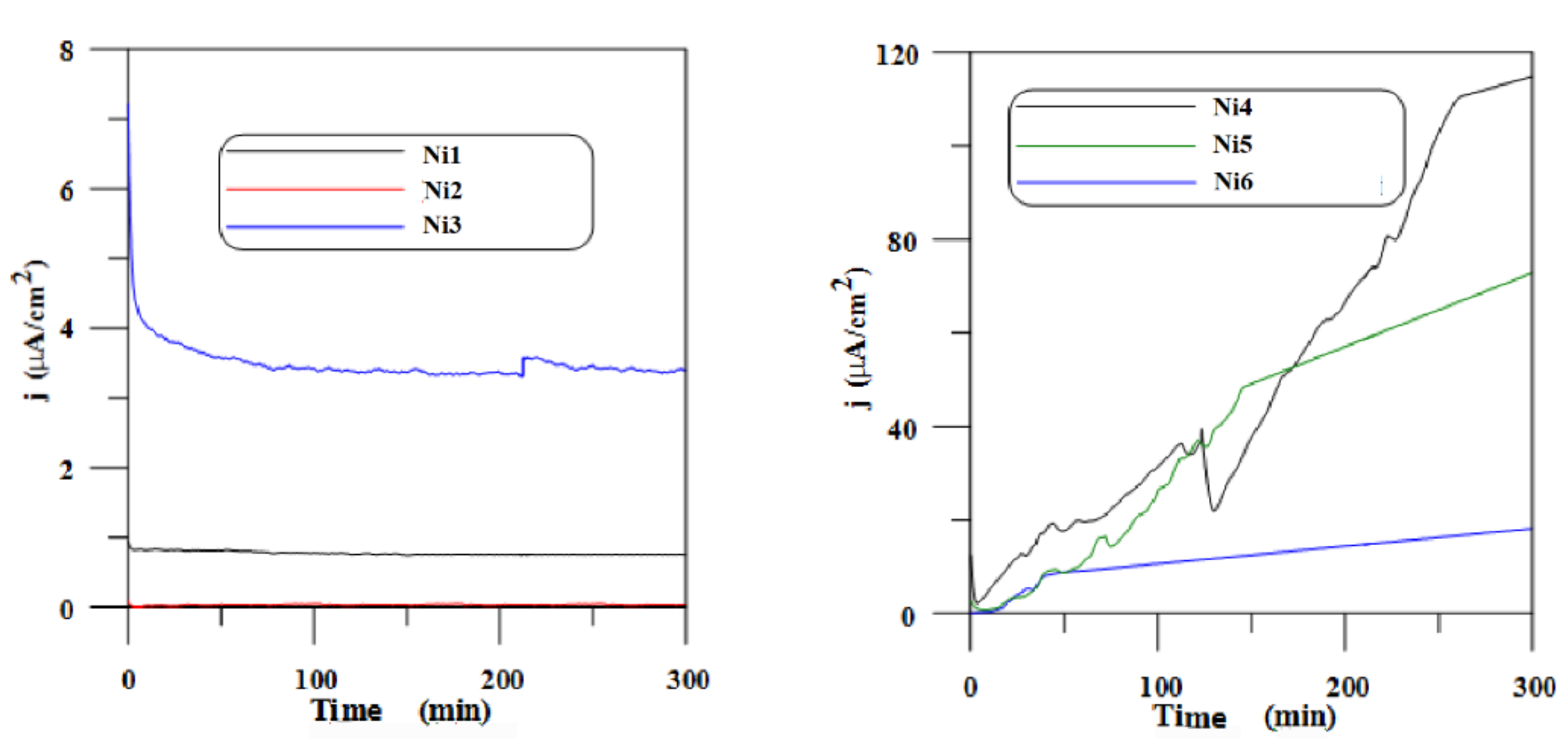
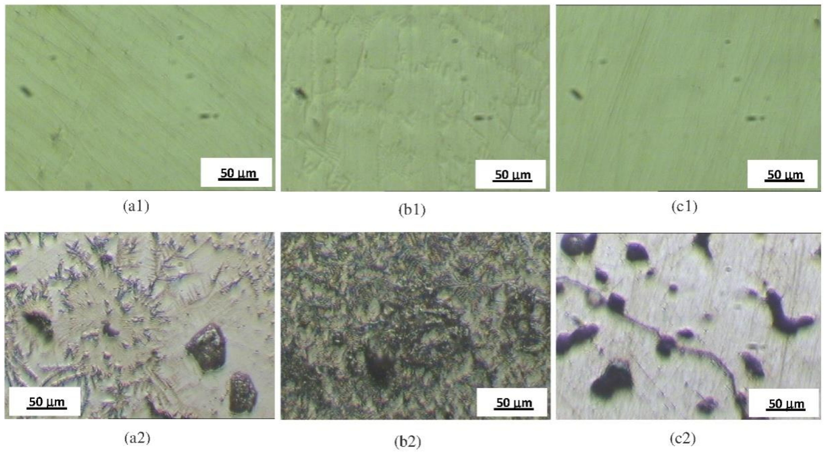
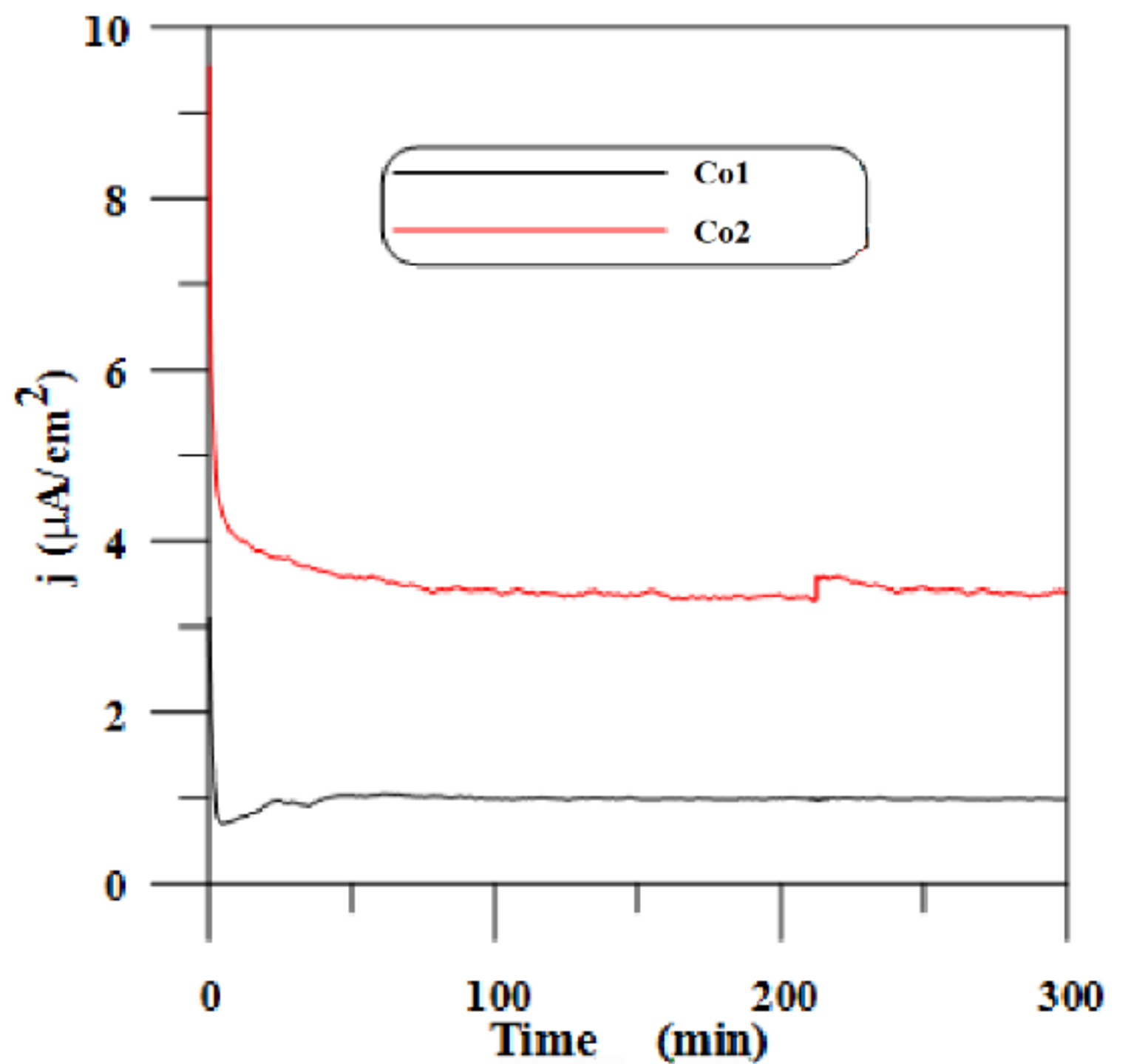
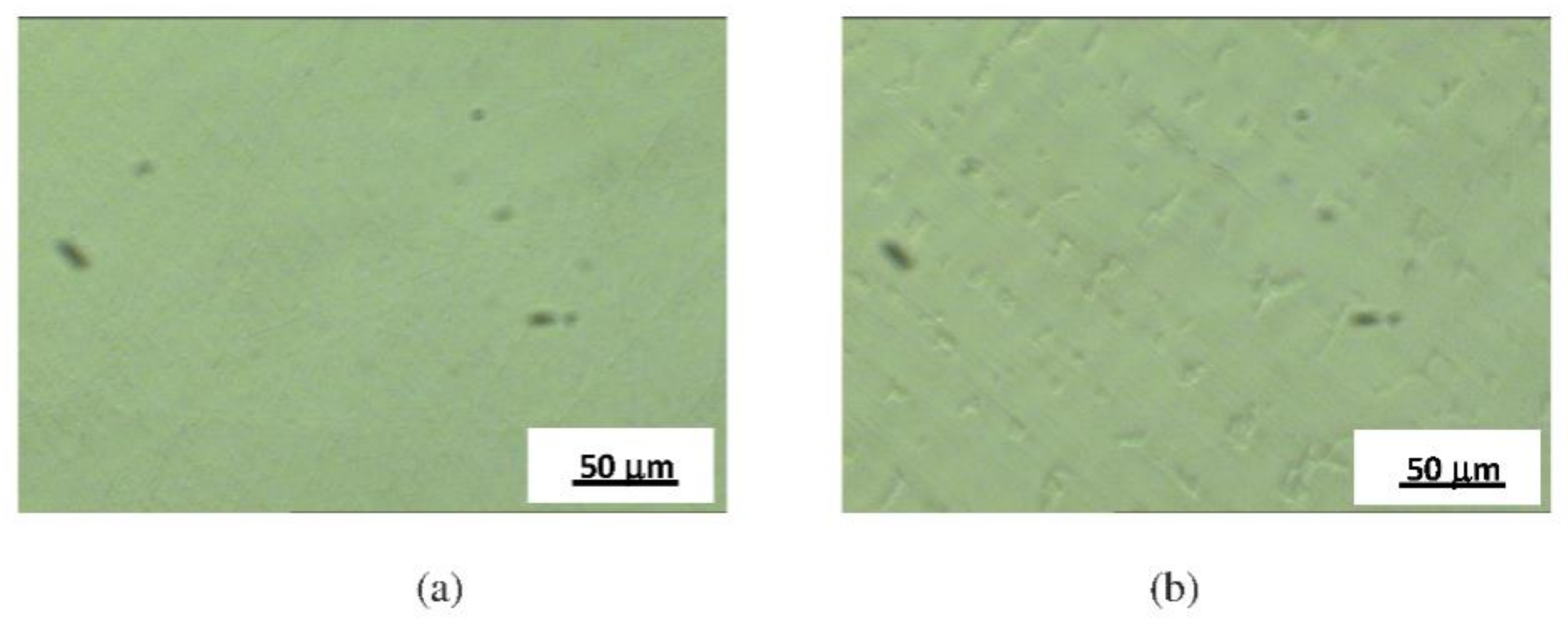
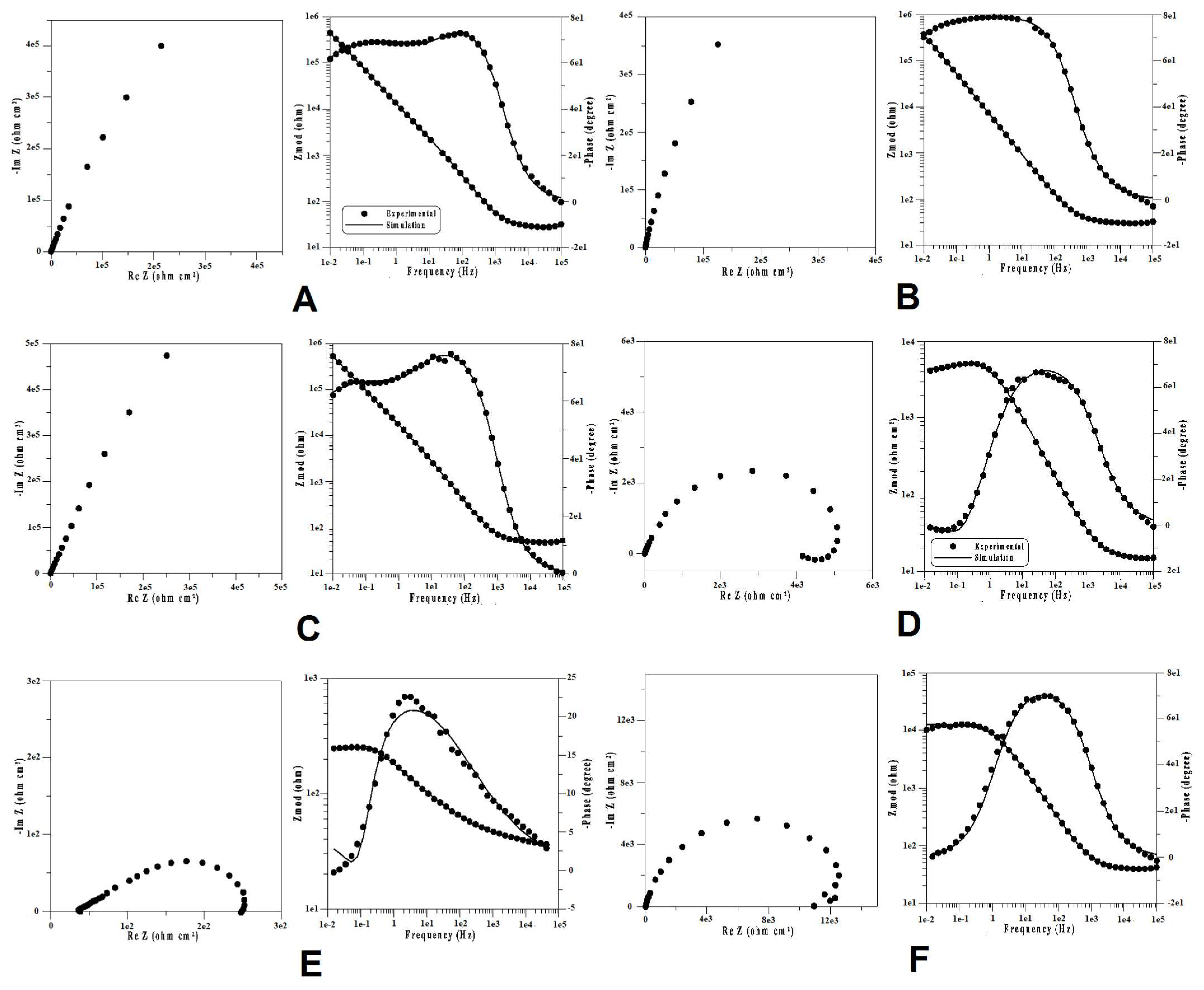
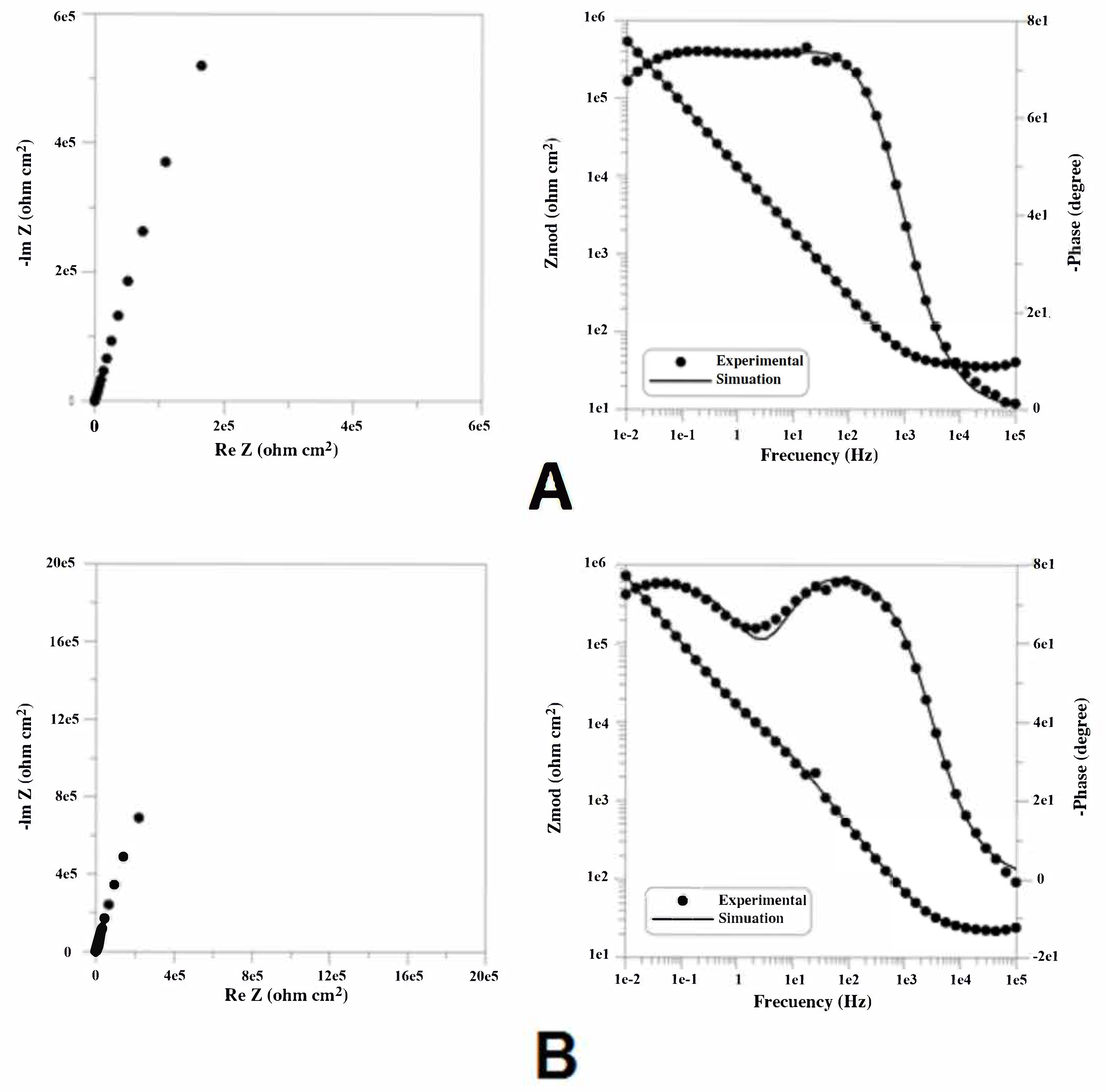
- The stability of the passive layer was not destroyed for the CoCr-based specimens Co1 and Co2, or the NiCr-based specimens Ni1, Ni2, and Ni3. This fact was confirmed by potentiostatic polarization curves and surface microscopy after polarization;
- In the cases of specimens Ni4, Ni5, and Ni6, it was found that the passive layer was destroyed after polarization. Therefore, there was no longer a protective passive layer on these alloys;
- Findings from the micrographs of the different NiCr and CoCr dental alloys studied after electrochemical treatments showed that there was no degradation for specimens Ni1, Ni2, Ni3, Co1, and Co2, but the development and growth of stable pits was discovered on the surfaces of specimens Ni4, Ni5, and Ni6;
- According to the results obtained, in terms of susceptibility to corrosion from the spectral data, the NiCr and CoCr dental alloys were divided in two different groups. A first group which included the two CoCr (Co1 and Co2) and three of the six NiCr alloys studied (Ni1, Ni2, and Ni3), where the polarization resistance showed high values. In this group, the most stable alloy was specimen Co2, with a polarization resistance in the order of 106 Ω cm2, characteristic of alloys highly resistant to corrosion. A second group with the other NiCr alloys investigated, Ni4, Ni5, and Ni6, where the passive layers were destroyed after polarization and the polarization resistance determinations were significantly lower than those exhibited by the first group. In this second group, specimen Ni5 had the highest stability and specimen Ni6 the lowest, based on polarization resistance values.
Author Contributions
Funding
Institutional Review Board Statement
Informed Consent Statement
Data Availability Statement
Conflicts of Interest
References
- Warlimont, H. Nickel and Nickel Alloys. In Springer Handbook of Materials Data; Warlimont, H., Martienssen, W., Eds.; Springer International Publishing: Cham, Switzerland, 2018. [Google Scholar]
- Moris, I.; Sakuma, M.; Faria, A.; Macedo, A.; Ribeiro, R.; Rodrigues, R. The dental alloys determine the choice of composite resins to be used. Braz. Dent. Sci. 2017, 20, 92–98. [Google Scholar] [CrossRef]
- Cha, M.S.; Huh, Y.H.; Cho, L.R.; Park, C.J. A comparative study of the wear of dental alloys against monolithic zirconia. J. Prosthet. Dent. 2020, 123, 866–873. [Google Scholar] [CrossRef]
- Chen, Q.; Thouas, G.A. Metallic implant biomaterials. Mater. Sci. Eng. R 2015, 87, 1–57. [Google Scholar] [CrossRef]
- Torabinejad, V.; Aliofkhazraei, M.; Assareh, S.; Allahyarzadeh, M.H.; SabourRouhaghdam, A. Electrodeposition of Ni-Fe alloys, composites, and nano coatings—A review. J. Alloys Compd. 2017, 691, 841–859. [Google Scholar] [CrossRef]
- Musa, A.Y.; Behazin, M.; Wren, J.C. Potentiostatic oxide growth kinetics on Ni–Cr and Co–Cr alloys. Potential and pH dependences. Electrochim. Acta 2015, 162, 185–197. [Google Scholar] [CrossRef]
- Kuznetsov, V.V.; Filatova, E.A.; Telezhkina, A.V.; Kruglikov, S.S. Corrosion resistance of Co−Cr−W coatings obtained by electrodeposition. J. Solid State Electrochem. 2018, 22, 2267–2276. [Google Scholar] [CrossRef]
- Chen, Y.; Li, Y.; Kurosu, S.; Meng, Q.; Tang, N.; Koizumi, Y.; Chiba, A. Analysis of in vitro wear behaviour and contact mechanisms of metal-on-metal hip joint bearings with different radial clearances. Mater. Trans. 2015, 56, 826–834. [Google Scholar] [CrossRef] [Green Version]
- Hornez, J.C.; Lefèvre, A.; Joly, D.; Hildebrand, H.F. Multiple parameter cytotoxicity index on dental alloys and pure metals. Biomol. Eng. 2002, 19, 103–117. [Google Scholar] [CrossRef]
- Messer, R.L.; Lucas, L.C. Cytotoxic evaluation of ions released from nickel-chromium dental alloys. J. Dent. Res. 1996, 75, 255. [Google Scholar]
- Messer, R.L.; Lucas, L.C. Evaluation of metabolic activities as biocompatibility tools: A study of individual ions’ effects on fibroblasts. Dent. Mater. 1999, 15, 1–6. [Google Scholar] [CrossRef]
- Elshahawy, W.M.; Watanabe, I.; Kramer, P. In vitro cytotoxicity evaluation of elemental ions released from different prosthodontic materials. Dent. Mater. 2009, 25, 1551–1555. [Google Scholar] [CrossRef]
- Nelson, S.K.; Wataha, J.C.; Lockwood, P.E. Accelerated toxicity testing of casting alloys and reduction of intraoral release of elements. J. Prosthet. Dent. 1999, 81, 715–720. [Google Scholar] [CrossRef]
- Alp, G.; Çakmak, G.; Sert, M.; Burgaz, Y. Corrosion potential in artificial saliva and possible genotoxic and cytotoxic damage in buccal epithelial cells of patients who underwent Ni-Cr based porcelain-fused-to-metal fixed dental prostheses. Mutat. Res. Genet. Toxicol. Environ. Mutagen 2018, 827, 19–26. [Google Scholar] [CrossRef] [PubMed]
- Craig, R.G.; Hanks, C.T. Reaction of fibroblasts to various dental casting alloys. J. Oral Pathol. 1988, 17, 341–347. [Google Scholar] [CrossRef] [PubMed] [Green Version]
- Craig, R.G.; Hanks, C.T. Cytotoxicity of experimental casting alloys evaluated by cell culture tests. J. Dent. Res. 1990, 69, 1539–1542. [Google Scholar] [CrossRef] [Green Version]
- Friend, W.Z. Corrosion of Nickel and Nickel-Based Alloys; John Wiley and Sons: New York, NY, USA, 1980. [Google Scholar]
- Wylie, C.M.; Shelton, R.M.; Fleming, G.J.P.; Davenport, A.J. Corrosion of nickel-based dental casting alloys. Dent. Mater. 2007, 23, 714–723. [Google Scholar] [CrossRef] [PubMed]
- Moslehifard, E.; Ghasemzadeh, S.; Nasirpouri, F. Influence of pH level of artificial saliva on corrosion behavior and nickel ion release of a Ni-Cr-Mo alloy: An in vitro study. Anti-Corros. Methods Mater. 2019, 66, 746–756. [Google Scholar] [CrossRef]
- Pan, Y.; Lin, Y.; Jiang, L.; Lin, H.; Xu, C.; Lin, D.; Cheng, H. Removal of dental alloys and titanium attenuates trace metals and biological effects on liver and kidney. Chemosphere 2020, 243, 125205. [Google Scholar] [CrossRef]
- Rodrigues, W.C.; Broilo, L.R.; Schaeffer, L.; Knörnschild, G.; Espinoza, F.R.M. Powder metallurgical processing of Co–28%Cr–6%Mo for dental implants: Physical, mechanical and electrochemical properties. Powder Technol. 2011, 206, 233–238. [Google Scholar] [CrossRef]
- Xiao, M.; Chen, Y.M.; Biao, M.N.; Zhang, X.D.; Yang, B.C. Bio-functionalization of biomedical metals. Mater. Sci. Eng. C 2017, 70 Pt 2, 1057–1070. [Google Scholar] [CrossRef]
- Gurappa, I. Development of appropriate thickness ceramic coatings on 316 L stainless steel for biomedical applications. Surf. Coat. Technol. 2002, 161, 70–78. [Google Scholar] [CrossRef]
- Hamidi, M.F.F.A.; Harun, W.S.W.; Samykano, M.; Ghani, S.A.C.; Ghazalli, Z.; Ahmadd, F.; Sulong, A.B. A review of biocompatible metal injection moulding process parameters for biomedical applications. Mater. Sci. Eng. C 2017, 78, 1263–1276. [Google Scholar] [CrossRef] [Green Version]
- Chen, Y.; Li, Y.; Kurosu, S.; Yamanaka, K.; Tang, N.; Chiba, A. Effects of microstructures on the sliding behavior of hot-pressed CoCrMo alloys. Wear 2014, 319, 200–210. [Google Scholar] [CrossRef]
- Chenakin, S.P.; Filatova, V.S.; Makeeva, I.N.; Vasylyev, M.A. Ultrasonic impact treatment of CoCrMo alloy: Surface composition and properties. Appl. Surf. Sci. 2017, 408, 11–20. [Google Scholar] [CrossRef]
- Gong, X.; Li, Y.; Nie, Y.; Huang, Z.; Liu, F.; Huang, L.; Jiang, L.; Mei, H. Corrosion behaviour of CoCrMo alloy fabricated by electron beam melting. Corros. Sci. 2018, 139, 68–75. [Google Scholar] [CrossRef]
- Qian, C.; Wu, X.; Zhang, F.; Yu, W. Electrochemical impedance investigation of Ni-free Co-Cr-Mo and Co-Cr-Mo-Ni dental casting alloy for partial removable dental prosthesis frameworks. J. Prosthet. Dent. 2016, 116, 112–118. [Google Scholar] [CrossRef]
- Henriques, B.; Bagheri, A.; Gasik, M.; Souza, J.C.M.; Carvalho, O.; Silva, F.S.; Nascimento, R.M. Mechanical properties of hot pressed CoCrMo alloy compacts for biomedical applications. Mater. Design 2015, 83, 829–834. [Google Scholar] [CrossRef]
- Park, W.U.; Park, H.G.; Hwang, K.H.; Zhao, J.; Lee, J.K. Interfacial Property of Dental Cobalt–Chromium Alloys and Their Bonding Strength with Porcelains. J. Nanosci. Nanotechnol. 2017, 17, 2585–2588. [Google Scholar] [CrossRef]
- Ramírez-Ledesma, A.L.; Roncagliolo, P.; Álvarez-Pérez, M.A.; Lopez, H.F.; Juárez-Islas, J.A. Corrosion Assessment of an Implantable Dental Co-Cr Alloy in Artificial Saliva and Biocompatibility Behavior. J. Mater. Eng. Perform. 2020, 29, 1657–1670. [Google Scholar] [CrossRef]
- Lucchetti, M.C.; Fratto, G.; Valeriani, F.; De Vittori, E.; Giampaoli, S.; Papetti, P.; Spica, V.R.; Manzon, L. Cobalt-chromium alloys in dentistry: An evaluation of metal ion release. J. Prosthet. Dent. 2015, 114, 602–608. [Google Scholar] [CrossRef] [PubMed]
- de Freitas, B.X.; Nunes, C.A.; dos Santos, C. Sintering behaviour of Co-28%Cr-6%Mo compacted blocks for dental prosthesis. J. Mater. Res. Technol. 2019, 8, 2052–2062. [Google Scholar] [CrossRef]
- Alharbi, N.; Wismeijer, D.; Osman, R.B. Additive manufacturing techniques in prosthodontics: Where do we currently stand? A critical review. Int. J. Prosthodont. 2017, 30, 474–484. [Google Scholar] [CrossRef] [PubMed]
- Svanborg, P.; Hjalmarsson, L.A. systematic review on the accuracy of manufacturing techniques for cobalt chromium fixed dental prostheses. Biomater. Investig. Dent. 2020, 7, 31–40. [Google Scholar] [CrossRef] [PubMed]
- Kono, H.; Kikuchi, M. Analysis of orthodontic wire springback to simplify wire bending. Orthod. Waves 2020, 79, 57–63. [Google Scholar] [CrossRef]
- Hanawa, T. Novel Structured Metallic and Inorganic Materials; Springer: Singapore, 2019. [Google Scholar]
- Park, J.; Lee, H.; Kang, S.; Kim, J.; Kim, J. Effect of core materials for core fabrication for dental implants on in-vitro cytocompatibility of MC3T3-E1 cells. BMC Oral Health 2019, 19, 284. [Google Scholar] [CrossRef] [Green Version]
- Bilgin, M.S.; Erdem, A.; Dilber, E.; Ersoy, İ. Comparison of fracture resistance between cast, CAD/CAM milling, and direct metal laser sintering metal post systems. J. Prosthodont. Res. 2016, 60, 23–28. [Google Scholar] [CrossRef]
- Xia, Y.; Zhao, J.; Dong, Z.; Guo, X.; Tian, Q.; Liu, Y. A Novel Method for Making Co-Cr-Mo Alloy Spherical Powder by Granulation and Sintering. JOM J. Miner. Met. Mater. Soc. 2020, 72, 1279–1285. [Google Scholar] [CrossRef]
- Wataha, J.C.; Messer, R.L. Casting alloys. Dent. Clin. N. Am. 2004, 48, 499–512. [Google Scholar] [CrossRef]
- Roberts, H.W.; Berzins, D.W.; Moore, B.K.; Charlton, D.G. Metal–ceramic alloys in dentistry: A review. J. Prosthodont. 2009, 18, 188–194. [Google Scholar] [CrossRef]
- Yang, Y.; Lu, C.; Shen, L.; Zhao, Z.; Peng, S.; Shuai, C. In-situ deposition of apatite layer to protect Mg-based composite fabricated via laser additive manufacturing. J. Magnes. Alloy. 2021; in press. [Google Scholar] [CrossRef]
- Vyas, R.; Issaid, M.A.; Idris, B.A. Biocompatibility and corrosive resistance. Cairo Dent. J. 2009, 25, 361–365. [Google Scholar]
- Rupp, F.; Liang, L.; Geis-Gerstorfer, J.; Scheideler, L.; Hüttig, F. Surface characteristics of dental implants. Dent. Mater. 2018, 34, 40–57. [Google Scholar] [CrossRef]
- Manam, N.S.; Harun, W.S.W.; Shri, D.N.A.; Ghani, S.A.C.; Kurniawan, T.; Ismail, M.H.; Ibrahim, M.H.I. Study of corrosion in biocompatible metals for implants: A review. J. Alloys Compd. 2017, 701, 698–715. [Google Scholar] [CrossRef] [Green Version]
- Mareci, D.; Ungureanu, G.; Aelenei, N.; Chelariu, R.; Mirza-Rosca, J.C. EIS diagnosis of some dental alloys in artificial saliva. Environ. Eng. Manag. J. 2007, 6, 313–317. [Google Scholar] [CrossRef]
- Mareci, D.; Ungureanu, G.; Aelenei, N.; Mirza-Rosca, J.C. Comparative corrosion study of non-precious Ni/Cr-based soft alloys in view of dental applications. Environ. Eng. Manag. J. 2008, 7, 41–49. [Google Scholar] [CrossRef]
- Mareci, D.; Sutiman, D.; Cailean, A.; Bolat, G. Comparative corrosion study of Ag-Pd and Co-Cr alloys used in dental applications. Bull. Mater. Sci. 2010, 33, 491–500. [Google Scholar] [CrossRef] [Green Version]
- Garcia-Falcon, C.M.; Gil-Lopez, T.; Verdu-Vazquez, A.; Mirza-Rosca, J.C. Electrochemical characterization of some cobalt base alloys in Ringer solution. Mater. Chem. Phys. 2021, 260, 124164. [Google Scholar] [CrossRef]
- Geru, N.; Bane, M.; Gurgu, C. Analiza Structurii Materialelor Metalice (The Analysis of the Structure of Metallic Materials); Editura Tehnica: Bucharest, Romania, 1991. [Google Scholar]
- Rondelli, G.; Vicentini, B. Effect of copper on the localized corrosion resistance of Ni-Ti shape memory alloy. Biomaterials 2002, 23, 639–644. [Google Scholar] [CrossRef]
- Black, J. Biological Performance of Materials: Fundamentals of Biocompatibility; CRC Press: Boca Raton, FL, USA, 2006. [Google Scholar]
- Newman, R.C.; Mehta, A. An ac impedance study of the de-alloying of Fe Ni alloys, and its relevance to chloride scc of stainless steels. Corros. Sci. 1998, 28, 1183–1187. [Google Scholar] [CrossRef]
- Turdean, G.L.; Craciun, A.; Popa, D.; Constantiniuc, M. Study of electrochemical corrosion of biocompatible Co–Cr and Ni–Cr dental alloys in artificial saliva. Influence of pH of the solution. Mater. Chem. Phys. 2019, 233, 390–398. [Google Scholar] [CrossRef]
- Meticos-Hukovic, M.; Pilic, Z.; Babic, R.; Omanovic, D. Influence of alloying elements on the corrosion stability of CoCrMo implant alloy in Hank’s solution. Acta Biomater. 2006, 2, 693–700. [Google Scholar] [CrossRef]
- Vander Voort, G.F. ASM Handbook Volume 9: Metallography and Microstructures; ASM International: Materials Park, OH, USA, 2004. [Google Scholar]
- Jones, D.A. Principles and Prevention of Corrosion; Harlow Pearson Education: Harlow, UK, 2014. [Google Scholar]
- Huang, H.H. Electrochemical impedance spectroscopy study of strained titanium in fluoride media. Electrochim. Acta 2002, 47, 2311–2318. [Google Scholar] [CrossRef]
- Pan, J.; Thierry, D.; Leygraf, C. Electrochemical impedance spectroscopy study of the passive oxide film on titanium for implant application. Electrochim. Acta 1996, 41, 1143–1153. [Google Scholar] [CrossRef]


| Composition (in wt.%) | Specimens | |||||
|---|---|---|---|---|---|---|
| Ni1 | Ni2 | Ni3 | Ni4 | Ni5 | Ni6 | |
| Ni | 60.1 | 60.8 | 63.4 | 72.1 | 64.9 | 53.4 |
| Cr | 24.3 | 23.9 | 23.2 | 20 | 17.9 | 14.4 |
| Mo | 10.1 | 8.8 | 3 | |||
| Fe | 2.1 | 2.4 | 9 | 7.5 | ||
| Nb | 1 | 3.8 | ||||
| Si | 1 | 1.8 | 1.5 | |||
| Cu | 9.9 | 9.5 | ||||
| Mn | 2 | 3.6 | 19.4 | |||
| Al | 1.5 | 1.6 | ||||
| Composition (in wt.%) | Specimens | |
|---|---|---|
| Co1 | Co2 | |
| Co | 63.5 | 63.4 |
| Cr | 27 | 29.0 |
| Mo | 5.5 | 5.2 |
| Fe | 2 | |
| Ni | 1 | |
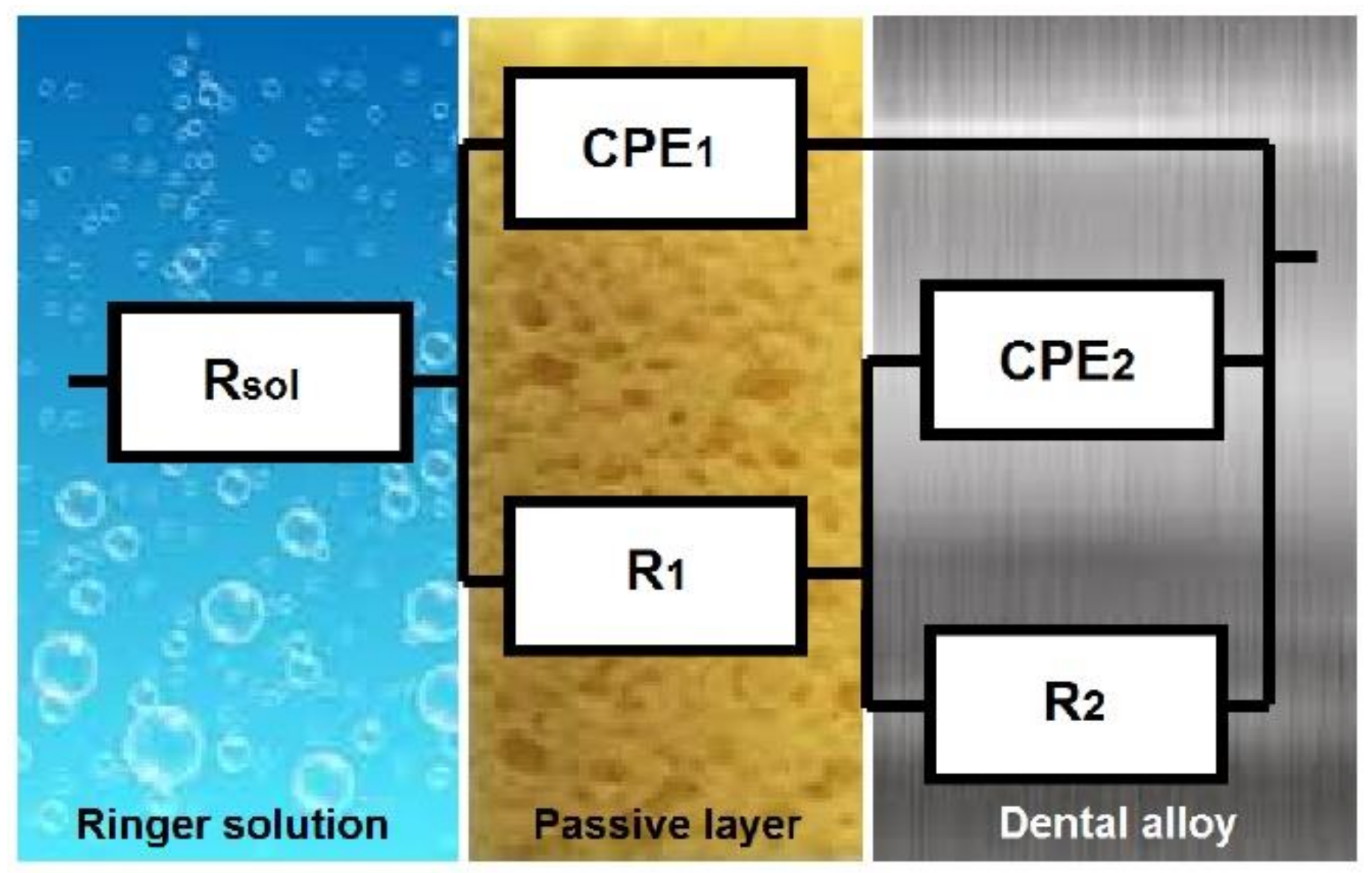
| Specimens | Rsol Ω cm2 | R1 Ω cm2 | Y01 Scm−2sn | n1 | R2 Ω cm2 | Y02 Scm−2sn | n2 | χ2 |
|---|---|---|---|---|---|---|---|---|
| Ni1 | 28 | 5 × 10³ | 8.9 × 10⁻⁶ | 0.83 | 5.5 × 10⁵ | 9.7 × 10⁻⁶ | 0.8 | 2 × 10⁻⁴ |
| Ni2 | 35 | 3 × 10³ | 1.9 × 10⁻⁵ | 0.9 | 6.2 × 10⁵ | 1 × 10⁻⁵ | 0.88 | 4 × 10⁻⁴ |
| Ni3 | 49 | 1.5 × 10⁴ | 8.4 × 10⁻⁶ | 0.89 | 5.9 × 10⁵ | 7.1 × 10⁻⁶ | 0.82 | 5 × 10⁻⁴ |
| Co1 | 37 | 1.5 × 10⁴ | 7.8 × 10⁻⁶ | 0.9 | 9.1 × 10⁵ | 8.3 × 10⁻⁶ | 0.83 | 2 × 10⁻⁴ |
| Co2 | 55 | 1.4 × 10⁴ | 6.1 × 10⁻⁶ | 0.9 | 1.2 × 10⁶ | 8.6 × 10⁻⁶ | 0.83 | 6 × 10⁻⁴ |
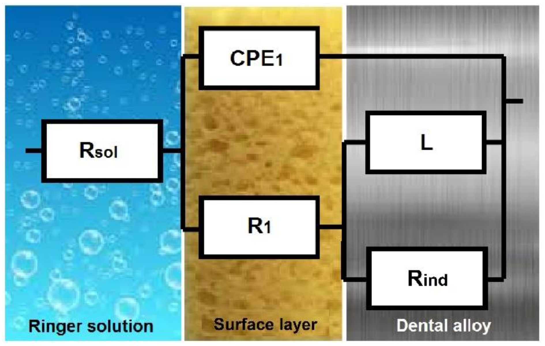
| Specimens | Rsol Ω cm2 | R1 Ω cm2 | Y01 Scm−2sn | n1 | Rind Ω cm2 | L Henri cm2 | χ2 |
|---|---|---|---|---|---|---|---|
| Ni4 | 20 | 4.5 × 10³ | 3.1 × 10⁵ | 0.8 | 1.7 × 10³ | 1.4 × 10³ | 8 × 10⁻⁴ |
| Ni5 | 38 | 1.1 × 10⁴ | 1.5 × 10⁻⁵ | 0.84 | 9 × 10² | 3 × 10³ | 6 × 10⁻⁴ |
| Ni6 | 34 | 260 | 2.4 10−3 | 0.4 | 240 | 231 | 6 × 10⁻⁴ |
Publisher’s Note: MDPI stays neutral with regard to jurisdictional claims in published maps and institutional affiliations. |
© 2021 by the authors. Licensee MDPI, Basel, Switzerland. This article is an open access article distributed under the terms and conditions of the Creative Commons Attribution (CC BY) license (https://creativecommons.org/licenses/by/4.0/).
Share and Cite
Garcia-Falcon, C.M.; Gil-Lopez, T.; Verdu-Vazquez, A.; Mirza-Rosca, J.C. Analysis and Comparison of the Corrosive Behavior of Nickel-Based and Cobalt-Based Dental Alloys. Materials 2021, 14, 4949. https://0-doi-org.brum.beds.ac.uk/10.3390/ma14174949
Garcia-Falcon CM, Gil-Lopez T, Verdu-Vazquez A, Mirza-Rosca JC. Analysis and Comparison of the Corrosive Behavior of Nickel-Based and Cobalt-Based Dental Alloys. Materials. 2021; 14(17):4949. https://0-doi-org.brum.beds.ac.uk/10.3390/ma14174949
Chicago/Turabian StyleGarcia-Falcon, Carmen Marina, Tomas Gil-Lopez, Amparo Verdu-Vazquez, and Julia Claudia Mirza-Rosca. 2021. "Analysis and Comparison of the Corrosive Behavior of Nickel-Based and Cobalt-Based Dental Alloys" Materials 14, no. 17: 4949. https://0-doi-org.brum.beds.ac.uk/10.3390/ma14174949