Article

Synthesis, NMR Characterization, and Antileukemic Activity of N-Nonanoylpiperazinyl-5α-Androstane-3α,17β-Diol A-Ring Derivatives

1
Laboratory of Medicinal Chemistry, Endocrinology and Nephrology Unit, CHU de Québec—Research Center, Québec, QC G1V 4G2, Canada
2
Department of Molecular Medicine, Faculty of Medicine, Université Laval, Québec, QC G1V 0A6, Canada
*
Author to whom correspondence should be addressed.
Submission received: 28 November 2020 / Revised: 19 December 2020 / Accepted: 21 December 2020 / Published: 23 December 2020
(This article belongs to the Section Magnetic Resonances)

Abstract

:
The combination of an androstane-3,17-diol nucleus and a 2β-N-alkylamidopiperazino sidechain is important for the anticancer activity of a new family of steroid derivatives. As the structure-activity relationship studies have so far been limited to the beta orientation of the substituent at position 2 of the steroid nucleus, a series of analogs (compounds 14) were synthesized to investigate the impact on biological activity of A-ring substitution. Nuclear magnetic resonance (NMR) analysis, especially using a series of 2D experiments, such as correlation spectroscopy (COSY), homonuclear Overhauser effect spectroscopy (NOESY), heteronuclear single-quantum correlation (HSQC), and heteronuclear multiple-bond correlation (HMBC) provided crucial information that was found essential in confirming the sidechain position and orientation of compounds 14. Assessment of their antiproliferative activity on leukemia HL-60 cells confirmed the best efficiency of the 2β-sidechain/3α-OH orientation (compound 1) compared to the other configurations tested (compounds 24).

Graphical Abstract

1. Introduction

Steroid derivatives with a N-substituted piperazino sidechain at position C2β of 5α-androstane-3α,17β-diol showed antiproliferative activity on different cancer cell lines [1,2,3,4,5,6] and promising results were obtained with two representative candidates tested in mouse xenograft tumor models [6,7,8]. They were designed by combining two crucial elements: A steroid core and a lateral piperazino sidechain in A-ring (Figure 1A). Structure-activity relationship (SAR) studies have made it possible to optimize the composition of the sidechain added at position 2β of the 5α-androstane-3α, 17β-diol steroid nucleus [2,3,4], but the impact on the biological activity of the sidechain and hydroxyl group positioning on A-ring has never been studied. In fact, the work focused only on the 2β-sidechain (R) and 3α-OH orientations (compound 1; Figure 1B), i.e., the configuration most easily chemically accessible by the regioselective and stereoselective opening (aminolysis) of the 2α,3α-steroidal epoxide [9,10,11,12,13]. Since the therapeutic target was not known for these aminosteroids, which precluded the use of molecular modeling, it was therefore crucial to extend the SAR study. To do this, it was necessary to obtain other derivatives with the same sidechain (N-nonanoylpiperazinyl), albeit in another orientation or position.
In addition to their chemical synthesis and biological evaluation, our challenge was to fully characterize the four steroid derivatives 14 by 1D and 2D nuclear magnetic resonance (NMR) experiments and to confirm both sidechain and OH orientations in A-ring.

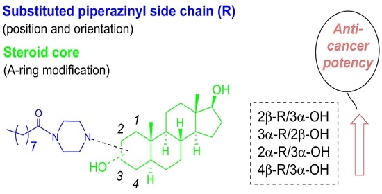
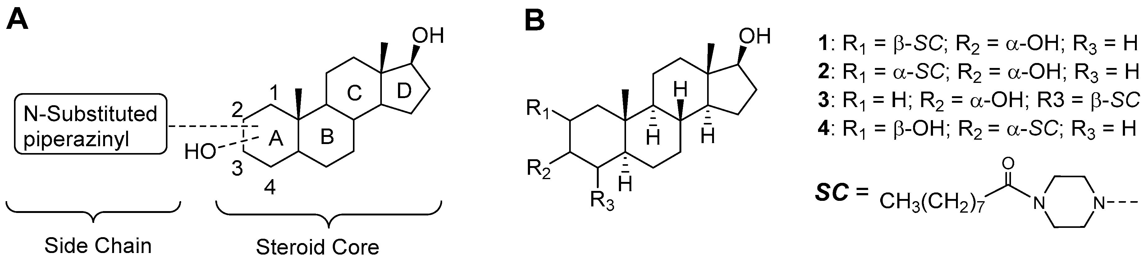
2. Results and Discussion
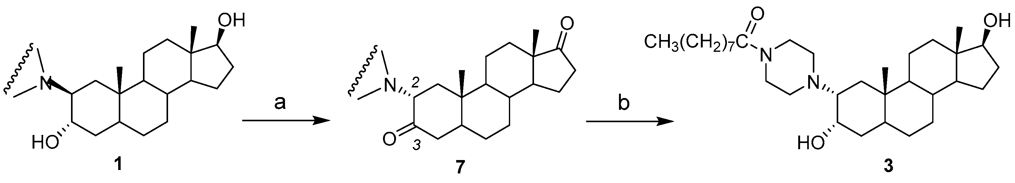
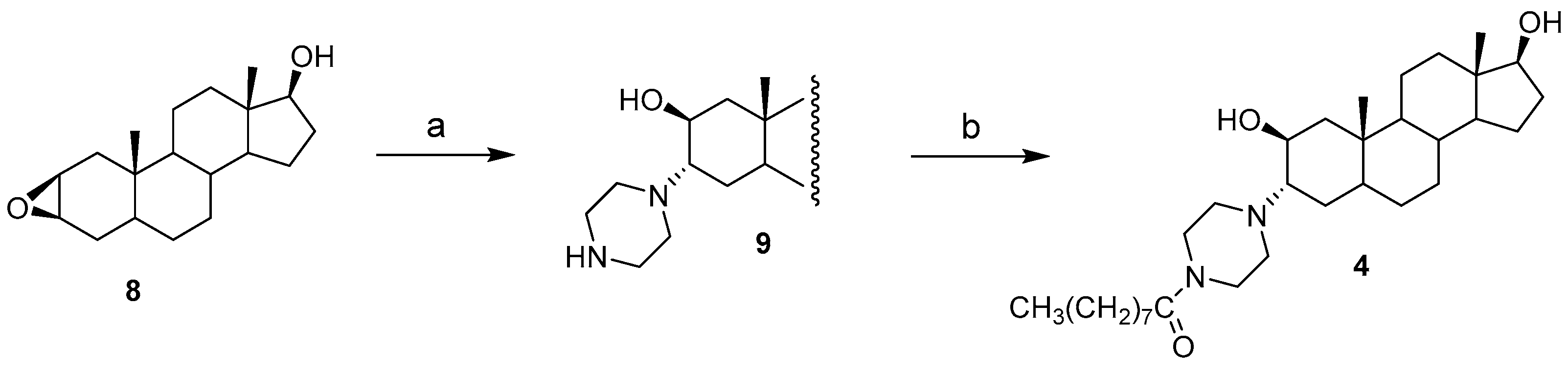
2.1. Chemical Synthesis of Compounds 14

Compounds 1 and 2 (2β-sidechain/3α-OH and 4β-sidechain/3α-OH) were obtained by an epoxide opening (Figure 2). We first generated the mixture of 2α,3α and 3α,4α-epoxides 5a and 5b (76:24) from the corresponding mixture of alkenes, which had been prepared from commercially available epiandrosterone by tosylation, elimination and reduction steps [3]. It is possible to perform a partial selective opening of 2α,3α-epoxide vs 3α,4α-epoxide using mild aminolysis conditions (2 equivalents of piperazine, ethylene glycol, 130 °C) [13], but we chose to use more severe aminolysis conditions (50 equivalents of piperazine, H2O, 166 °C [14,15]) to generate a mixture of amino alcohols. In fact, from the four possible piperazine alcohols, the opening of such an epoxide mixture is known to give the compounds resulting from the two trans-diaxial openings in agreement with Barton’s generalization [11,12,15]. Thus, opening 2α,3α-epoxide led to compound 6a, whereas opening 3α,4α-epoxide led to compound 6b. The amidation of the mixture of amines 6a and 6b with nonanoic acid, with O-(benzotriazol-1)-yl-N,N,N’,N’-tetramethyluronium hexafluorophosphate (HBTU) as coupling agent, and diisopropylethylamine (DIPEA) as base provided 1 and 2, respectively, after separation by chromatography. The former had already been prepared from the opening of pure 2α,3α-epoxide followed by amidation [2], but confirmation of the stereochemistry at positions 2 and 3 had not been performed by an NMR study and it was not possible to obtain a crystal to perform the X-ray analysis of compound 1.
Compound 3 (2α-sidechain/3α-OH) was obtained in two steps from compound 1 (Figure 3). Swern’s oxidation first gave the 3,17-diketone 7 in moderate yield. The presence of a carbonyl in position 3, the acidity of the proton in position 2, and the triethylamine(TEA) used in the Swern’s reaction [16] favored the isomerization at carbon 2 (C2), which went from a 2β-sidechain orientation (less stable; 60.1 kcal/mol) to 2α (more stable; 59.0 kcal/mol). The α-configuration of the substituent at C2 position of compound 7 was determined by a homonuclear Overhauser effect (NOE) spectroscopy (NOESY) experiment showing a correlation between CH3-19 and CH-2β. In the next step, the two C3- and C17-carbonyls were stereoselectively reduced using NaBH4 to obtain the desired compound 3. Considering the presence of the axial methyl-18 close to carbonyl-17, the reduction is highly stereoselective and produces the expected 17β-OH [17]. The reduction of the carbonyl-3 proceeded by the less hindered β-steroid face and generated the 3α-OH alcohol 3 as confirmed by the subsequent 2D NMR analysis of 3.
Finally, compound 4 (3α-sidechain/2β-OH) was also prepared by a stereoselective aminolysis according to the generalization of Barton [11,12], but using a key 2β,3β-epoxide (Figure 4). In fact, the attack of piperazine takes place at position 3 [15] by the α-steroid face of epoxide 8 to generate the amino alcohol 9 resulting from the trans-diaxial opening. The amidation of the secondary amine of 9 with nonanoic acid, HBTU, and DIPEA thus provided compound 4.
The yields for the synthesis of compounds 14 were low, but the quantities obtained were considered sufficient for characterization and biological evaluation, so they were not optimized. In the context of a previous study [2], compound 1 had been obtained with a 70% yield for the opening of the epoxide and an 82% yield for the amidation, leading to an overall 57% yield for the 2 steps. In this study, however, the fact that the two reactions were performed without purifying the intermediate compound, which may have contributed to the low overall yields of respectively 18%, 9.9% and 22% for compounds 1, 2, and 4. However, the 22% yield for compound 3 may be explained by an incomplete reduction of the two carbonyls and the formation of a mixture of monoketonic compounds (42%).

2.2. Nuclear Magnetic Resonance (NMR) Characterization

2.2.1. Assignments of Carbons and Protons

The assignment of NMR signals corresponding to the carbons and protons of aminosteroids 14 is an essential step before the characterization of the positioning and stereochemistry of the two A-ring substituents (Table 1). The signals of carbons 6–18 (B, C and D-rings) were easily assigned considering data from the literature [18,19,20,21]. From the JCH correlations in the heteronuclear single-quantum correlation (HSQC) spectra we then identified the signals of the corresponding protons. In addition, the chemical shifts (δ) of carbons 6–18 do not differ between compounds 14 since the modifications on the A-ring do not affect them. However, a slight difference of 1.2 ppm is observed for C-7 (34.0 ppm for 2 vs 32.8 ppm for 1, 3, and 4). For the nonanoyl chain located on the piperazine ring, 13C and 1H NMR signals were also identical between the four compounds. Signals were assigned using HSQC (JCH) and heteronuclear multiple-bond correlation (HMBC) (JCCH and JCCCH). Thus, in 1H NMR, the CH3-9″ signal at 0.92 ppm, which is the only triplet among the three methyl signals, was linked to the 14.4 ppm signal in 13C NMR. This later was next used to identify C-8″ (JHH and JCCH) and C-7″ (JCCCH). Similarly, the identification of CH2-2″ at 2.38–2.40 ppm (shielded by the carbonyl) leads to C-2″ (JCH), CH2-3″ (JHH) and C-4″ (JCCCH), this latter methylene with an identical δ to those of CH2-5″ and 6″. For the CH2 of the piperazine nucleus, they appear as a broad multiplet at about 3.5 ppm for the protons near the amide group (CH2-3′ and 5′), which correlate (JCH) with two signals at around 43 and 47 ppm. For the neighboring protons of the amine group (CH2-2′ and 6′), they appear in the form of two multiplets around 2.5–2.7 ppm in 1H NMR, which correlate (JCH) with two signals around 50.9–51.9 ppm in 13C NMR for compounds 1, 3, and 4, but 53.8 and 54.4 ppm for compound 2. For this later, the C4 positioning of the sidechain (close to the substituted C-5) is responsible for this deshielding effect of about 3 ppm.
Having already identified the CH3-9″ and CH3-18, then the singlet between 0.86–1.11 ppm (1H NMR) and the signal between 12.9–15.6 ppm (13C NMR) corresponds to CH3-19 at the junction of rings A and B. From this signal, we observed four important correlations in the HMBC spectrum that make it possible to identify the carbons CH2-1, CH-5, CH-9, and C-10. The JCCH correlation with C-10 (37.1–37.7 ppm for 14) indicates that the positioning of the chain has little influence on the chemical shift. It is the same for CH-9 (56.3–57.4 ppm) identified by a JCCCH correlation, but not for CH-5 (JCCCH, 40.0–45.7 ppm). For the latter, the presence of the C4-sidechain (compound 2) causes a significant deshielding effect (~5 ppm). The fourth JCCCH correlation allows the identification of CH2-1 (34.0–36.9 ppm for 1–3 and 42.4 ppm for 4), whose chemical shift is greatly affected (8 ppm) by the presence of a chain on C-3α. For the assignment of the three remaining carbons (C2, C3 and C4), the significant deshielding effect for CH-OH (66.6–68.5 ppm) and CH-NR1R2 (63.0–70.5 ppm) in 13C NMR should be noted, and is not the case with the CH2 at C2 or C4 (26.6–36.5 ppm). The remaining CH2 was thus easily identified since this is the last unassigned signal and because of the use of attached proton test (APT) and HSQC spectra (two separate signals for the two protons). For the two A-ring CHs, it is well known that a CH carrying an OH is more shielded (4.09–4.15 ppm) in 1H NMR than that carrying an NR1R2 (1.90–2.40 ppm) and the HSQC spectra (JCH) made it possible to discriminate the two CH. In the case of compound 1, and contrary to the three other compounds, the 2-CH and 3-CH signals are identical in 13C NMR.

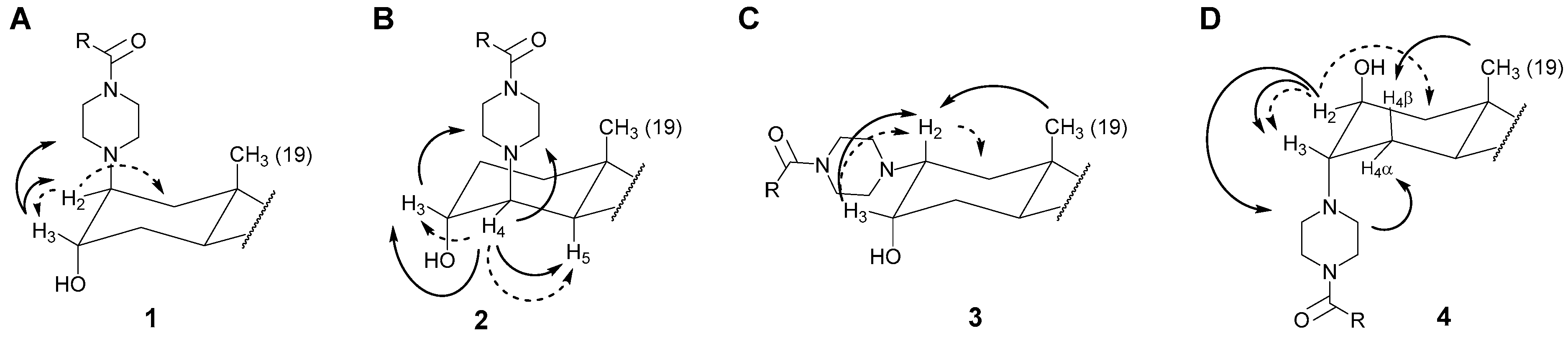
2.2.2. Positioning and Orientation of A-Ring Sidechain and OH

Compounds 14 differ only by the positioning and orientation of A-ring substituents, which were determined by a careful analysis of NMR data. For compound 1 (2β-R/3α-OH; Figure 5A), two JHH correlations (COSY) between CH-2 and CH2-1, as well as CH-2 and CH-3, made it possible to confirm the positioning of the sidechain and of the OH in C2 and C3, respectively. In the NOESY spectrum, an absence of correlation between CH3-19 and CH-2 suggests the presence of the chain on the β side of the steroid, while a correlation between CH-3 and CH2-2′/CH2-6′ indicated the β orientation of H-3 and, consequently, an α orientation for the 3-OH. The NMR analysis therefore confirmed the 2β-R/3α-OH configuration, as predicted by theory [11,12]. For compound 2 (4β-R/3α-OH; Figure 5B), the COSY spectrum showed correlations between CH-4 and CH-5, as well as CH-4 and CH-3. In the NOESY spectrum, no correlation was observed between CH3-19 and CH-4, suggesting the presence of the sidechain on 4β-steroid face. On the other hand, three correlations between CH-4 and CH-5, CH-4 and CH-3, CH-4 and CH2-2′/CH2-6′, as well as CH-3β and CH2-2′/CH2-6′ in the NOESY spectrum confirmed the obtention of the 4β-R/3α-OH configuration as predicted by the trans-diaxial opening of 3α,4α-epoxide. As for compounds 1 and 2, the two compounds with a 4β and 2β sidechain, respectively, no correlation was observed in the NOESY spectra between the piperazine protons of the sidechain and the axial CH3-19. These two results could be explained by a conformation of the piperazine nucleus which moves the CH2-2′/CH2-6′ and CH2-3′/CH2-5′ away from the CH3-19.
For compound 3 (2α-R/3α-OH; Figure 5C), two JHH correlations between CH-2 and CH2-1′, as well as CH-2 and CH-3 are observed in the COSY spectrum. In addition, in the NOESY spectrum, a correlation between CH3-19 and CH-2 demonstrated the 2α orientation (equatorial) of the chain while another correlation between CH-2β and CH-3 confirms the presence of OH at 3α (axial). Finally, for compound 4 (3α-R/2β-OH; Figure 5D), a JHH correlation between the CH carrying an OH group and CH2-1′ confirms the positioning of OH on C-2, unlike compounds 13. A JHH correlation between CH-2 and CH-3 then demonstrates the positioning of the chain on C-3. Since any correlation was observed between CH3-19 and CH-2 in the NOESY spectrum, the OH is therefore in 2α-orientation. On the other hand, a correlation in the NOESY spectrum was observed between CH-2α and CH2-2′/CH2-6′ supporting a 3α orientation of the sidechain, which would agree with a trans-diaxial opening of the 2β,3β-epoxide 9. This 3α-sidechain orientation is also confirmed by a NOE correlation between CH2-2′/CH2-6′ of the piperazine nucleus and H-4α located on the α side of the steroid nucleus. H-4α had previously been differentiated from H-4β, which showed an NOE correlation with CH3-19.

2.3. Inhibition of HL-60 Cell Proliferation

Aminosteroids were tested to evaluate their effect on human myeloid leukemia HL-60 cell growth. The assay was performed at a concentration of 10 µM and the results are expressed as the percentage of cell growth inhibition (Table 2). Interesting SAR results were obtained, since the antiproliferative activity varies depending on the positioning, as well as the orientation of the chain and the OH group present in the steroid A-ring. In fact, the 2β-R/3α-OH orientation of compound 1 generates a better cell proliferation inhibition (84%) than those of aminosteroids 24. In contrast, the antiproliferative activity is completely lost (1%) by the displacement of the chain from position 2 to 4 (compound 2; 4β-R/3α-OH). Inversing the chain at position 2 with alpha configuration (compound 3; 2α-R/3α-OH) halves the antiproliferative activity (42% vs. 84%). Finally, when the chain is positioned in 3α and the OH in 2β (compound 4), the activity is reduced to 51%. It can be assumed that the diaxial orientation of compound 1 promotes the formation of hydrophobic interactions between a section of the alkyl group and a suspected hydrophobic pocket of the protein targeted by this family of aminosteroids whose exact mechanism of action is not yet fully elucidated. On the other hand, the chair form of the piperazine ring at C4 seems to move the alkyl group away from the hydrophobic pocket, eliminating all favorable interactions. We also note that the equatorial positioning of the C2 chain (vs axial) decreases the activity, but the flexibility of the nonanoyl group seems to maintain several favorable interactions. For compound 4, it is interesting to note that a rotation of 180° around the longitudinal axis of the steroid (Table 2) allows a diaxial positioning and orientation similar to compound 1, which could explain that despite a C3α chain (or pseudo C2β) and an OH in C2β (or pseudo C3α) interactions with the hydrophobic pocket are not fully lost and that it retains an acceptable antiproliferative activity. Either way, it is evident that the 2β-R/3α-OH configuration is the one that causes the best inhibition of cancer cell proliferation, and that this diaxial arrangement should be prioritized over other configurations.

3. Materials and Methods

3.1. General

Chemical reagents and solvents were purchased from commercial suppliers and used as received. Thin-layer chromatography (TLC) and flash-column chromatography were performed on 0.20-mm silica gel 60 F254 plates (E. Merck; Darmstadt, Germany) and with 230–400 mesh silica gel 60 (Silicycle, Quebec, QC, Canada), respectively. Infrared (IR) spectra were recorded with a Horizon MB 3000 ABB FTIR spectrometer (Quebec, QC, Canada). NMR spectra were recorded at room temperature in CD3OD with a 5-mm NMR tube on a Bruker AVANCE 400 spectrometer (Billerica, MA, USA). 1H and 13C NMR chemical shifts were referenced to the residual central peak of MeOH (3.33 and 49.0 ppm, respectively). For characterization, we used the following experiments: APT, COSY, NOESY, HSQC, and HMBC. Low-resolution mass spectra (LRMS) were recorded on a Shimadzu Prominence apparatus (Shimadzu Corporation, Kyoto, Japan) equipped with a Shimadzu LCMS-2020 mass spectrometer and an atmospheric-pressure chemical ionization (APCI) probe.

3.2. Synthesis of Compounds 1 and 2

Aminolysis: A mixture of epoxides 5a and 5b in a 76:24 ratio (318 mg, 1.09 mmol), piperazine (2.45 g, 28.5 mmol) and H2O (2 mL) was refluxed 24 h. The reaction mixture was then cooled, poured into water and the precipitate was filtered. The solid was dissolved in CH2Cl2 and the solution dried over Na2SO4, filtered, and evaporated to dryness to give a crude mixture of 6a and 6b (372 mg). Amidation: Under an argon atmosphere at 0 °C, O-(benzotriazol-1-yl)-N,N,N′,N′-tetramethyluronium hexafluorophosphate (HBTU; 358 mg, 0.94 mmol) and nonanoic acid (164.5 mg, 1.04 mmol) were dissolved in dimethylformamide (DMF; 15 mL), diisopropylethylamine (DIPEA; 244 mg, 1.89 mmol) was added and the mixture allowed to react for 5 min. Thereafter, the mixture of 6a and 6b (356 mg, 0.94 mmol) dissolved in DMF (20 mL) was added and the temperature was raised to room temperature for 4 h. The reaction mixture was concentrated, and water was added before extraction with diethyl ether. The organic phase was washed with water, dried over Na2SO4, filtered, and evaporated under reduced pressure. Purification by flash-column chromatography with a gradient of CH2Cl2:MeOH (100:0) to CH2Cl2:MeOH (98:2) yielded the desired amides 1 (98.6 mg, 18% corrected yield for 2 steps) and 2 (53.6 mg, 9.9% corrected yield for 2 steps). 2β-(4-nonanoylpiperazinyl-5α-androstane-3α,17β-diol (1); IR (film): 3406 (OH), 2925, 2854, 1628 (NC = O), 1445, 1248, 1023 cm−1; 1H NMR (400 MHz) and 13C NMR (100.6 MHz) in CD3OD: data reported in Table 1. LRMS for C32H57N2O3 [M + H]+: 517.4 (calc) and 517.5 (found) m/z. 4β-(4-nonanoylpiperazinyl-5α-androstane-3α,17β-diol (2); IR (film): 3406 (OH), 2925, 2854, 1627 (NC = O), 1445, 1245, 1027 cm−1; 1H NMR (400 MHz) and 13C NMR (100.6 MHz) in CD3OD: data reported in Table 1. LRMS for C32H57N2O3 [M + H]+: 517.4 (calc) and 517.5 (found) m/z.

3.3. Synthesis of Compound 3

Oxidation: Under an argon atmosphere, DMSO (1.29 g, 16.0 mmol) in anhydrous CH2Cl2 (1.5 mL) was added dropwise to a solution of oxalyl chloride (1.01 g, 8.0 mmol) in anhydrous CH2Cl2 (1.5 mL) at 60 °C. This solution was added dropwise to a solution of compound 1 (343 mg, 0.66 mmol) in anhydrous CH2Cl2 (7 mL) to −60 °C and the reaction mixture was warmed to −20 °C. The mixture was left to react for 30 min, trimethylamine (TEA) (1.61 g, 16.0 mmol) was added, and the mixture was warmed to 0 °C and left to react 15 min. A saturated solution of NaHCO3 was added before extraction with CH2Cl2, the organic layer was filtered and evaporated under reduced pressure. The crude compound was purified by flash-column chromatography using a gradient of hexanes/acetone (9:1 to 7:3) to give the 3,17-diketone 7 (151 mg, 44% yield). Reduction: To a solution of 7 (118 mg, 0.23 mmol) in MeOH/ CH2Cl2 (9:1) (30 mL) was added NaBH4 (18 mg, 0.46 mmol) and the mixture was stirred under inert atmosphere at 0 °C. After 1 h, solvents were evaporated, water was added, and the mixture extracted with EtOAc. The organic layer was dried with MgSO4, filtered, and evaporated under reduced pressure. The crude compound was purified by flash-column chromatography with CH2Cl2/MeOH (98:2) to give the amide 3 (26 mg, 22% yield). 2α-(4-nonanoylpiperazinyl-5α-androstane-3α,17β-diol (3); IR (film): 3418 (OH), 2925, 2853, 1633 (NC = O), 1445, 1242, 1051 cm−1; 1H NMR (400 MHz) and 13C NMR (100.6 MHz) in CD3OD: data reported in Table 1. LRMS for C32H57N2O3 [M + H]+: 517.4 (calc) and 517.4 (found) m/z.

3.4. Synthesis of Compound 4

Aminolysis: The epoxide 8 (70 mg, 0.24 mmol), piperazine (540 mg, 6.26 mmol) and H2O (1 mL) were refluxed 24 h. The reaction mixture was then cooled, poured in water, and extracted with CH2Cl2. The organic layer was dried with Na2SO4, filtered, and evaporated under reduced pressure to give the desired piperazine derivative 9 (84 mg). Amidation: Under an argon atmosphere at 0 °C, HBTU (74 mg, 0.19 mmol) and nonanoic acid (34 mg, 0.22 mmol) were dissolved in DMF (7 mL), DIPEA (50 mg, 0.39 mmol) was added and the mixture allowed to react for 5 min. Thereafter, the crude aminoalcohol 9 (73 mg, 0.19 mmol) dissolved in DMF (17 mL) was added and the temperature was raised to room temperature for 3 h. The resulting mixture was diluted in water and extracted with CH2Cl2. The organic layer was washed, filtered, and evaporated under reduced pressure. Purification of the crude product by flash chromatography with CH2Cl2:MeOH (100:0) to CH2Cl2:MeOH (98:2) yielded the amide 4 (24 mg, 22% corrected yield for 2 steps). 3α-(4-nonanoylpiperazinyl-5α-androstane-2β,17β-diol (4); IR (film): 3410 (OH), 2926, 2853, 1628 (NC = O, 1444, 1242, 1024) cm−1; 1H NMR (400 MHz) and 13C NMR (100.6 MHz) in CD3OD: data reported in Table 1. LRMS for C32H57N2O3 [M + H]+: 517.4 (calc) and 517.4 (found) m/z.

3.5. Cell Proliferation Assay

Human promyelocytic leukemia cells HL-60 (ATCC, Rockville, MD, USA) were routinely grown in suspension in 90% RPMI-1640 (Sigma, St. Louis, MO, USA) containing L-glutamine (2 nM), antibiotics (100 IU penicillin/mL, 100 µg streptomycin/mL) and supplemented with 10% (v/v) foetal bovine serum (FBS), in a 5% CO2 humidified atmosphere at 37 °C. Cells were maintained twice a week by diluting the cells in RPMI 1640 medium containing 10% FBS. The cell proliferation assay was performed using 3-(4,5-dimethylthiazol-2-yl)-5-(3-carboxymethoxyphenyl)2-(4-sulfophenyl)-2H-tetrazolium (MTS) (Cell Titer 96 Aqueous, Promega, Madison, WI, USA), which allowed us to measure the number of viable cells. In brief, triplicate cultures of 1 × 10⁴ cells in a total of 100 µL medium in 96-well microtiter plates (Becton Dickinson and Company, Lincoln Park, NJ, USA) were incubated at 37 °C, 5% CO2. Compounds were dissolved in ethanol to prepare the stock solution of 1 × 10⁻² M. These compounds and doxorubicin (Novapharm, Toronto, ON, Canada) were diluted at multiple concentrations with culture media, added to each well and incubated for 3 days. Following each treatment, MTS (20 µL) was added to each well and incubated for 4 h. MTS is converted to water-soluble colored formazan by a dehydrogenase enzyme present in metabolically active cells. Subsequently, the plates were read at 490 nm using a microplate reader (Molecular Devices, Sunnyvale, CA, USA).

4. Conclusions

Our previous structure–activity relationship study had only focused on steroid position 2β as well as on sidechain composition. Although interesting results had been obtained at this position, from which certain compounds more active than compound 1 were subsequently identified, it was crucial to explore other sidechain positions and orientations by testing new compounds. After we synthesized compounds 14, a careful 1H and 13C NMR analysis using NOESY, COSY, HSQC, and HMBC experiments was necessary to confirm the right positioning and stereochemistry of the sidechain at C2, C3, or C4, as well as the OH at C3 or C2. The 13C and 1H chemical shifts of the B-, C-, and D-rings did not show any differences between compounds 14, but some A-ring signals were more characteristic and turned out to be good NMR markers. Thus, the identical chemical shifts of CH-2 and 3 (66.5 ppm) make it possible to differentiate compound 1 (2β-R/3α-OH) from the others. For compound 2 (4β-R/3α-OH), positioning the chain in position 4 causes a significant deshielding effect for CH-4 (70.5 vs. 63.0–66.5 ppm) and CH-5 (45.7 vs. 40.0–41.1 ppm). For compound 3 (2α-R/3α-OH), reversing the orientation of the chain at position 2 causes a shielding effect of 3.5 ppm for CH-2, but particularly affects CH3-19. Indeed, a shielding effect makes it possible to differentiate it from other compounds as much in 13C NMR (12.9 vs. 14.5–15.6 ppm) as in 1H NMR 0.86 vs. 1.04–1.11 ppm). Finally, two markers are assigned to compound 4 (3α-R/2β-OH), i.e., 42.4 (CH2-1) and 15.6 (CH3-19) ppm.
Through NMR analysis, it was then possible to validate which of the four chain configurations and OH in the A-ring of compounds 14 was most important for antileukemia activity. As demonstrated with a viability cell assay with the HL-60 cancer cell line, the 2β-R/3α-OH configuration of compound 1 was shown to be the most advantageous among the four configurations.

Supplementary Materials

The following are available online at https://0-www-mdpi-com.brum.beds.ac.uk/2312-7481/7/1/3/s1, 1H NMR, 13C NMR (APT), NOESY, COSY, HSQC, and HMBC spectra for compounds 14.

Author Contributions

D.P. analyzed the NMR data and wrote the paper; I.R. performed chemical synthesis; J.R. tested the compounds; R.M. analyzed the NMR data; R.M. collaborated on the chemical synthesis and revised the paper. All authors have read and agreed to the published version of the manuscript.

Funding

This research was funded by the Canadian Institutes of Health Research grant number DDP-82934.

Data Availability Statement

The data presented in this study are available in supplementary material.

Acknowledgments

We are grateful to Marie-Claude Trottier for NMR spectra and to Micheline Harvey for careful reading of this manuscript.

Conflicts of Interest

D.P., J.R. and R.M. have patent rights on US8653054 and CA2,744,369 (2-(N-Substituted piperazinyl) steroid derivatives).

References

  1. He, Q.; Na, X. The effects and mechanisms of a novel 2-aminosteroid on murine WEHI-3B leukemia cells in vitro and in vivo. Leukemia Res. 2001, 25, 455–461. [Google Scholar] [CrossRef]
  2. Thibeault, D.; Roy, J.; DeRoy, P.; Poirier, D. Chemical synthesis of 2β-amino-5α-androstane-3α,17β-diol N-derivatives and their antiproliferative effect on HL-60 human leukemia cells. Bioorg. Med. Chem. 2008, 16, 5062–5077. [Google Scholar] [CrossRef] [PubMed]
  3. Roy, J.; Maltais, R.; Jegham, H.; Poirier, D. Libraries of 2β-(N-substituted piperazino)-5α-androstane-3α,17β-diols: Chemical synthesis and cytotoxic effects on human leukemia HL-60 cells and on normal lymphocytes. Mol. Divers. 2011, 15, 317–339. [Google Scholar] [CrossRef] [PubMed]
  4. Ayan, D.; Maltais, R.; Hospital, A.; Poirier, D. Chemical synthesis, cytotoxicity, selectivity and bioavailability of 5alpha-androstane-3alpha,17beta-diol derivatives. Bioorg. Med. Chem. 2014, 22, 5847–5859. [Google Scholar] [CrossRef] [PubMed]
  5. Gupta, A.; Kumar, B.S.; Negi, A.S. Current status on development of steroids as anticancer agents. J. Steroid Biochem. Mol. Biol. 2013, 137, 242–270. [Google Scholar] [CrossRef] [PubMed]
  6. Jegham, H.; Maltais, R.; Roy, J.; Doillon, C.; Poirier, D. Biological evaluation of a new family of aminosteroids that display a selective toxicity for various malignant cell lines. Anti-Cancer Drugs 2012, 23, 803–814. [Google Scholar] [CrossRef] [PubMed]
  7. Kenmogne, L.C.; Ayan, D.; Roy, J.; Maltais, R.; Poirier, D. The aminosteroid derivative RM-133 shows in vitro and in vivo antitumor activity in human ovarian and pancreatic cancers. PLoS ONE 2015, 10, e0144890. [Google Scholar] [CrossRef] [PubMed] [Green Version]
  8. Maltais, R.; Hospital, A.; Delhomme, A.; Roy, J.; Poirier, D. Chemical synthesis, NMR analysis and evaluation on a cancer xenograft model (HL-60) of the aminosteroid derivative RM-133. Steroids 2014, 82, 68–76. [Google Scholar] [CrossRef] [PubMed]
  9. Roy, J.; DeRoy, P.; Poirier, D. 2β-(N-substituted piperazino)-5α-androstane-3α,17β-diols: Parallel solid-phase synthesis and antiproliferative activity on human leukemia HL-60 mcells. J. Comb. Chem. 2007, 9, 347–358. [Google Scholar] [CrossRef] [PubMed]
  10. Thibeault, D.; Poirier, D. An efficient method for the regioselective aminolysis of 2,3α-steroidal epoxyde. Synlett 2003, 8, 1192–1194. [Google Scholar]
  11. Barton, D.H.R. The stereochemistry of cyclohexane derivatives. J. Chem. Soc. 1953, 1027–1040. [Google Scholar] [CrossRef]
  12. Matthews, G.J.; Hassner, A. Organic Reactions in Organic Chemistry; Fried, J., Edwards, J.A., Eds.; Van Nostrand Reinhold Company: New York, NY, USA, 1972; Volume 2, pp. 1–53. [Google Scholar]
  13. Anderson, A.; Boyd, A.C.; Byford, A.; Campbell, A.C.; Gemmell, D.K.; Hamilton, N.M.; Hill, D.R.; Hill-Venning, C.; Lambert, J.J.; Maidment, M.S.; et al. Anestetic activity of novel water-soluble 2β-morpholinyl steroids and their modulatory effects at GABAA receptors. J. Med. Chem. 1997, 40, 1668–1681. [Google Scholar] [CrossRef]
  14. He, Q.; Xu, Y.H. Synthesis of 2β-(4’-methyl-1’-piperazino)-3α-hydroxyl-16,17-substituted-5α-androstanes. Acta Pharm Sin. 1992, 27, 101–106. [Google Scholar]
  15. Lewett, C.L.; Savage, D.S. Amino-steroids. Part III.1 2- and 3-Amino-5α-androstanes. J. Chem. Soc. 1968, 1134–1140. [Google Scholar] [CrossRef]
  16. Mancuso, A.J.; Huang, S.L.; Swern, D. Oxidation of long-chain and related alcohols to carbonyls by dimethylsulfoxide “activated” by oxalyl chloride. J. Org. Chem. 1978, 43, 2480–2482. [Google Scholar] [CrossRef]
  17. Cadot, C.; Laplante, Y.; Kamal, F.; Luu-The, V.; Poirier, D. C6-(N,N-butyl-methyl-heptanamide) derivatives of estrone and estradiol as inhibitors of type 1 17β-hydroxysteroid dehydrogenase: Chemical synthesis and biological evaluation. Bioorg. Med. Chem. 2007, 15, 714–726. [Google Scholar] [CrossRef] [PubMed]
  18. Blunt, J.W.; Stothers, J.B. 13C N.m.r. spectra of steroids—A survey and commentary. J. Magn. Reson. 1977, 9, 439–464. [Google Scholar] [CrossRef]
  19. Poirier, D.; Maltais, R. NMR-assisted structure elucidation of an anticancer steroid-β-enaminone derivative. Magnetochemistry 2017, 3, 37. [Google Scholar] [CrossRef] [Green Version]
  20. Tchédam-Ngatcha, B.; Trottier, M.C.; Poirier, D. 13C Nuclear magnetic resonance spectroscopy data of a variety of androsterone and epi-androsterone derivatives substituted at position 3beta or/and 3alpha. Curr. Top. Steroid Res. 2011, 8, 35–46. [Google Scholar]
  21. Fielding, L. 1H and 13C NMR studies of some steroidal neuromuscular blocking drugs: Solution conformations and dynamics. Magn. Reson. Chem. 1998, 36, 387–397. [Google Scholar] [CrossRef]
Figure 1. (A) Representation of a new family of anticancer agents showing their crucial structural elements. Both sidechain and steroid core are needed to inhibit cancer cell proliferation. (B) New A-ring steroid derivatives 14 needed to extend the SAR study.
Magnetochemistry 07 00003 g001
Figure 2. Synthesis of compounds 1 and 2. Reagents and conditions: (a) Piperazine, H2O, 166 °C; (b) Nonanoic acid, HBTU, diisopropylethylamine (DIPEA), dimethylformamide (DMF), room temperature; (c) Silica gel chromatography purification (18% and 9.9% of 1 and 2, respectively, for 2 steps).
Magnetochemistry 07 00003 g002
Figure 3. Synthesis of compounds 3. Reagents and conditions: (a) Oxalyl chloride, DMSO, triethylamine (Et3N), CH2Cl2, −60 °C (44% of 7); (b) NaBH4, MeOH/CH2Cl2 (9:1), 0 °C (22% of 3).
Magnetochemistry 07 00003 g003
Figure 4. Synthesis of compound 4. Reagents and conditions: (a) Piperazine, H2O, 160 °C; (b) Nonanoic acid, HBTU, diisopropylamine (DIPEA), dimethylformamide (DMF), 0 °C, room temperature.
Magnetochemistry 07 00003 g004
Figure 5. Key correlations observed in NMR experiments and confirming the positioning and orientation (α or β) of the two substituents (sidechain and OH) on steroid A-ring of compounds 14 (AD). Nuclear Overhauser effect (NOE) correlations from NOESY spectra are reported by plain arrows and JHH correlations from COSY spectra are reported by dashed arrows.
Magnetochemistry 07 00003 g005
Table 1. Chemical shifts (δ in ppm) and assignment of 1H and 13C from compounds 14 in CD3OD.
Magnetochemistry 07 00003 i001
C and H
Assignment
1 (1H)1 (13C)2 (1H)2 (13C)3 (1H)3 (13C)4 (1H)4 (13C)
2β-Chain--3α-OH4β-Chain--3α-OH2α-Chain--3α-OH3α-Chain--2β-OH
CH2-11.40/1.8234.51.40/1.5234.01.30/1.7336.91.54/1.6842.4
CH-2 or CH2-22.4066.51.9026.92.2463.04.0968.5
CH-34.1066.64.1066.64.1566.82.2565.8
CH-4 or CH2-41.3534.22.3470.51.5536.51.42/1.8226.6
CH-51.6640.61.7745.71.6040.01.4841.1
CH2-61.3029.01.4029.21.3028.91.2829.2
CH2-70.92/1.7132.70.91/1.8034.00.95/1.8832.80.92/1.7132.8
CH-81.4236.61.4037.01.4536.61.4636.5
CH-90.7556.90.7357.40.8256.30.7257.1
C-10-37.3-37.2-37.7-37.1
CH2-111.38/1.6221.81.33/1.5821.11.38/1.7021.61.33/1.6021.7
CH2-121.05/1.8438.11.04/1.8438.11.10/1.8538.01.05/1.8438.1
C-13-44.2-44.0-44.1-44.2
CH-140.9552.40.9352.41.0052.40.9652.4
CH2-151.25/1.6024.31.27/1.6224.31.30/1.6224.31.26/1.6024.3
CH2-161.46/1.9830.61.45/1.9830.61.50/2.0030.61.47/1.9830.6
CH-173.5782.53.5782.53.5782.53.5782.5
CH3-180.7411.70.7411.60.7511.70.7411.7
CH3-191.0414.61.1114.50.8612.91.0515.6
CH2-2′/CH2-6′2.53/2.6351.4/51.92.63/2.6853.8/54.42.65/2.7050.9/51.32.50/2.5651.4/51.9
CH2-3′/CH2-5′3.5743.1/47.23.50/3.5743.6/47.73.5843.0/47.13.5743.1/47.2
C-1″-174.1-174.1-174.1-174.0
CH2-2″2.4034.02.3834.02.4034.02.4034.0
CH2-3″1.6126.61.6126.61.6226.61.6126.6
CH2-4″1.3430.31.3430.31.3630.31.3530.3
CH2-5″1.3430.51.3430.51.3630.51.3530.5
CH2-6″1.3430.51.3430.41.3630.41.3530.4
CH2-7″1.3433.01.3433.01.3633.01.3333.0
CH2-8″1.3423.71.3423.71.3523.71.3323.7
CH2-9″0.9214.40.9214.40.9214.40.9214.5
1 After a full assignment of all carbons (heteronuclear single-quantum correlation (HSQC), heteronuclear multiple-bond correlation (HMBC), correlation spectroscopy (COSY), homonuclear Overhauser effect spectroscopy (NOESY), and literature data), the chemical shift of each corresponding proton was obtained from the JCH correlation observed in HSQC spectra. 1H NMR, 13C NMR, NOESY, COSY, HSQC, and HMBC spectra for compounds 14 are available in Supplementary Materials.
Table 2. A-ring substitution and antiproliferative activity (AA).
Compound1234
Structure Magnetochemistry 07 00003 i002 Magnetochemistry 07 00003 i003 Magnetochemistry 07 00003 i004 Magnetochemistry 07 00003 i005
Substitution2β-R (axial) 3α-OH (axial)4β-R (axial) 3α-OH (axial)2α-R (equatorial) 3α-OH (axial)3α-R (axial) 2β-OH (axial)
AA (%)a8214251
a Inhibition of the proliferation of leukemia HL-60 cells treated 3 days with compounds 14 in comparison to the untreated cells (control). The positive control Doxorubicin inhibited 95% of cell proliferation at 10 µM.
Publisher’s Note: MDPI stays neutral with regard to jurisdictional claims in published maps and institutional affiliations.

