Article

Field-Induced SMM and Vis/NIR Luminescence on Mononuclear Lanthanide Complexes with 9-Anthracenecarboxylate and 2,2′:6,2″-Terpyridine

1
Departament de Química Inorgànica i Orgànica, Secció de Química Inorgànica and and Institut de Nanociència i Nanotecnologia (IN2UB), Universitat de Barcelona, Martí i Franquès 1-11, 08028 Barcelona, Spain
2
Departament de Mineralogia, Cristal·Lografia i Dipòsits Minerals and Unitat de Difracció de RX, Centre Científic i Tecnològic de la Universitat de Barcelona (CCiTUB), Universitat de Barcelona, C/Solé i Sabarís, 1-3, 08028 Barcelona, Spain
*
Authors to whom correspondence should be addressed.
Submission received: 25 July 2021 / Revised: 24 August 2021 / Accepted: 6 September 2021 / Published: 10 September 2021
(This article belongs to the Special Issue Reviews on Slow-Relaxation Molecules)

Abstract

:
Five new mononuclear lanthanide complexes are synthesized by adding the several lanthanide nitrate hexahydrate salts, which for lanthanide (Ln) are Eu, Tb, Dy, Er, and Yb, with 9-anthracenecarboxylic acid (9-Hanthc) and 2,2′:6,2″-terpyridine (TPY) in mixed solution of methanol and dimethylformamide (DMF). The general formula is [Eu(9-anthc)3(TPY)(DMF)]·H2O (1Eu) where Eu(III) is ennea-coordinated or [Ln(9-anthc)3(TPY)(H2O)]·H2O·DMF (Ln = Tb (2Tb), Dy (3Dy), Er (4Er), and Yb (5Yb)) where Ln(III) is octa-coordinated. For compounds 3Dy, 4Er, and 5Yb, the dynamic ac magnetic study indicated field-induced single molecule magnet (SMM) behavior. The photoluminescence studies in the solid state of these complexes show the sensitization of 4f-4f transitions for 4Er and 5Yb in the NIR region.

1. Introduction

In recent years, coordination complexes with lanthanide ions have been extensively studied because of their interesting magnetic and luminescent properties. The magnetic interest arises from the high magnetic momentum of the lanthanide ions and strong magnetic anisotropy of its ground state widely used in the molecular magnetism area to design single-molecule magnets (SMMs) that provide potential in information storage, quantum computation, and spintronics [1,2,3,4,5,6,7,8]. In SMMs lanthanide compounds, the energy needed to reverse the magnetization, and the gaps energy between the emission contributions, result in the sublevel structure of the Stark components. In addition, the magnetic relaxation occurs through different process such as Orbach one, direct relaxation, Raman mechanism, or Quantum Tunneling of the Magnetization (QTM). In many cases, these processes can occur conjunctly. Therefore, to optimize the SMMs characteristics, the involved mechanism in the magnetic relaxation and the factors that affect them have to be understood. One of the used strategies to better understand the magnetic properties is to take advantage of the correlation between the magnetic and optical properties of the luminescent SMM compounds. Photoluminescent studies may allow spectroscopic detection of the energy splitting of the mJ states of Ln(III) that can be compared with the magnetic results obtained [9,10,11,12,13,14,15,16,17,18,19,20,21,22,23,24,25].
Regarding the optical aspect, Ln(III)-based materials manifest exceptional luminescence characteristics, with high emission quantum yields, narrow bandwidths, ligand-induced large Stokes shifts, long-lived emission, and ligand-dependent luminescence sensitization [26]. Nevertheless, due to the limited extinction degree of the ff transitions prohibited by Laporte rules [27], the Lanthanide ions must be excited indirectly by the energy transfer proceedings of organic ligands known as antenna that have a large extinction degree (ε). Then, the excitation energy is transmitted as characteristic narrow ff emission bands in the visible and/or in near-infrared (NIR) spectral ranges [28]. Lanthanide complexes emitting in the NIR region present interesting applications as solid-state laser materials [29], in telecommunications [30], in optical communication systems [31], in optical amplifiers [32], and in fluoroimmunoassay [33,34]. The intense luminescence from complexes emitting in visible region as Eu(III) and Tb(III) has generated the development of multiple applications [35,36,37,38,39,40].
Recently, we have reported three series of dinuclear lanthanide compounds with formulas [Ln2(2-FBz)4(NO3)2(phen)2] [41], [Ln22-9-AC)4(9-AC)2(bpy)2] [42], and [Ln2(μ-2-FBz)2(2-FBz)4(terpy)2]·2(H-2-FBz)·2(H2O) [43]. In these series, Nd, Er, and Yb dinuclear compounds exhibit field-induced SMM behavior and NIR-luminescence; thus, they can be considered as multifunctional compounds. We aim to enhance and expand the results obtained in the synthesis of new complexes and induce structural change that may cause variations in magnetic and luminescence behavior. Another reason that led us to such an investigation is the scarce lanthanide metals with 9-anthracenecarboxylate compounds described in the literature. According to the Cambridge Structural Database (CSD) [44], barely twenty-two coordination complexes have been structurally characterized with only lanthanide metals and 9-anthracenecarboxylate: one Yb(III) compound with mononuclear structure, [45] twenty dinuclear compounds with different lanthanide metals where ten of them are reported by us [41,42,43] and the other ten are described by Liu [46,47,48], Wang [49], and Wu [50], and finally one dinuclear compound of Er(III) showing monodimensional structure [45]. Thus, this time, we have combined the ligand 2,2′:6′,2″-terpyridine (TPY), 9-anthracenecarboxylic acid (9-Hanthc), and lanthanide sales. This has allowed the isolation of five new mononuclear complexes of formula [Eu(9-anthc)3(TPY)(DMF)]·H2O (1Eu) where Eu(III) is ennea-coordinated or [Ln(9-anthc)3(TPY)(H2O)]·H2O·DMF (Ln = Tb (2Tb), Dy (3Dy), Er (4Er), and Yb (5Yb)) where the Ln(III) ions are octa-coordinated. The new compounds were characterized by X-ray diffraction, and their magnetic and luminescent properties were studied.

2. Results and Discussion

2.1. X-ray Crystal Structures

Complexes 1Eu5Yb present two different structures depending on the atomic radii of the lanthanide ion. For the largest Eu(III), the lanthanide ion is ennea-coordinated and presents a type I structure. On the other hand, for small lanthanides, the coordination is eight and presents a type II structure. As complexes 2Tb5Yb are isostructural, only the structures of compounds 1Eu and 4Er will be discussed in detail. The most relevant parameters for single crystal determination are collected in Table 1 in the Materials and Methods section.

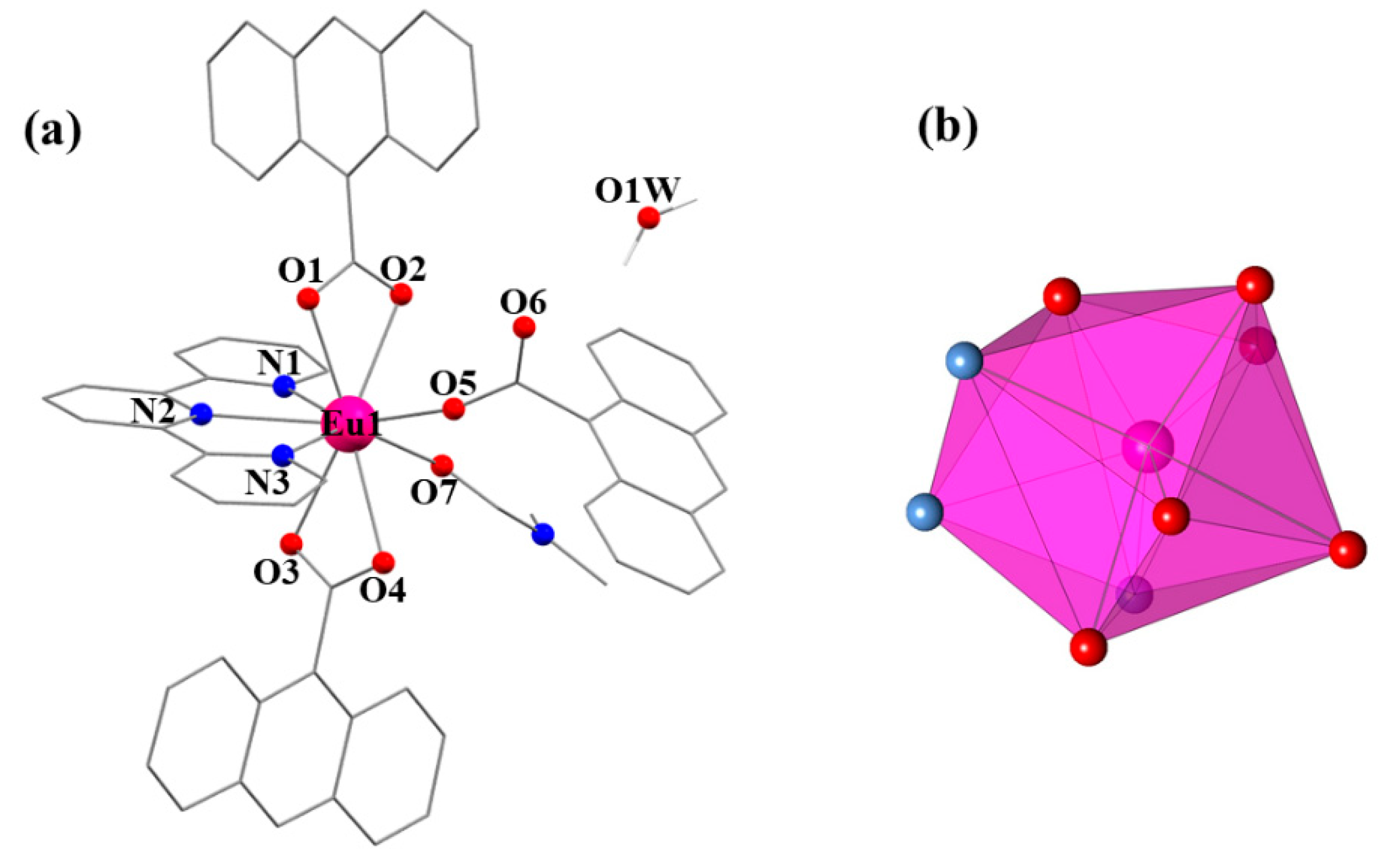
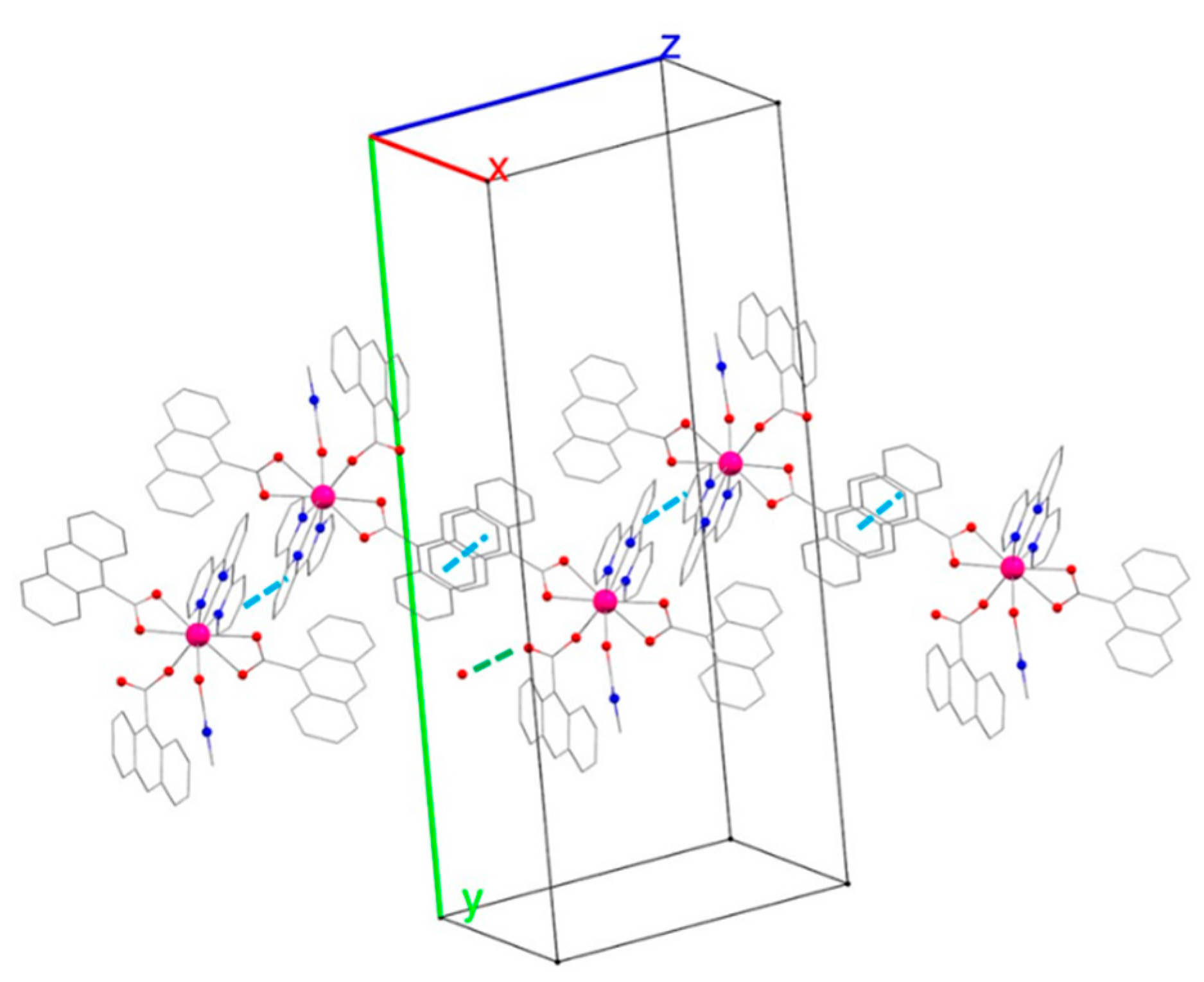
2.1.1. Structural Type I: [Eu(9-anthc)3(TPY)(DMF)]·H2O (1Eu)

Complex 1Eu of formula [Eu(9-anthc)3(TPY)(DMF)]·H2O crystallizes in the monoclinic space group P21/c. A partially labeled plot of the Eu(III) mononuclear complex is presented in Figure 1a. Selected bond distances are listed in Table 2. The structure of 1Eu consists of [Eu(9-anthc)3(TPY)(DMF)] molecules in which each Eu(III) ion is ennea-coordinated with a EuN3O6 coordination environment formed by a tridentate TPY ligand, two chelating 9-anthc ligands, one monodentate 9-anthc ligand, and a coordinated DMF solvent molecule. Three of the coordination sites around the central atom are occupied by the N1, N2, and N3 atoms of a TPY ligand, with Eu–N bond distances equal to 2.552 (3), 2.606 (3), and 2.545 (3) Å, respectively. In addition, the Eu(III) central atom is coordinated to five oxygen atoms from three different 9-anthc ligands. Two of the 9-anthc ligands show a chelating coordination mode with Eu1–O1 and Eu1–O2 bond distances of 2.421 (3) and 2.574 (3) Å and Eu1–O3 and Eu1–O4 distances of 2.465 (3) and 2.599 (3) Å, while the third 9-anthc ligand is coordinated in the monodentate mode with an Eu1–O5 bond distance of 2.279 (3) Å. The ennea-coordinated sphere of Eu(III) is completed by the O7 oxygen atom from a DMF solvent molecule with an Eu1–O7 bond distance of 2.410 (3) Å.
The estimation of the coefficients of distortion of the EuN3O6 coordination polyhedron for 1Eu in reference to the ideal nine vertex polyhedral was made by employing the continuous shape measure theory and SHAPE software [51,52], showing that the N3O6 disposition for 1Eu is intermediate between different coordination polyhedra. The best SHAPE estimation led to Muffin (MFF-9, C2v), spherical-capped square antiprism (CSAPR-9, C4v), and spherical-tricapped trigonal prism (TCTPR-9, D3h) geometries with continuous shape measure (CShM) values of 1.678, 2.006, and 2.407, respectively. A graphical representation of the metal coordination geometry of 1Eu is shown in Figure 1b.
The coordination of [Eu(9-anthc)3(TPY)(DMF)] forms a hydrogen bond with a molecule of water through the hydrogen atom H1W and the oxygen atom O6 of the monodentate 9-anthc ligand (Supplementary Materials, Table S2). In addition, each [Eu(9-anthc)3(TPY)(DMF)] molecule forms a ππ type interaction with a neighboring molecule between the ring containing the N1 atom (Cg(1) from the Supplementary Materials, Table S3) and the one containing the N2 atom (Cg(2) from the Supplementary Materials, Table S3) of the respective TPY ligands. In addition, the mononuclear compound also forms an intermolecular interaction of the same nature with another neighboring molecule via two anthracene groups of the two corresponding 9-anthc ligands containing O1–O2 oxygens (contact Cg(4)-Cg(4)’ from the Supplementary Materials, Table S3). The sum of the two intermolecular contacts forms chains of [Eu(9-anthc)3(TPY)(DMF)] units along the direction [110] of the crystalline network with Eu(III)⋯Eu(III) distances of 8.748 Å (Figure 2).

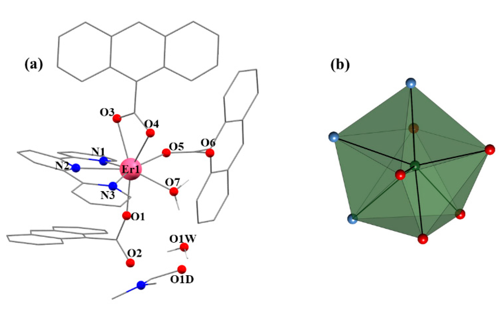
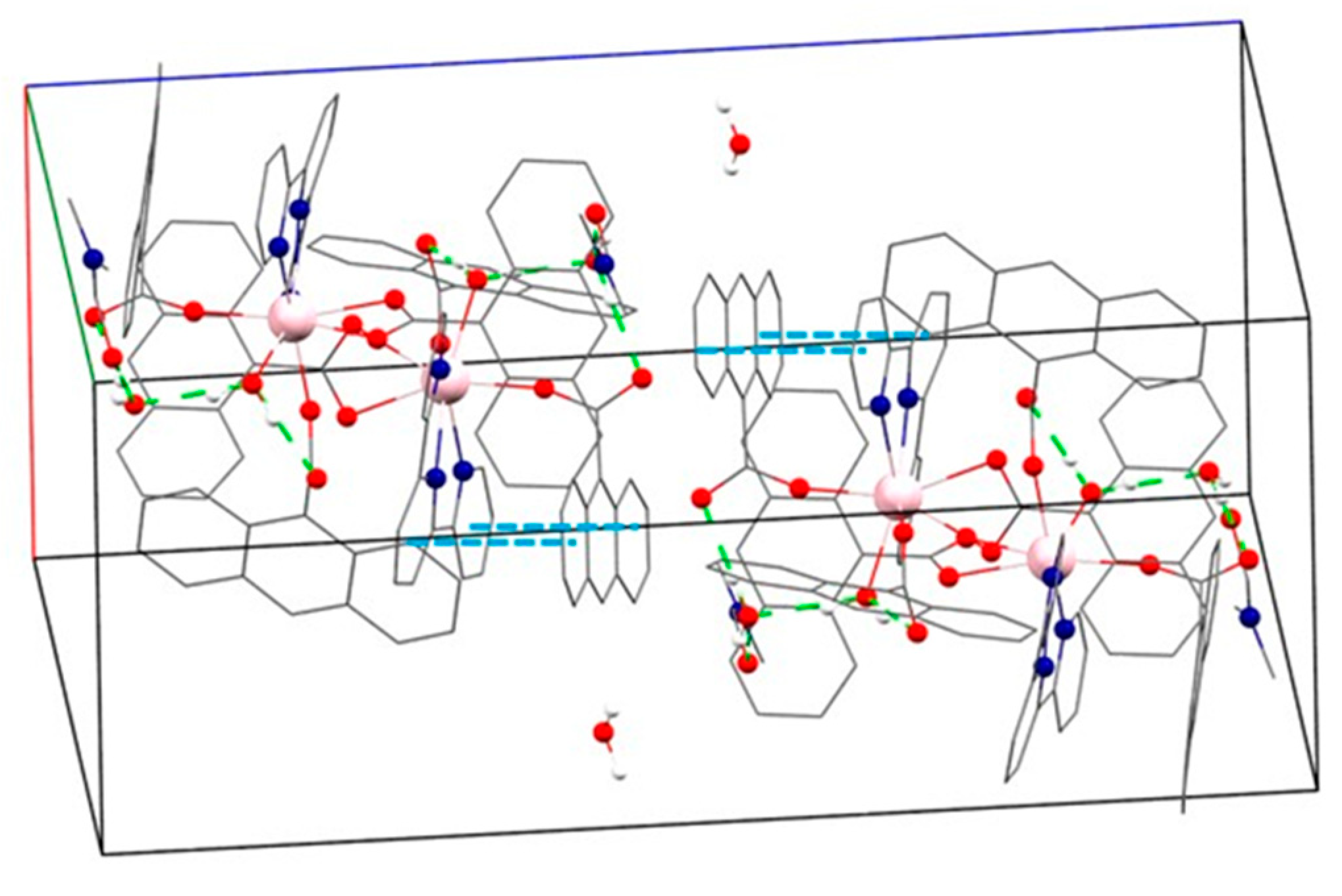
2.1.2. Structural Type II: [Ln(9-anthc)3(TPY)(H2O)]·H2O·DMF (Ln = Tb (2Tb), Dy (3Dy), Er (4Er) and Yb (5Yb))

Complexes 2Tb5Yb with formula [Ln(9-anthc)3(TPY)(H2O)]·H2O·DMF (Ln = Tb (2Tb), Dy (3Dy), Er (4Er), and Yb (5Yb)) crystallize in the monoclinic space group P21/n. Selected bond distances for compounds 2Tb5Yb are listed in Table 1. As an example of the structural type II, 4Er will be described below (Figure 3).
The structure of compound 4Er consists of [Er(9-anthc)3(TPY)(H2O)] molecules in which each Er(III) ion is octa-coordinated. The Er(III) ion has an ErN3O5 octa-coordination environment formed by a tridentate TPY ligand, a chelating 9-anthc ligand, two monodentate 9-anthc ligands, and a water molecule. The octa-coordinated environment of the Er(III) ion corresponds to four oxygen atoms of three different 9-anthc ligands (O1 and O5 from two monodentate 9-anthc ligands and O3 and O4 from the chelating 9-anthc ligand, with Er-O average distance of 2.314 Å), three nitrogen atoms of the TPY ligand (N1, N2 and N3, with an average Er–N distance of 2.492 Å), and the oxygen atom from the coordinated water molecule (O7, Er1–O7 bond distance of 2.344 Å).
The estimation of the coefficient of distortion of the ErN3O5 coordination polyhedron for 4Er in reference to the ideal eight vertex polyhedral was made by employing the continuous shape measure theory and SHAPE software and shows that the N3O5 distribution in 4Er is intermediate between different coordination polyhedra. The best SHAPE estimation led to triangular dodecahedron (TDD-8, D2d), biaugmented trigonal prism (BTPR-8, C2v), or Johnson-biaugmented trigonal prism (JBTP-8, C2v) geometries with continuous shape measure (CShM) values of 2.342, 2.426, and 2.516, respectively. A graphical depiction of the metal coordination geometry of 4Er is shown in Figure 3b. The result of the estimation of the coefficient of distortion of the LnN3O5 coordination polyhedron for 2Tb5Yb in reference to the ideal eight vertex polyhedral are reported in Table S1 in the Supplementary Materials.
Compound 4Er presents an intramolecular hydrogen bond between H7A from the water molecule coordinated to Er(III) and O6 from one of the 9-anthc ligands coordinated in the monodentate mode. On the other hand, the other hydrogen of this H2O molecule, H7B, forms another H bond with the crystallization water molecule (O1W). At the same time, both hydrogen atoms of this water molecule also form hydrogen bonds, H1WA with O2 of a monodentate 9-anthc ligand and H1WB with O1D from a DMF crystallization molecule (Figure 4). The contact distances and angles for compounds 2Tb5Yb are listed in Table S2 from the Supplementary Materials.
The compounds also present ππ stacking interactions between different aromatic rings. One of them is given within each molecule [Ln(9-anthc)3(TPY)(H2O)] between the TPY ligand rings (Cg(1) from the Supplementary Materials, Table S3) and the anthracene group of the monodentate ligand containing the oxygen atoms O1 and O2 (Cg (6)′or Cg (7)′ from the Supplementary Materials, Table S3). The intermolecular contacts vary slightly according to the Ln(III) ion. In the case of the Tb(III) and Dy(III) compounds, the TPY rings of each mononuclear unit interact with two different neighbor molecules via the corresponding terpyridine ligands. Thus, there is an interaction between the ring that contains the N1 atom with the ring that contains the N2 atom of a neighboring molecule (Cg (1)–Cg (2)′ from the Supplementary Materials, Table S3) and another supramolecular contact of the TPY ligand ring that contains the N3 nitrogen with the same ring of another adjacent molecule (contact Cg (3)–Cg (3)′ from the Supplementary Materials, Table S3). On the other hand, the compounds [Ln(9-anthc)3(TPY)(H2O)] with Ln (III) = Er and Yb have a single supramolecular contact type ππ, which is also through the TPY ligands, aromatic ring containing the N1 atom of the reference molecule with the ring containing the nitrogen N3 of the neighboring molecule (Supplementary Materials, Figure S1).

2.1.3. Structural Discussion

The key to obtaining the structural types I and II for the described compounds by using the same synthetic method is the contraction of the ionic radius within the series of the lanthanide elements. As described, for the Eu(III) ion, with the largest ionic radius, the nona-coordinate coordination sphere is due to the coordination of three carboxylate ligands 9-anthc, two in chelating mode and one in monodentate mode, and for the coordination of a DMF molecule. For the Tb(III), Dy(III), Er(III), and Yb(III) ions, the compounds have been obtained with an octa-coordinate environment since the decrease in ionic radius causes two of the three 9-anthc anions to be coordinated in monodentate mode and the last to be coordinated in chelating mode. This structural change is also accompanied by the coordination of a molecule of water instead of a DMF molecule.

2.2. Magnetic Properties

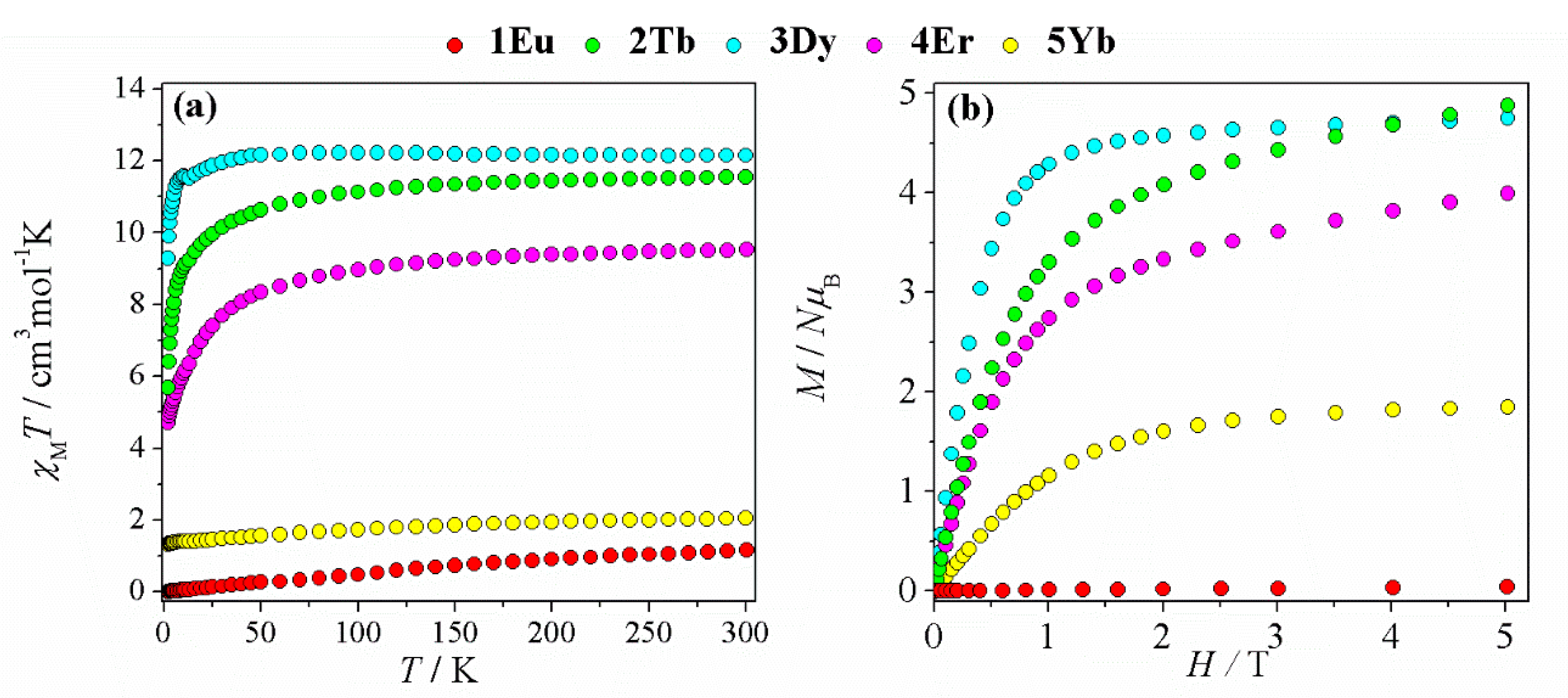
2.2.1. dc Magnetic Studies

Magnetic susceptibility (χM) data on polycrystalline powder samples of 1Eu5Yb were collected under fields of 3000 G (300–30 K) and 200 G approximately (30–2 K). The data are represented as χMT vs. T plot in Figure 5a.
At 300 K, the χMT values for compounds 1Eu5Yb are 1.16, 11.55, 12.15, 9.52, and 2.05 cm3 mol−1 K, respectively. These data are in good agreement with the expected room temperature values for the corresponding Ln(III) centers [53]. In the case of the 1Eu compound, the non-zero value of χMT reveals the presence of excited states that are very close in energy to the fundamental one [54]. For compounds 2Tb and 4Er, the χMT values remain practically constant as the samples are cooled to T ≈ 150 K. Below this temperature, the susceptibilities gradually decrease to values of χMT = 9.12 cm3 mol−1 K a T = 11 K for 2Tb and χMT = 7.68 cm3 mol−1 K a T = 30 K for 4Er. From these temperatures, the fall of the χMT values accelerates to reach minimum values equal to 5.69 cm3 mol−1 K for 2Tb and 4.70 cm3 mol−1 K for 4Er at T = 2 K. For the 3Dy complex, it is observed that the χMT values are practically constant when decreasing the temperature. From T = 8 K, the χMT values decrease quickly to χMT = 9.28 cm3 mol−1 K at 2 K.
Magnetization measurements at 2 K were also performed for complexes 1Eu5Yb and are depicted in Figure 5b. The magnetization saturation under an applied field of 5T is only observed in compounds 3Dy and 5Yb, with M values equal to 4.75 and 1.85 B, respectively.

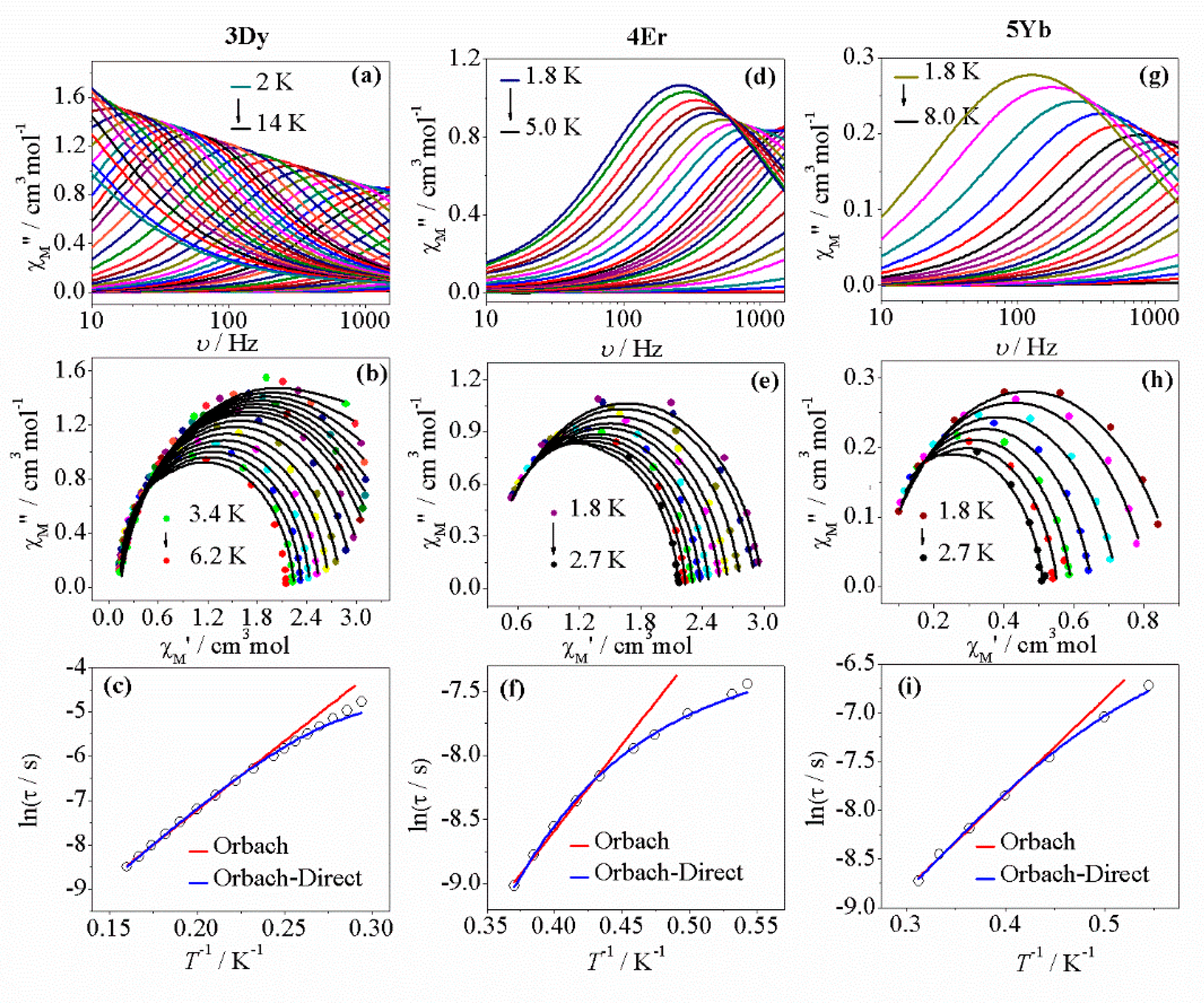
2.2.2. ac Magnetic Studies

Magnetic susceptibility measurements were performed on compounds 2Tb5Yb at a 4 G ac field oscillating between 10 and 1488 Hz. Under zero dc magnetic field, no maxima for the in-phase (χM′) and out-of-phase (χM″) susceptibility components were observed for any of the compounds, which was probably due to an important quantum tunneling of the magnetization (QTM) process present in these systems. To suppress the QTM relaxation process, an optimal external dc field of 1000G was applied for the complexes 2Tb5Yb after checking with low fields and basing on recent works reported by the authors [41,42,43]. Then, slow relaxation of the magnetization was revealed (Figure 6 and Supplementary Materials, Figure S2) for the compounds 3Dy5Yb [55,56].
For 2Tb, even under applied dc fields up to 4000 G, no frequency or thermal dependency of χM′ and/or χM″ were observed.
The ac magnetic susceptibility of compound 3Dy has been measured between 2 and 14 K. Figure 6a shows the out-of-phase component of the susceptibility (χM″) vs. the frequency of the ac field, with maxima for temperatures below 6.4 K. The curves measured at temperatures between 6.2 and 3.4 K have been adjusted to the generalized Debye equation [57]. The corresponding Cole–Cole diagrams (Figure 6b) show slightly elongated semi-circular shapes, with α values between 0.13 and 0.06. The low distribution of the relaxation times (Figure 6c from the Supplementary Materials, Table S4) indicates the presence of a single relaxation process. The fitting of the thermal dependence of τ with the Arrhenius law (Orbach relaxation mechanism) for temperatures higher than 4.5 K reveals values of τ0 = 1.43 × 10⁻⁶ s and Ueff = 21.7 cm−1. To fit the values throughout the range of temperatures, equation (Equation (1) was used, which also takes into account a direct relaxation. The obtained parameters were τ0 = 8.47 × 10⁻⁷ s, Ueff = 24.0 cm−1, and A = 30 K−1 s−1.
τ−1 = τ0−1 [exp(−Ueff/kBT)] + AT
For compound 4Er, the representation of χM″ as a function of the frequency between temperatures of 5.0 and 1.8 K shows maxima for temperatures below 2.7 K (Figure 6d). The fitting of the Cole–Cole diagrams (Figure 6e) shows a narrow distribution of relaxation times with α values between 0.10 and 0.05 (from the Supplementary Materials, Table S5). The relaxation time follows a linear dependence at temperatures above 2.3 K and can be fitted according to the Arrhenius law. The parameters obtained were τ0 = 8.68 × 10⁻⁷ s and Ueff = 9.33 cm−1 (Figure 6f). On the other hand, the data can be fitted throughout all the range of temperatures, taking into account Orbach and direct relaxation processes (Figure 6f). The parameters obtained from this second fitting are τ0 = 4.67 × 10⁻⁸ s, Ueff = 15.4 cm−1, and A = 903 K−1 s−1.
Compost 5Yb shows a maxima of χM″ frequency dependents for temperatures lower than 2.7 K (Figure 6g). The Cole–Cole plots for temperatures between 2.7 and 1.8 K (Figure 6h) have a semi-circular shape. The fitting with the generalized Debye model indicates a narrow distribution of the relaxation times (from the Supplementary Materials, Table S6) with values of α between 0.12 and 0.24. Figure 6i shows the thermal dependence of the relaxation times. For temperatures between 3.2 and 2.5 K, the τ values can be modeled considering an Orbach relaxation mechanism with fitting parameters of τ0 = 7.94 × 10⁻⁶ s and Ueff = 6.79 cm−1. At temperatures below 2.5 K, the values are deviated from linearity, but they can be fitted considering a relaxation of the magnetization by Orbach and direct processes. The parameter obtained are τ0 = 4.16 × 10⁻⁶ s, Ueff = 8.6 cm−1, and A = 323 K−1 s−1.

2.3. Photoluminescence Properties

The emission properties of 1Eu5Yb were studied in solid state at r.t. and 77 K.

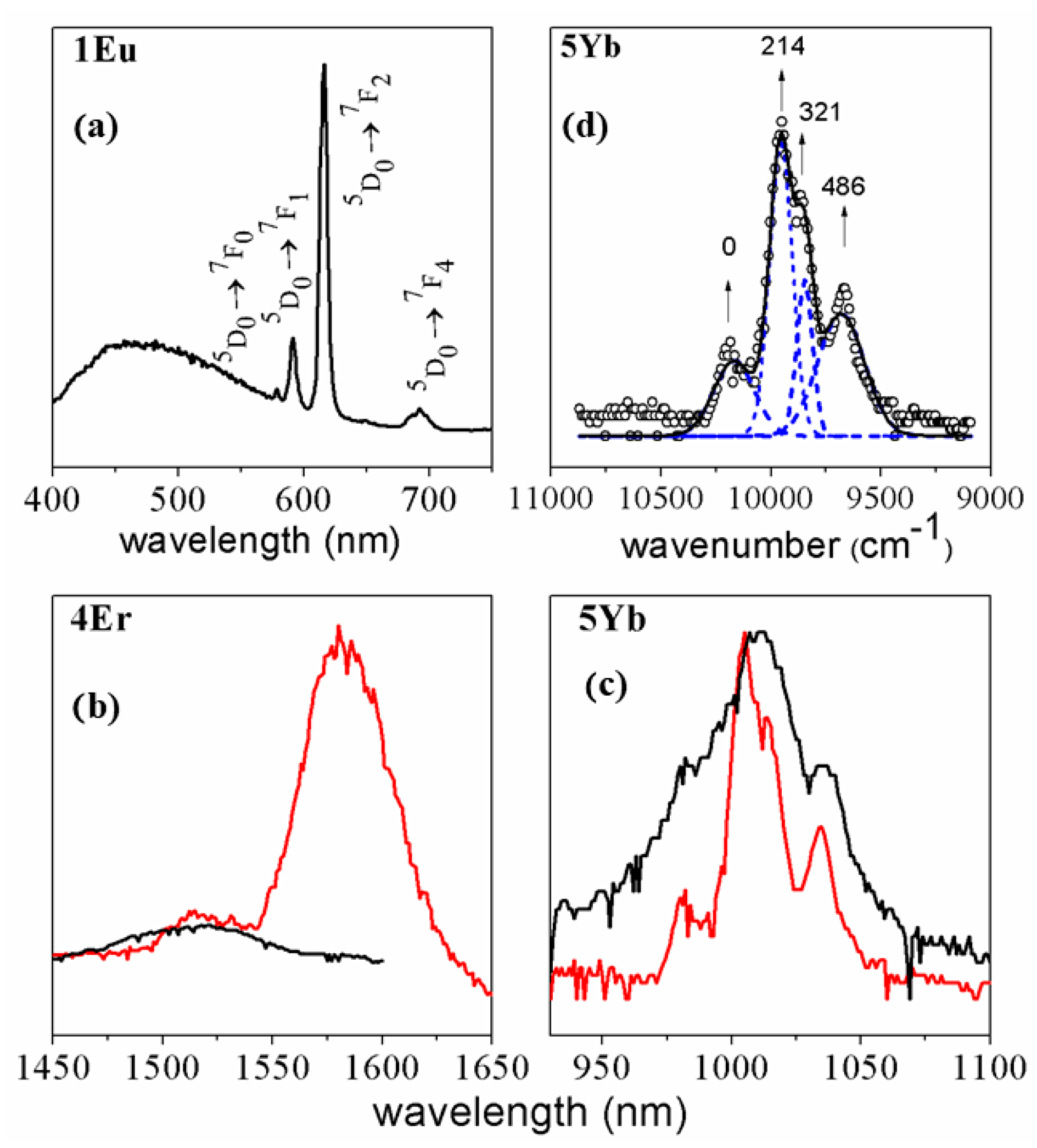
2.3.1. Visible Emission

When compound 1Eu is excited at λex = 319 nm, the resulting emission spectrum shows the characteristic Eu(III) transitions centered at 578 nm (5D07F0), 591 nm (5D07F1), 616 nm (5D07F2), and 692 nm (5D07F4), as well as the residual emission of the 9-anthc ligand as a broad band centered at 470 nm (Figure 7a). Meanwhile, the emission spectra of compounds 2Tb and 3Dy (Supplementary Materials, Figure S3) only show a broad band between 400 and 500 nm that corresponds to the 9-anthc ligand. This may be because there is a water molecule directly bonded to the lanthanide atom, which normally quenches the luminescence of such metals.

2.3.2. NIR Emission

Complex 4Er shows a principal peak corresponding to the 4I13/24I15/2 transition with a maximum at 1580 nm (Figure 7b). Decreasing the temperature to 77 K does not permit us to increase sufficiently the resolution to have access to the crystal field splitting. Compound 4Er, when measured at room temperature and at 77 K under an excitation wavelength of 360 nm, shows broad peaks of residual emission of the ligands between 390 and 550 nm, in the absorption range of the 9-anthc ligand (Figure S4). As can be seen in Figure 7b, under this same excitation energy and at T = 77 K, the compound also emits in the area of the near IR, with a maximum of 1580 nm, which correspond to the transition 4I13/24I15/2.
Complex 5Yb shows the characteristic Yb(III) transition emission profile, with the principal transition 2F5/22F7/2 at 1005 nm (Figure 7c, black spectrum). When decreasing the temperature to 77 K (Figure 7c, red line), the emission band splits into four bands, with the maxima at 980, 1005, 1014, and 1035 nm, which correspond to the four expected Stark levels for a J = 7/2 in low symmetry [56,58]. This transition offers us information of the crystal field energy diagram of the ytterbium ion in complex 5Yb and permits us to estimate the energy between the ground and the first excited mJ states of the fundamental 2F7/2 level of about 210 cm−1 (Figure 7d). The difference between this value and the effective energy barrier value obtained from the Arrhenius fit of the ac data (≈7.0 cm−1) confirms that relaxation of the magnetization takes place by direct and/or Raman processes instead of only the Orbach one [59].

3. Experimental Section

3.1. Starting Materials

Ln(NO3)3·xH2O salts, 9-anthracenecarboxylic acid (9-Hanthc), 2,2′:6,2″-terpyridine (TPY), methanol, and N,N-dimethylformamide were obtained from Sigma Aldrich and used without further purification.

3.2. General Syntheses

Compounds 1Eu5Yb have been synthetized by following the same experimental procedure. To a solution consisting of 1.5 mmol 9-Hanthc and 0.3 mmol TPY in 15 mL of MeOH, a solution of 10 mL DMF containing 0.25 mmol of Ln(NO3)3·xH2O is added (Ln(III) = Eu (1Eu), Tb (2Tb), Dy (3Dy), Er (4Er), and Yb (5Yb). In all the cases, slow evaporation of the methanol/dimethylformamide solution of the product gave prism-shaped yellow monocrystals suitable for X-ray determination.
Data for 1Eu: Selected IR bands (KBr pellet, cm−1): 3418 (m, υH2O(O-H)), 1650 (s, υas(COO-)a), 1597 (vs, υas(COO-)b), 1574 (vs, υ(C = N)), 1436 (m, υs(COO-)b), 1386 (s, υs(COO-)a). C63H47EuN4O (1140.01): Calc. (%) C 66.37, H 4.15, N 4.91; Found C 67.1, H 4.5, N 4.8.
Data for 2Tb: Selected IR bands (KBr pellet, cm−1): 3440 (s, υH2O(O-H)), 1664 (m, υas(COO-)a), 1598 (s, υas(COO-)b), 1556 (s, υ(C = N)), 1443 (m, υs(COO-)b), 1385 (m, υs(COO-)a). C63H49N4O9Tb (1164.99): Calc. (%) C 64.95, H 4.24, N 4.81; Found C 64.9, H 4.6, N 4.8.
Data for 3Dy: Selected IR bands (KBr pellet, cm−1): 3444 (m, υH2O(O-H)), 1659 (s, υas(COO-)a), 1596 (s, υas(COO-)b), 1559 (m, υ(C = N)), 1445 (m, υs(COO-)b), 1385 (m, υs(COO-)a). C63H49DyN4O9 (1168.56) Calc. (%) C 64.75, H 4.23, N 4.79; Found C 64.5, H 4.3, N 4.7.
Data for 4Er: Selected IR bands (KBr pellet, cm−1): 3442 (s, υH2O(O-H)), 1675 (s, υas(COO-)a), 1598 (vs, υas(COO-)b), 1535 (s, υ(C = N)), 1448 (s, υs(COO-)b), 1385 (m, υs(COO-)a). C63H49ErN4O9 (1173.32): Calc. (%) C 64.49, H 4.21, N 4.77; Found C 64.3, H 4.3, N 4.9.
Data for 5Yb: Selected IR bands (KBr pellet, cm−1): 3455 (m, υH2O(O-H)), 1658 (vs, υas(COO-)a), 1596 (vs, υas(COO-)b), 1555 (s, υ(C = N)), 1452 (s, υs(COO-)b), 1384 (m, υs(COO-)a).C63H49N4O9Yb (1179.10): Calc. (%) C 64.17, H 4.19, N 4.75; Found C 63.9, H 4.4, N 4.8.
(a) is 9-anthcencarboxylate in monodentate coordination mode and (b) is 9-anthcencarboxylate in chelate coordination mode.

3.3. Physical Measurements

Elemental analyses of the compounds were done at the Serveis Científics i Tecnològics of the Universitat de Barcelona.
Infrared spectra (4000–400 cm−1) were registered from KBr pellets on a Perkin-Elmer 380-B spectrophotometer.
Luminescence spectra were registered on a Horiba Jobin Yvon SPEX Nanolog fluorescence spectrophotometer at rt. and at 77 K.
Magnetic measurements were done on polycrystalline samples in a Quantum Design MPMS-XL SQUID magnetometer at the Unitat de Mesures Magnètiques (CCiTUB) of the Universitat de Barcelona. To estimate the diamagnetic corrections, Pascal’s constants were used and subtracted from the experimental susceptibilities to give the corrected molar magnetic susceptibilities.

3.4. X-ray Crystallography

Crystal data, conditions retained for the intensity data collection, and some features of the structure refinements for 1Eu5Yb are summarized in Table 2. Crystals of 1Eu5Yb were selected and mounted in air on a D8VENTURE (Bruker) diffractometer with a CMOS detector. All the structures were refined by the least-squares method. Intensities were collected with a multilayer monochromated Mo-Kα radiation. Lorentz polarization and absorption corrections were made in all the compounds. The structures were solved by direct methods, using the SHELXS-97 software [60] and refined by the full-matrix least-squares method, using the SHELXL software [61]. The non-H atoms were located in successive difference Fourier syntheses and refined with anisotropic thermal parameters on F2. In the case of H atoms, isotropic temperature factors have been assigned as 1.2 or 1.5 times of the respective parent.
CCDC 2095083 (for 1Eu), 2095084 (for 2Tb), 2095085 (for 3Dy), 2095081 (for 4Er), and 2095082 (for 5Yb) contain the supplementary crystallographic data for this paper. These data can be obtained free of charge via http://www.ccdc.cam.ac.uk/conts/retrieving.html, accessed on 7 July 2021.

4. Conclusions

Here, we have presented the structural, magnetic, and luminescence studies of new five mononuclear Ln(III) complexes by employing 9-anthracenecarboxylic acid (9-Hanthc) and 2,2′:6,2″-terpyridine ligands. We have used a straightforward room-temperature synthetic procedure, and the viability of the experimental processes adopted to obtain low-nuclear coordination compounds with Ln(III) has been corroborated.
Structurally, the decrease on the ionic radii across the 4f row is translated into two different structures obtained with formulae [Eu(9-anthc)3(TPY)(DMF)]·H2O (1Eu) where Eu(III) is ennea-coordinated or [Ln(9-anthc)3(TPY)(H2O)]·H2O·DMF (Ln = Tb (2Tb), Dy (3Dy), Er (4Er), and Yb (5Yb)) where Ln(III) are octa-coordinated.
From the same stoichiometric relationship between Ln (III) salt, 9-antracencarboxylic acid, the secondary ligand 2,2′:6′,2″-terpyridine (3 N-donors atoms), favors the production of mononuclear structures while the ligands 2,2’-bipyridine or 1,10-phenanthroline (2 N-donors atoms) favor the dinuclear structures.
To the best of our knowledge, the complexes 1Eu5Yb reported in this work are the first examples of mononucleares complexes of lanthanide(III) in combination with secondary organic ligands.
Dynamic magnetic measurements indicate field-induced SMM behavior for complexes 3Dy, 4Er, and 5Yb.
Not all the compounds discussed in this work show the ff emission luminescence. The Eu(III) (1Eu), Tb(III) (2Tb), and Dy(III) (3Dy) compounds emit in the visible range, while the Er(III) (4Er) and Yb(III) (5Yb) compounds emit in the NIR range.
Thus, compounds 4Er and 5Yb present both field-induced SMM and luminescent behaviors; therefore, they can be considered as multifunctional compounds with possible potential biomedical applications.
The results reported certainly will attract the interest of scientists working in the area of multifunctional lanthanide compounds.
More and further investigations in this area with other lanthanide metal ions and chiral organic ligands could be very interesting. Such investigations are under development in our laboratories with promising results.

Supplementary Materials

The following are available online at https://0-www-mdpi-com.brum.beds.ac.uk/article/10.3390/magnetochemistry7090124/s1, Figure S1: Supramolecular arrangement representation in (a) 4Er, (b) 5Yb. ππ stacking interactions are indicated in blue, Figure S2: χM′ and χM″ vs. T plots for compounds 3Dy5Yb, Figure S3: Emission spectra for 2Tb and 3Dy at rt., Figure S4: Emission spectra for 4Er at r.t. (back line) and at 77 K (red line), Table S1: Continuous shape measures (CShMs) using SHAPE software for 2Tb5Yb,Table S2: Hydrogen bonds distances (d; D = donor atom, A = acceptor atom) and angles of 1Eu5Yb, Table S3: Supramolecular ππ stacking interactions for 1Eu5Yb, Tables S4–S6: Relaxation parameters values for the best fit of χM″ and χM′ vs. frequency using generalized Debye model for 3Dy5Yb.

Author Contributions

Synthesis and characterization of the compounds, O.P. and B.C.; performance of the photoluminescence measurements and analysis of the results of magnetic studies for all compounds, B.C. and S.S.; contribution on the writing—original draft preparation, B.C., S.S. and R.V.; conceptualization, R.V.; performance of single-crystal X-ray diffraction experiments and refinement, M.F.-B.; supervision and preparation—editing of the final version of the manuscript, M.S.E.F. All authors have read and agreed to the published version of the manuscript.

Funding

This work was supported by the Spanish Ministry of Science and Innovation (MICINN) Project PGC2018-094031-B-100.

Data Availability Statement

The data presented in this study are available in this article and in the Supplementary Materials.

Conflicts of Interest

There is no conflict to declare.

References

  1. Gatteschi, D.; Sessoli, R.; Villain, J. Molecular Nanomagnets; Oxford University Press: Oxford, UK, 2007; Volume 5, ISBN 9780191718298. [Google Scholar]
  2. Bogani, L.; Wernsdorfer, W. Molecular spintronics using single-molecule magnets. Nat. Mater. 2008, 7, 179–186. [Google Scholar] [CrossRef] [PubMed]
  3. Mannini, M.; Pineider, F.; Sainctavit, P.; Danieli, C.; Otero, E.; Sciancalepore, C.; Talarico, A.M.; Arrio, M.-A.; Cornia, A.; Gatteschi, D.; et al. Magnetic memory of a single-molecule quantum magnet wired to a gold surface. Nat. Mater. 2009, 8, 194–197. [Google Scholar] [CrossRef]
  4. Leuenberger, M.N.; Loss, D. Quantum computing in molecular magnets. Nature 2001, 410, 789–793. [Google Scholar] [CrossRef] [PubMed] [Green Version]
  5. Lehmann, J.; Gaita-Arino, A.; Coronado, E.; Loss, D. Spin qubits with electrically gated polyoxometalate molecules. Nat. Nanotechnol. 2007, 2, 312–317. [Google Scholar] [CrossRef] [PubMed] [Green Version]
  6. Ganzhorn, M.; Klyatskaya, S.; Ruben, M.; Wernsdorfer, W. Strong spin–phonon coupling between a single-molecule magnet and a carbon nanotube nanoelectromechanical system. Nat. Nanotechnol. 2013, 8, 165–169. [Google Scholar] [CrossRef]
  7. Long, J.; Guari, Y.; Ferreira, R.A.S.; Carlos, L.D.; Larionova, J. Recent advances in luminescent lanthanide based Single-Molecule Magnets. Coord. Chem. Rev. 2018, 363, 57–70. [Google Scholar] [CrossRef]
  8. Pointillart, F.; le Guennic, B.; Cador, O.; Maury, O.; Ouahab, L. Lanthanide Ion and Tetrathiafulvalene-Based Ligand as a “Magic” Couple toward Luminescence, Single Molecule Magnets, and Magnetostructural Correlations. Acc. Chem. Res. 2015, 48, 2834–2842. [Google Scholar] [CrossRef]
  9. Bi, Y.; Chen, C.; Zhao, Y.-F.; Zhang, Y.-Q.; Jiang, S.-D.; Wang, B.-W.; Han, J.-B.; Sun, J.-L.; Bian, Z.-Q.; Wang, Z.-M.; et al. Thermostability and photoluminescence of Dy(III) single-molecule magnets under a magnetic field. Chem. Sci. 2016, 7, 5020–5031. [Google Scholar] [CrossRef] [Green Version]
  10. Al Hareri, M.; Gavey, E.L.; Regier, J.; Ras Ali, Z.; Carlos, L.D.; Ferreira, R.A.S.; Pilkington, M. Encapsulation of a [Dy(OH2)8]3+ cation: Magneto-optical and theoretical studies of a caged, emissive SMM. Chem. Commun. 2016, 52, 11335–11338. [Google Scholar] [CrossRef]
  11. Li, Q.-W.; Liu, J.-L.; Jia, J.-H.; Chen, Y.-C.; Liu, J.; Wang, L.-F.; Tong, M.-L. “Half-sandwich” Yb III single-ion magnets with metallacrowns. Chem. Commun. 2015, 51, 10291–10294. [Google Scholar] [CrossRef] [PubMed]
  12. Gavey, E.L.; Al Hareri, M.; Regier, J.; Carlos, L.D.; Ferreira, R.A.S.; Razavi, F.S.; Rawson, J.M.; Pilkington, M. Placing a crown on Dy III–A dual property Ln III crown ether complex displaying optical properties and SMM behaviour. J. Mater. Chem. C 2015, 3, 7738–7747. [Google Scholar] [CrossRef]
  13. Soussi, K.; Jung, J.; Pointillart, F.; Le Guennic, B.; Lefeuvre, B.; Golhen, S.; Cador, O.; Guyot, Y.; Maury, O.; Ouahab, L. Magnetic and photo-physical investigations into Dy III and Yb III complexes involving tetrathiafulvalene ligand. Inorg. Chem. Front. 2015, 2, 1105–1117. [Google Scholar] [CrossRef]
  14. Yi, X.; Bernot, K.; Le Corre, V.; Calvez, G.; Pointillart, F.; Cador, O.; Le Guennic, B.; Jung, J.; Maury, O.; Placide, V.; et al. Unraveling the Crystal Structure of Lanthanide-Murexide Complexes: Use of an Ancient Complexometry Indicator as a Near-Infrared-Emitting Single-Ion Magnet. Chem. A Eur. J. 2014, 20, 1569–1576. [Google Scholar] [CrossRef]
  15. Shintoyo, S.; Murakami, K.; Fujinami, T.; Matsumoto, N.; Mochida, N.; Ishida, T.; Sunatsuki, Y.; Watanabe, M.; Tsuchimoto, M.; Mrozinski, J.; et al. Crystal Field Splitting of the Ground State of Terbium(III) and Dysprosium(III) Complexes with a Triimidazolyl Tripod Ligand and an Acetate Determined by Magnetic Analysis and Luminescence. Inorg. Chem. 2014, 53, 10359–10369. [Google Scholar] [CrossRef]
  16. Ren, M.; Bao, S.-S.; Ferreira, R.A.S.; Zheng, L.-M.; Carlos, L.D. A layered erbium phosphonate in pseudo-D5h symmetry exhibiting field-tunable magnetic relaxation and optical correlation. Chem. Commun. 2014, 50, 7621. [Google Scholar] [CrossRef] [PubMed]
  17. Long, J.; Vallat, R.; Ferreira, R.A.S.; Carlos, L.D.; Almeida Paz, F.A.; Guari, Y.; Larionova, J. A bifunctional luminescent single-ion magnet: Towards correlation between luminescence studies and magnetic slow relaxation processes. Chem. Commun. 2012, 48, 9974. [Google Scholar] [CrossRef]
  18. Cucinotta, G.; Perfetti, M.; Luzon, J.; Etienne, M.; Car, P.-E.; Caneschi, A.; Calvez, G.; Bernot, K.; Sessoli, R. Magnetic Anisotropy in a Dysprosium/DOTA Single-Molecule Magnet: Beyond Simple Magneto-Structural Correlations. Angew. Chem. Int. Ed. 2012, 51, 1606–1610. [Google Scholar] [CrossRef] [PubMed]
  19. Pointillart, F.; Guennic, B.L.; Golhen, S.; Cador, O.; Maury, O.; Ouahab, L. A redox-active luminescent ytterbium based single molecule magnet. Chem. Commun. 2013, 49, 615–617. [Google Scholar] [CrossRef]
  20. Chen, Y.-C.; Liu, J.-L.; Lan, Y.; Zhong, Z.-Q.; Mansikkamäki, A.; Ungur, L.; Li, Q.-W.; Jia, J.-H.; Chibotaru, L.F.; Han, J.-B.; et al. Dynamic Magnetic and Optical Insight into a High Performance Pentagonal Bipyramidal Dy III Single-Ion Magnet. Chem. A Eur. J. 2017, 23, 5708–5715. [Google Scholar] [CrossRef] [Green Version]
  21. Ruiz, J.; Lorusso, G.; Evangelisti, M.; Brechin, E.K.; Pope, S.J.A.; Colacio, E. Closely-Related ZnII2 LnIII2 Complexes (LnIII = Gd, Yb) with Either Magnetic Refrigerant or Luminescent Single-Molecule Magnet Properties. Inorg. Chem. 2014, 53, 3586–3594. [Google Scholar] [CrossRef] [PubMed] [Green Version]
  22. Yamashita, K.; Miyazaki, R.; Kataoka, Y.; Nakanishi, T.; Hasegawa, Y.; Nakano, M.; Yamamura, T.; Kajiwara, T. A luminescent single-molecule magnet: Observation of magnetic anisotropy using emission as a probe. Dalton Trans. 2013, 42, 1987. [Google Scholar] [CrossRef] [PubMed]
  23. Li, Q.-W.; Liu, J.-L.; Jia, J.-H.; Leng, J.-D.; Lin, W.-Q.; Chen, Y.-C.; Tong, M.-L. Fluorescent single-ion magnets: Molecular hybrid (HNEt3)[DyxYb1−x(bpyda)2] (x = 0.135–1). Dalton Trans. 2013, 42, 11262. [Google Scholar] [CrossRef]
  24. Boulon, M.-E.; Cucinotta, G.; Luzon, J.; Degl’Innocenti, C.; Perfetti, M.; Bernot, K.; Calvez, G.; Caneschi, A.; Sessoli, R. Magnetic Anisotropy and Spin-Parity Effect Along the Series of Lanthanide Complexes with DOTA. Angew. Chemie Int. Ed. 2013, 52, 350–354. [Google Scholar] [CrossRef]
  25. Marin, R.; Brunet, G.; Murugesu, M. Shining New Light on Multifunctional Lanthanide Single-Molecule Magnets. Angew. Chem. Int. Ed. 2021, 60, 1728–1746. [Google Scholar] [CrossRef]
  26. Bünzli, J.-C.G.; Piguet, C. Taking advantage of luminescent lanthanide ions. Chem. Soc. Rev. 2005, 34, 1048. [Google Scholar] [CrossRef] [PubMed]
  27. Vleck, J.H.V. The Puzzle of Rare-earth Spectra in Solids. J. Phys. Chem. 1937, 41, 67–80. [Google Scholar] [CrossRef]
  28. D′Aléo, A.; Pointillart, F.; Ouahab, L.; Andraud, C.; Maury, O. Charge transfer excited states sensitization of lanthanide emitting from the visible to the near-infra-red. Coord. Chem. Rev. 2012, 256, 1604–1620. [Google Scholar] [CrossRef]
  29. So, S.; Mackenzie, J.I.; Shepherd, D.P.; Clarkson, W.A.; Betterton, J.G.; Gorton, E.K.; Terry, J.A.C. Intra-cavity side-pumped Ho:YAG laser. Opt. Express 2006, 14, 10481. [Google Scholar] [CrossRef] [Green Version]
  30. Kuriki, K.; Koike, Y.; Okamoto, Y. Plastic Optical Fiber Lasers and Amplifiers Containing Lanthanide Complexes. Chem. Rev. 2002, 102, 2347–2356. [Google Scholar] [CrossRef]
  31. Zang, F.X.; Hong, Z.R.; Li, W.L.; Li, M.T.; Sun, X.Y. 1.4 μm band electroluminescence from organic light-emitting diodes based on thulium complexes. Appl. Phys. Lett. 2004, 84, 2679–2681. [Google Scholar] [CrossRef]
  32. Feng, J.; Zhou, L.; Song, S.-Y.; Li, Z.-F.; Fan, W.-Q.; Sun, L.-N.; Yu, Y.-N.; Zhang, H.-J. A study on the near-infrared luminescent properties of xerogel materials doped with dysprosium complexes. Dalton Trans. 2009, 33, 6593–6598. [Google Scholar] [CrossRef] [PubMed]
  33. Puntus, L.N.; Schenk, K.J.; Bünzli, J.-C.G. Intense Near-Infrared Luminescence of a Mesomorphic Ionic Liquid Doped with Lanthanide $β$-Diketonate Ternary Complexes. Eur. J. Inorg. Chem. 2005, 2005, 4739–4744. [Google Scholar] [CrossRef]
  34. Glover, P.B.; Bassett, A.P.; Nockemann, P.; Kariuki, B.M.; Van Deun, R.; Pikramenou, Z. Fully Fluorinated Imidodiphosphinate Shells for Visible- and NIR-Emitting Lanthanides: Hitherto Unexpected Effects of Sensitizer Fluorination on Lanthanide Emission Properties. Chem. A Eur. J. 2007, 13, 6308–6320. [Google Scholar] [CrossRef]
  35. Leonard, J.P.; dos Santos, C.M.G.; Plush, S.E.; McCabe, T.; Gunnlaugsson, T. pH driven self-assembly of a ternary lanthanide luminescence complex: The sensing of anions using a β-diketonate-Eu(III) displacement assay. Chem. Commun. 2007, 2, 129–131. [Google Scholar] [CrossRef]
  36. Binnemans, K.; Görller-Walrand, C. Lanthanide-Containing Liquid Crystals and Surfactants. Chem. Rev. 2002, 102, 2303–2346. [Google Scholar] [CrossRef] [PubMed]
  37. Faulkner, S.; Pope, S.J.A.; Burton-Pye, B.P. Lanthanide Complexes for Luminescence Imaging Applications. Appl. Spectrosc. Rev. 2005, 40, 1–31. [Google Scholar] [CrossRef]
  38. Yam, V.W.-W.; Lo, K.K.-W. Recent advances in utilization of transition metal complexes and lanthanides as diagnostic tools. Coord. Chem. Rev. 1999, 184, 157–240. [Google Scholar] [CrossRef]
  39. De Sá, G.F.; Malta, O.L.; de Mello Donegá, C.; Simas, A.M.; Longo, R.L.; Santa-Cruz, P.A.; da Silva, E.F. Spectroscopic properties and design of highly luminescent lanthanide coordination complexes. Coord. Chem. Rev. 2000, 196, 165–195. [Google Scholar] [CrossRef]
  40. Pietraszkieyvicz, M.; Karpiuk, J.; Rout, A.K. Lanthanide complexes of macrocyclic and macrobicyclic N-oxides; light-converting supramolecular devices. Pure Appl. Chem. 1993, 65, 563–566. [Google Scholar] [CrossRef]
  41. Casanovas, B.; Font-Bardía, M.; Speed, S.; El Fallah, M.S.; Vicente, R. Field-Induced SMM and Visible/NIR-Luminescence Behaviour of Dinuclear Ln III Complexes with 2-Fluorobenzoate. Eur. J. Inorg. Chem. 2018, 2018, 1928–1937. [Google Scholar] [CrossRef]
  42. Casanovas, B.; Speed, S.; Maury, O.; El Fallah, M.S.; Font-Bardía, M.; Vicente, R. Dinuclear LnIII Complexes with 9-Anthracenecarboxylate Showing Field-Induced SMM and Visible/NIR Luminescence. Eur. J. Inorg. Chem. 2018, 2018, 3859–3867. [Google Scholar] [CrossRef]
  43. Casanovas, B.; Speed, S.; Vicente, R.; Font-Bardía, M. Sensitization of visible and NIR emitting lanthanide(III) ions in a series of dinuclear complexes of formula [Ln2(μ-2-FBz)2(2-FBz)4(terpy)2]·2(2-HFBz)·2(H2O). Polyhedron 2019, 173, 114113. [Google Scholar] [CrossRef]
  44. Groom, C.R.; Bruno, I.J.; Lightfoot, M.P.; Ward, S.C. The Cambridge Structural Database. Acta Crystallogr. Sect. B Struct. Sci. Cryst. Eng. Mater. 2016, 72, 171–179. [Google Scholar] [CrossRef] [PubMed]
  45. Utochnikova, V.V.; Kalyakina, A.S.; Bushmarinov, I.S.; Vashchenko, A.A.; Marciniak, L.; Kaczmarek, A.M.; Van Deun, R.; Bräse, S.; Kuzmina, N.P. Lanthanide 9-anthracenate: Solution processable emitters for efficient purely NIR emitting host-free OLEDs. J. Mater. Chem. C 2016, 4, 9848–9855. [Google Scholar] [CrossRef]
  46. Liu, C.-S.; Guo, L.-Q.; Yan, L.-F.; Wang, J.-J. Tetrakis(μ-anthracene-9-carboxylato)-κ4O:O ′;κ3O,O ′:O′;κ3O:O,O′-bis[(anthracene-9-carboxylato-κ2O,O′)(1,10-phenanthroline-κ2N,N′)erbium(III)]: Effects of a noncoordinating anthracene ligand ring system on the final structure of a coordination complex. Acta Crystallogr. Sect. C Cryst. Struct. Commun. 2008, 64, m292–m295. [Google Scholar] [CrossRef] [PubMed]
  47. Liu, C.-S.; Yan, L.-F.; Chang, Z.; Wang, J.-J. Tetrakis(μ-anthracene-9-carboxylato)bis[(anthracene-9-carboxylato)(2,2′-bipyridyl)lanthanum(III)]. Acta Crystallogr. Sect. E Struct. Rep. Online 2008, 64, m15–m16. [Google Scholar] [CrossRef] [PubMed] [Green Version]
  48. Liu, C.-S.; Hu, M.; Zhang, Q. Synthesis and Crystal Structure of a New Dinuclear Holmium(III) Complex with a Bulky Anthracene-Based Carboxylate Ligand. J. Chem. Crystallogr. 2010, 40, 1002–1005. [Google Scholar] [CrossRef]
  49. Wang, Y.-L.; Han, C.-B.; Zhang, Y.-Q.; Liu, Q.-Y.; Liu, C.-M.; Yin, S.-G. Fine-Tuning Ligand to Modulate the Magnetic Anisotropy in a Carboxylate-Bridged Dy 2 Single-Molecule Magnet System. Inorg. Chem. 2016, 55, 5578–5584. [Google Scholar] [CrossRef]
  50. Kusrini, E.; Adnan, R.; Saleh, M.I.; Yan, L.-K.; Fun, H.-K. Synthesis and structure of dimeric anthracene-9-carboxylato bridged dinuclear erbium(III) complex, [Er2(9-AC)6(DMF)2(H2O)2]. Spectrochim. Acta Part A Mol. Biomol. Spectrosc. 2009, 72, 884–889. [Google Scholar] [CrossRef] [PubMed]
  51. Alvarez, S.; Alemany, P.; Casanova, D.; Cirera, J.; Llunell, M.; Avnir, D. Shape maps and polyhedral interconversion paths in transition metal chemistry. Coord. Chem. Rev. 2005, 249, 1693–1708. [Google Scholar] [CrossRef]
  52. Cirera, J.; Ruiz, E.; Alvarez, S. Stereochemistry and Spin State in Four-Coordinate Transition Metal Compounds. Inorg. Chem. 2008, 47, 2871–2889. [Google Scholar] [CrossRef]
  53. Benelli, C.; Gatteschi, D. Introduction to Molecular Magnetism; Wiley-VCH Verlag GmbH & Co. KGaA: Weinheim, Germany, 2015; ISBN 9783527690541. [Google Scholar]
  54. Ballentine, L.E.; Griffiths, D. Quantum Mechanics. Am. J. Phys. 1991, 59, 1153–1154. [Google Scholar] [CrossRef]
  55. Zhu, W.-H.; Xiong, X.; Gao, C.; Li, S.; Zhang, Y.; Wang, J.; Zhang, C.; Powell, A.K.; Gao, S. A family of one-dimensional lanthanide complexes bridged by two distinct carboxylate ligands with the Dy analogue displaying magnetic relaxation behaviour. Dalton Trans. 2017, 46, 14114–14121. [Google Scholar] [CrossRef] [PubMed]
  56. Liu, T.-Q.; Yan, P.-F.; Luan, F.; Li, Y.-X.; Sun, J.-W.; Chen, C.; Yang, F.; Chen, H.; Zou, X.-Y.; Li, G.-M. Near-IR Luminescence and Field-Induced Single Molecule Magnet of Four Salen-type Ytterbium Complexes. Inorg. Chem. 2015, 54, 221–228. [Google Scholar] [CrossRef] [PubMed]
  57. Aubin, S.M.J.; Sun, Z.; Pardi, L.; Krzystek, J.; Folting, K.; Brunel, L.-C.; Rheingold, A.L.; Christou, G.; Hendrickson, D.N. Reduced Anionic Mn 12 Molecules with Half-Integer Ground States as Single-Molecule Magnets. Inorg. Chem. 1999, 38, 5329–5340. [Google Scholar] [CrossRef]
  58. Speed, S.; Feng, M.; Fernandez Garcia, G.; Pointillart, F.; Lefeuvre, B.; Riobé, F.; Golhen, S.; Le Guennic, B.; Totti, F.; Guyot, Y.; et al. Lanthanide complexes involving multichelating TTF-based ligands. Inorg. Chem. Front. 2017, 4, 604–617. [Google Scholar] [CrossRef]
  59. Pointillart, F.; Cador, O.; Le Guennic, B.; Ouahab, L. Uncommon lanthanide ions in purely 4f Single Molecule Magnets. Coord. Chem. Rev. 2017, 346, 150–175. [Google Scholar] [CrossRef]
  60. Sheldrick, G.M. A short history of SHELX. Acta Crystallogr. Sect. A Found. Crystallogr. 2008, 64, 112–122. [Google Scholar] [CrossRef] [Green Version]
  61. Sheldrick, G.M. Crystal structure refinement with SHELXL. Acta Crystallogr. Sect. C Struct. Chem. 2015, 71, 3–8. [Google Scholar] [CrossRef]
Figure 1. (a) Structure of 1Eu partially labeled. Hydrogen atoms are omitted for clarity. Color code: gray = C, blue = N, red = O and pink = Eu. (b) Coordination polyhedron of the Eu(III) ion in 1Eu.
Magnetochemistry 07 00124 g001
Figure 2. Hydrogen bond (green dashed line) and ππ stacking (blue dashed lines) in 1Eu.
Magnetochemistry 07 00124 g002
Figure 3. (a) Partially labeled structure of 4Er. H atoms are omitted for clarity. Color code: gray = C, blue = N, red = O, and light pink = Er. (b) Coordination polyhedron of the Er(III) ion in 4Er.
Magnetochemistry 07 00124 g003
Figure 4. Hydrogen bond (green dashed line) and ππ stacking (blue dashed lines) in 4Er.
Magnetochemistry 07 00124 g004
Figure 5. Plots of (a) χMT vs. T, (b) field dependence of the magnetization plots for 1Eu5Yb.
Magnetochemistry 07 00124 g005
Figure 6. Plot of the ac susceptibility νs. frequency (out of phase susceptibility) for (a) 3Dy, (d) 4Er, and (g) 5Yb; Cole–Cole plots of (b) 3Dy, (e) 4Er, and (h) 5Yb; Magnetization relaxation time, ln(τ) vs. T−1 for (c) 3Dy, (f) 4Er, and (i) 5Yb. Red and blue lines represent the fitting using the Arrhenius equation and a combination of direct relaxation processes (see text).
Magnetochemistry 07 00124 g006
Figure 7. (a) Emission spectra at r.t. at λex = 350 nm in solid state of 1Eu; (b,c) Emission spectra of 4Er and 5Yb at r.t (black spectra) and 77 K (red spectra) at λex = 360 nm; (d) Gaussian deconvolution of the emission spectrum of 5Yb, circles: Experimental spectrum, dashed lines: Gaussian deconvolutions, bold line: summed spectrum. Energy of the Stark sublevels.
Magnetochemistry 07 00124 g007
Table 1. Crystallographic data and data collection details for the X-ray structures of 1Eu5Yb.
1Eu2Tb3Dy4Er5Yb
FormulaC63H47EuN4O8C63H49N4O9TbC63H49DyN4O9C63H49ErN4O9C63H49N4O9Yb
FW, g·mol−11140.011164.991168.561173.321179.10
Systemmonoclinicmonoclinicmonoclinicmonoclinicmonoclinic
Space groupP21/cP21/nP21/nP21/nP21/n
a, Å10.3824 (6)13.9192 (8)13.9408 (7)11.262 (1)11.2612 (4)
b, Å33.0002 (17)25.6243 (15)25.5845 (12)17.3804 (16)17.3724 (5)
c, Å14.9696 (8)14.5847 (9)14.5560 (6)26.931 (2)26.9285 (9)
β, deg99.981 (2)98.792 (2)98.892 (2)90.316 (3)90.313 (1)
V, Å35051.3 (5)5140.8 (5)5129.3 (4)5271.3 (8)5268.1 (3)
Z44444
T, K100 (2)100 (2)100 (2)100 (2)100 (2)
λ(MoKα), Å0.710730.710730.710730.710730.71073
Dcalc, g·cm−31.4991.5051.5131.4791.487
µ, mm−11.3061.4411.5231.6561.839
F(000)23202368237223802388
Collected48902145691647301085925695
Unique (Rint)9615 (0.06)15595 (0.024)10446 (0.043)10859 (0.053)6774 (0.041)
parameters693725699692715
R10.04490.01880.02300.03670.0312
wR20.07870.05350.06680.07560.0841
Goodness of fit1.180.970.991.181.09
Table 2. Selected bond distances (Å) for 1Eu5Yb.
1Eu2Tb3Dy4Er5Yb
Ln1–O12.421 (3)2.2336 (11)2.2156 (16)2.199 (2)2.176 (4)
Ln1–O22.574 (3)----
Ln1–O32.465 (3)2.3974 (10)2.3772 (17)2.358 (2)2.336 (3)
Ln1–O42.599 (3)2.4660 (9)2.4552 (15)2.430 (2)2.407 (3)
Ln1–O52.279 (3)2.2789 (11)2.2698 (16)2.269 (2)2.256 (3)
Ln1–O62.410 (3)2.4196 (11)2.4084 (17)2.344 (2)2.314 (3)
Ln1–N12.552 (3)2.5154 (10)2.511 (2)2.488 (3)2.472 (3)
Ln1–N22.606 (3)2.5522 (11)2.5374 (19)2.479 (2)2.457 (3)
Ln1–N32.545 (3)2.5098 (12)2.497 (2)2.508 (3)2.489 (3)
Publisher’s Note: MDPI stays neutral with regard to jurisdictional claims in published maps and institutional affiliations.

