Review

Seaweed Protein Hydrolysates and Bioactive Peptides: Extraction, Purification, and Applications

by
Javier Echave
1,†,
Maria Fraga-Corral
1,2,†,
Pascual Garcia-Perez
1,
Jelena Popović-Djordjević
3,
Edina H. Avdović
4,
Milanka Radulović
5,
Jianbo Xiao
1,6,
Miguel A. Prieto
1,2,* and
Jesus Simal-Gandara
1,*
1
Nutrition and Bromatology Group, Analytical and Food Chemistry Department, Faculty of Food Science and Technology, Ourense Campus, University of Vigo, E-32004 Ourense, Spain
2
Centro de Investigação de Montanha (CIMO), Instituto Politécnico de Bragança, Campus de Santa Apolonia, 5300-253 Bragança, Portugal
3
Department of Chemistry and Biochemistry, Faculty of Agriculture, University of Belgrade, 11080 Belgrade, Serbia
4
Department of Science, Institute for Information Technologies Kragujevac, University of Kragujevac, 34000 Kragujevac, Serbia
5
Department of Bio-Medical Sciences, State University of Novi Pazar, Vuka Karadžića bb, 36300 Novi Pazar, Serbia
6
International Research Center for Food Nutrition and Safety, Jiangsu University, Zhenjiang 212013, China
*
Authors to whom correspondence should be addressed.
These authors contributed equally to the publication.
Submission received: 13 August 2021 / Revised: 28 August 2021 / Accepted: 28 August 2021 / Published: 31 August 2021
(This article belongs to the Special Issue Nutra-Cosmeceuticals from Algae for Health and Wellness)

Abstract

:
Seaweeds are industrially exploited for obtaining pigments, polysaccharides, or phenolic compounds with application in diverse fields. Nevertheless, their rich composition in fiber, minerals, and proteins, has pointed them as a useful source of these components. Seaweed proteins are nutritionally valuable and include several specific enzymes, glycoproteins, cell wall-attached proteins, phycobiliproteins, lectins, or peptides. Extraction of seaweed proteins requires the application of disruptive methods due to the heterogeneous cell wall composition of each macroalgae group. Hence, non-protein molecules like phenolics or polysaccharides may also be co-extracted, affecting the extraction yield. Therefore, depending on the macroalgae and target protein characteristics, the sample pretreatment, extraction and purification techniques must be carefully chosen. Traditional methods like solid–liquid or enzyme-assisted extraction (SLE or EAE) have proven successful. However, alternative techniques as ultrasound- or microwave-assisted extraction (UAE or MAE) can be more efficient. To obtain protein hydrolysates, these proteins are subjected to hydrolyzation reactions, whether with proteases or physical or chemical treatments that disrupt the proteins native folding. These hydrolysates and derived peptides are accounted for bioactive properties, like antioxidant, anti-inflammatory, antimicrobial, or antihypertensive activities, which can be applied to different sectors. In this work, current methods and challenges for protein extraction and purification from seaweeds are addressed, focusing on their potential industrial applications in the food, cosmetic, and pharmaceutical industries.

1. Introduction

Seaweeds are considered an important source of macronutrients, especially proteins and lipids, and micronutrients, represented by vitamins and minerals, together with dietary fiber and other minoritarian constituents, as it is the case of polyphenols. This rich variety of biomolecules turns macroalgae into a well appreciated resource for the extraction of natural ingredients aimed to the development of nutraceuticals, functional food, cosmetics, pharmaceutical products, or animal feeding, among others [1]. Furthermore, the estimated rise of the global population for 2050 is an international concern since it is expected a parallel increase of protein demand. In this sense, seaweed may stand for a potential source of proteins [2]. Indeed, macroalgae have been reported to annually produce higher protein yield per surface area (2.5–7.5 annual tons per hectare) than terrestrial plants (around 1 to 2 tons per hectare for soybean, legumes, or wheat) [3]. The protein content in seaweeds is variable among red (Rhodophyta), brown (Ochrophyta), and green (Chlorophyta) seaweeds. For instance, red seaweeds are considered the most prominent source of proteins, with protein content representing between 19 and 44% of dry weight (dw), while the green and brown ones exhibit lower protein amounts, around 20% or 10% of dry weight, respectively [4]. These values are comparable and even slightly higher than those of legumes (20–30%), cereals (10–15%), or nuts (20–30%) [5].
As previously reported, the protein concentration in algal sources depends on several factors, including interspecific variations, geographical location, environmental conditions, and seasonal variations [6]. The maximal protein contents have been described between winter and the beginning of spring, while minimal levels were reported by summer and early fall [7]. Besides, macroalgae, as it occurs with terrestrial plants, are highly susceptible to the presence of different biotic and abiotic stress signals, which contribute to the regulation of protein expression and the biosynthesis of specialized metabolites.
Regarding the variability of the protein profile of macroalgae, they show a rich source of several types of seaweed protein (SP) and derivatives—such as peptides, enzymes, glycoproteins, lectins, and mycosporine-like amino acids (MAAs), as well as phycobiliproteins, characteristic of red seaweeds [4]. Understanding the quality of proteins, in terms of amino acid composition and digestibility, is a fundamental step facing their use for human consumption. Thus, concerning amino acid composition, proteins of high quality are those holding high proportions of essential amino acids (EAAs), due to the impossibility of being synthesized by the human body. In the case of SP, the most abundant amino acids are glycine, alanine, arginine, proline, glutamic, and aspartic acids, while tryptophan, cysteine and lysine are present in a lower extent. The sum of aspartic and glutamic acids content in macroalgae may stand for about 30% of the total amino acids (Table 1). Consequently, the combination of macroalgae ingest with other protein-enriched foods is regarded as an optimal approach for a high-quality intake of proteins [8]. On the other hand, protein quality depends on their digestibility. Indeed, the higher proportion of poorly digestible amino acids the lower nutritional value of SP. Besides, digestibility can be altered by distinct factors, such as the presence of anti-nutritional molecules like some polysaccharides or tannin derivates, among others [3,8]. In recent years, scientific works have analyzed both the amino acids profile and the digestibility of macroalgal proteins to accurately predict their nutritional quality (Table 1). In general terms, algal proteins display a rich free amino acid profile and remarkable digestibility rates, mostly higher than 70% and thus, comparable to those of grains (69–84%), legumes (72–92%), fruits (72–92%), and vegetables (68–80%) [3].
Therefore, macroalgae stand for a sustainable source of proteins that can be applied as ingredients for human and animal consumption. In fact, seaweed have been consumed since ancient times, mostly in Asian countries, and nowadays, they may be used for fortifying food or feed matrixes either with low protein content or poor amino acidic profile. They can be also used as natural food preservatives or additives to improve the organoleptic properties of food products while minimizing the side effects associated to their synthetic analogues [9]. Indeed, food grade phycobiliproteins are generally recognized as safe (GRAS), which point them as target compounds for their direct application in food industry.
For instance, phycobiliproteins from Neoporphyra haitanensis (formerly Porphyra haitanensis) were investigated for their further inclusion in liposome–meat systems. They have a high EAA content (43%) and significant antioxidant capacity, able to reduce lipid peroxidation [16]. In the same way, SP have been involved in the development of functional products, as reported for the protein hydrolysates from Palmaria palmata, which were incorporated into bread to keep its renin inhibitory activity after baking, thus preserving its cardiovascular protective properties. Hence, the fortification of commonly consumed products makes SP excellent carriers of bioactive compounds, showing a wide range of health benefits [17].
In this sense, several biological activities have been associated to SP, hydrolysates, or peptides that are of great interest for other industrial sectors, such as pharmacology or cosmetics. SP can be found inside cell cytoplasm and/or attached to macroalgae cell wall polysaccharides (e.g., ulvan, alginate, carrageenan…) forming diverse complexes. These polymers are mostly composed of glucans of different nature depending on the species, and preeminently xylan-based polysaccharides, which are highly resistant to hydrolysis [18]. Hence, disrupting these cell wall polysaccharides is an essential process that must be conducted to obtain and further process SP. To obtain SP hydrolysates, these proteins must be degraded either by physical (energy) or chemical methods (e.g., endoproteases, pH-induced degradation). For example, P. palmata, was used to obtain a protein hydrolysate with the ability of improving glycemia and insulin production when assessed in vivo, as the specific Alcalase/Flavourzyme protein hydrolysate enhanced the glucose tolerance and satiety. Thus, these P. palmata protein hydrolysates were suggested as potential molecules for managing two chronic diseases with a worldwide increasing prevalence: obesity and type-2 diabetes mellitus [19]. Another work based on proteins extracted from the same species proved their antiproliferative capacity against five human cancer cell lines derived from breast (MCF-7), liver (HepG-2), gastric (SGC-7901), lung (A549), and colon cancers (HT-29), with half inhibitory concentrations (IC50) ranging between 192 and 317 μg/mL [20]. In the field of cosmetics, MAAs are of great interest for the formulation of sunscreens, thanks to their low molecular weight, hydrosolubility, and chemical stability towards light and heat. Arginine, which can be found at higher concentrations in Palmaria and Porphyra species, is also a highly appreciated amino acid in cosmetics for being a precursor of urea, a widely used cosmetic ingredient [21].
Therefore, macroalgae represent a sustainable and natural source of high-quality proteins and protein-derived molecules. Their multiple applications as food, nutraceutical or drug ingredients revealed them as cost-effective and profitable molecules for their use in various industries. Moreover, SP hydrolysates have displayed increased bioactive properties in comparison to whole proteins. Herein, available methods of SP extraction, purification, and hydrolysate production will be discussed in the following sections.

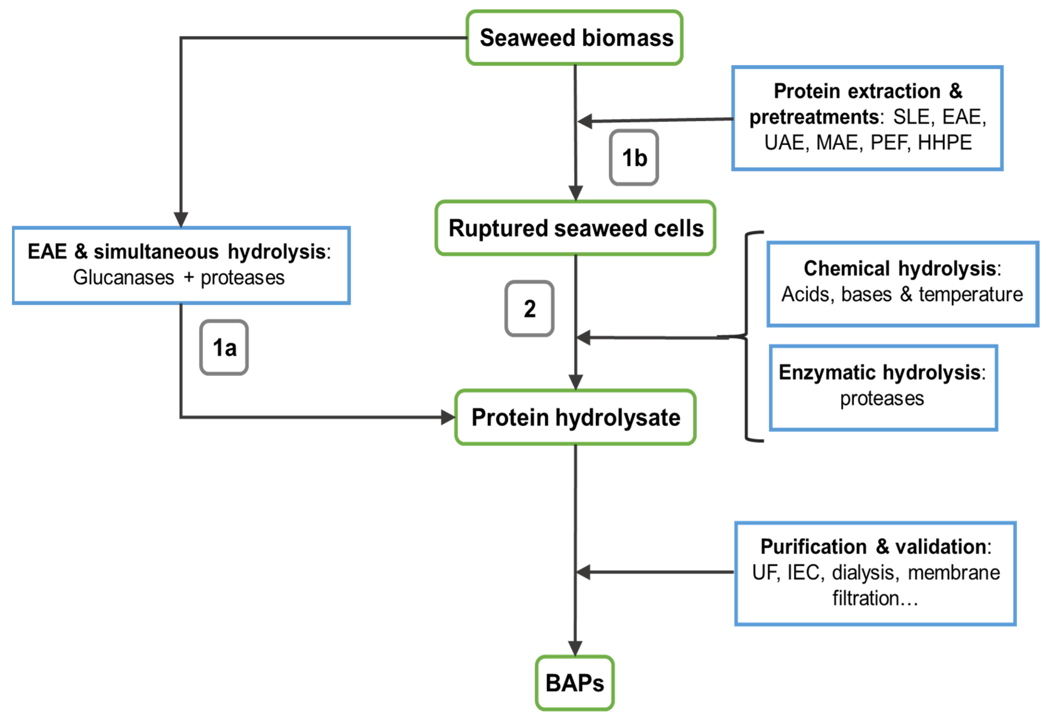
2. Extraction Technologies

Prior to hydrolysate or peptide production, SP must be released and isolated from the rest of biomass components, which requires disruption of the seaweed cell wall polysaccharides. This implies that macroalgae biomass must be subjected to a pretreatment stage prior extraction, involving different disruptive techniques, that would aid to improve the extraction yield. A combinatorial approach including a coordinate pretreatment and extraction technique increases protein recovery [22]. Pretreatment methods aim at breaking cell walls to release the intracellular fraction of biological samples, including free and cell wall-attached proteins, depending on the system of choice. Extensively used pretreatment methods thus include mechanical grinding, osmotic shock (OS), alkaline treatment, freeze-thaw, or ultrasonic sonication [4]. Since seaweeds are marine organisms, they are susceptible to strong osmotic pressures, and their cell walls may be broken by allowing the seaweed biomass to be transferred into hypotonic solutions. This may be achieved using ultrapure or de-ionized water to induce OS [23]. In the same manner, a combination of sonication and OS has been proven to increase SP yield and extractability [24]. Nonetheless, one of the most valuable pretreatment methods involves the application of glucanases to the extraction mix to maximize SP yield [25]. Using fresh, dried or freeze-dried seaweed samples may also influence the resulting SP yield, although yield variations are not significant. Drying tests on several Sargassum species revealed that freeze-dried seaweeds subjected to classical SLE yielded slightly more extractable protein in comparison to oven-dried [26]. However, Angell et al. found out that using fresh pulped Ulva ohnoi blades yielded as much as by two-fold the extractable proteins, applying the same SLE method, but following a sample management closer to that of terrestrial plants [27]. In the same manner, heat treatment exerts a differential effect on SP extractability depending on the algal biomass. For example, in the study carried out by O’ Connor et al., autoclave treatment (>121 °C) resulted in higher SP yields for P. palmata but not for F. vesiculosus or Alaria esculenta [24]. In summary, diverse pretreatment and extraction options must be specifically studied and assessed for each seaweed species, reaching meaningful rates of SP yield, stability, or digestibility.
Extraction methods currently available for protein extraction include solid–liquid extraction (SLE), enzyme-assisted extraction (EAE), pulse-electric field (PEF), high hydrostatic pressure extraction (HHPE), ultrasound-assisted extraction (UAE), and microwave-assisted extraction (MAE). In general terms, solubilization is a paramount factor that modulates protein extraction, which depends on different physicochemical conditions. In fact, proteins can be extracted by their solubilization at different pH values, involving the sequential use of differently buffered solutions. Nevertheless, proteins are generally co-extracted with other interferents, such as sugar or phenolic compounds [25,28], forcing the application of protein precipitation to achieve the isolation and purification of the extracted SP. Moreover, except for EAE, extraction methods promote an unspecific protein hydrolysis, which is also accompanied by the liberation of intracellular proteases from cell walls that may further degrade protein structures. Protein integrity is also affected by the precipitation method of choice, aimed at reaching the protein isoelectric point (IP) by acidifying the medium pH and causing the ‘salting-out’ effect, improving protein solubility using salts like ammonium sulphate, (NH4)2SO4 [29]. Yet, protein stability is not a top priority feature for hydrolysate production. Thereafter, more disruptive methods may be used to that aim, such as MAE, PFE, or UAE.

2.1. Solid–Liquid Extraction

SLE is the most traditional method used for SP extraction, involving the use of different solvents, such as distilled water, buffered solutions, and lysis surfactant-containing solutions [30]. Nonetheless, about food applications, the use of either non-toxic or easy-to-remove reagents must be employed for SP extraction. Thus, demineralized and de-ionized water are the most extensively used solvents for the application of SLE methods, as they lead to performance of OS, helping protein extraction in a cost-effective and easy manner [31]. However, this method should be optimized to improve its efficiency, modulating different factors, such as the algal biomass/solvent ratio (w:v), temperature, or time.
Temperature is a key parameter in SP extraction, as it influences protein integrity, enzymatic activity, and solubility of other cellular constituents, such as cell wall polysaccharides [24]. Indeed, hot water has been classically selected to extract algal polysaccharides by SLE, also promoting the co-extraction of proteins. Therefore, SP aqueous extraction requires low temperatures (around 4 °C) to ensure protein integrity, whereas higher temperatures can be applied for protein hydrolysate production, thus involving heat-assisted extraction (HAE) or a combination with enzymatic pretreatments [32]. Due to the heterogeneous protein composition of algal extracts, SLE procedures are generally based on two sequential extraction procedures to meet the solubility requirements of different acidic and alkaline proteins, which includes an initial OS stage followed by the application of an alkaline NaOH solution [16,27]. The process is considered food-grade, as NaOH is used for protein extraction from various food matrices [28]. A reducing agent, normally 2-mercaptoethanol, is usually added to the alkaline solution to minimize potential protein degradation, although it has been reported as a toxic compound and has been increasingly replaced by N-acetyl-L-cysteine (NAC) for food purposes [22]. That shift on pH extraction has shown beneficial results on protein extraction, obtaining enhanced extraction yields, as it was seen on the combination of acidic SLE followed by alkaline SP extraction from Ascophyllum nodosum [33]. Moreover, the application of pH-shift combined with IP precipitation, reaching pH values between 2 and 4 ensures a maximum yield with this extraction method [29]. Combining these methods, a 14% protein yield (w/w) was obtained from Porphyra dioica [34]. Besides the combination of OS and an SLE extraction of alkaline-soluble proteins with NaOH, the use of a lysis solution holding urea, detergent and other reactants, allowed for extracting 11.8% (w/w) of protein from Ulva sp. [31]. While SP extraction yields by SLE may be near the total protein content of seaweed species, it is also a very time-consuming method [32].

2.2. Enzyme-Assisted Extraction

EAE is one of the most studied techniques used to disrupt macroalgal cell walls. The application of targeted polysaccharide-digesting enzymes, such as cellulases, hemicellulases, β-glucanases, and xylanases has been described as a food-grade approach to breakdown the macroalgae cell wall. Thus, commercial enzyme cocktails have been proven to be successful for this purpose [25]. However, seaweed cell wall composition can vary among phyla and species and, therefore, a right choice of carbohydrase(s), together with the optimization of operating conditions (enzyme:substrate ratio (E:S), temperature, pH) of individual enzymes or cocktails must be proved prior to large-scale extraction to maximize protein recovery. In general, EAE has been mostly studied on red and green seaweeds, because of their simpler composition with respect to brown seaweeds. A greater protein yield has been reported on Solieria chordalis, Ulva sp. and Sargassum muticum seaweeds when using EAE and traditional SLE [35]. In accordance with a study comparing EAE using cellulase, hemicellulose, and a mixture of both to P. palmata and S. chordalis while testing HHPE, showed that cellulase alone was generally more effective and further enhanced SP yield in combination with HHPE [36]. In other study, the use of cellulase-assisted extraction (using commercially available CellicCTec3®) on brown and red seaweeds, Macrocystis pyrifera and Chondracanthus chamissoi, led to significantly higher protein yields compared to SLE, 36.10% and 74.60% respectively (Table 2), as a result of the optimization of the extraction process through a central composite experimental design [37]. Other authors reported that a higher yield of alkaline soluble protein was recovered from P. palmata following treatment with a combination of commercial glucanase cocktails (Shearzyme and Celluclast) [22]. This resulted in a total protein recovery of 8.39% when compared to that obtained following OS and mechanical shear (6.77% and 6.92%, respectively). Therefore, EAE employing glucanases may achieve high protein yield, although the co-extraction of other components (i.e., phenolics) may also occur [25].
Besides glucanases, proteases are also used in EAE of SP, to achieve the release of proteins, peptides, and amino acids present in the supernatant of extracted samples [28].

2.3. Pulse-Electric Field Assisted Extraction

The use of electric pulses to break cell membranes and walls down has been growingly investigated to aid biomolecules’ extraction. The application of PEFs can help in protein extraction from macroalgae by generating high voltage (kV) electric pulses of different length, ranging between micro and milliseconds, which result in reversible or irreversible electroporation of cell wall and membrane [43]. PEF is considered as a rapid and effective green technology that should cope with the own limitations, since conductivity and electrode gap may limit the widespread application of PEF at a larger scale [44]. PEF has been extensively applied to improve protein extraction from green seaweed species. For example, protein extraction from Ulva sp. was optimized using OS combined with PEF followed by hydraulic press treatment, increased protein extraction from 2.25% to 5.38% (w/v) [45]. Similar protein yields were reported in the same genus when using PEF combined with hydraulic pressure. Another study reported the application of PEF with a custom-made insulated gate bipolar transistor-pulsed generator coupled with a gravitation press-electrode to aid protein extraction from U. ohnoi, improving protein yield from 3.16% to 14.94% [23]. PEF coupled with mechanical pressing was employed for protein extraction from Ulva sp. An optimized protocol resulted in a seven-fold increase in total protein yield (~20% protein in the extract) when compared to an extract obtained using an OS [23]. Moreover, PEF has been regarded as a workable method for SP extraction and simultaneous hydrolyzation, as extracted proteins by this method display higher antioxidant activity than non-PFE extracted ones.

2.4. High Hydrostatyc Pressure Extraction

High hydrostatic pressure (HHP) improves extraction efficiency because of the application of pressurized conditions (up to 1000 bar) to induce cellular disruption [43]. Factors affecting HHPE efficiency include the choice of solvent along with the operating pressure, temperature, and duration. HHPE is considered as an effective green extraction technology due to its short processing time, mild operating temperature, and high recovery yields. Therefore, this technique may be suitable for heat sensitive compounds, although pressure-induced conformation changes/denaturation of proteins may need to be taken into consideration. The application of HHP (600 MPa for 4 min) to aid protein extraction from two brown seaweeds, F. vesiculosus and Alaria esculenta, and two red seaweeds, P. palmata and C. crispus was investigated [24]. HHP treatment appeared to be the most effective for protein extraction from F. vesiculosus (23.70% of total protein). On the contrary, autoclave treatment yielded higher protein content (21.50%) than the HHP treatment (14.90%) in the case of P. palmata [24]. However, in the case of S. chordalis, HPP treatment (400 MPa for 20 min) only resulted in a 2.60% increase in protein yield [24]. The use of HHPE in combination with other extraction techniques has also been investigated, particularly HHP-assisted enzyme extraction. Interestingly, it has been reported that lower temperatures and higher pressures could yield lower proportion of undesirable artifacts, such as polyphenols, and higher protein yields [36]. HHP treatment (400 MPa for 20 min) alone did not increase protein yield from dried ground P. palmata, while HHP-assisted hydrolysis with cellulase and hemicellulase resulted in a 17% increase in protein yield compared to the control. Managing extraction parameters to lower polyphenol extraction would contribute to the optimization and enhancement of SP yield, although further research is still needed to assess HHPE as a workable, specific SP extraction method.

2.5. Ultrasound-Assisted Extraction

The use of UAE whether as sonication pretreatment or central component of the extraction process has gained significant interest in maximizing algal protein extraction. This technique cause cell wall breakdown by the acoustic cavitation phenomenon, in which the implosion of air bubbles results in the generation of a potent mechanical energy that disrupts the cell wall [46]. The main advantages of this extraction method are its short processing time, the independence of temperature and the use of water as the solvent of choice, which also plays a meaningful factor in the case of algal samples, thanks to their susceptibility to OS [39]. However, the application of high ultrasound power for extended periods of time can lead to an excessive heat production that may significantly alter protein structure. UAE can be used simultaneously with other techniques, such as OS and EAE. These integrated approaches have led to increasing protein extraction yields. Combination of UAE and EAE with a cellulase cocktail promoted a significantly higher phycobiliprotein yield from the red seaweed Grateloupia turuturu, compared to treatment with EAE alone [32] (Table 2). UAE using NaOH as solvent allowed for obtaining a 3.4% protein yield (w/w) from the red seaweed N. haitanensis in just 20 min of sonication at room temperature [38]. In addition, O’ Connor et al. proved that sonication combined with ammonium sulfate precipitation resulted in the highest protein recovery from F. vesiculosus and Chondrus crispus, with yields of 35.1% and 35.5% of total protein, respectively [24] (Table 2). This was compared to other approaches, like HHPE or laboratory autoclave treatment that yielded protein recoveries ranging from 16.1% to 24.3% out of total protein. Application of UAE as a food-grade extraction protocol at a larger scale has been previously proved in the protein extraction from Ulva sp. and Gracilaria sp., allowing to recover 70% and 86% of total proteins, respectively [39] (Table 2).

2.6. Microwave-Assisted Extraction

During the application of MAE, microwave energy is converted into heat by ion conduction and dipole rotation, and non-polar compounds are not heated [43]. Therefore, MAE may not be suitable for the extraction of heat-sensitive bioactive compounds. Although it has been recognized as an efficient low-energy extraction approach, MAE has to date been more widely used to extract carbohydrate or phenolic compounds rather than bioactive proteins/peptides from seaweed samples [40]. Microwave-derived heating and ionization can promote the release of intracellular components from the matrix into the extracting solution, partly because of the damage effect of microwave on cell wall and cell membrane. Because of the high ash content of edible seaweeds contributing to ionic conduction, MAE constitutes a powerful technique to be applied to macroalgae protein extraction. Compared with conventional extraction, MAE shortens extraction time and reduces solvent consumption, as reported for the application of acid/alkali SLE and MAE to U. ohnoi, where MAE achieved higher SP yield (23% dw) after 20 min, while SLE extraction required at least 24 h to yield a significant amount of protein from seaweeds [22] (Table 2). Most importantly, although SLE reached higher yields, in this study MAE was conducted in just 5 min and only with the aqueous SP fraction [40]. In summary, although MAE efficiency was reported to be lower than SLE in terms of SP yield, processing times can be reduced saving energy consumption and thus improving the efficiency of this technique [47].

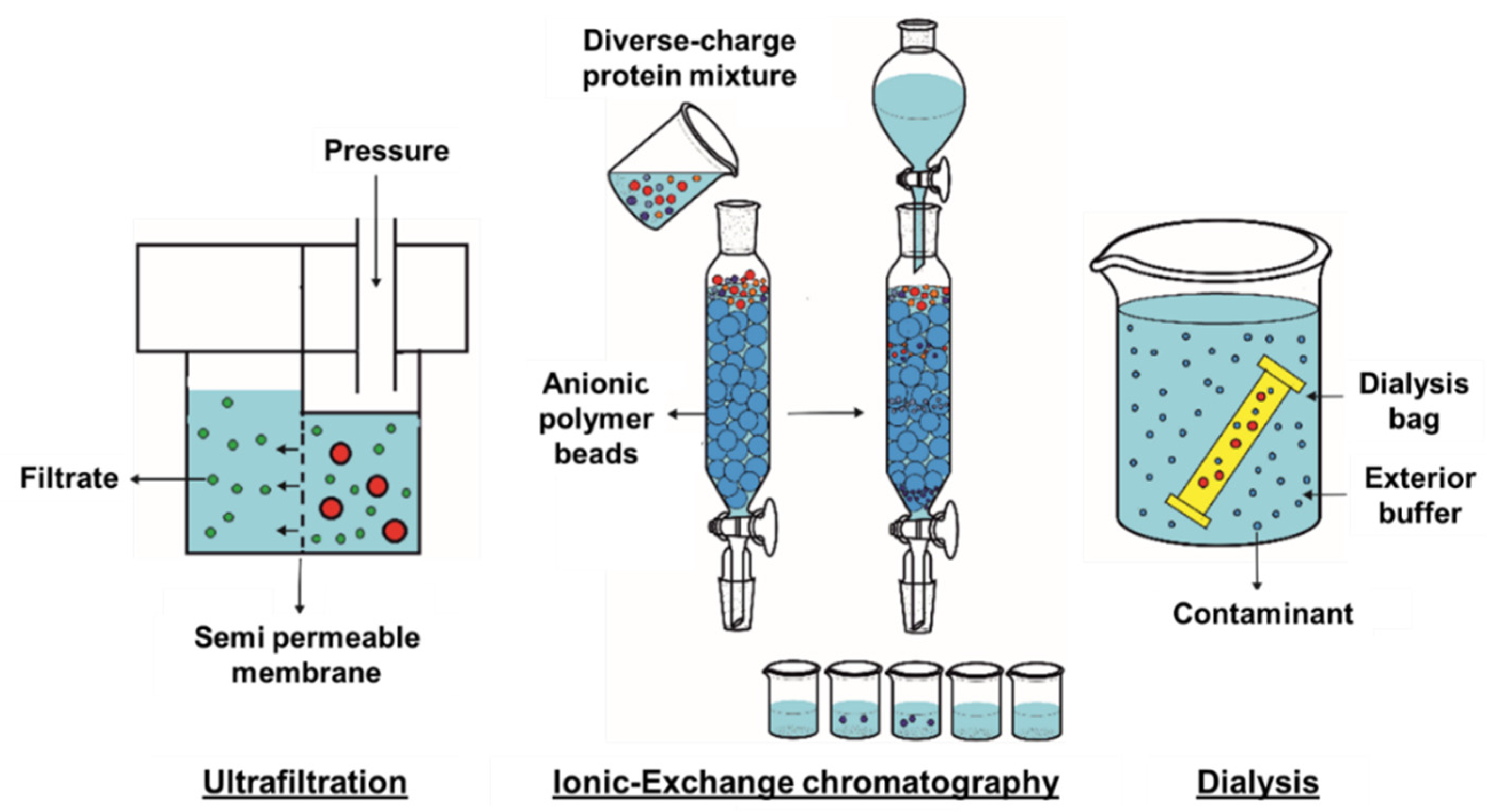
3. Protein Purification

After extraction, SP should be purified to drop other co-extracted components, represented by polysaccharides, minerals, and phenolic compounds. Protein purification methods are based on molecular charge and size differences, and the most extensively used methods are ultrafiltration (UF), ionic-exchange chromatography (IEC), and dialysis.

3.1. Ultrafiltration

UF is an important and widespread purification technique developed by membrane filtration and based on the separation of biological molecules according to their molecular mass [48,49]. The method is suitable for purifying proteins from low-mass contaminating molecules and can be used to concentrate protein solutions at the same time (Figure 1). UF membranes must supply great separation ability, high flux value, and good mechanical and chemical properties. The membrane carrier must be chemically inert with excellent mechanical properties. The device should provide an easy way to remove the deposited layer as well as easy washing and sanitation of the membrane [50]. By the action of pressure force, solvents and molecules of low molecular weight pass through the membrane, while larger molecules stay trapped. If finer separation is needed, the UF method needs to be coupled to more selective methods, such as chromatography or electrophoresis [48,51].

3.2. Ionic-Exchange Chromatography

Regarding its use on SP purification, this method is more extensively used in SP hydrolysates fractionation. Therefore, use of UF allows separating very low-molecular weight compounds from the extracted matrix and is especially useful for purifying BAPs after enzymatic hydrolysis of SP. Indeed, different studies used UF membranes of <1 kDa molecular weight for purifying an enzymatic hydrolysate of Ulva sp. and yielded fractions with greater anti-inflammatory potential and inhibitory capacity against the angiotensin I-converting enzyme (ACE) [52]. Use of UF to separate low-molecular weight peptides from Ulva rigida hydrolysates doubled the ACE inhibitory activity of the sample in comparison to non-filtered hydrolysate [53]. Therefore, this technique may prove useful for concentrating BAPs and SP hydrolysates.
IEC is widely used for purification of charged biomolecules such as proteins, peptides, or amino acids [54,55]. IEC presents a high performance, because it can be applied to a large number of different proteins using high-affinity and cost-effective buffers. The basis of IEC is the interaction of charged molecules flowing in the mobile phase with charged groups of the stationary phase (column packing matrix). Amino acids present charges of different polarity, for example lysine and arginine have a positive charge, while aspartic acid and glutamic acid are negatively charged at physiological pH [56]. Thus, each protein has variable net charges based on its aminoacidic composition but also depending on the pH of the solution they are dissolved in. Thus, the proteins’ IP is key to purify these by IEC, since it will find the best initial conditions for protein purification. Ion exchangers are divided into two classes: strong and weak. Strong ion exchange ligands keep their charge characteristics and ion exchange capacity in a wide range of pH, while weak ion exchange ligands show a more pronounced change in their exchange capacity with pH modifications [57]. If purification is conducted at a pH above 9 for anion exchange or below 6 for cation exchange, then a strong ion exchanger must be used. However, if purification is performed at less extreme pH values, then both strong and weak ion exchangers should be used, and the obtained results should be compared to optimize the purification process. Protein binding and elution is based on the competition between the charged amino acid groups on the protein surface and the charged conditions in the binding buffer for the oppositely charged groups in the stationary phase (Figure 1).
The protein sample is applied to an ion exchange column in a low salt solution [55,56,57,58]. The charged groups on the protein have a high binding affinity for the charged counterions on the ion exchange resin. Different affinity columns may be used to keep proteins and peptides, depending on their functional group. For example, sulfopropyl functional groups, as strong cation exchangers may keep molecules on a range of pH 4 to 13. Conversely, diethylaminoethyl is a weak anionic exchanger and may retain molecules in pH from 2 to 9 [59]. Regarding the use of this technique in SP, the most commonly used columns are anionic exchangers [60]. For example, IEC allowed for accurate fractionation of anticoagulant peptides from a Neopyropia yezoensis hydrolysate [61].

3.3. Dyalisis

Dialysis is a common method for removing contaminants by selective and passive diffusion through a semipermeable membrane, such as a dialysis tube. Dialysis is most used to remove small molecules in solution, such as salts, reducing agents, or preservatives, among others. This technique is a simple but time-demanding method since separation depends on diffusion rate. The dialysis system consists of a tube, which holds a semipermeable membrane, e.g., porous cellulose, closed on both sides to contain a protein solution [62]. The tube is immersed in a much larger vessel filled with buffer (Figure 1). Low-molecular weight particles will diffuse through the semipermeable membrane, while protein molecules, due to their larger size, will remain inside the tube. On the other hand, the buffers inside and outside the dialysis tube will change during diffusion until equilibrium is reached. This equilibrium obeys the Donnan effect, which keeps electrical neutrality on both sides of the membrane [63]. The process of diffusion of small molecules through the membrane is accompanied by the increased movement of protein molecules inside the dialysis tube. As they cannot diffuse due to their size, proteins remain inside the membrane. It should be noted that polyvalent proteins do not allow charged particles to move away from the membrane surface, thus preventing them from setting up an equilibrium potential [64]. On the other hand, the Donnan effect has a significant effect on the colligative properties of ions in the case of high concentrations of proteins or buffers with low ionic strength. Finally, during the dialysis process, the buffer inside the dialysis tube is gradually replaced with the solution in the outer vessel. In this way, efficient removal of small contaminants from the tube is achieved [48]. In summary, dialysis is indeed one of the most used techniques for separating peptides and proteins from natural matrixes. Hence, pairing high-yield extraction techniques with dialysis allows obtaining SP isolates with greater protein content compared to other purification methods [24,65].

4. Hydrolysis and Peptide Production

Protein hydrolysis allows producing protein hydrolysates and BAPs from seaweed. Figure 2 schematically depicts the steps involved in seaweed BAPs production. As mentioned above, pre-treatment strategies may be used to both simplify the extraction process and enhance hydrolyzed SP yield. While some SP hydrolysates have been reported to be biologically active, SP-derived BAPs have gained much attention due to their higher bioactive potential. Hence, once extracted and isolated, SP need to be hydrolyzed into smaller peptide sequences to become biologically active [66]. Hydrolysis can be done either chemically or using proteases. Chemical hydrolysis is typically performed using acids and/or alkali at temperatures over 40 °C. While the procedure is simple and inexpensive, the composition of the hydrolysate is difficult to control, also yielding products with modified amino acids in their peptide sequence [67]. For example, strong organic acids can efficiently hydrolyze hydrophobic peptide bonds, as a mixture of HCl and trichloroacetic acid was found to destroy tryptophan [68]. Conversely, alkaline hydrolysis with NaOH can reduce cystine, arginine, threonine, serine, and isoleucine; form unusual amino acid residues such as lysinoalanine or lanthionine; as well as induce isomerization of L-lysine residues into D-lysine ones [69]. Similarly, high temperatures may be used to induce hydrolyzation, without need of aiding chemicals [42].
In contrast, enzymatic hydrolysis is performed at lower temperature in order to preserve the native structure of the used protease conducting the hydrolysis process. In addition, enzymatic hydrolysis is more specific to the desired peptides and results in higher peptide yield. Due to the lack of organic solvents, the purification steps are less labor-intensive and side reactions are less pronounced [70,71]. The experimental conditions of enzymatic hydrolysis are typically governed by the needs of the enzyme. The process can be done in a single or, at times, in multiple steps [72]. Substrate-to-enzyme ratio as well as the duration of the hydrolysis have a significant effect on the nature and yield of the final products [70]. Table 3 illustrates a range of bioactive peptides obtained by enzymatic hydrolysis from seaweed with the aid of a number of proteases. The most extensively used proteases to perform protein hydrolysis are trypsin, pepsin, papain, and α-chymotrypsin. They are used both individually and in combination with other proteases. More recently, commercially available enzyme cocktails including proteases and carbohydrases are used (Table 3).
A range of experimental conditions (temperature, pH, agitation speed, enzyme-to-substrate ratio, hydrolysis time) need to be optimized for best bioactivity. Typically, a mixture of peptides is obtained, of which only a few are biologically active. The active ones are typically isolated from the mixture using membrane filtration and reverse-phase chromatography techniques [90,91]. SP bioactive peptides range in sizes from 2 to 20 amino acids with the cut-off masses usually ranging from 5 kDa to 30 kDa [92]. Nonetheless, as the nature of SP varies for each seaweed, the resulting BAP profile will also be different depending on the hydrolysis conditions and protease of choice, as these account for different cut-off points [93]. The use of pepsin or trypsin may be also useful to obtain insight on in vivo SP degradation and potential bioavailability of BAPs. Indeed, a wide range of commercially available enzymes has been evaluated to screen out for the possible BAPs produced, such as Flavourzyme, Corolase, or fungal and bacterial proteases [75,76]. The type of the chosen proteolytic enzyme has been shown to have impact on the resulting bioactivity, as it defines the resulting BAP profile. In a recent study, Gracilariopsis lemaneiformis proteins were hydrolyzed by different proteases such as trypsin, pepsin, papain, α-chymotrypsin, or Alcalase and hydrolysates obtained by α-chymotrypsin hydrolysis displayed the highest antioxidant activity [81]. Studies performed by Fitzgerald et al. and Harnedy et al. proved that using papain or Corolase yielded highly different BAP in P. palmata protein isolates hydrolysis [73,75]. Hydrolysis is stopped by enzyme denaturation, often achieved with pH shifts or heating. One disadvantage of thermal deactivation is the fact that higher temperatures might speed up the kinetics of unwanted processes. An approach to overcome this problem is enzyme immobilization on solid substrates. The use of immobilized enzymes enables the enzymes to be separated from the hydrolysis mixture by filtration instead of increased temperature. An additional advantage is that immobilized enzymes are prevented from being aggregated with the peptides [94]. One challenge of enzymatic hydrolysis is the fact that the algal proteome needs to be known prior to hydrolysis so that enzymes with according cut sites are used. To improve the purity and yield of BAPs, enzymatic hydrolysis is often preceded by physical cell disruption methods, including PEF and bead milling [95,96]. Nonetheless, the hydrolytic activity of the chosen protease can be inhibited by co-extracted compounds such as polysaccharides or phenolic compounds, and optimization studies must be conducted [82,97]. Hydrolysates may be purified by the above-mentioned techniques to ensure concentration of lower molecular weight peptides or to obtain purified BAPs. In this sense, sequential purification processes seem to work best for obtaining high-purity peptides [39].

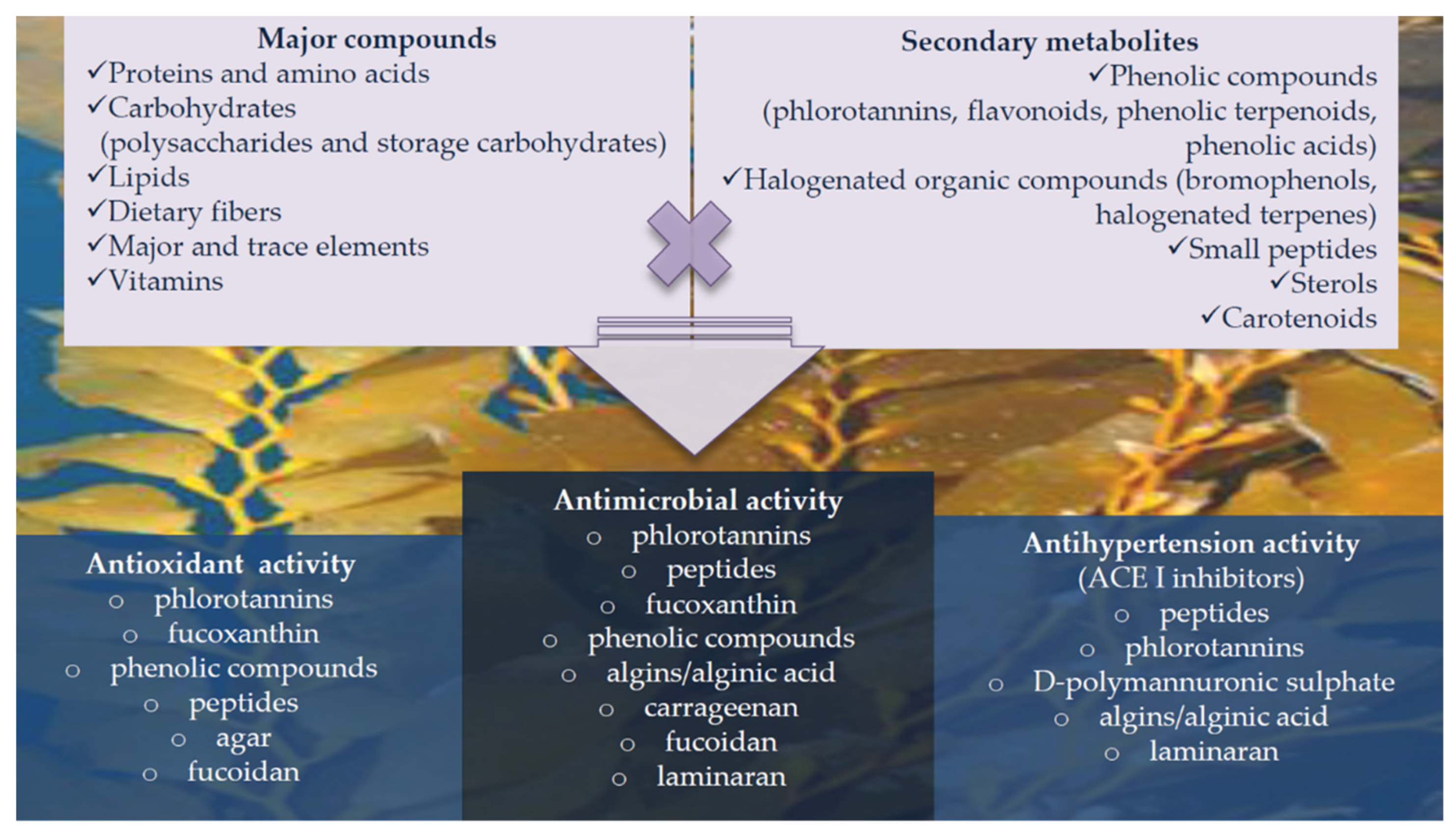
5. Bioactive Properties and Applications of Seaweed Proteins and Derived Products

As mentioned throughout this work, proteins and derived products obtained from seaweed show various biological activities, as reported by in vitro and a few in vivo tests, such as antioxidant, antimicrobial, anti-inflammatory, antihypertensive, or even antitumor [1]. Regarding SP, the extent of their displayed bioactivities varies upon protein composition, season, but also hydrolysis degree and purification methods applied. The three main bioactivities consistently reported for SP and BAPs from different species are antioxidant, anti-inflammatory and antihypertensive [97,98]. In vitro antioxidant activity is usually tested against oxidation-sensible molecules, such as 2,2-diphenyl-1-picrylhydrazyl (DPPH) or 2,2’-Azino-bis(3-ethylbenzothiazoline-6-sulfonic acid) (ABTS). It has been reported that SP derived peptides display higher antioxidant activity than SP hydrolysates [76]. These could be due to the concentration and better accessibility of released amino acids from folded proteins. Concentrated 1 < kDa BAPs from P. dioica hydrolysates showed significant antioxidant activity (20.88 µmol Trolox equivalents) in a wide range of assays, including DPPH and ABTS [78]. Comparing results with free aminoacidic composition of the isolated fractions, the authors suggested that higher presence of sulfated amino acids, like cysteine or methionine, could be related to these results, as proved by these in vitro assays. Different results were reported for Macrocystis pyrifera and Chondracanthus chamissoi BAPs, accounting for 193 and 167 µmol Trolox equivalents respectively, in a DPPH assay [37]. Antioxidant assays with P. palmata hydrolysates also suggested that a higher protein hydrolysis degree was related to higher antioxidant activity [93].
Anti-inflammatory assays are often conducted on cell culture tests, assessing production or inhibition of pro-inflammatory mediators, such as interleukins. SP hydrolysates produced using several proteases from Ulva sp. were found to exert significant anti-inflammatory activity by upregulating interleukin 10 and inhibiting tumor necrosis factor alpha (TNF-α) expression [52]. Hydrolysates obtained from Pyropia columbina also displayed anti-inflammatory properties in vitro with similar tested mechanisms, while also inhibiting expression of necrosis factor κB [77].
Yet, the most well studied bioactive property of SP, derived hydrolysates, and BAPs is their hypotensive activity [86]. These properties may be assessed in vitro by checking the inhibition capacity of these molecules against key cardiac tension enzymes such as ACE, being the most extensively used model, but also renin or endothelin I in a lesser extent [86]. Conversely, in vivo tests may be conducted monitoring blood pressure and cardiac rhythm in animal models. The previously mentioned study of Vasquez et al. showed that the M. pyrifera extract displayed an ACE inhibition of 38.8% compared to control and that the smaller the size of the purified peptides, the greater inhibition degree was [37]. However, C. chamissoi extracts did not show a detectable ACE-inhibitory activity. Authors proposed that this could be due to interactions between co-extracted compounds and ACE enzymatic reaction products [37]. Protein hydrolysates from P. palmata, showed that ACE inhibitory activity was independent from the time of harvesting, but was related to its hydrolysis degree [93]. Nine ACE-inhibitory peptides derived from phycobiliproteins were found, and the oligopeptide LRY demonstrated particularly high inhibitory activities, with an IC50 of 0.044 mM [99]. Similarly, the peptide PAFG, obtained from U. clathrata protein, showed an IC50 value of 35.9 mM on ACE-inhibitory activity [83] (Table 3). Moreover, since BAPs are obtained through different hydrolysis methods, these have shown to be stable towards gastrointestinal proteases like pepsin or trypsin, as well as the acidic pH of digestive reactions [37,83].
Other reported bioactivities include antidiabetic or antimicrobial, associated to phycobiliproteins and lectins, but also some SP hydrolysates [100]. Antidiabetic activity was characterized in P. palmata hydrolysates through in vitro inhibition of dipeptidyl peptidase IV (DPP-IV). Results showed that Corolase-hydrolyzed SP accounted for a much significant DPP-IV inhibition (IC50 = 1.65 mg/mL) [93]. BAPs from Saccharina longricuris displayed antimicrobial activity against several bacterial pathogens, such as Escherichia coli, Staphylococcus aureus, or Pseudomonas aeruginosa, achieving a 40% growth inhibition at 2.5 mg peptides/mL [88].
Therefore, based on their reported bioactivities, these macroalgae molecules may be used for diverse industrial applications (Figure 3).
Both antioxidant and antimicrobial properties make SP hydrolysates a desirable choice for their use as food additives, preservatives, and protein fortifiers. Additionally, the ACE-inhibitory and anti-inflammatory activities widely apparent in several SP, could make them to be assessed as nutraceuticals, functional ingredients for food and feed, or cosmetic ingredients [101]. The fact that this effect has been assessed in vivo, with Undaria pinnatifida BAPs allowing for reduced blood pressure in rats, could be argued to also be the basis for developing new hypotensive pharmaceuticals [42,87]. Moreover, SP hydrolysates keep their bioactive properties when combined with other food matrices [17,102]. In the same way, the antioxidant properties attributed to BAPs make them excellent natural ingredients for cosmetic applications, as SP also hold significantly higher levels of taurine compared to other vegetal sources [91]. In summary, the full set of already reported bioactivities and the ongoing research to fully characterize the properties and production of SP and derived molecules will unveil the full extent of the potential applications of these natural molecules.
Development of such applications would thus confer an added value to this underexploited natural resource. Furthermore, these properties are also well accounted for co-extracted bioactive compounds in SP extractions, such as cell wall polysaccharides (e.g., carrageenan, fucoidan…) or phenolic compounds, such as brown seaweeds phlorotannins [103]. Altogether, seaweed hold several different bioactive molecules with similar reported activities, that in the case of some extracts, could act in cooperation [104] Thus, SP could be considered as part of the whole composition of health-promoting compounds of seaweeds and their extracts be applied for a wide diversity of applications [105] (Figure 3).

6. Conclusions

SP and especially their hydrolysates and several identified BAPs, have been repeatedly reported to show significant bioactivities such as antioxidant or anti-inflammatory, but most importantly, antihypertensive. Their bioactivities raise potential applications in food and animal feed, whether as functional additives or colorants in the case of phycobiliproteins. On the other hand, BAPs may find use as cosmetic ingredients with antioxidant activity, while thanks to their antihypertensive activity, they can be used as nutraceuticals in the pharmaceutical industry. Nonetheless, these seaweed components need more extensive and in-depth research to fully characterize their bioactive properties, production methods, and potential in vivo effects.

Author Contributions

Conceptualization, J.E., M.F.-C., P.G.-P., J.X. and M.A.P.; Methodology, J.E., M.F.-C., J.X. and P.G.-P.; Software, J.E., M.F.-C. and P.G.-P.; Validation, M.F.-C. and P.G.-P.; Formal analysis, J.E., M.F.-C., J.P.-D., E.H.A. and M.R.; Investigation, J.E., M.F.-C., J.P.-D., E.H.A. and M.R.; Data curation, J.X.; Writing—original draft preparation, J.E. and M.A.P.; Writing—review and editing, J.E., M.F.-C., P.G.-P., J.P.-D., E.H.A. and M.R., Visualization, J.S.-G., J.X. and M.A.P.; Supervision, J.S.-G., J.X. and M.A.P. All authors have read and agreed to the published version of the manuscript.

Funding

The authors are grateful to the program Grupos de Referencia Competitiva (GRUPO AA1-GRC 2018) that supports the work of J. Echave, to the Bio Based Industries Joint Undertaking (JU) under grant agreement No. 888003 UP4HEALTH Project (H2020-BBI-JTI-2019) that supports the work of P. Garcia-Perez. The JU receives support from the European Union’s Horizon 2020 research and innovation program and the Bio Based Industries Consortium. The project SYSTEMIC Knowledge Hub on Nutrition and Food Security has received funding from national research funding parties in Belgium (FWO), France (INRA), Germany (BLE), Italy (MIPAAF), Latvia (IZM), Norway (RCN), Portugal (FCT), and Spain (AEI) in a joint action of JPI HDHL, JPI-OCEANS, and FACCE-JPI, launched in 2019 under the ERA-NET ERA-HDHL (No. 696295).

Institutional Review Board Statement

Not applicable.

Data Availability Statement

Not applicable.

Acknowledgments

We would like to thank MICINN for supporting the Ramón y Cajal grant for M.A. Prieto (RYC-2017-22891), and Xunta de Galicia for supporting the program EXCELENCIA-ED431F 2020/12, the post-doctoral grant of M. Fraga-Corral (ED481B-2019/096). The authors acknowledge the Ministry of Education, Science and Technological Development of the Republic of Serbia for the support (grants nos. 451-03-9/2021-14/200026 and 451-03-09/2021-14/200378).

Conflicts of Interest

The authors declare no conflict of interest.

Abbreviations

SPSeaweed proteins
dwDry weight
SLESolid–liquid extraction
HAEHeat-assisted extraction
EAEEnzyme-assisted extraction
UAEUltrasound-assisted extraction
MAEMicrowave-assisted extraction
PEFPulse-electric field
HHPHigh-hydrostatic pressure
HHPEHigh-hydrostatic pressure extraction
HAEHeat-assisted extraction
EAEEnzyme-assisted extraction
UAEUltrasound-assisted extraction
MAEMicrowave-assisted extraction
IPIsoelectric point
PrPrecipitation
PuPurification
DIDialysis
OSOsmotic shock
EprEnzymatic pretreatment
UprUltrasound pretreatment
dWDeionized water
AKAlkaline
MAAsMycosporine-like amino acids
EAAEssential amino acids
GRASGenerally recognized as safe
RAASRenin–angiotensin–aldosterone system
IC50Half inhibitory concentration
DPP-IVDipeptidyl peptidase IV
MCF-7Human breast cancer cell line
HepG2Human liver cancer cell line
SGC-7901Human gastric cancer cell line
A549Human lung cancer cell line
HT-29Human colon cancer cell line
UFUltrafiltration
IECIonic-exchange chromatography
BAPBioactive peptide
DPPH2,2-diphenyl-1-picrylhydrazyl
ABTS2,2’-Azino-bis(3-ethylbenzothiazoline-6-sulfonic acid)
ACEAngiotensin I converter enzyme

References

  1. Carpena, M.; Caleja, C.; Pereira, E.; Pereira, C.; Ćirić, A.; Soković, M.; Soria-Lopez, A.; Fraga-Corral, M.; Simal-Gandara, J.; Ferreira, I.C.F.R.; et al. Red Seaweeds as a Source of Nutrients and Bioactive Compounds: Optimization of the Extraction. Chemosensors 2021, 9, 132. [Google Scholar] [CrossRef]
  2. Garcia-Oliveira, P.; Fraga-Corral, M.; Pereira, A.G.; Prieto, M.A.; Simal-Gandara, J. Solutions for the sustainability of the food production and consumption system. Crit. Rev. Food Sci. Nutr. 2020, 1–17. [Google Scholar] [CrossRef]
  3. Bleakley, S.; Hayes, M. Algal proteins: Extraction, application, and challenges concerning production. Foods 2017, 6, 33. [Google Scholar] [CrossRef] [Green Version]
  4. Pliego-Cortés, H.; Wijesekara, I.; Lang, M.; Bourgougnon, N.; Bedoux, G. Current knowledge and challenges in extraction, characterization and bioactivity of seaweed protein and seaweed-derived proteins. In Advances in Botanical Research; Elsevier Ltd.: Amsterdam, The Netherlands, 2020; Volume 95, pp. 289–326. ISBN 9780081027103. [Google Scholar]
  5. Ohanenye, I.C.; Tsopmo, A.; Ejike, C.E.C.C.; Udenigwe, C.C. Germination as a bioprocess for enhancing the quality and nutritional prospects of legume proteins. Trends Food Sci. Technol. 2020, 101, 213–222. [Google Scholar] [CrossRef]
  6. Lorenzo, J.M.; Agregán, R.; Munekata, P.E.S.; Franco, D.; Carballo, J.; Şahin, S.; Lacomba, R.; Barba, F.J. Proximate composition and nutritional value of three macroalgae: Ascophyllum nodosum, Fucus vesiculosus and Bifurcaria bifurcata. Mar. Drugs 2017, 15, 360. [Google Scholar] [CrossRef] [PubMed] [Green Version]
  7. Denis, C.; Morançais, M.; Li, M.; Deniaud, E.; Gaudin, P.; Wielgosz-Collin, G.; Barnathan, G.; Jaouen, P.; Fleurence, J. Study of the chemical composition of edible red macroalgae Grateloupia turuturu from Brittany (France). Food Chem. 2010, 119, 913–917. [Google Scholar] [CrossRef]
  8. Fleurence, J.; Morançais, M.; Dumay, J. Seaweed Proteins, 2nd ed.; Yada, R.Y.B.T.-P., Ed.; Elsevier Ltd.: Amsterdam, The Netherlands, 2018; ISBN 9780081007297. [Google Scholar]
  9. Dumay, J.; Morançais, M.; Munier, M.; Le Guillard, C.; Fleurence, J. Phycoerythrins. In Advances in Botanical Research; Elsevier: Amsterdam, The Netherlands, 2014; Volume 71, pp. 321–343. ISBN 9780124080621. [Google Scholar]
  10. Batista, S.; Pereira, R.; Oliveira, B.; Baião, L.F.; Jessen, F.; Tulli, F.; Messina, M.; Silva, J.L.; Abreu, H.; Valente, L.M.P. Exploring the potential of seaweed Gracilaria gracilis and microalga Nannochloropsis oceanica, single or blended, as natural dietary ingredients for European seabass Dicentrarchus labrax. J. Appl. Phycol. 2020, 32, 2041–2059. [Google Scholar] [CrossRef]
  11. Bjarnadóttir, M.; Aðalbjörnsson, B.V.; Nilsson, A.; Slizyte, R.; Roleda, M.Y.; Hreggviðsson, G.Ó.; Friðjónsson, Ó.H.; Jónsdóttir, R. Palmaria palmata as an alternative protein source: Enzymatic protein extraction, amino acid composition, and nitrogen-to-protein conversion factor. J. Appl. Phycol. 2018, 30, 2061–2070. [Google Scholar] [CrossRef]
  12. Dawczynski, C.; Schubert, R.; Jahreis, G. Amino acids, fatty acids, and dietary fibre in edible seaweed products. Food Chem. 2007, 103, 891–899. [Google Scholar] [CrossRef]
  13. Biancarosa, I.; Espe, M.; Bruckner, C.G.; Heesch, S.; Liland, N.; Waagbø, R.; Torstensen, B.; Lock, E.J. Amino acid composition, protein content, and nitrogen-to-protein conversion factors of 21 seaweed species from Norwegian waters. J. Appl. Phycol. 2017, 29, 1001–1009. [Google Scholar] [CrossRef]
  14. Ortiz, J.; Uquiche, E.; Robert, P.; Romero, N.; Quitral, V.; Llantén, C. Functional and nutritional value of the Chilean seaweeds Codium fragile, Gracilaria chilensis and Macrocystis pyrifera. Eur. J. Lipid Sci. Technol. 2009, 111, 320–327. [Google Scholar] [CrossRef] [Green Version]
  15. Ortiz, J.; Romero, N.; Robert, P.; Araya, J.; Lopez-Hernández, J.; Bozzo, C.; Navarrete, E.; Osorio, A.; Rios, A. Dietary fiber, amino acid, fatty acid and tocopherol contents of the edible seaweeds Ulva lactuca and Durvillaea antarctica. Food Chem. 2006, 99, 98–104. [Google Scholar] [CrossRef]
  16. Chen, X.; Wu, M.; Yang, Q.; Wang, S. Preparation, characterization of food grade phycobiliproteins from Porphyra haitanensis and the application in liposome-meat system. LWT 2017, 77, 468–474. [Google Scholar] [CrossRef]
  17. Fitzgerald, C.; Gallagher, E.; Doran, L.; Auty, M.; Prieto, J.; Hayes, M. Increasing the health benefits of bread: Assessment of the physical and sensory qualities of bread formulated using a renin inhibitory Palmaria palmata protein hydrolysate. LWT-Food Sci. Technol. 2014, 56, 398–405. [Google Scholar] [CrossRef]
  18. Deniaud, E.; Fleurence, J.; Lahaye, M. Preparation and chemical characterization of cell wall fractions enriched in structural proteins from Palmaria palmata (Rhodophyta). Bot. Mar. 2003, 46, 366–377. [Google Scholar] [CrossRef]
  19. McLaughlin, C.M.; Harnedy-Rothwell, P.A.; Lafferty, R.A.; Sharkey, S.; Parthsarathy, V.; Allsopp, P.J.; McSorley, E.M.; FitzGerald, R.J.; O’Harte, F.P.M. Macroalgal protein hydrolysates from Palmaria palmata influence the ‘incretin effect’ in vitro via DPP-4 inhibition and upregulation of insulin, GLP-1 and GIP secretion. Eur. J. Nutr. 2021. [Google Scholar] [CrossRef]
  20. Fan, X.; Bai, L.; Mao, X.; Zhang, X. Novel peptides with anti-proliferation activity from the Porphyra haitanesis hydrolysate. Process Biochem. 2017, 60, 98–107. [Google Scholar] [CrossRef]
  21. Lourenço-Lopes, C.; Fraga-Corral, M.; Jimenez-Lopez, C.; Pereira, A.G.; Garcia-Oliveira, P.; Carpena, M.; Prieto, M.A.; Simal-Gandara, J. Metabolites from macroalgae and its applications in the cosmetic industry: A circular economy approach. Resources 2020, 9, 101. [Google Scholar] [CrossRef]
  22. Harnedy, P.A.; FitzGerald, R.J. Extraction of protein from the macroalga Palmaria palmata. LWT-Food Sci. Technol. 2013, 51, 375–382. [Google Scholar] [CrossRef]
  23. Robin, A.; Kazir, M.; Sack, M.; Israel, A.; Frey, W.; Mueller, G.; Livney, Y.D.; Golberg, A. Functional Protein Concentrates Extracted from the Green Marine Macroalga Ulva sp., by High Voltage Pulsed Electric Fields and Mechanical Press. ACS Sustain. Chem. Eng. 2018, 6, 13696–13705. [Google Scholar] [CrossRef]
  24. O’ Connor, J.; Meaney, S.; Williams, G.A.; Hayes, M. Extraction of Protein from Four Different Seaweeds Using Three Different Physical Pre-Treatment Strategies. Molecules 2020, 25, 2005. [Google Scholar] [CrossRef]
  25. Mendez, R.L.; Kwon, J.Y. Effect of extraction condition on protein recovery and phenolic interference in Pacific dulse (Devaleraea mollis). J. Appl. Phycol. 2021, 33, 2497–2509. [Google Scholar] [CrossRef]
  26. Wong, K.; Chikeung Cheung, P. Influence of drying treatment on three Sargassum species 2. Protein extractability, in vitro protein digestibility and amino acid profile of protein concentrates. J. Appl. Phycol. 2001, 13, 51–58. [Google Scholar] [CrossRef]
  27. Angell, A.R.; Paul, N.A.; de Nys, R. A comparison of protocols for isolating and concentrating protein from the green seaweed Ulva ohnoi. J. Appl. Phycol. 2017, 29, 1011–1026. [Google Scholar] [CrossRef]
  28. Naseri, A.; Marinho, G.S.; Holdt, S.L.; Bartela, J.M.; Jacobsen, C. Enzyme-assisted extraction and characterization of protein from red seaweed Palmaria palmata. Algal Res. 2020, 47, 101849. [Google Scholar] [CrossRef]
  29. Veide Vilg, J.; Undeland, I. pH-driven solubilization and isoelectric precipitation of proteins from the brown seaweed Saccharina latissima—effects of osmotic shock, water volume and temperature. J. Appl. Phycol. 2017, 29, 585–593. [Google Scholar] [CrossRef] [PubMed] [Green Version]
  30. Garcia-Vaquero, M.; Lopez-Alonso, M.; Hayes, M. Assessment of the functional properties of protein extracted from the brown seaweed Himanthalia elongata (Linnaeus) S. F. Gray. Food Res. Int. 2017, 99, 971–978. [Google Scholar] [CrossRef] [Green Version]
  31. Wijesekara, I.; Lang, M.; Marty, C.; Gemin, M.P.; Boulho, R.; Douzenel, P.; Wickramasinghe, I.; Bedoux, G.; Bourgougnon, N. Different extraction procedures and analysis of protein from Ulva sp. in Brittany, France. J. Appl. Phycol. 2017, 29, 2503–2511. [Google Scholar] [CrossRef]
  32. Le Guillard, C.; Dumay, J.; Donnay-Moreno, C.; Bruzac, S.; Ragon, J.Y.; Fleurence, J.; Bergé, J.P. Ultrasound-assisted extraction of R-phycoerythrin from Grateloupia turuturu with and without enzyme addition. Algal Res. 2015, 12, 522–528. [Google Scholar] [CrossRef] [Green Version]
  33. Kadam, S.U.; Álvarez, C.; Tiwari, B.K.; O’Donnell, C.P. Extraction and characterization of protein from Irish brown seaweed Ascophyllum nodosum. Food Res. Int. 2017, 99, 1021–1027. [Google Scholar] [CrossRef]
  34. Cermeño, M.; Stack, J.; Tobin, P.R.; O’Keeffe, M.B.; Harnedy, P.A.; Stengel, D.B.; FitzGerald, R.J. Peptide identification from a Porphyra dioica protein hydrolysate with antioxidant, angiotensin converting enzyme and dipeptidyl peptidase IV inhibitory activities. Food Funct. 2019, 10, 3421–3429. [Google Scholar] [CrossRef] [PubMed]
  35. Hardouin, K.; Burlot, A.S.; Umami, A.; Tanniou, A.; Stiger-Pouvreau, V.; Widowati, I.; Bedoux, G.; Bourgougnon, N. Biochemical and antiviral activities of enzymatic hydrolysates from different invasive French seaweeds. J. Appl. Phycol. 2014, 26, 1029–1042. [Google Scholar] [CrossRef]
  36. Suwal, S.; Perreault, V.; Marciniak, A.; Tamigneaux, É.; Deslandes, É.; Bazinet, L.; Jacques, H.; Beaulieu, L.; Doyen, A. Effects of high hydrostatic pressure and polysaccharidases on the extraction of antioxidant compounds from red macroalgae, Palmaria palmata and Solieria chordalis. J. Food Eng. 2019, 252, 53–59. [Google Scholar] [CrossRef]
  37. Vásquez, V.; Martínez, R.; Bernal, C. Enzyme-assisted extraction of proteins from the seaweeds Macrocystis pyrifera and Chondracanthus chamissoi: Characterization of the extracts and their bioactive potential. J. Appl. Phycol. 2019, 31, 1999–2010. [Google Scholar] [CrossRef]
  38. Wen, L.; Tan, S.; Zeng, L.; Wang, Z.; Ke, X.; Zhang, Z.; Tang, H.; Guo, H.; Xia, E. Ultrasound-assisted extraction and in vitro simulated digestion of Porphyra haitanensis proteins exhibiting antioxidative and α-glucosidase inhibitory activity. J. Food Meas. Charact. 2020, 14, 3291–3298. [Google Scholar] [CrossRef]
  39. Kazir, M.; Abuhassira, Y.; Robin, A.; Nahor, O.; Luo, J.; Israel, A.; Golberg, A.; Livney, Y.D. Extraction of proteins from two marine macroalgae, Ulva sp. and Gracilaria sp., for food application, and evaluating digestibility, amino acid composition and antioxidant properties of the protein concentrates. Food Hydrocoll. 2019, 87, 194–203. [Google Scholar] [CrossRef]
  40. Magnusson, M.; Glasson, C.R.K.; Vucko, M.J.; Angell, A.; Neoh, T.L.; de Nys, R. Enrichment processes for the production of high-protein feed from the green seaweed Ulva ohnoi. Algal Res. 2019, 41, 101555. [Google Scholar] [CrossRef]
  41. Kandasamy, G.; Karuppiah, S.K.; Rao, P.V.S. Salt- and pH-induced functional changes in protein concentrate of edible green seaweed Enteromorpha species. Fish. Sci. 2012, 78, 169–176. [Google Scholar] [CrossRef]
  42. Suetsuna, K.; Maekawa, K.; Chen, J.R. Antihypertensive effects of Undaria pinnatifida (wakame) peptide on blood pressure in spontaneously hypertensive rats. J. Nutr. Biochem. 2004, 15, 267–272. [Google Scholar] [CrossRef] [PubMed]
  43. Silva, A.; Silva, S.A.; Lourenço-Lopes, C.; Jimenez-Lopez, C.; Carpena, M.; Gullón, P.; Fraga-Corral, M.; Domingues, V.F.; Fátima Barroso, M.; Simal-Gandara, J.; et al. Antibacterial use of macroalgae compounds against foodborne pathogens. Antibiotics 2020, 9, 712. [Google Scholar] [CrossRef]
  44. Ade-Omowaye, B.I.O.; Rastogi, N.K.; Angersbach, A.; Knorr, D. Combined effects of pulsed electric field pre-treatment and partial osmotic dehydration on air drying behaviour of red bell pepper. J. Food Eng. 2003, 60, 89–98. [Google Scholar] [CrossRef]
  45. Polikovsky, M.; Fernand, F.; Sack, M.; Frey, W.; Müller, G.; Golberg, A. Towards marine biorefineries: Selective proteins extractions from marine macroalgae Ulva with pulsed electric fields. Innov. Food Sci. Emerg. Technol. 2016, 37, 194–200. [Google Scholar] [CrossRef]
  46. Silva, A.; Silva, S.A.; Carpena, M.; Garcia-Oliveira, P.; Gullón, P.; Barroso, M.F.; Prieto, M.A.; Simal-Gandara, J. Macroalgae as a source of valuable antimicrobial compounds: Extraction and applications. Antibiotics 2020, 9, 642. [Google Scholar] [CrossRef]
  47. Singh, S.; Gaikwad, K.K.; Park, S.I.; Lee, Y.S. Microwave-assisted step reduced extraction of seaweed (Gelidiella aceroso) cellulose nanocrystals. Int. J. Biol. Macromol. 2017, 99, 506–510. [Google Scholar] [CrossRef]
  48. Janson, J.-C. Protein Purification: Principles, High Resolution Methods, and Applications, 3rd ed.; John Wiley & Sons: Hoboken, NJ, USA, 2012; Volume 54, ISBN 9780471746614. [Google Scholar]
  49. Baldasso, C.; Barros, T.C.; Tessaro, I.C. Concentration and purification of whey proteins by ultrafiltration. Desalination 2011, 278, 381–386. [Google Scholar] [CrossRef]
  50. Chaturvedi, B.K.; Ghosh, A.K.; Ramachandhran, V.; Trivedi, M.K.; Hanra, M.S.; Misra, B.M. Preparation, characterization and performance of polyethersulfone ultrafiltration membranes. Desalination 2001, 133, 31–40. [Google Scholar] [CrossRef]
  51. Nordin, J.Z.; Lee, Y.; Vader, P.; Mäger, I.; Johansson, H.J.; Heusermann, W.; Wiklander, O.P.B.; Hällbrink, M.; Seow, Y.; Bultema, J.J.; et al. Ultrafiltration with size-exclusion liquid chromatography for high yield isolation of extracellular vesicles preserving intact biophysical and functional properties. Nanomed. Nanotechnolo. Biol. Med. 2015, 11, 879–883. [Google Scholar] [CrossRef] [PubMed] [Green Version]
  52. Cian, R.; Hernández-Chirlaque, C.; Gámez-Belmonte, R.; Drago, S.; Sánchez de Medina, F.; Martínez-Augustin, O. Green Alga Ulva spp. Hydrolysates and Their Peptide Fractions Regulate Cytokine Production in Splenic Macrophages and Lymphocytes Involving the TLR4-NFκB/MAPK Pathways. Mar. Drugs 2018, 16, 235. [Google Scholar] [CrossRef] [Green Version]
  53. Paiva, L.; Lima, E.; Neto, A.I.; Baptista, J. Isolation and characterization of angiotensin I-converting enzyme (ACE) inhibitory peptides from Ulva rigida C. Agardh protein hydrolysate. J. Funct. Foods 2016, 26, 65–76. [Google Scholar] [CrossRef]
  54. Levison, P.R. Large-scale ion-exchange column chromatography of proteins. J. Chromatogr. B 2003, 790, 17–33. [Google Scholar] [CrossRef]
  55. Cutler, P. Protein Purification Protocols, 2nd ed.; Humana Press Inc.: Totowa, NJ, USA, 2003; Volume 244, ISBN 1-59259-655-X. [Google Scholar]
  56. Rathore, A.S.; Kumar, D.; Kateja, N. Recent developments in chromatographic purification of biopharmaceuticals. Biotechnol. Lett. 2018, 40, 895–905. [Google Scholar] [CrossRef]
  57. Singh, C.; Sharma, C.S.; Kamble, P.R. Amino acid analysis using ion-exchange chromatography: A review. Int. J. Pharmacogn. 2014, 3, 3559–3567. [Google Scholar] [CrossRef]
  58. Lenhoff, A.M. Ion-exchange chromatography of proteins: The inside story. Mater. Today Proc. 2016, 3, 3559–3567. [Google Scholar] [CrossRef]
  59. Ljunglöf, A.; Lacki, K.M.; Mueller, J.; Harinarayan, C.; van Reis, R.; Fahrner, R.; Van Alstine, J.M. Ion exchange chromatography of antibody fragments. Biotechnol. Bioeng. 2007, 96, 515–524. [Google Scholar] [CrossRef]
  60. Kim, E.Y.; Choi, Y.H.; Lee, J.I.; Kim, I.H.; Nam, T.J. Antioxidant Activity of Oxygen Evolving Enhancer Protein 1 Purified from Capsosiphon fulvescens. J. Food Sci. 2015, 80, H1412–H1417. [Google Scholar] [CrossRef]
  61. Indumathi, P.; Mehta, A. A novel anticoagulant peptide from the Nori hydrolysate. J. Funct. Foods 2016, 20, 606–617. [Google Scholar] [CrossRef]
  62. Tan, Z.; Li, F.; Xu, X.; Xing, J. Simultaneous extraction and purification of aloe polysaccharides and proteins using ionic liquid based aqueous two-phase system coupled with dialysis membrane. Desalination 2012, 286, 389–393. [Google Scholar] [CrossRef]
  63. Whitford, D. Proteins: Structure and Function; John Wiley & Sons: Chichester, UK, 2005; ISBN 1118685725. [Google Scholar]
  64. Filoti, D.I.; Shire, S.J.; Yadav, S.; Laue, T.M. Comparative Study of Analytical Techniques for Determining Protein Charge. J. Pharm. Sci. 2015, 104, 2123–2131. [Google Scholar] [CrossRef] [PubMed]
  65. Fitzgerald, C.; Mora-Soler, L.; Gallagher, E.; O’Connor, P.; Prieto, J.; Soler-Vila, A.; Hayes, M. Isolation and Characterization of Bioactive Pro-Peptides with in Vitro Renin Inhibitory Activities from the Macroalga Palmaria palmata. J. Agric. Food Chem. 2012, 60, 7421–7427. [Google Scholar] [CrossRef] [PubMed]
  66. Kim, S.-K.; Wijesekara, I. Development and biological activities of marine-derived bioactive peptides: A review. J. Funct. Foods 2010, 2, 1–9. [Google Scholar] [CrossRef]
  67. Tavano, O.L. Protein hydrolysis using proteases: An important tool for food biotechnology. J. Mol. Catal. B Enzym. 2013, 90, 1–11. [Google Scholar] [CrossRef]
  68. Tsugita, A.; Scheffler, J.-J. A Rapid Method for Acid Hydrolysis of Protein with a Mixture of Trifluoroacetic Acid and Hydrochloric Acid. Eur. J. Biochem. 2005, 124, 585–588. [Google Scholar] [CrossRef]
  69. Provansal, M.M.P.; Cuq, J.L.A.; Cheftel, J.C. Chemical and nutritional modifications of sunflower proteins due to alkaline processing. Formation of amino acid crosslinks and isomerization of lysine residues. J. Agric. Food Chem. 1975, 23, 938–943. [Google Scholar] [CrossRef]
  70. Hammed, A.M.; Jaswir, I.; Amid, A.; Alam, Z.; Asiyanbi-H, T.T.; Ramli, N. Enzymatic Hydrolysis of Plants and Algae for Extraction of Bioactive Compounds. Food Rev. Int. 2013, 29, 352–370. [Google Scholar] [CrossRef]
  71. Castro, H.C.; Abreu, P.A.; Geraldo, R.B.; Martins, R.C.A.; dos Santos, R.; Loureiro, N.I.V.; Cabral, L.M.; Rodrigues, C.R. Looking at the proteases from a simple perspective. J. Mol. Recognit. 2011, 24, 165–181. [Google Scholar] [CrossRef]
  72. Cian, R.E.; Martínez-Augustin, O.; Drago, S.R. Bioactive properties of peptides obtained by enzymatic hydrolysis from protein byproducts of Porphyra columbina. Food Res. Int. 2012, 49, 364–372. [Google Scholar] [CrossRef]
  73. Fitzgerald, C.; Aluko, R.E.; Hossain, M.; Rai, D.K.; Hayes, M. Potential of a Renin Inhibitory Peptide from the Red Seaweed Palmaria palmata as a Functional Food Ingredient Following Confirmation and Characterization of a Hypotensive Effect in Spontaneously Hypertensive Rats. J. Agric. Food Chem. 2014, 62, 8352–8356. [Google Scholar] [CrossRef] [PubMed]
  74. Fitzgerald, C.; Gallagher, E.; O’Connor, P.; Prieto, J.; Mora-Soler, L.; Grealy, M.; Hayes, M. Development of a seaweed derived platelet activating factor acetylhydrolase (PAF-AH) inhibitory hydrolysate, synthesis of inhibitory peptides and assessment of their toxicity using the Zebrafish larvae assay. Peptides 2013, 50, 119–124. [Google Scholar] [CrossRef] [PubMed]
  75. Harnedy, P.A.; O’Keeffe, M.B.; Fitzgerald, R.J. Purification and identification of dipeptidyl peptidase (DPP) IV inhibitory peptides from the macroalga Palmaria palmata. Food Chem. 2015, 172, 400–406. [Google Scholar] [CrossRef]
  76. Harnedy, P.A.; O’Keeffe, M.B.; FitzGerald, R.J. Fractionation and identification of antioxidant peptides from an enzymatically hydrolysed Palmaria palmata protein isolate. Food Res. Int. 2017, 100, 416–422. [Google Scholar] [CrossRef] [Green Version]
  77. Cian, R.E.; López-Posadas, R.; Drago, S.R.; Sánchez De Medina, F.; Martínez-Augustin, O. A Porphyra columbina hydrolysate upregulates IL-10 production in rat macrophages and lymphocytes through an NF-κB, and p38 and JNK dependent mechanism. Food Chem. 2012, 134, 1982–1990. [Google Scholar] [CrossRef]
  78. Pimentel, F.B.; Cermeño, M.; Kleekayai, T.; Harnedy-Rothwell, P.A.; Fernandes, E.; Alves, R.C.; Oliveira, M.B.P.P.; FitzGerald, R.J. Enzymatic Modification of Porphyra dioica-Derived Proteins to Improve their Antioxidant Potential. Molecules 2020, 25, 2838. [Google Scholar] [CrossRef] [PubMed]
  79. Admassu, H.; Gasmalla, M.A.A.; Yang, R.; Zhao, W. Identification of Bioactive Peptides with α-Amylase Inhibitory Potential from Enzymatic Protein Hydrolysates of Red Seaweed (Porphyra spp.). J. Agric. Food Chem. 2018, 66, 4872–4882. [Google Scholar] [CrossRef]
  80. Kumagai, Y.; Kitade, Y.; Kobayashi, M.; Watanabe, K.; Kurita, H.; Takeda, H.; Yasui, H.; Kishimura, H. Identification of ACE inhibitory peptides from red alga Mazzaella japonica. Eur. Food Res. Technol. 2020, 246, 2225–2231. [Google Scholar] [CrossRef]
  81. Zhang, X.; Cao, D.; Sun, X.; Sun, S.; Xu, N. Preparation and identification of antioxidant peptides from protein hydrolysate of marine alga Gracilariopsis lemaneiformis. J. Appl. Phycol. 2019, 31, 2585–2596. [Google Scholar] [CrossRef]
  82. Cao, D.; Lv, X.; Xu, X.; Yu, H.; Sun, X.; Xu, N. Purification and identification of a novel ACE inhibitory peptide from marine alga Gracilariopsis lemaneiformis protein hydrolysate. Eur. Food Res. Technol. 2017, 243, 1829–1837. [Google Scholar] [CrossRef]
  83. Pan, S.; Wang, S.; Jing, L.; Yao, D. Purification and characterisation of a novel angiotensin-I converting enzyme (ACE)-inhibitory peptide derived from the enzymatic hydrolysate of Enteromorpha clathrata protein. Food Chem. 2016, 211, 423–430. [Google Scholar] [CrossRef]
  84. Sun, S.; Xu, X.; Sun, X.; Zhang, X.; Chen, X.; Xu, N. Preparation and identification of ACE inhibitory peptides from the marine macroalga Ulva intestinalis. Mar. Drugs 2019, 17, 179. [Google Scholar] [CrossRef] [Green Version]
  85. Garcia-Vaquero, M.; Mora, L.; Hayes, M. In Vitro and In Silico Approaches to Generating and Identifying Angiotensin-Converting Enzyme I Inhibitory Peptides from Green Macroalga Ulva lactuca. Mar. Drugs 2019, 17, 204. [Google Scholar] [CrossRef] [PubMed] [Green Version]
  86. Zheng, Y.; Zhang, Y.; San, S. Efficacy of a novel ACE-inhibitory peptide from Sargassum maclurei in hypertension and reduction of intracellular endothelin-1. Nutrients 2020, 12, 653. [Google Scholar] [CrossRef] [Green Version]
  87. Sato, M.; Hosokawa, T.; Yamaguchi, T.; Nakano, T.; Muramoto, K.; Kahara, T.; Funayama, K.; Kobayashi, A.; Nakano, T. Angiotensin I-Converting Enzyme Inhibitory Peptides Derived from Wakame (Undaria pinnatifida) and Their Antihypertensive Effect in Spontaneously Hypertensive Rats. J. Agric. Food Chem. 2002, 50, 6245–6252. [Google Scholar] [CrossRef] [PubMed]
  88. Beaulieu, L.; Bondu, S.; Doiron, K.; Rioux, L.E.; Turgeon, S.L. Characterization of antibacterial activity from protein hydrolysates of the macroalga Saccharina longicruris and identification of peptides implied in bioactivity. J. Funct. Foods 2015, 17, 685–697. [Google Scholar] [CrossRef]
  89. Chen, J.-C.; Wang, J.; Zheng, B.-D.; Pang, J.; Chen, L.-J.; Lin, H.; Guo, X. Simultaneous Determination of 8 Small Antihypertensive Peptides with Tyrosine at the C-Terminal in Laminaria japonica Hydrolysates by RP-HPLC Method. J. Food Process. Preserv. 2016, 40, 492–501. [Google Scholar] [CrossRef]
  90. Chabeaud, A.; Vandanjon, L.; Bourseau, P.; Jaouen, P.; Chaplain-Derouiniot, M.; Guerard, F. Performances of ultrafiltration membranes for fractionating a fish protein hydrolysate: Application to the refining of bioactive peptidic fractions. Sep. Purif. Technol. 2009, 66, 463–471. [Google Scholar] [CrossRef]
  91. Harnedy, P.A.; Fitzgerald, R.J. Bioactive proteins, peptides, and amino acids from macroalgae. J. Phycol. 2011, 47, 218–232. [Google Scholar] [CrossRef]
  92. Jeon, Y.-J.; Samarakoon, K. Recovery of Proteins and Their Biofunctionalities from Marine Algae; John Wiley & Sons, Ltd.: Chichester, UK, 2013. [Google Scholar]
  93. Harnedy, P.A.; FitzGerald, R.J. In vitro assessment of the cardioprotective, anti-diabetic and antioxidant potential of Palmaria palmata protein hydrolysates. J. Appl. Phycol. 2013, 25, 1793–1803. [Google Scholar] [CrossRef]
  94. Homaei, A.A.; Sariri, R.; Vianello, F.; Stevanato, R. Enzyme immobilization: An update. J. Chem. Biol. 2013, 6, 185–205. [Google Scholar] [CrossRef] [Green Version]
  95. Akaberi, S.; Gusbeth, C.; Silve, A.; Senthilnathan, D.S.; Navarro-López, E.; Molina-Grima, E.; Müller, G.; Frey, W. Effect of pulsed electric field treatment on enzymatic hydrolysis of proteins of Scenedesmus almeriensis. Algal Res. 2019, 43, 101656. [Google Scholar] [CrossRef]
  96. Alavijeh, R.S.; Karimi, K.; Wijffels, R.H.; van den Berg, C.; Eppink, M. Combined bead milling and enzymatic hydrolysis for efficient fractionation of lipids, proteins, and carbohydrates of Chlorella vulgaris microalgae. Bioresour. Technol. 2020, 309, 123321. [Google Scholar] [CrossRef]
  97. Admassu, H.; Gasmalla, M.A.A.; Yang, R.; Zhao, W. Bioactive Peptides Derived from Seaweed Protein and Their Health Benefits: Antihypertensive, Antioxidant, and Antidiabetic Properties. J. Food Sci. 2018, 83, 6–16. [Google Scholar] [CrossRef] [Green Version]
  98. Lee, D.; Nishizawa, M.; Shimizu, Y.; Saeki, H. Anti-inflammatory effects of dulse (Palmaria palmata) resulting from the simultaneous water-extraction of phycobiliproteins and chlorophyll a. Food Res. Int. 2017, 100, 514–521. [Google Scholar] [CrossRef] [PubMed]
  99. Nakamori, T.; Nagai, M.; Maebuchi, M.; Furuta, H.; Park, E.Y.; Nakamura, Y.; Sato, K. Identification of peptides in sediments derived from an acidic enzymatic soy protein hydrolysate solution. Food Sci. Technol. Res. 2014, 20, 301–307. [Google Scholar] [CrossRef] [Green Version]
  100. Fontenelle, T.P.C.; Lima, G.C.; Mesquita, J.X.; de Souza Lopes, J.L.; de Brito, T.V.; das Chagas Vieira Júnior, F.; Sales, A.B.; Aragão, K.S.; Souza, M.H.L.P.; dos Reis Barbosa, A.L.; et al. Lectin obtained from the red seaweed Bryothamnion triquetrum: Secondary structure and anti-inflammatory activity in mice. Int. J. Biol. Macromol. 2018, 112, 1122–1130. [Google Scholar] [CrossRef]
  101. Cermeño, M.; Kleekayai, T.; Amigo-Benavent, M.; Harnedy-Rothwell, P.; FitzGerald, R.J. Current knowledge on the extraction, purification, identification, and validation of bioactive peptides from seaweed. Electrophoresis 2020, 41, 1694–1717. [Google Scholar] [CrossRef]
  102. Cian, R.E.; Caballero, M.S.; Sabbag, N.; González, R.J.; Drago, S.R. Bio-accessibility of bioactive compounds (ACE inhibitors and antioxidants) from extruded maize products added with a red seaweed Porphyra columbina. LWT-Food Sci. Technol. 2014, 55, 51–58. [Google Scholar] [CrossRef]
  103. Popović-Djordjević, J.B.; Katanić Stanković, J.S.; Mihailović, V.; Pereira, A.G.; Garcia-Oliveira, P.; Prieto, M.A.; Simal-Gandara, J. Algae as a Source of Bioactive Compounds to Prevent the Development of Type 2 Diabetes Mellitus. Curr. Med. Chem. 2021, 28, 4592–4615. [Google Scholar] [CrossRef]
  104. Charoensiddhi, S.; Conlon, M.A.; Franco, C.M.M.; Zhang, W. The development of seaweed-derived bioactive compounds for use as prebiotics and nutraceuticals using enzyme technologies. Trends Food Sci. Technol. 2017, 70, 20–33. [Google Scholar] [CrossRef] [Green Version]
  105. Ibañez, E.; Cifuentes, A. Benefits of using algae as natural sources of functional ingredients. J. Sci. Food Agric. 2013, 93, 703–709. [Google Scholar] [CrossRef] [Green Version]
Figure 1. Depiction of ultrafiltration, ionic exchange chromatography and dialysis in protein purification.
Marinedrugs 19 00500 g001
Figure 2. Schematic depiction of production of SP hydrolysates and bioactive peptides (BAPs) from seaweed. Proteins are extracted from seaweed cells and subsequently hydrolyzed in a single (1a) or multiple steps (1b and 2) to produce a protein hydrolysate. The protein hydrolysate is further separated into peptide fractions using diverse purification methods.
Marinedrugs 19 00500 g002
Figure 3. Reported bioactivities of different seaweed biomolecules with potential industrial application.
Marinedrugs 19 00500 g003
Table 1. Average protein content, essential amino acidic composition, and digestibility of few representative seaweed sources.
SpeciesProtein
(% dw)
EAA Composition
(% TAA)
EAA
(% prot.)
DigestibilityReference
Rhodophyta
Gracilaria gracilis31–45%R 1.3, H 0.2, K 1.6, T 1.7, I 2.3, L 1.9, V 3.1, M 0.2, F 1.7, C 0.4, P 1.0, A 1.9, Y 1.3, D 2.6, E 2.4, G 1.1, S 1.614%68% (in vivo)[8,10]
Palmaria palmata55%T 4.1, V 5.4, M 2.0, I 4.3, L 7.2, K 5.7, F 4.7, W 0.9, H 1.7, S 6.2, Q + E 14.9, P 7.9, G 5.8, A 8.8, C 2.5, D + N 9.7, Y 2.7, R 5.636%56% (pancreatin)[8,11]
Porphyra sp.31%D 8.5, T 5.3, S 4.9, E 10.2, G 5.1, A 6.2, V 5.2, I 3.3, L 5.9, Y 3.4, F 3.5, H 2.6, K 5.2, R 5.9, P 3.6, C 1.3, M 1.8, W 0.751%57% (pepsin),
56% (pancreatin), 78% (pronase)
[8,12]
Clorophyta
Cladophora rupestris12%A 5.5, R 6.5, N 15.3, E 15.3, G 6.7, H 1.4, I 3.6, L 7.0, K 7.4, M 1.8, F 4.5, P 5.7, S 4.3, T 5.1, Y 4.3, V 5.813.9%N.A.[13]
Codium fragile11%D 0.8, E 1.1, S 0.5, H 0.1, G 0.5, T 0.6, R 0.4, A 0.6, Y 0.4, V 1.4, M 0.9, C 0.1, I 0.4, L 0.7, F 0.5, L 0.55.4%N.A.[14]
Ulva sp.27%D 1.5, E 1.5, S 0.8, H 0.1, G 0.8, T 0.8, R 0.5, A 1.1, Y 0.4, V 0.3, M 0.7, I 0.5, L 1.0, F 1.2, K 0.712%17% (pepsin),
67% (pancreatin), 95% (pronase)
[8,15]
Ochrophyta
Fucus serratus4%A 6.8, R 4.4, N 14.0, E 1596, G 5.8, H 1.7, I 4.0, L 6.8, K 5.5, M 1.9, F 5.0, P 4.0, S 5.6, T 5.5, Y 3.7, V 5.64.6%N.A.[13]
Sargassum fusiformis12%D 9.1, T 4.1, S 5.6, E 18.7, G 4.8, A 4.3, V 4.9, I 4.0, L 6.7, Y 2.8, F 4.6, H 2.6, K 3.1, R 4.5, P 3.8, C 0.9, M 1.6, W 0.410.9%N.A.[12]
Undaria pinnatifida19.8%D 8.7, T 4.4, S 4.0, E 14.5, G 5.1, A 4.7, V 5.2, I 4.1, L 7.4, Y 2.9, F 4.7, H 2.5, K 5.6, R 5.2, P 3.6, C 0.9, M 1.7, W 0.735.5%24% (pepsin), 48% (pancreatin), 87% (pronase)[8,12]
Abbreviations: dw: Dry weight, N.A: Not analyzed, prot: Protein, EAA: Essential amino acids, TAA: Total amino acids.
Table 2. Source, protein yield, pre-treatment, extraction, and purification methods described. Protein yields are indicated as % of algal biomass dw.
SourcePretreatmentExtraction MethodPrecipitation and PurificationYield
(% dw)
Reference
Rhodophyta
Palmaria palmataFreeze-dried
OS, (1: 20), 16 h, 4 °C // EPr, Celluclast + Shearzyme (E:S 4.8 × 10³ U/100 g), pH 5, 24 h, 40 °C
SLE ak, 0.12 M NaOH + 0.1 mg/L NAC, 1 h, 25 °CPr: IP, pH 4, 1N HCl11.57%[22]
Freeze driedEAE (E:S 0.5) Celluclast 0.2% + Alcalase 0.2%, pH 4.5, 14 h, 50 °C // SLE ak, 0.1 M NaOH + 1 g/L NAC, 1.5 h, 25 °CPr: IP, pH 3, 5M HCl13.7%[28]
Dried, milled
Hydrated (6%) Tris-HCl, pH 5, 16 h, 4 °C
EAE-HHPE, Hemicellulase (E:S 0.05), pH 4.5, 400 MPa, 20 min 40 °C-6.3%[36]
Soliera chordalisDried, milled
Hydrated (6%) Tris-HCl, pH 5, 16 h, 4 °C
EAE-HHPE, Hemicellulase (E:S 0.05), pH 4.5, 400 MPa, 20 min, 40 °C-3.4%[36]
Porphyra dioicaFreeze dried
OS, (1: 20), 16 h, 4 °C
SLE ak, NaOH 0.12 M, 1 h, 25 °CPr: IP, pH 4.5 1M HCl14.28%[34]
Neoporphyra haitanensisFreeze driedUAE ak, 400 W, 40 kHz, 0.01% NaOH, 20 min, 35 °CPr: (NH4)2SO4, 40%, 4 h, 4 °C3.8%[38]
Chondrus crispusFreeze driedUAE, dW (1:20), 42 Hz, 1 h, 4 °CPr: (NH4)2SO4, 80%, 1 h, 4 °C
Pu: DI, 3.5 kDa
~6.7%[24]
Freeze driedHHPE, dW (1:20) 600 MPa, 4 min, 4 °CPu: Filtered, 100 μm nylon bag~3.1%
Condracanthus chamissoiOven dried (60 °C)
0.1 M NaOAc buffer, pH 4.5, 10 min, 50 °C
EAE Cellic CTec3 (E:S 0.1, 1.64 U/mg), pH 4.5, 16 h, 50 °CPr: Cold acetone (1: 4), 2 h6.35%[37]
Clorophyta
Ulva sp.Oven dried (60 °C), freeze dried, milledUAE ak (2×), (1:10), 1M NaOH, sonication (Hz non specified), 2 h, 25 °CPu: Filtered (0.45 μm) // DI, 2 kDa // IEC, Tris buffer, pH 9.5 // DI 2 kDa5.4%[39]
Freeze dried, milledSLE (1:20), lysis solution (8 M urea, 2% Tween, 1% PVP, 30 mM DTT), 16 h, 4 °CPu: DI, 6–8 kDa, 4 °C, 16 h11.88%[31]
UntreatedPFE aq, dW, 50 kV, 50 pulses, 0.5 Hz, 34 kJ // Mechanical pressPr: DI, 100–500 kDa4.7%[23]
Ulva ohnoiOven dried (55 °C), milledSLE aq, dW (1: 20), 16 h, 30 °C // SLE 1M NaOH, pH 12, 30 °C, 2 hPr: IP, pH 2.25, 10% v/v HCl12.28%[27]
Fresh, pulpedSLE aq, dW (1:20), 16 h, 30 °C // Filtration (100 μm) // SLE 1M NaOH, pH 12, 30 °C, 2 h // Filtration (100 μm)Pr: IP, pH 2.25, 10% v/v HCl17.13%
OS (1:10), 30 min, 40 °C // 0.05M HCl, 1 h, 85 °CMAE aq, dW (1:34), 5 min., 123 °CPr: IP, pH 2.25, 10% v/v HCl11.3%[40]
Ulva compressaOven dried (60 °C), milled
OS (1: 20), 16 h, 35 °C
SLE ak, 1M NaOH, pH 12 + 0.5% 2-mercaptoethanol, 2 h, 25 °CPr: (NH4)2SO4 80%
Pu: DI (kDa n.s.)
6.48%[41]
Ochrophyta
Ascophyllum nodosumOven dried (40 °C)
UPr, dW (1: 20), 750 W, 20kHz, 10 min, 4 °C
SLE ak (1:15) 0.4M NaOH 1 h, 4°C // SLE ac (1:15) 0.4M HCl 1 h, 4 °CPu: HPSEC, 150–300 Å, 15 min, 40 °C4.23%[33]
Alaria esculentaFreeze dried, milledHAE aq Autoclave, dW (1:20), 0.101 MPa, 2 × 15 min, 124 °CPu: Filtered, 100 μm muslin bag~2.4%[24]
Sargassum patensFreeze dried
OS, (1:20), 16 h, 35 °C
SLE ak, 1M NaOH, pH 12 + 0.5% 2-mercaptoethanol, 2 h, 25 °CPr: (NH4)2SO4 85%
Pu: DI (kDa n.s.)
8.2%[26]
Macrocystis pyriferaOven dried (60 °C)
0.1 M NaOAc buffer, pH 4.5, 10 min, 50 °C
EAE Cellic CTec3 (E:S 0.1, 1.64 U/mg), pH 4.5, 16 h, 50 °CPr: Cold acetone (1:4), 2 h7.39%[37]
Undaria pinnatifidaDried, powderedSLE aq, dW (1:3), 20 min, 93 °CPu: HPLC, Develosil ODS-5 column, 25% CH3CN + 0.05% CF3COOH12%[42]
Fucus vesiculosusFreeze dried, milledUAE aq, dW (1:20), 42 Hz, 1 h, 4 °CPr: (NH4)2SO4, 80%, 1 h, 4 °C
Pu: DI 3.5 kDa
~1.8%[24]
Abbreviations: OS: Osmotic shock, EPr: Enzymatic pretreatment, Upr: Ultrasonic treatment, //: sequential procedures, SLE: Solid-liquid extraction, aq: Aquose, ak: Alkaline, ac: Acid, Pr: Precipitation, Pu: Purification, dW: Deionized water, DI: Dialysis, NAC: N-acetyl-L-cysteine, EAE: Enzyme-assisted extraction, UAE: Ultrasound-assisted extraction, HHPE: High hydrostatic pressure extraction, PEF: Pulse-electric field, PBS: Sodium phosphate buffer, PVP: polyvinyl propylene, DTT: dithiothretol, IEC: Ionic exchange chromatography, HPSEC: High performance size exclusion chromatography, IP: Isoelectric precipitation. n.s.: Not stated.
Table 3. Bioactive SP-derived hydrolysates, peptides, aminoacidic sequence of bioactive peptides, and proteolytic methods used.
SeaweedHydrolysis MethodPeptide SequenceBioactivity ReportedReference
Rhodophyta
Palmaria palmataPapain, (E:S 20.7), pH 6, 24 h, 60 °CIRLIIVLMPILMA, NIGK, IRRenin, DPP IV, PAF-AH inhibition[65,73,74]
Corolase PP (E:S 1), pH 7, 2 h, 50 °CILAP, LLAP, MAGVDHI, FITDGNK., NAATIIK, ANAATIIK, SDITRPGGQM, DNIQGITKPA., LITGA., LITGAA., LITGAAQA., LGLSGK., LTLAPK, LTIAPK, ITLAPK ITIAPK, VVPT, QARGAAQAAntioxidant, DPP IV inhibition[75,76]
Pyropia columbinaFungal protease concentrate (E: S 5) pH 4.3, 3 h, 55 // Flavourzyme (E:S 2), pH 7, 4 h, 55 °CN.A.Antitumor, anti-inflammatory, antioxidant[77]
Neopyropia yezoensisPepsin (E:S 0.025), 5 h, 45 °CNMEKGSSSVVSSRMAnticoagulant[61]
Porphyra dioicaAlcalase + Flavourzyme (E:S 1), pH 7, 4 h, 50 °CDYYLR, AGFY, YLVA, AFIT, SFLPDLTDQ, MKTPITE, TYIA, LDLWACE, DPP IV inhibition[34]
Prolyve 1000, (E:S 1), 2 h, 50 °CN.A. (higher < 1 kDa peptides proportion)Antioxidant[78]
Porphyra sp.Pepsin (E:S 8), pH 2, 4 h, 37 °CGGSK, ELSα-amylase inhibition[79]
Mazzaella japonicaThermolysin (E:S 1), pH 7, 5 h, 37 °CYRD, VSEGLD, TIMPHPR, GGPAT, SSNDYPI, SRIYNVKSNG, VDAHY, YGDPDHY, NLGN, DFGVPGHEPACE inhibition[80]
Grateulopia lemaneiformisα-chymotrypsin (E:S 4), pH 8, 2 h, 37 °CELWKTFAntioxidant[81]
Trypsin (E:S 4), pH 8, 8 h, 37 °CQVEYACE inhibition[82]
Chlorophyta
Ulva°C lathrataAlcalase (E:S 5), pH 7.6, 90 min, 25 °C // 10 min, 100 °CPAFGACE inhibition[83]
Ulva. intestinalisTrypsin + Pepsin + Papain (E:S 4), pH 8.42, 5 h, 28.5 °CFGMPLDR, MELVLRACE inhibition[84]
Ulva rigidaPepsin (E:S 1), pH 2, 20 h, 37 °C // Bromelain (E:S 1), pH 7, 20 h, 37 °CIP, AFLACE, Renin inhibition[53]
Ulva lactucaPapain (E:S 1), pH 6, 24 h, 60 °CTotal of 58 non-allergenic, ACE inhibitory peptides identifiedACE inhibition[85]
Ulva sppPurazyme + Flavourzyme // Alkaline protease-Protex 6L + FlavourzymeN.A.Anti-inflammatory (IL10 expression & TNF-α inhibition)[52]
Ochrophyta
Sargassum maclureiPepsin (80 U/g S), pH 2, 2 h, 37 °C // Papain (60 U/g S), pH 7, 3 h, 50 °CRVLSAAFNTR, IMNILEK, GGVQAIR, KAALMEK, GVFDGPCGT, SGVFDGPCGT, QNIGDPR, AYSSGVSFK, RWDISQPY, LVYIVQGR, KPGGSGR, LGLSAKNYGR, KEAWLIEK, REVADDK, ENFFFAGIDK, QEMVDK, EEEEEEQQQAntyhypertensive (ACE & ET-1 inhibition)[86]
Undaria pinnatifidaProtease S “Amano” (E:S 0.01), pH 8, 18 h, 70 °CVY, IY, AW, FY, VW, IW, LWAntihypertensive (ACE inhibition & in vivo)[87]
dW (3:20), 20 min, 93 °CYH, KY, FY, IYAntihypertensive (ACE inhibition & in vivo)[42]
Saccharina longicrurisTrypsin (E:S 0.05), pH 7, 24 h, 30 °CTITLDVEPSDTIDGVK, ISGLIYEETR, MALSSLPR, ILVLQSNQIR, ISAILPSR, IGNGGELPR, LPDAALNR, EAESSLTGGNGCAK, QVHPDTGISKAntimicrobial[88]
Saccharina japonicaAlcalase + Papain + Trypsin (E:S n.s.), time n.s., pH 7.5, 55 °CKY, GKY, STKY, AKY, AKYSY, KKFY, FY, KFKYACE inhibition[89]
Abbreviations: //, Subsequent procedures; n.s., Not stated; N.A.: Not analyzed; DPP, Dipeptidyl peptidase; PAF-AH, Platelet activating factor acetylhydrolase; ACE, Angiotensin 1 converter enzyme; ET-I, Endothelin-1; dW, Deionized Water.
Publisher’s Note: MDPI stays neutral with regard to jurisdictional claims in published maps and institutional affiliations.

