Sarcoma as Second Cancer in a Childhood Cancer Survivor: Case Report, Large Population Analysis and Literature Review

 , ,
, ,
Abstract
:1. Introduction
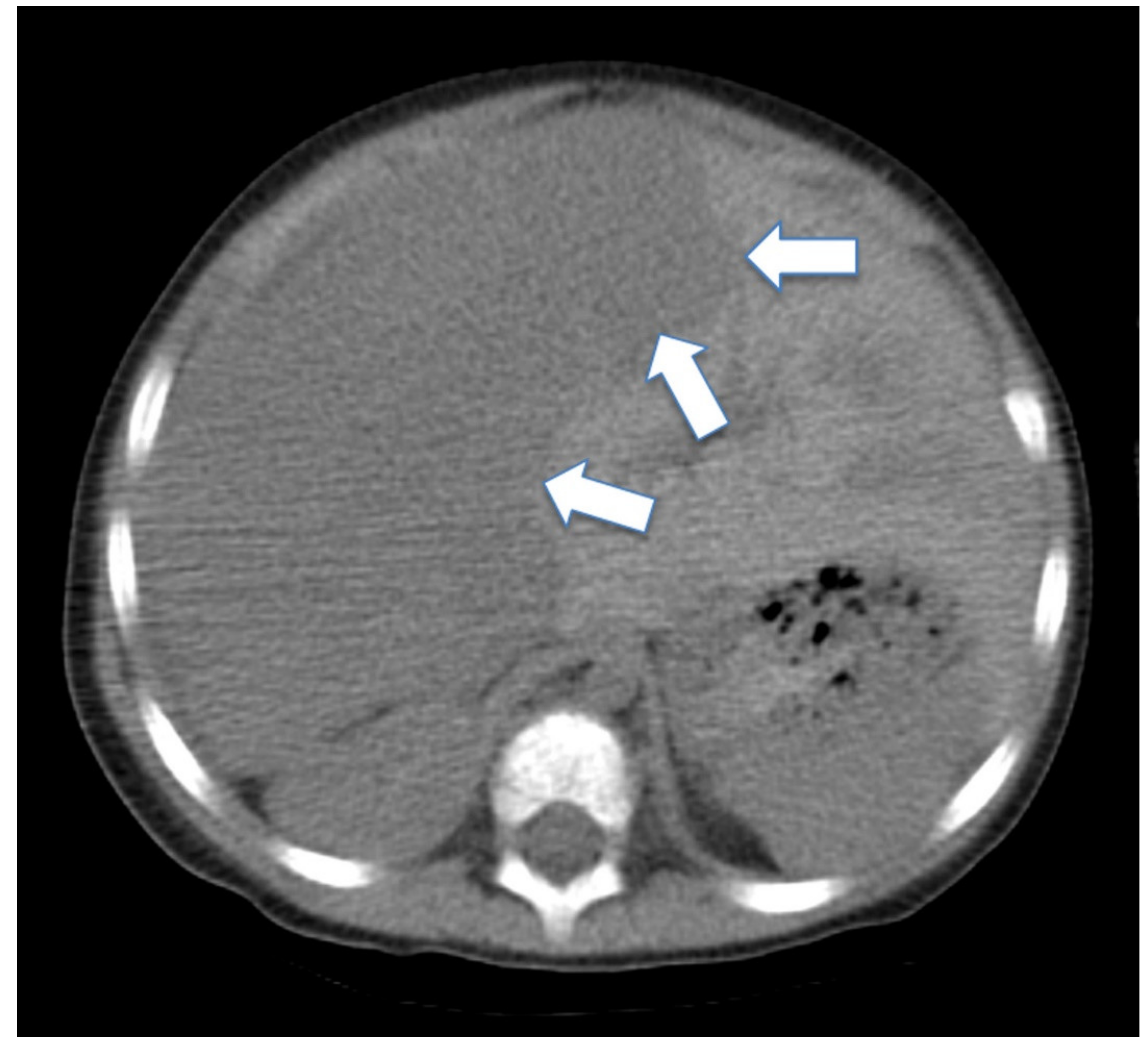
2. Case Report
3. Literature Review
4. SEER Database Analysis
5. Large Population Analysis from the SEER Database
6. Discussion
7. Conclusions
Author Contributions
Funding
Acknowledgments
Conflicts of Interest
References
- Perkins, J.L.; Chen, Y.; Harris, A.; Diller, L.; Stovall, M.; Armstrong, G.T.; Yasui, Y.; Robison, L.L.; Sklar, C.A. Childhood Cancer Survivor Study Infections among long-term survivors of childhood and adolescent cancer: a report from the Childhood Cancer Survivor Study. Cancer 2014, 120, 2514–2521. [Google Scholar] [CrossRef] [PubMed] [Green Version]
- Armstrong, G.T.; Chen, Y.; Yasui, Y.; Leisenring, W.; Gibson, T.M.; Mertens, A.C.; Stovall, M.; Oeffinger, K.C.; Bhatia, S.; Krull, K.R.; et al. Reduction in Late Mortality among 5-Year Survivors of Childhood Cancer. New Engl. J. Med. 2016, 374, 833–842. [Google Scholar] [CrossRef] [PubMed]
- Koh, K.-N.; Yoo, K.H.; Im, H.J.; Sung, K.W.; Koo, H.H.; Kim, H.S.; Han, J.W.; Yoon, J.H.; Park, H.J.; Park, B.-K.; et al. Characteristics and Outcomes of Second Malignant Neoplasms after Childhood Cancer Treatment: Multi-Center Retrospective Survey. J. Korean Med. Sci. 2016, 31, 1254–1261. [Google Scholar] [CrossRef] [PubMed]
- Ishida, Y.; Qiu, D.; Maeda, M.; Fujimoto, J.; Kigasawa, H.; Kobayashi, R.; Sato, M.; Okamura, J.; Yoshinaga, S.; Rikiishi, T.; et al. Secondary cancers after a childhood cancer diagnosis: a nationwide hospital-based retrospective cohort study in Japan. Int. J. Clin. Oncol. 2015, 21, 506–516. [Google Scholar] [CrossRef]
- Felice, M.S.; Rossi, J.G.; Alonso, C.N.; Rubio, P.; Gallego, M.S.; Galluzzo, M.L.; Lubieniecki, F.; Gutiérrez, G.; Guitter, M.R.; Alderete, D.H.; et al. Second Neoplasms in Children Following a Treatment for Acute Leukemia and/or Lymphoma. J. Pediatr. Hematol. 2017, 39, e406–e412. [Google Scholar] [CrossRef]
- Fidler, M.M.; Reulen, R.C.; Winter, D.L.; Allodji, R.S.; Bagnasco, F.; Bárdi, E.; Bautz, A.; Bright, C.J.; Byrne, J.; Feijen, E.A.M.; et al. Risk of Subsequent Bone Cancers Among 69 460 Five-Year Survivors of Childhood and Adolescent Cancer in Europe. J. Natl. Cancer Inst. 2017, 110, 183–194. [Google Scholar] [CrossRef] [Green Version]
- MacCarthy, A.; Bayne, A.M.; Brownbill, P.A.; Bunch, K.J.; Diggens, N.L.; Draper, G.J.; Hawkins, M.M.; Jenkinson, H.C.; Kingston, J.E.; Stiller, C.A.; et al. Second and subsequent tumours among 1927 retinoblastoma patients diagnosed in Britain 1951–2004. Br. J. Cancer. 2013, 108, 2455–2463. [Google Scholar] [CrossRef] [Green Version]
- Royle, J.S.; Baade, P.D.; Joske, D.; Fritschi, L. Risk of second cancer after lymphohematopoietic neoplasm. Int. J. Cancer 2010, 129, 910–919. [Google Scholar] [CrossRef]
- Friedman, D.L.; Whitton, J.; Leisenring, W.; Mertens, A.C.; Hammond, S.; Stovall, M.; Donaldson, S.S.; Meadows, A.T.; Robison, L.L.; Neglia, J. Subsequent neoplasms in 5-year survivors of childhood cancer: the Childhood Cancer Survivor Study. J. Natl. Cancer Inst. 2010, 102, 1083–1095. [Google Scholar] [CrossRef]
- Henderson, T.O.; Whitton, J.; Stovall, M.; Mertens, A.C.; Mitby, P.; Friedman, D.; Strong, L.C.; Hammond, S.; Neglia, J.; Meadows, A.T.; et al. Secondary Sarcomas in Childhood Cancer Survivors: A Report From the Childhood Cancer Survivor Study. J. Natl. Cancer Inst. 2007, 99, 300–308. [Google Scholar] [CrossRef]
- Wolpert, F.; Grotzer, M.A.; Niggli, F.; Zimmermann, D.; Rushing, E.; Bode-Lesniewska, B. Ewing’s Sarcoma as a Second Malignancy in Long-Term Survivors of Childhood Hematologic Malignancies. Sarcoma 2016, 2016, 1–11. [Google Scholar] [CrossRef] [PubMed] [Green Version]
- Neglia, J.; Friedman, D.L.; Yasui, Y.; Mertens, A.C.; Hammond, S.; Stovall, M.; Donaldson, S.S.; Meadows, A.T.; Robison, L.L. Second Malignant Neoplasms in Five-Year Survivors of Childhood Cancer: Childhood Cancer Survivor Study. J. Natl. Cancer Inst. 2001, 93, 618–629. [Google Scholar] [CrossRef] [PubMed] [Green Version]
- Bjerkehagen, B.; Småstuen, M.C.; Hall, K.S.; Skjeldal, S.; Bruland Øyvind, S.; Smeland, S.; Johannesen, T.B.; Fosså, S.D. Incidence and mortality of second sarcomas – A population-based study. Eur. J. Cancer 2013, 49, 3292–3302. [Google Scholar] [CrossRef]
- Bjerkehagen, B.; Småstuen, M.C.; Hall, K.S.; Skjeldal, S.; Smeland, S.; Fosså, S.D. Why do patients with radiation-induced sarcomas have a poor sarcoma-related survival? Br. J. Cancer 2011, 106, 297–306. [Google Scholar] [CrossRef]
- Koshy, M.; Paulino, A.C.; Mai, W.Y.; Teh, B.S. Radiation-induced osteosarcomas in the pediatric population. Int. J. Radiat. Oncol. 2005, 63, 1169–1174. [Google Scholar] [CrossRef]
- Schwartz, B.; Benadjaoud, M.A.; Cléro, É.; Haddy, N.; El-Fayech, C.; Guibout, C.; Teinturier, C.; Oberlin, O.; Veres, C.; Pacquement, H.; et al. Risk of second bone sarcoma following childhood cancer: role of radiation therapy treatment. Radiat. Environ. Biophys. 2014, 53, 381–390. [Google Scholar] [CrossRef]
- Tubiana, M. Can we reduce the incidence of second primary malignancies occurring after radiotherapy? A critical review. Radiother. Oncol. 2009, 91, 4–15. [Google Scholar] [CrossRef] [PubMed]
- Tukenova, M.; Guibout, C.; Hawkins, M.; Quiniou, E.; Mousannif, A.; Pacquement, H.; Winter, D.; Bridier, A.; Lefkopoulos, D.; Oberlin, O.; et al. Radiation Therapy and Late Mortality From Second Sarcoma, Carcinoma, and Hematological Malignancies After a Solid Cancer in Childhood. Int. J. Radiat. Oncol. 2011, 80, 339–346. [Google Scholar] [CrossRef] [PubMed]
- Kawashima, H.; Ogose, A.; Hotta, T.; Imai, C.; Imamura, M.; Endo, N. Secondary osteosarcoma arising from osteochondroma following autologous stem cell transplantation with total-body irradiation for neuroblastoma: A case report. Oncol. Lett. 2015, 10, 1026–1030. [Google Scholar] [CrossRef] [PubMed] [Green Version]
- Menu-Branthomme, A.; Rubino, C.; Shamsaldin, A.; Hawkins, M.M.; Grimaud, E.; Dondon, M.-G.; Hardiman, C.; Vassal, G.; Campbell, S.; Panis, X.; et al. Radiation dose, chemotherapy and risk of soft tissue sarcoma after solid tumours during childhood. Int. J. Cancer 2004, 110, 87–93. [Google Scholar] [CrossRef]
- Tucker, M.A.; D’Angio, G.J.; Boice, J.D.; Strong, L.C.; Li, F.P.; Stovall, M.; Stone, B.J.; Green, D.M.; Lombardi, F.; Newton, W.; et al. Bone Sarcomas Linked to Radiotherapy and Chemotherapy in Children. New Engl. J. Med. 1987, 317, 588–593. [Google Scholar] [CrossRef] [PubMed]
- Zhang, A.Y.; Judson, I.; Benson, C.; Wunder, J.S.; Ray-Coquard, I.; Grimer, R.J.; Quek, R.; Wong, E.; Miah, A.B.; Ferguson, P.C.; et al. Chemotherapy with radiotherapy influences time-to-development of radiation-induced sarcomas: a multicenter study. Br. J. Cancer 2017, 117, 326–331. [Google Scholar] [CrossRef] [PubMed]
- Teepen, J.C.; Van Leeuwen, F.E.; Tissing, W.J.; Broeder, E.V.D.-D.; Heuvel-Eibrink, M.M.V.D.; Van Der Pal, H.J.; Loonen, J.; Bresters, D.; Versluys, B.; Neggers, S.J.; et al. Long-Term Risk of Subsequent Malignant Neoplasms After Treatment of Childhood Cancer in the DCOG LATER Study Cohort: Role of Chemotherapy. J. Clin. Oncol. 2017, 35, 2288–2298. [Google Scholar] [CrossRef] [PubMed] [Green Version]
- Kang, M.H.; Wang, J.; Makena, M.R.; Lee, J.-S.; Paz, N.; Hall, C.P.; Song, M.M.; Calderon, R.I.; Cruz, R.E.; Hindle, A.; et al. Activity of MM-398, nanoliposomal irinotecan (nal-IRI), in Ewing’s family tumor xenografts is associated with high exposure of tumor to drug and high SLFN11 expression. Clin. Cancer Res. 2015, 21, 1139–1150. [Google Scholar] [CrossRef] [PubMed] [Green Version]
- Embree, L.J.; Azuma, M.; Hickstein, D.D. Ewing sarcoma fusion protein EWSR1/FLI1 interacts with EWSR1 leading to mitotic defects in zebrafish embryos and human cell lines. Cancer Res. 2009, 69, 4363–4371. [Google Scholar] [CrossRef] [Green Version]
- Desai, S.S.; Jambhekar, N.A. Pathology of Ewing’s sarcoma/PNET: Current opinion and emerging concepts. Indian J. Orthop. 2010, 44, 363–368. [Google Scholar]
- Magro, G.; Brancato, F.; Musumeci, G.; Alaggio, R.; Parenti, R.; Salvatorelli, L. Cyclin D1 is a useful marker for soft tissue Ewing’s sarcoma/peripheral Primitive Neuroectodermal Tumor in children and adolescents: A comparative immunohistochemical study with rhabdomyosarcoma. Acta Histochem. 2015, 117, 460–467. [Google Scholar] [CrossRef]
- Gazzeri, S.; Gouyer, V.; Vour’Ch, C.; Brambilla, C.; Brambilla, E. Mechanisms of p16INK4A inactivation in non small-cell lung cancers. Oncogene 1998, 16, 497–504. [Google Scholar] [CrossRef] [Green Version]
- Feenstra, J.D.M.; Nivarthi, H.; Gisslinger, H.; Leroy, E.; Rumi, E.; Bagienski, K.; Kubesova, B.; Pietra, D.; Gisslinger, B.; Milanesi, C.; et al. Whole-exome sequencing identifies novel MPL and JAK2 mutations in triple-negative myeloproliferative neoplasms. Blood 2016, 127, 325–332. [Google Scholar] [CrossRef]
- Zakaria, D.; Shaw, A.; Xie, L. Risk of a second cancer in Canadians diagnosed with a first cancer in childhood or adolescence. EClinicalMedicine 2019, 16, 107–120. [Google Scholar] [CrossRef]
- Ishida, Y.; Maeda, M.; Adachi, S.; Rikiishi, T.; Sato, M.; Kawaguchi, H.; Manabe, A.; Tokuyama, M.; Hori, H.; Okamura, J.; et al. Secondary bone/soft tissue sarcoma in childhood cancer survivors: a nationwide hospital-based case-series study in Japan. Jpn. J. Clin. Oncol. 2018, 48, 806–814. [Google Scholar] [CrossRef] [PubMed]
- Wilson, C.L.; Cohn, R.J.; A Johnston, K.; Ashton, L.J. Late mortality and second cancers in an Australian cohort of childhood cancer survivors. Med J. Aust. 2010, 193, 258–261. [Google Scholar] [CrossRef] [PubMed]
- Yamanaka, R.; Hayano, A. Secondary Craniofacial Sarcomas Following Retinoblastoma: A Systematic Review. World Neurosurg. 2017, 101, 722–730. [Google Scholar] [CrossRef] [PubMed]
- Sandoval, C.; Dunbar, J.; Ozkaynak, M.; Jayabose, S. Osteosarcoma Following Growth Hormone Therapy in Recurrent Acute Lymphoblastic Leukemia and Rapadilino Syndrome. Pediatr. Hematol. Oncol. 2012, 29, 270–271. [Google Scholar] [CrossRef] [PubMed]
- Thakral, B.; Khoury, J.D. Histiocytic sarcoma: secondary neoplasm or “transdifferentiation” in the setting of B-acute lymphoblastic leukemia. Blood 2016, 128, 2475. [Google Scholar] [CrossRef] [PubMed] [Green Version]
- Kebudi, R.; Ozger, H.; Kızılocak, H.; Bay, S.B.; Bilgic, B. Osteosarcoma After Hematopoietic Stem Cell Transplantation in Children and Adolescents: Case Report and Review of the Literature. Pediatr. Blood Cancer 2016, 63, 1664–1666. [Google Scholar] [CrossRef]
- Zhu, W.; Hu, F.; Zhao, T.; Wang, C.; Tao, Q.; Information, P.E.K.F.C. Clinical Characteristics of Radiation-Induced Sarcoma of the Head and Neck: Review of 15 Cases and 323 Cases in the Literature. J. Oral Maxillofac. Surg. 2016, 74, 283–291. [Google Scholar] [CrossRef]
- Ehrhardt, M.J.; Howell, C.R.; Hale, K.; Baassiri, M.J.; Rodriguez, C.; Wilson, C.L.; Joshi, S.S.; Lemond, T.C.; Shope, S.; Howell, R.M.; et al. Subsequent Breast Cancer in Female Childhood Cancer Survivors in the St Jude Lifetime Cohort Study (SJLIFE). J. Clin. Oncol. 2019, 37, 1647–1656. [Google Scholar] [CrossRef]
- Yavvari, S.; Makena, Y.; Sukhavasi, S.; Makena, M.R. Large Population Analysis of Secondary Cancers in Pediatric Leukemia Survivors. Children 2019, 6, 130. [Google Scholar] [CrossRef] [Green Version]
- Suarez, C.R.; Bertolone, S.J.; Raj, A.; Coventry, S. Second malignant neoplasms in childhood acute lymphoblastic leukemia: Primitive neuroectodermal tumor of the chest wall with germline p53 mutation as a second malignant neoplasm. Am. J. Hematol. 2004, 76, 52–56. [Google Scholar] [CrossRef]
- Khadwal, A.; Biswas, G.; Arora, B.; Kurkure, P.A.; Deshmukh, C.; Shetty, V. Primitive neuroectodermal tumor (PNET) as second malignancy after treatment of Hodgkin’s disease. Indian J. Pediatr. 2006, 73, 437–438. [Google Scholar] [CrossRef] [PubMed]
- Travis, L.B. Therapy-associated solid tumors. Acta Oncol. 2002, 41, 323–333. [Google Scholar] [CrossRef] [PubMed] [Green Version]
- Choi, D.K.; Helenowski, I.; Hijiya, N. Secondary malignancies in pediatric cancer survivors: Perspectives and review of the literature. Int. J. Cancer 2014, 135, 1764–1773. [Google Scholar] [CrossRef] [PubMed]
- Ng, A.K.; Kenney, L.B.; Gilbert, E.S.; Travis, L.B. Secondary malignancies across the age spectrum. Semin. Radiat. Oncol. 2010, 20, 67–78. [Google Scholar] [CrossRef] [Green Version]


| Cancer | Number of Cases | % of Total SMNs |
|---|---|---|
| Leukemia | 183 | 15.0 |
| Sarcoma | 175 | 14.4 |
| Thyroid carcinoma | 166 | 13.6 |
| Lymphoma | 51 | 4.2 |
| Astrocytoma | 48 | 3.9 |
| Melanoma | 48 | 3.9 |
| Renal carcinoma | 38 | 3.1 |
| Intracranial and intraspinal embryonal tumors | 19 | 1.6 |
| Gliomas | 18 | 1.5 |
| Others | 472 | 38.8 |
| Total | 1218 | 100.0 |
| Cancer | Number of Cases | % of Total Sarcoma |
|---|---|---|
| Other specified soft tissue sarcomas | 50 | 28.6 |
| Osteosarcoma | 41 | 23.4 |
| Unspecified soft tissue sarcomas | 21 | 12.0 |
| Fibrosarcoma | 20 | 11.4 |
| Rhabdomyosarcoma | 18 | 10.3 |
| Ewing sarcoma | 10 | 5.7 |
| Chondrosarcoma | 9 | 5.1 |
| Other specified malignant bone tumors | 4 | 2.3 |
| Unspecified malignant bone tumors | 2 | 1.1 |
| Total | 175 | 100.0 |
| Primary Cancer | Distribution % | Age at Primary Cancer (Mean, SD, Range) | Age at SMN (Sarcoma) (Mean, SD, Range) | Years to Sarcoma from Primary Cancer (Mean, SD, Range) |
|---|---|---|---|---|
| Retinoblastoma | 16.6 | 0.7, 1.1, (0–5) | 14.9, 9.8, (2–41) | 14.3, 9.4, (2–38) |
| Lymphoid leukemias | 13.7 | 6.8, 4.9, (0–16) | 14.6, 5.1, (5–22) | 7.8, 4.8, (1–17) |
| Hodgkin lymphomas | 12.0 | 13, 3.5, (5–16) | 28.5, 10.8, (13–50) | 15.5, 10.3, (3–34) |
| Astrocytoma | 10.3 | 9.6, 5.5, (1–16) | 13.6, 7.0, (1–26) | 4.1, 5.3, (0–20) |
| Renal tumors | 7.4 | 5.3, 4, (1–15) | 13.5, 8.1, (4–36) | 8.2, 9.2, (0–34) |
© 2020 by the authors. Licensee MDPI, Basel, Switzerland. This article is an open access article distributed under the terms and conditions of the Creative Commons Attribution (CC BY) license (http://creativecommons.org/licenses/by/4.0/).
Share and Cite
Nguyen, T.H.; Makena, M.R.; Yavvari, S.; Kaur, M.; Pham, T.; Urias, E.; Panapitiya, N.; Al-Rahawan, M.M. Sarcoma as Second Cancer in a Childhood Cancer Survivor: Case Report, Large Population Analysis and Literature Review. Medicina 2020, 56, 224. https://0-doi-org.brum.beds.ac.uk/10.3390/medicina56050224
Nguyen TH, Makena MR, Yavvari S, Kaur M, Pham T, Urias E, Panapitiya N, Al-Rahawan MM. Sarcoma as Second Cancer in a Childhood Cancer Survivor: Case Report, Large Population Analysis and Literature Review. Medicina. 2020; 56(5):224. https://0-doi-org.brum.beds.ac.uk/10.3390/medicina56050224
Chicago/Turabian StyleNguyen, Thinh H., Monish Ram Makena, Siddhartha Yavvari, Maninder Kaur, Teresia Pham, Eduardo Urias, Narendra Panapitiya, and Mohamad M. Al-Rahawan. 2020. "Sarcoma as Second Cancer in a Childhood Cancer Survivor: Case Report, Large Population Analysis and Literature Review" Medicina 56, no. 5: 224. https://0-doi-org.brum.beds.ac.uk/10.3390/medicina56050224